Embed Size (px)
Citation preview
European Journal of Histochemistry 2015; volume 59:2467
[page 98] [European Journal of Histochemistry 2015; 59:2467]
Apigenin induces dermal collagen synthesis via smad2/3signaling pathwayY. Zhang,1 J. Wang,1 X. Cheng,2 B. Yi,3X. Zhang,4 Q. Li11Department of Plastic andReconstructive Surgery, Shanghai 9th
People’s Hospital, School of Medicine,Shanghai Jiao Tong University2Department of Urology, Renji Hospital,School of Medicine, Shanghai Jiao TongUniversity3Clinical College of General Hospital ofBeijing Military Region, Anhui MedicalUniversity, Hefei4The Key Laboratory of Stem CellBiology, Institute of Health Sciences,Shanghai Institutes for BiologicalSciences, Chinese Academy of Sciencesand Shanghai Jiao Tong University Schoolof Medicine, Shanghai, China
Abstract
Decrease in fibroblast-produced collagenhas been proven to be the pivotal cause of skinaging, but there is no satisfactory drug whichdirectly increases dermal thickness and col-lage density. Here we found that a flavonoidnatural product, apigenin, could significantlyincrease collagen synthesis. NIH/3T3 and pri-mary human dermal fibroblasts (HDFs) wereincubated with various concentrations of api-genin, with dimethyl sulfoxide (DMSO) serv-ing as the negative control. Real-time reverse-transcription polymerase chain reaction(PCR), Western Blot, and Toluidine blue stain-ing demonstrated that apigenin stimulatedtype-I and type-III collagen synthesis of fibrob-lasts on the mRNA and protein levels.Meanwhile, apigenin did not induce expres-sion of alpha smooth muscle actin (α-SMA) invitro and in vivo, a fibrotic marker in living tis-sues. Then the production of collagen was con-firmed by Masson’s trichrome stain,Picrosirius red stain and immunohistochem-istry in mouse models. We also clarified thatthis compound induced collagen synthesis byactivating smad2/3 signaling pathway. Takentogether, without obvious influence on fibrob-lasts’ apoptosis and viability, apigenin couldpromote the type-I and type-III collagen synthe-sis of dermal fibroblasts in vitro and in vivo,thus suggesting that apigenin may serve as apotential agent for esthetic and reconstructiveskin rejuvenation.
Introduction
Dermis consists of several structural compo-nents, and collagen takes the major part. Inaddition to glycosaminoglycans and elastinfibers, dermal matrix in adult skin are com-posed of type I (80-85%) and type III collagen(10-15%).1 It is of great importance that colla-gen plays a main role in the texture andappearance of skin. Skin aging is inevitablyassociated with a disturbance in collagenmetabolism2 (due to the decreased activity offibroblasts and their collagen synthesis), aswell as elastin. Increasing the collagen contentof dermis has been regarded as a well-effectiveway for anti-aging in skin.Collagen is a kind of biomacromolecule and
it cannot be absorbed through the stratumcorneum. Recently, some researches demon-strated that collagen hydrolysate ingestionmight be beneficial to slow chronological skinaging3 and photoaging4 in rats, and the densityof collagen fibrils increased compared with lac-talbumin and water controls.5 But the clinicaleffect of oral collagen hydrolysate still lacksconvincing evidences. Up to now, injectablecollagen1 or analogous composition6 fillingimplants are recognized as the well-acceptedtreatment modality for cosmetic purposes.However, maintaining skin appearance relieson expansive and complex treatment repeated-ly because of the short-term duration of exo-genic injected collagen.Apigenin (4,5,7-trihydroxyflavone), a
flavone subclass of flavonoid widely distributedin many herbs, fruits, and vegetables, is a sub-stantial component of the human diet and hasbeen shown to possess a variety of biologicalcharacteristics, including anti-oxidative,7 anti-inflammatory effect,8 tumor growth inhibition9
and promoting neurogenesis.10 It has beenshown that apigenin could enhance woundhealing and tissue repair in diabetic rat skin.11
In the process of wound healing, fibroblastssecreted collagen and the formation of colla-gen-rich granulation tissue are vital patho-physiological mechanisms for wound closure.12
Given this, we wonder what effect wouldapigenin have on fibroblasts and whether api-genin could induce collagen synthesis in nor-mal human dermal fibroblasts. Consequently,we examined its effects on collagen synthesisin normal human dermal fibroblasts in vitroand tested its function in the skin aging mousemodel induced by D-Galactose. Furthermore,we investigated the potential mechanisminvolved in the positive effects of apigenin oncollagen expression in fibroblasts.
Materials and MethodsCell culturePrimary human dermal fibroblasts were
obtained from adolescent foreskin tissue of tenpeople (aged 8-12 years), and none of themhad a history with skin diseases. Skin tissuewas obtained after obtaining informed consentfrom the patients, with the approval of theethics committee of Shanghai 9th People’sHospital and in conformity with the Helsinkiguidelines. NIH/3T3 and human dermal fibrob-lasts (HDFs) were maintained in DMEM(Hyclone, Thermo Fisher Scientific, Waltham,MA, USA) supplemented with 10% fetal bovine
Correspondence: Xiaoling Zhang and QingfengLi, Department of Plastic and ReconstructiveSurgery, Shanghai 9th People’s Hospital,Shanghai Jiao Tong University School ofMedicine, 639 Zhizaoju Road, Shanghai 200011,China. Tel. +86.21.23271699-5615 - Fax: +86.21.63089567. E-mails: (X. Zhang) [email protected]; (Q. Li)[email protected]
Key words: Apigenin, flavonoid, collagen I/III,fibroblasts, smad2/3.
Contributions: YZ and JW contributed equally tothis work; YZ, JW, experimental work, data collec-tion and interpretation; XC, participation inexperimental work design and coordination, dataacquisition; BY, participation in study design,data collection, analysis of data and manuscriptpreparation; QL, XZ, study design, data analysisand interpretation, manuscript drafting.
Conflict of interest: the authors declare no con-flict of interest.
Funding: this study was supported by grants fromthe key project of the National Natural ScienceFoundation (No. 81230042), the National KeyProject of Scientific and Technical SupportingPrograms Funded by Ministry of Science &Technology of China (No. 2012BAI11B03) (Q. Li)and the Chinese Academy of Sciences (No.XDA01030102), Shanghai Municipal Commissionof Health and Family Planning (No.2013ZYJB0501) (X. Zhang).
Received for publication: 28 November 2014.Accepted for publication: 9 March 2015.
This work is licensed under a Creative CommonsAttribution NonCommercial 3.0 License (CC BY-NC 3.0).
©Copyright Y. Zhang et al., 2015Licensee PAGEPress, ItalyEuropean Journal of Histochemistry 2015; 59:2467doi:10.4081/ejh.2015.2467
EJH_2015_02-article.qxp_Hrev_master 22/06/15 12:54 Pagina 98
[European Journal of Histochemistry 2015; 59:2467] [page 99]
serum (Hyclone, Thermo Fisher Scientific),100 U/mL penicillin, and 100 mg/L strepto-mycin. Both HDFs and NIH/3T3 were incubatedat 37°C in a humidified atmosphere with 5%CO2. Primary fibroblasts of passages 6-8 wereused. Toluidine blue (Sigma-Aldrich, St. Louis,MO, USA) staining was used to assess extra-cellular matrix synthesis.13
Real-time PCR analysisThe total RNA of cells was isolated using
TRIzol reagent (Invitrogen, Carlsbad, CA, USA)and subjected to reverse transcription withOligo (dT) and M-MLV Reverse Transcriptase(Thermo Fisher Scientific). Synthesized com-plementary DNA (cDNA) was analysed withquantitative real-time PCR using SYBR®Premix (Takara, Dalian, China) and Roche480system. Glyceraldehyde 3-phospate dehydroge-nase (GAPDH) was used as a reference gene.Primers sequences were as follows: collagen,type I, alpha 2 (Col1a2; mouse), 5�-GGTGAGC-CTGGTCAAACGG-3� (forward) and 5�- ACTGT-GTCCTTTCACGCCTTT-3� (reverse); Col1a2(human), 5�-GGCCCTCAAGGTTTCCAAGG-3�(forward) and 5�-CACCCTGTGGTCCAA-CAACTC-3� (reverse); collagen, type III, alpha 1(Col3a1; mouse), 5�- CTGTAACATG-GAAACTGGGGAAA-3� (forward) and 5�-CCATAGCTGAACTGAAAACCACC-3� (reverse);Col3a1 (human), 5�-TTGAAGGAGGATGTTCC-CATCT-3� (forward) and 5�- ACAGACA-CATATTTGGCATGGTT-3� (reverse); matrixmetalloproteinases 1 (MMP1; human), 5�-AAAATTACACGCCAGATTTGCC-3� (forward)and 5�-GGTGTGACATTACTCCAGAGTTG-3�(reverse); matrix metalloproteinases 2(MMP2; human), 5�-TACAGGATCATTGGCTA-CACACC-3� (forward) and 5�-GGTCA-CATCGCTCCAGACT-3� (reverse); matrix metal-loproteinases 9 (MMP9; human), 5�-TGTAC-CGCTATGGTTACACTCG-3� (forward) and 5�-GGCAGGGACAGTTGCTTCT-3� (reverse); tissueinhibitor of metalloproteinases1 (TIMP1;human), 5�-CTTCTGCAATTCCGACCTCGT-3�(forward) and 5�-ACGCTGGTATAAGGTG-GTCTG-3� (reverse); α-SMA (human), 5�-AAAAGACAGCTACGTGGGTGA-3� (forward) and5�-GCCATGTTCTATCGGGTACTTC-3� (reverse).
Cell viability assayFor the cell viability assay, HDFs were seed-
ed on 96-well plates (100 mL per well), fol-lowed by apigenin (cat. no. 42251; Sigma-Aldrich) or DMSO (Sigma-Aldrich) treatment.After 3 or 5 days, cell culture medium wasreplaced by Thiazolyl Blue TetrazoliumBromide (MTT) working solution, followed bya 4-hour incubation at 37°C in a 5% CO2 incu-bator. After MTT working solution wasremoved and DMSO added, the absorbances at490 nm were detected.
Flow cytometric analysisCell apoptosis was assessed by flow cytome-
try using the Alexa Fluor® 488 Annexin V/DeadCell Apoptosis Kit (Invitrogen, Carlsbad, CA,USA). Following apigenin (5 μmol/L or 1μmol/L) or dimethyl sulfoxide (DMSO) treat-ment, harvested cells were suspended in 100μL Annexin-binding buffer. Then, 5 μL AlexaFluor® 488 Annexin V and 1 μL PI workingsolution were added and incubated with thecells for 15 min in the dark. After the incuba-tion period, 400 μL 1X Annexin-binding bufferwas added and mixed gently. The stained cellswere analysed directly by flow cytometry usingthe Cell Quest program (Becton Dickinson, CA,USA). Data were analysed using FlowJo soft-ware.
Colony formation assayAnchorage-dependent growth of HDFs were
investigated by monolayer colony formationassay.14 Cells were cultured in a 6-well plate(500 per well) and treated with 5 μmol/L or 1μmol/L apigenin or DMSO. After cultured for14 days, surviving colonies were stained 5 minwith Gentian Violet (Sigma-Aldrich) after 4%paraformaldehyde fixation.
Immunofluorescence cell stainingHuman dermal fibroblasts at a density of
2×10³ cells per well were seeded on coverslides in 24-well plates and incubatedovernight. Cells were fixed with 4%paraformaldehyde and blocked with 5% goatserum in PBST (0.1% TritonX-100 in phos-phate buffered saline) for 1 h. For F-actinstaining, cells were incubated with Alexa Fluor488 Phalloidin (Cytoskeleton Inc., Denver, CO,USA; 1:200) for 1 h at room temperature. Forα-SMA staining, cells were incubated with pri-mary antibodies against α-SMA (Abcam,Cambridge, UK, 1:200) for 2 h at room temper-ature, followed by an Alexa Fluor 555-conjugat-ed secondary antibody. For smad3 staining,cells were incubated with primary antibodiesagainst smad3 (Cell Signaling Technology,Beverly, MA, USA; 1:200) for 2 h at room tem-perature, followed by an Alexa Fluor 488-conju-gated secondary antibody. Immunofluore -scence signals were captured using confocalmicroscopy (LSM 510, META Laser ScanningMicroscope; Zeiss, Jena, Germany).
Smad2/3 knockdown by siRNARNA interference was performed using
smad2/3 siRNA (human) (sc-37238; SantaCruz Biotechnology, Dallas, TX, USA), target-ing human smad2/3 and control siRNA (sc-37007) as negative control. Transfection forHDFs was conducted using LipofectamineRNAiMAX reagent (Invitrogen, Carlsbad, CA,USA) according to the manufacturer’s protocol.
Western blotCultured cells were lysed using radioim-
munoprecipitation assay (RIPA) lysis buffer.Protein concentrations were determined usinga micro bicinchoninic acid (BCA) assay(Thermo Fisher Scientific). Twenty micro-grams total protein extract was separated by8% or 10% sodium dodecyl sulfate-polyacry-lamide gel electrophoresis (SDS-PAGE) underreducing conditions and electroblotted ontopolyvinylidene difluoride membranes(Millipore, Bedford, MA, USA). The membranewas then blocked and then incubated with pri-mary antibodies overnight at 4°C.The primary antibodies used included the
followings: anti-Col1a2, anti-Col3a1, anti-α-SMA (Abcam; 1:1000), anti-cyclin-dependentkinase (CDK) family, anti-cyclin D1, anti-cyclin E1, anti-smads, anti-MAPK family (CellSignaling Technology, Beverly, MA, 1:1000),anti-GAPDH (Sigma-Aldrich; 1:10,000).Immunoreactive bands were quantitativelyanalyzed with ImageJ software.
D-galactose-induced skin agingmouse model and animal experimentsSix-week-old female C57BL/6 mice were
purchased from the Shanghai Slac LaboratoryAnimal (Slac, Shanghai, China). All animalstudies have been approved by the AnimalCare and Use Committee of Shanghai JiaoTong University. All efforts were made to mini-mize animal suffering.A total of 18 mice were randomly assigned to
three groups (n=6). Two groups of animalsreceived daily subcutaneous injection of D-galactose (D-gal; 1000 mg/kg) for 8 weeks.15
The third group received phosphate bufferedsaline (PBS) as a negative control. Two weekslater, DMSO and apigenin (5 μmol/L) wasdelivered by microneedles16 [MTS-RollerModel: CR2 (0.2 mm)] to the dermis of D-galactose treated mice once a day for 4 weeks,respectively. Mice were sacrificed at the end oftreatment, and skin tissue was harvested forfurther analyses.
Histology and immunohistochemistryParaformaldehyde-fixed paraffin-embedded
tissue sections (5 μm) were stained withhematoxylin and eosin (H&E), Masson’strichrome (Trichrome stain LG solution,HT10316; Sigma-Aldrich) and Picrosirius red(Fluka, Buchs, Switzerland). For immunohis-tochemical staining, the sections were detect-ed with primary antibodies against collagenI/III (Millipore; 1:1,000) and α-SMA (Abcam;1:200) overnight at 4°C. After incubation withthe appropriate secondary antibodies, the sec-tions were developed with diaminobenzidineand counterstained with hematoxylin.
Original Paper
EJH_2015_02-article.qxp_Hrev_master 22/06/15 12:54 Pagina 99
[page 100] [European Journal of Histochemistry 2015; 59:2467]
Statistical analysisStatistical differences were calculated using
Friedman’s analysis of variance (ANOVA), withpost-hoc least significant difference (LSD) testas appropriate. A significant difference amonggroups was set at P<0.05.
ResultsApigenin stimulated collagen synthesis but had no effect onmatrix metalloproteinases in vitroFibroblasts are the predominant mesenchy-
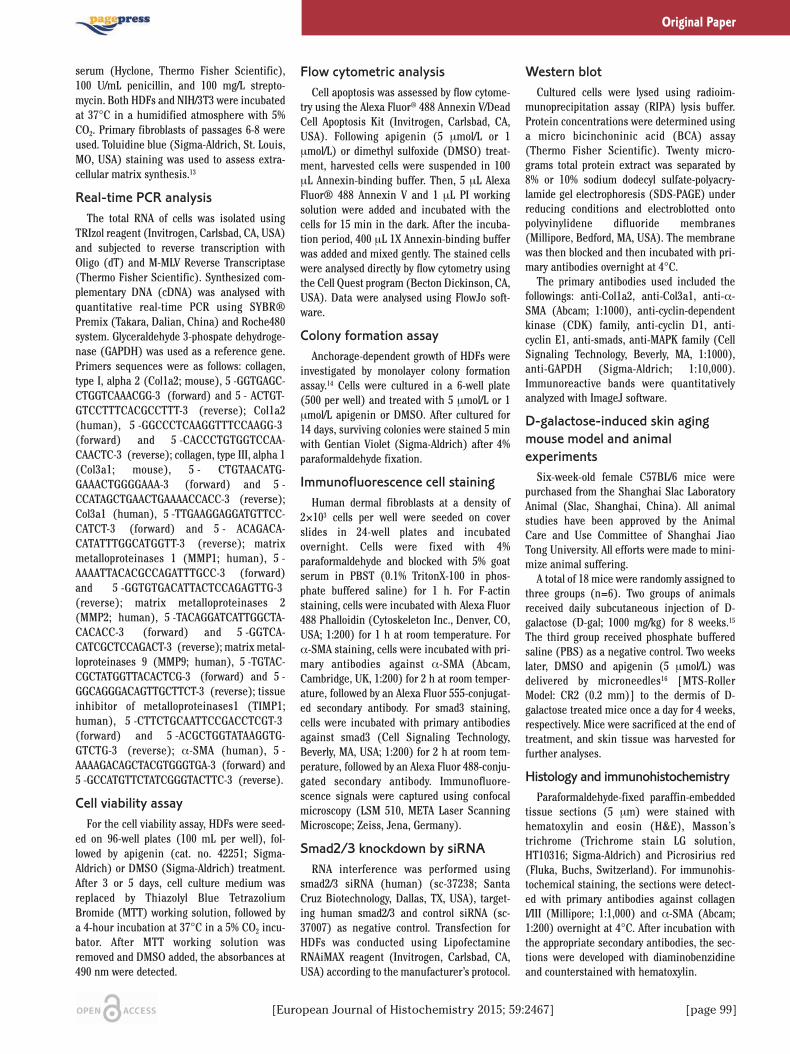
mal cells in the dermis, and their function isstrongly implicated in dermatology. To studyapigenin’s effect on fibroblasts (Figure 1A),NIH/3T3 and HDFs were administered withapigenin at the concentrations of 5 μmol/L. Asshown in Figure 1B, apigenin could potentlyincrease Toluidine blue staining after api-genin treatment for 5 days, which mean the
Original Paper
Figure 1. Apigenin stimulated collagen synthesis of fibroblasts. A) The molecular structure of apigenin. B) Toluidine blue staining inNIH/3T3 and HDFs for 5 days. C) Dose-dependent effects of apigenin on mRNA expression of Col1a2 and Col3a1 in NIH/3T3 andHDFs for 3 days. D) The protein level of Col1a2 and Col3a1 was measured by Western Blot at 5 days after apigenin was applied atconcentrations of 0.1 μmol/L to 10 μmol/L. E) The expression of MMP1, MMP2, MMP9 and TIMP1 were also assessed by real-timePCR. Data are presented as mean ± SD, n≥3; NS, not significant; *P<0.05; **P< 0.01; ***P<0.001.
EJH_2015_02-article.qxp_Hrev_master 22/06/15 12:54 Pagina 100
[European Journal of Histochemistry 2015; 59:2467] [page 101]
synthesis of extracellular matrix wasincreased.13 Further real-time PCR analysisshowed that, in NIH/3T3 and HDFs, apigenin(0.1 μmol/L - 10 μmol/L) dose-dependentlystimulated endogenous expression of Col1a2and Col3a1. The most significant changes
were observed when NIH/3T3 and HDFs weretreated with apigenin at the concentration of 5μmol/L, and the increase of Col3a1 was moreobvious than Col1a2 (Figure 1C). These upreg-ulation effect of apigenin on collagen expres-sion were then confirmed by Western blot
analysis. When HDFs were treated with api-genin for 5 days, the protein level of Col1a2and Col3a1 were higher than that of cells treat-ed with DMSO (Figure 1D). In addition, wenext examined the effect of apigenin onmatrix metalloproteinases (MMPs) and
Original Paper
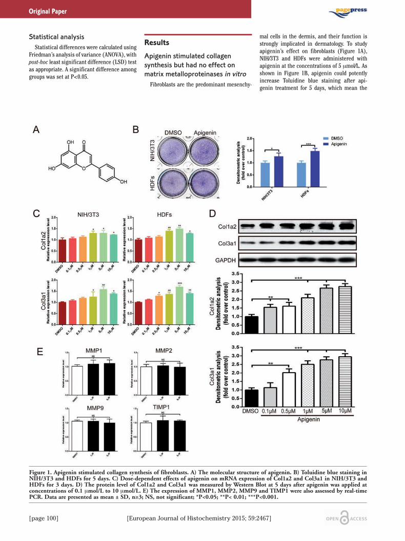
Figure 2. No obvious cytotoxicity exerted by apigenin on fibroblasts viability, apoptosis, proliferation, cell cycle and activation. A) Cellviability was examined by MTT assays at 3 or 5 days after apigenin was applied in HDFs. B) Apoptosis was evaluated after treatingHDFs with 5 μmol/L or 1 μmol/L apigenin or DMSO; flow cytometry profile represents Alexa Fluor® 488 Annexin V staining in Xaxis and PI in Y axis. C) The effect of apigenin on fibroblasts growth was investigated by monolayer colony formation assay. D) Theexpression of cyclin E1, CDK4, cyclin D1, CDK2 and p-CDK2 proteins was analysed using Western blot in HDFs. E-F) The levels ofα-SMA mRNA and protein expression were measured by real-time PCR and Western blot. G) Immunofluorescence cell staining for α-SMA and F-actin in cultured HDFs after incubation with apigenin or DMSO for 72 h; F-actin is shown by green fluorescence and α-SMA is shown by red fluorescence; nucleus (blue) was stained with DAPI; scale bar: 50 μm. Data are presented as mean ± SD, n≥3;NS, not significant; **P<0.01; ***P<0.001.
EJH_2015_02-article.qxp_Hrev_master 22/06/15 12:55 Pagina 101
TIMP1, well-known proteases that degrade col-lagen proteins. The expression of MMP1,MMP2, MMP9, and their inhibitor TIMP1 wereunchanged (Figure 1E).
Apigenin did not effect fibroblastsviability and activityAfterwards, we studied the effect of api-
genin on fibroblasts viability, apoptosis, prolif-eration and activation. In vitro, MTT assaysshowed that the viability of HDFs was similarwith those incubated with DMSO, when incu-bated with apigenin (1 to 10 μmol/L) for 3 or 5days, respectively (Figure 2A). In addition, nosignificant differences in percentages of apop-totic cells was observed after the exposure offibroblasts to apigenin (Figure 2B). To investi-gate the effects of apigenin on proliferationand cell cycle, colony formation assay andWestern Blot analysis of cell cycle related pro-teins were performed. Colony forming abilityof HDFs was similar in apigenin-treated groupto DMSO-treated group (Figure 2C). Theexpression levels of cell cycle associated pro-teins remained unchanged between apigeninand DMSO treated groups (Figure 2D). Theseresults suggested that apigenin had no obvi-ous cytotoxicity on fibroblasts’ viability, apop-tosis and proliferation.Fibroblast overactivation leads to pathologi-
cal collagen deposition or scar formation.17
Myofibroblasts, known as activated fibroblasts,are marked by α-SMA expression. To deter-mine the effects of apigenin on fibroblasts’activation, we evaluated the levels of α-SMAmRNA and protein expression (Figure 2 E,F)in cultured HDFs treated with apigenin. Wefound that α-SMA mRNA expression had noobvious change in apigenin-treated cells com-pared with DMSO-treated cells. We alsoshowed that apigenin did not affect α-SMAexpression in vitro by immunofluorescencestaining (Figure 2G). These findings suggest-ed that apigenin did not cause fibroblasts tooveractivate into myofibroblasts while colla-gen synthesis was increasing.
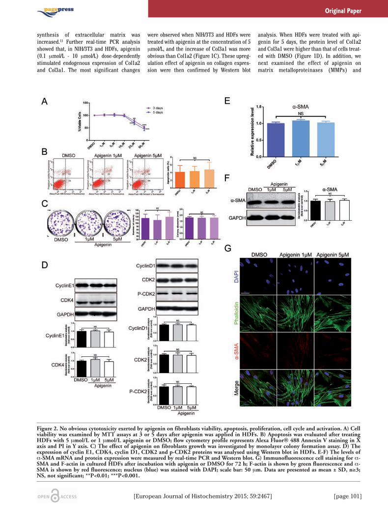
Induction of collagen synthesis wasmediated by smad2/3 activationTo further explore the underlying mecha-
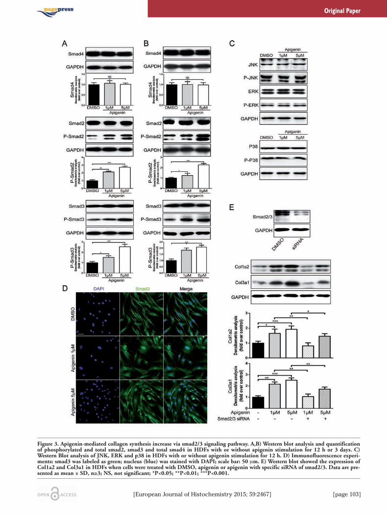
nism of how apigenin activated type-I andtype-III collagen gene expression, transform-ing growth factor beta 1 (TGF-β1) and mito-gen-activated protein kinase (MAPK) signal-ing pathway were analysed. TGF-β1 is a proto-typic fibrogenic cytokine, enhancing extracel-lular matrix gene expression. Previous studiesproved that Col1a2 and Col3a1 were directTGF-β1/smad3 targets in human dermalfibroblasts.18 As shown in Figure 3A, whenHDFs were treated for 12 h, apigenin (1μmol/L or 5 μmol/L) markedly increased theexpression of phosphorylated smad2 and
smad3 in a dose-dependent manner, whereastotal smad2, smad3 and smad4 did not obvi-ously alter. It also showed that apigenin hadsustained effect on promoting phosphorylationof smad2 and smad3 after a 3-day treatment(Figure 3B). Yoon et al.19 revealed that MAPKpathway was involved with peptide-inducedcollagen synthesis of fibroblasts. However,when fibroblasts were treated with apigeninfor 12 h, the expression of total and phospho-rylated JNK, ERK and p38 protein remainedunchanged, compared with DMSO (Figure3C). Immunofluorescence experiments de -monstrated that after treatment with apigeninfor 12 h, smad3 protein (labeled by green) wassignificantly increased and mostly translocat-ed into the nucleus (labeled by blue) (Figure3D). By contrast, in the DMSO groups, smad3were retained in the cytoplasm. Once targetedknockdown smad2/3 by specific siRNA, the up-regulation effect of apigenin on the expres-sion of collagen type-I and type-III protein wasobviously reduced (Figure 3E), which con-firmed that smad2/3 is required for the trans-duction of apigenin effect on collagen expres-sions.
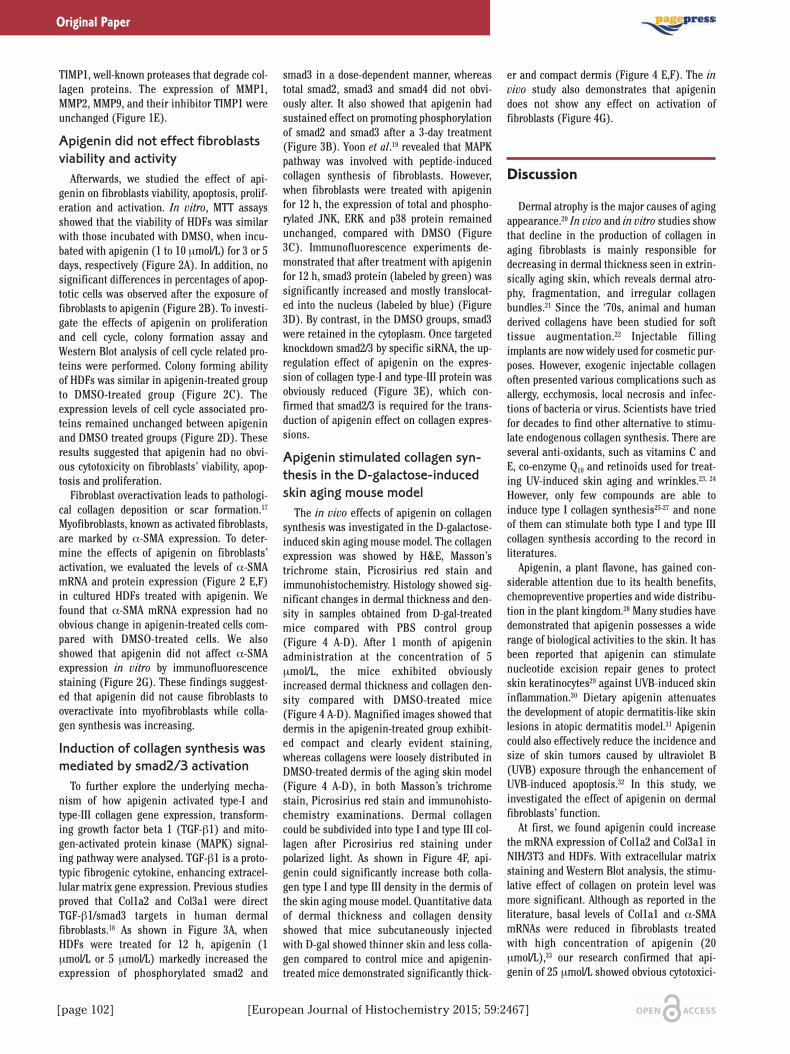
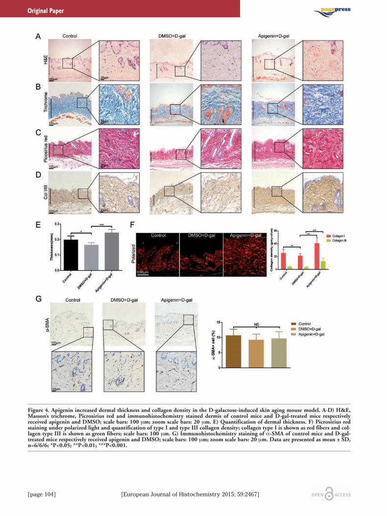
Apigenin stimulated collagen syn-thesis in the D-galactose-inducedskin aging mouse modelThe in vivo effects of apigenin on collagen
synthesis was investigated in the D-galactose-induced skin aging mouse model. The collagenexpression was showed by H&E, Masson’strichrome stain, Picrosirius red stain andimmunohistochemistry. Histology showed sig-nificant changes in dermal thickness and den-sity in samples obtained from D-gal-treatedmice compared with PBS control group(Figure 4 A-D). After 1 month of apigeninadministration at the concentration of 5μmol/L, the mice exhibited obviouslyincreased dermal thickness and collagen den-sity compared with DMSO-treated mice(Figure 4 A-D). Magnified images showed thatdermis in the apigenin-treated group exhibit-ed compact and clearly evident staining,whereas collagens were loosely distributed inDMSO-treated dermis of the aging skin model(Figure 4 A-D), in both Masson’s trichromestain, Picrosirius red stain and immunohisto-chemistry examinations. Dermal collagencould be subdivided into type I and type III col-lagen after Picrosirius red staining underpolarized light. As shown in Figure 4F, api-genin could significantly increase both colla-gen type I and type III density in the dermis ofthe skin aging mouse model. Quantitative dataof dermal thickness and collagen densityshowed that mice subcutaneously injectedwith D-gal showed thinner skin and less colla-gen compared to control mice and apigenin-treated mice demonstrated significantly thick-
er and compact dermis (Figure 4 E,F). The invivo study also demonstrates that apigenindoes not show any effect on activation offibroblasts (Figure 4G).
Discussion
Dermal atrophy is the major causes of agingappearance.20 In vivo and in vitro studies showthat decline in the production of collagen inaging fibroblasts is mainly responsible fordecreasing in dermal thickness seen in extrin-sically aging skin, which reveals dermal atro-phy, fragmentation, and irregular collagenbundles.21 Since the ‘70s, animal and humanderived collagens have been studied for softtissue augmentation.22 Injectable fillingimplants are now widely used for cosmetic pur-poses. However, exogenic injectable collagenoften presented various complications such asallergy, ecchymosis, local necrosis and infec-tions of bacteria or virus. Scientists have triedfor decades to find other alternative to stimu-late endogenous collagen synthesis. There areseveral anti-oxidants, such as vitamins C andE, co-enzyme Q10 and retinoids used for treat-ing UV-induced skin aging and wrinkles.23, 24
However, only few compounds are able toinduce type I collagen synthesis25-27 and noneof them can stimulate both type I and type IIIcollagen synthesis according to the record inliteratures.Apigenin, a plant flavone, has gained con-
siderable attention due to its health benefits,chemopreventive properties and wide distribu-tion in the plant kingdom.28 Many studies havedemonstrated that apigenin possesses a widerange of biological activities to the skin. It hasbeen reported that apigenin can stimulatenucleotide excision repair genes to protectskin keratinocytes29 against UVB-induced skininflammation.30 Dietary apigenin attenuatesthe development of atopic dermatitis-like skinlesions in atopic dermatitis model.31 Apigenincould also effectively reduce the incidence andsize of skin tumors caused by ultraviolet B(UVB) exposure through the enhancement ofUVB-induced apoptosis.32 In this study, weinvestigated the effect of apigenin on dermalfibroblasts’ function.At first, we found apigenin could increase
the mRNA expression of Col1a2 and Col3a1 inNIH/3T3 and HDFs. With extracellular matrixstaining and Western Blot analysis, the stimu-lative effect of collagen on protein level wasmore significant. Although as reported in theliterature, basal levels of Col1a1 and α-SMAmRNAs were reduced in fibroblasts treatedwith high concentration of apigenin (20μmol/L),33 our research confirmed that api-genin of 25 μmol/L showed obvious cytotoxici-
[page 102] [European Journal of Histochemistry 2015; 59:2467]
Original Paper
EJH_2015_02-article.qxp_Hrev_master 22/06/15 12:55 Pagina 102
[European Journal of Histochemistry 2015; 59:2467] [page 103]
Original Paper
Figure 3. Apigenin-mediated collagen synthesis increase via smad2/3 signaling pathway. A,B) Western blot analysis and quantificationof phosphorylated and total smad2, smad3 and total smad4 in HDFs with or without apigenin stimulation for 12 h or 3 days. C)Western Blot analysis of JNK, ERK and p38 in HDFs with or without apigenin stimulation for 12 h. D) Immunofluorescence experi-ments: smad3 was labeled as green; nucleus (blue) was stained with DAPI; scale bar: 50 μm. E) Western blot showed the expression ofCol1a2 and Col3a1 in HDFs when cells were treated with DMSO, apigenin or apigenin with specific siRNA of smad2/3. Data are pre-sented as mean ± SD, n≥3; NS, not significant; *P<0.05; **P<0.01; ***P<0.001.
EJH_2015_02-article.qxp_Hrev_master 22/06/15 12:55 Pagina 103
[page 104] [European Journal of Histochemistry 2015; 59:2467]
Original Paper
Figure 4. Apigenin increased dermal thickness and collagen density in the D-galactose-induced skin aging mouse model. A-D) H&E,Masson’s trichrome, Picrosirius red and immunohistochemistry stained dermis of control mice and D-gal-treated mice respectivelyreceived apigenin and DMSO; scale bars: 100 μm; zoom scale bars: 20 μm. E) Quantification of dermal thickness. F) Picrosirius redstaining under polarized light and quantification of type I and type III collagen density; collagen type I is shown as red fibers and col-lagen type III is shown as green fibers; scale bars: 100 μm. G) Immunohistochemistry staining of α-SMA of control mice and D-gal-treated mice respectively received apigenin and DMSO; scale bars: 100 μm; zoom scale bars: 20 μm. Data are presented as mean ± SD,n=6/6/6; *P<0.05; **P<0.01; ***P<0.001.
EJH_2015_02-article.qxp_Hrev_master 22/06/15 12:55 Pagina 104
[European Journal of Histochemistry 2015; 59:2467] [page 105]
ty in fibroblasts. We believed that the attenuat-ing effect of high concentration of apigenin onphenotypic transitions in the analyzed cell pop-ulations were not independent of its cytotoxicactivity. We detected the markers related toextracellular matrix degradation and foundthat apigenin had no effect on the balance ofMMPs/TIMPs. We further observed that apigenin directly
activated smad2/3-dependent signaling path-way. This is not surprising since this flavonoiddisplays considerable muti-effect. It targets anumber of secondary messengers, includingthose potentially involved in TGF-β1 signalingpathway, such as NF-kB,34 MAPK/ERK,35
FAK,36,37 PKC38 and PI3K-Akt39 in a cell context-dependent manner. We observed that apigeninmarkedly increased the expression of phos-phorylated smad2 and smad3 protein, whiletotal smad2, smad3 and smad4 protein allremained unaltered. So, a more meticulousnetwork may connect TGF-β1 signaling path-way and the abovementioned secondary mes-sengers.A previous study showed the accelerated
aging effect of D-gal injection on mouse skin,as well as changes in dermal thickness and col-lagen content.15 In order to confirm the effectof apigenin on collagen synthesis in vivo, theD-galactose-induced skin aging mouse modelwas established. Our data indicated that skinaging mice treated with apigenin showedmarkedly increasing dermal thickness and col-lagen expression, compared with DMSO-treat-ed mice. Hou et al.40 reported that topical api-genin improved epidermal permeability barrierfunction by stimulating epidermal differentia-tion, lipid synthesis and secretion, as well ascutaneous antimicrobial peptide production,and our result showed that dermal injection ofapigenin significantly increased dermal thick-ness and density. So we could conclude thatapigenin caused different biological functionswith two forms of drug administration by act-ing on epidermis or derma, which might indi-cate the importance of choosing suitableadministration methods to different skin dis-eases, even for the same drug.Our study demonstrates that apigenin could
induce both type I and type III collagen synthe-sis of fibroblasts in vitro and could increasedermal thickness and collagen deposition inthe dermis of mice. This compound is a poten-tial target for drug design and development foresthetic and reconstructive purpose.
References
1. Baumann L, Kaufman J, Saghari S.Collagen fillers. Dermatol Ther 2006;19:134-40.
2. Fisher GJ, Wang ZQ, Datta SC, Varani J,Kang S, Voorhees JJ. Pathophysiology ofpremature skin aging induced by ultravio-let light. N Engl J Med 1997;337:1419-28.
3. Liang JA, Pei XR, Zhang ZF, Wang N, WangJB, Li Y. The Protective Effects of Long-Term Oral Administration of MarineCollagen Hydrolysate from Chum Salmonon Collagen Matrix Homeostasis in theChronological Aged Skin of Sprague-Dawley Male Rats. J Food Sci 2010;75:H230-8.
4. Hou H, Li BF, Zhang ZH, Xue CH, Yu GL,Wang JF, et al. Moisture absorption andretention properties, and activity in allevi-ating skin photodamage of collagenpolypeptide from marine fish skin. FoodChem 2012;135:1432-9.
5. Matsuda N, Koyama YI, Hosaka Y, Ueda H,Watanabe T, Araya T, et al. Effects of inges-tion of collagen peptide on collagen fibrilsand Glycosaminoglycans in the dermis. JNutr Sci Vitaminol (Tokyo) 2006;52:211-5.
6. Iannitti T, Morales-Medina JC, Coacci A,Palmieri B. Experimental and ClinicalEfficacy of Two Hyaluronic Acid-basedCompounds of Different Cross-Linkageand Composition in the Rejuvenation ofthe Skin. Pharm Res Epub 2014 Jun 25.
7. Sharma H, Kanwal R, Bhaskaran N, GuptaS. Plant flavone apigenin binds to nucleicacid bases and reduces oxidative DNAdamage in prostate epithelial cells. PLoSOne 2014;9:e91588.
8. Wang J, Liu YT, Xiao L, Zhu L, Wang Q, YanT. Anti-Inflammatory Effects of Apigenin inLipopolysaccharide-Induced Inflammatoryin Acute Lung Injury by Suppressing COX-2 and NF-kB Pathway. Inflammation2014;37:2085-90.
9. Polier G, Giaisi M, Kohler R, Muller WW,Lutz C, Buss EC, et al. Targeting CDK9 bywogonin and related natural flavonespotentiates the anti-cancer efficacy of theBcl-2 family inhibitor ABT-263. Int JCancer 2015;136:688-98.
10. Taupin P. Apigenin and related compoundsstimulate adult neurogenesis. Mars, Inc.,the Salk Institute for Biological Studies:WO2008147483. Expert Opin Ther Pat2009;19:523-7.
11. Lodhi S, Singhai AK. Wound healing effectof flavonoid rich fraction and luteolin iso-lated from Martynia annua Linn. on strep-tozotocin induced diabetic rats. Asian PacJ Trop Med 2013;6:253-9.
12. Singer AJ, Clark RA. Cutaneous woundhealing. N Engl J Med 1999;341:738-46.
13. Yano F, Hojo H, Ohba S, Fukai A, Hosaka Y,Ikeda T, et al. A novel disease-modifyingosteoarthritis drug candidate targetingRunx1. Ann Rheum Dis 2013;72:748-53.
14. Jin H, Wang X, Ying J, Wong AH, Cui Y,
Srivastava G, et al. Epigenetic silencing ofa Ca(2+)-regulated Ras GTPase-activatingprotein RASAL defines a new mechanismof Ras activation in human cancers. ProcNatl Acad Sci U S A 2007;104:12353-8.
15. Zhang S, Dong Z, Peng Z, Lu F. Anti-agingeffect of adipose-derived stem cells in amouse model of skin aging induced by D-galactose. PLoS One 2014;9:e97573.
16. Prausnitz MR. Microneedles for transder-mal drug delivery. Adv Drug Deliv Rev2004;56:581-7.
17. Wang J, Dodd C, Shankowsky HA, Scott PG,Tredget EE, Wound Healing Research G.Deep dermal fibroblasts contribute tohypertrophic scarring. Lab Invest 2008;88:1278-90.
18. Verrecchia F, Chu ML, Mauviel A.Identification of novel TGF-beta/Smadgene targets in dermal fibroblasts using acombined cDNA microarray/promotertransactivation approach. J Biol Chem2001;276:17058-62.
19. Yoon JH, Kim J, Lee H, Kim SY, Jang HH,Ryu SH, et al. Laminin peptide YIGSRinduces collagen synthesis in Hs27 humandermal fibroblasts. Biochem Biophys ResCommun 2012;428:416-21.
20. Fenske NA, Lober CW. Structural and func-tional changes of normal aging skin. J AmAcad Dermatol 1986;15:571-85.
21. Lavker RM. Structural alterations inexposed and unexposed aged skin. J InvestDermatol 1979;73:59-66.
22. Klein AW, Elson ML. The history of sub-stances for soft tissue augmentation.Dermatol Surg 2000;26:1096-105.
23. Kwok HH, Yue PYK, Mak NK, Wong RNS.Ginsenoside Rb-1 induces type I collagenexpression through peroxisome prolifera-tor-activated receptor-delta. BiochemPharmacol 2012;84:532-9.
24. Winterfield L, Cather J, Cather J, MenterA. Changing paradigms in dermatology:Nuclear hormone receptors Clin Dermatol.2003;21:447-54.
25. Lee J, Jung E, Yu H, Kim Y, Ha J, Kim YS,et al. Mechanisms of carvacrol-inducedexpression of type I collagen gene. JDermatol Sci 2008;52:160-9.
26. Choi MS, Yoo MS, Son DJ, Jung HY, LeeSH, Jung JK, et al. Increase of collagensynthesis by obovatol through stimulationof the TGF-beta signaling and inhibition ofmatrix metalloproteinase in UVB-irradiat-ed human fibroblast. J Dermatol Sci2007;46:127-37.
27. Wang J, Zhou J, Zhang N, Zhang X, Li Q. Aheterocyclic molecule kartogenin inducescollagen synthesis of human dermalfibroblasts by activating the smad4/smad5pathway. Biochem Biophys Res Commun2014;450:568-74.
Original Paper
EJH_2015_02-article.qxp_Hrev_master 22/06/15 12:55 Pagina 105
[page 106] [European Journal of Histochemistry 2015; 59:2467]
28. Shukla S, Gupta S. Apigenin: a promisingmolecule for cancer prevention. PharmRes 2010;27:962-78.
29. Das S, Das J, Paul A, Samadder A, Khuda-Bukhsh AR. Apigenin, a bioactiveflavonoid from Lycopodium clavatum,stimulates nucleotide excision repairgenes to protect skin keratinocytes fromultraviolet B-induced reactive oxygenspecies and DNA damage. J AcupunctMeridian Stud 2013;6:252-62.
30. Byun S, Park J, Lee E, Lim S, Yu JG, Lee SJ,et al. Src kinase is a direct target of api-genin against UVB-induced skin inflam-mation. Carcinogenesis 2013;34:397-405.
31. Yano S, Umeda D, Yamashita S, Yamada K,Tachibana H. Dietary apigenin attenuatesthe development of atopic dermatitis-likeskin lesions in NC/Nga mice. J NutrBiochem 2009;20:876-81.
32. Abu-Yousif AO, Smith KA, Getsios S, GreenKJ, Van Dross RT, Pelling JC.Enhancement of UVB-induced apoptosisby apigenin in human keratinocytes and
organotypic keratinocyte cultures. CancerRes 2008;68:3057-65.
33. Ricupero DA, Poliks CF, Rishikof DC,Kuang PP, Goldstein RH. Apigenindecreases expression of the myofibroblastphenotype. FEBS Lett 2001;506:15-21.
34. Kang OH, Lee JH, Kwon DY. Apigenininhibits release of inflammatory media-tors by blocking the NF-kappaB activationpathways in the HMC-1 cells.Immunopharmacol Immunotoxicol 2011;33:473-9.
35. Hwang YP, Oh KN, Yun HJ, Jeong HG. Theflavonoids apigenin and luteolin suppressultraviolet A-induced matrix metallopro-teinase-1 expression via MAPKs and AP-1-dependent signaling in HaCaT cells. JDermatol Sci 2011;61:23-31.
36. Franzen CA, Amargo E, Todorovic V, DesaiBV, Huda S, Mirzoeva S, et al. TheChemopreventive Bioflavonoid ApigeninInhibits Prostate Cancer Cell Motilitythrough the Focal Adhesion Kinase/SrcSignaling Mechanism. Cancer Prev Res
(Phila) 2009;2:830-41.37. Hu XW, Meng D, Fang J. Apigenin inhibited
migration and invasion of human ovariancancer A2780 cells through focal adhesionkinase. Carcinogenesis 2008;29:2369-76.
38. Balasubramanian S, Zhu L, Eckert RL.Apigenin inhibition of involucrin geneexpression is associated with a specificreduction in phosphorylation of proteinkinase C delta Tyr(311). J Biol Chem2006;281:36162-72.
39. Shukla S, Gupta S. Apigenin-induced cellcycle arrest is mediated by modulation ofMAPK, PI3K-Akt, and loss of cyclin D1associated retinoblastoma dephosphoryla-tion in human prostate cancer cells. CellCycle 2007;6:1102-14.
40. Hou M, Sun R, Hupe M, Kim PL, Park K,Crumrine D, et al. Topical apigeninimproves epidermal permeability barrierhomoeostasis in normal murine skin bydivergent mechanisms. Exp Dermatol2013;22:210-5.
Original Paper
EJH_2015_02-article.qxp_Hrev_master 22/06/15 12:55 Pagina 106