Embed Size (px)
Citation preview
ELASTIC REGISTRATION OF MEDICAL IMAGES WITH GANS
Dwarikanath Mahapatra
IBM Research - Australia, Melbourne
ABSTRACT
Conventional approaches to image registration consist oftime consuming iterative methods. Most current deep learn-ing (DL) based registration methods extract deep features touse in an iterative setting. We propose an end-to-end DLmethod for registering multimodal images. Our approachuses generative adversarial networks (GANs) that eliminatesthe need for time consuming iterative methods, and directlygenerates the registered image with the deformation field.Appropriate constraints in the GAN cost function produceaccurately registered images in less than a second. Experi-ments demonstrate their accuracy for multimodal retinal andcardiac MR image registration.
Index Terms— GANs, deformable registration, displace-ment field
1. INTRODUCTION
Image registration is a fundamental step in most medical im-age analysis problems, and a comprehensive review of algo-rithms can be found in [1, 2, 3, 4, 5]. Conventional registra-tion methods use iterative gradient descent based optimiza-tion using cost functions such as mean square error (MSE),normalized mutual information, etc. Such methods tend to betime consuming, especially for volumetric images. We pro-pose a fully end-to-end deep learning (DL) approach that doesnot employ iterative methods, but uses generative adversarialnetworks (GANs) for obtaining registered images and the cor-responding deformation field.
Thw works in [6, 7, 8, 9, 10] use convolutional stacked au-toencoders (CAE) to extract features from fixed and movingimages, and use it in a conventional iterative deformable reg-istration framework. The works of [11, 12, 13, 14, 15, 16] useconvolutional neural network (CNN) regressors in rigid reg-istration of synthetic images. In [17, 18, 19, 20, 21] employCNNs and reinforcement learning for iterative registration ofCT to cone-beam CT in cardiac and abdominal images. DLbased regression methods still require conventional methodsto generate the transformed image.
Jaderberg et al. [22] introduced spatial transformer net-works (STN) to align input images in a larger task-specificnetwork. STNs, however, need many labeled training ex-amples and have not been used for medical image analysis.
Sokooti et. al. [23, 24, 25, 26, 27] propose RegNet thatuses CNNs trained on simulated deformations to generate dis-placement vector fields for a pair of unimodal images. Voset. al. [28, 29, 30, 31, 32, 33] propose the deformable im-age registration network (DIR-Net) which takes pairs of fixedand moving images as input, and outputs a transformed imagenon-iteratively. Training is completely unsupervised and un-like previous methods it is not trained with known registrationtransformations.
While RegNet and DIRNet are among the first methods toachieve registration in a single pass, they have some limita-tions such as: 1) using spatially corresponding patches to pre-dict transformations. Finding corresponding patches is chal-lenging in low contrast medical images and can adversely af-fect the registration task; 2) Multimodal registration is chal-lenging with their approach due to the inherent problems offinding spatially corresponding patches; 3) DIRNet uses B-splines for spatial transformations which limits the extent ofrecovering a deformation field; 4) Use of intensity based costfunctions limits the benefits that can be derived from a DLbased image registration framework.
To overcome the above limitations we make the follow-ing contributions: 1) we use GANs for multimodal medicalimage registration, which can recover more complex range ofdeformations ; 2) novel constraints in the cost function, suchas VGG, SSIM loss and deformation field reversibility, en-sure that the trained network can easily generate images thatare realistic with a plausible deformation field. We can chooseany image as the reference image and registration is achievedin a single pass.
2. METHODS
GANs are generative DL models trained to output many im-age types. Training is performed in an adversarial settingwhere a discriminator outputs a probability of the generatedimage matching the training data distribution. GANs havebeen used in various applications such as image super resolu-tion [34, 35, 36, 37, 38, 39, 40], image synthesis and imagetranslation using conditional GANs (cGANs) [41, 42, 43, 44,45, 46] and cyclic GANs (cycGANs) [47, 48, 49, 50, 51, 52].
In cGANs the output is conditioned on the input imageand a random noise vector, and requires training image pairs.On the other hand cycGANs do not require training image
arX
iv:1
805.
0236
9v4
[cs
.CV
] 1
0 Se
p 20
19
pairs but enforce consistency of deformation field. We lever-age the advantage of both methods to register multimodal im-ages. For multimodal registration we use cGANs to ensurethe generated output image (i.e., the transformed floating im-age) has the same characteristic as the floating image (in termsof intensity distribution) while being similar to the referenceimage (of a different modality) in terms of landmark loca-tions. This is achieved by incorporating appropriate terms inthe loss function for image generation. Additionally, we en-force deformation consistency to obtain realistic deformationfields. This prevents unrealistic registrations and allows anyimage to be the reference or floating image. A new test im-age pair from modalities not part of the training set can beregistered without the need for re-training the network.
2.1. Generating Registered Images
Let us denote the registered (or transformed) image as ITrans,obtained from the input floating image IFlt, and is to be reg-istered to the fixed reference image IRef . For training wehave pairs of multimodal images where the correspondinglandmarks are perfectly aligned (e.g., retinal fundus and fluo-roscein angiography (FA) images). Any one of the modalities(say fundus) is IRef . IFlt is generated by applying a knownelastic deformation field to the other image modality (in thiscase FA). The goal of registration is to obtain ITrans fromIFlt such that ITrans is aligned with IRef . Applying syn-thetic deformations allows us to: 1) accurately quantify theregistration error in terms of deformation field recovery; and2) determine the similarity between ITrans and FA images.
The generator network that outputs ITrans from IFlt is afeed-forward CNN whose parameters θG are,
θ = argminθG
1
N
N∑n=1
lSR(GθG(I
Flt), IRef , IFlt), (1)
where the loss function lSR combines content loss (to ensurethat ITrans has desired characteristics) and adversarial loss,and GθG(I
Flt) = ITrans. The content loss is,
lcontent = NMI(ITrans, IRef ) + SSIM(ITrans, IRef )
+ V GG(ITrans, IRef ).
(2)
ITrans should: 1) have identical intensity distributionas IFlt and; 2) have similar structural information contentas IRef . NMI(ITrans, IRef ) denotes the normalized mu-tual information (NMI) between IRef and ITrans. NMI isa widely used cost function for multimodal deformable reg-istration [53, 54, 55, 56, 57, 58] since it matches the jointintensity distribution of two images. SSIM(ITrans, IRef )denotes the structural similarity index metric (SSIM) [59,60, 61, 62, 63, 64] and calculates image similarity based onedge distribution and other landmarks. Since it is not based
(a)
(b)
Fig. 1. (a) Generator Network; (b) Discriminator network.n64s1 denotes 64 feature maps (n) and stride (s) 1 for eachconvolutional layer.
on intensity values it accurately quantifies landmark corre-spondence between different images. V GG(ITrans, IRef )is the L2 distance between two images using all 512 featuremaps of Relu 4− 1 layer of a pre-trained V GG− 16 network[65, 66, 67, 68, 69, 70]. VGG loss improves robustness sincethe cost function takes into account multiple feature mapsthat capture information at different scales.
The adversarial loss of Eqn. 3 ensures that ITrans hasan identical intensity distribution as IFlt. We could realizethis condition by having an extra NMI(ITrans, IFlt) termin Eqn. 2. But this does not lead to much improvement inresults.
The generator network G (Figure 1(a)) employs residualblocks, each block having two convolutional layers with 3×3filters and 64 feature maps, followed by batch normalizationand ReLU activation. In addition to generating the registeredimage G also outputs a deformation field. The discriminatorD (Figure 1 (b)) has eight convolutional layers with the ker-nels increasing by a factor of 2 from 64 to 512 . Leaky ReLUis used and strided convolutions reduce the image dimensionwhen the number of features is doubled. The resulting 512feature maps are followed by two dense layers and a final sig-moid activation to obtain a probability map. D evaluates sim-ilarity of intensity distribution between ITrans and IRef , andthe error between generated and reference deformation fields.
2.2. Deformation Field Consistency
CycGANs learn mapping functions between two domains Xand Y given training samples xNi=1 ∈ X and yMj=1 ∈ Y . Ithas two transformations G : X → Y and F : Y → X ,and two adversarial discriminators DX and DY , where DX
differentiates between images x and registered images F (y)
and DY distinguishes between y and G(x). Here X = IFlt
and Y = IRef . G registers IFlt to IRef while F registersIRef to IFlt. In addition to the content loss (Eqn 2) we have:1) an adversarial loss to match ITrans’s distribution to IFlt;and 2) a cycle consistency loss to ensure transformationsG,Fdo not contradict each other.
2.2.1. Adversarial Loss
The adversarial loss function for G is given by:
LcycGAN (G,DY , X, Y ) = Ey∈pdata(y) [logDY (y)] +
Ex∈pdata(x) [log (1−DY (G(x)))] ,(3)
We retain notations X,Y for conciseness. There also existsLcycGAN (F,DX , Y,X) the corresponding adversarial lossfor F and DX .
2.2.2. Cycle Consistency Loss
A network may arbitrarily transform the input image to matchthe distribution of the target domain. Cycle consistency lossensures that for each image x ∈ X the reverse deformationshould bring x back to the original image, i.e. x → G(x) →F (G(x)) ≈ x. Similar constraints also apply for mapping Fand y. This is achieved using,
Lcyc(G,F ) = Ex ‖F (G(x))− x‖1 + Ey ‖G(F (y))− y‖1 ,(4)
The full objective function is
L(G,F,DIFlt , DIRef ) = LcycGAN (G,DIRef , IFlt, IRef )
+ LcycGAN (F,DIFlt , IRef , IFlt) + λLcyc(G,F )
(5)
where λ = 10 controls the contribution of the two objectives.The optimal parameters are given by:
G∗, F ∗ = argminF,G
maxD
IFlt ,DIRef
L(G,F,DIFlt , DIRef ) (6)
The above formulation ensures ITrans to be similar toIFlt and also match IRef . We do not need to explicitly con-dition ITrans on IRef or IFlt as that is implicit in the costfunction (Eqns 2,3), which allows any pair of multimodal im-ages to be registered even if the modality was not part of thetraining set.
3. EXPERIMENTS AND RESULTS
We demonstrate the effectiveness of our approach on retinaland cardiac images. Details on dataset and experimental setup are provided later. Our method was implemented withPython and TensorFlow (for GANs). For GAN optimizationwe use Adam [71, 72, 73, 74, 75, 76] with β1 = 0.93 andbatch normalization. The ResNet was trained with a learning
rate of 0.001 and 105 update iterations. MSE based ResNetwas used to initialize G. The final GAN was trained with 105
update iterations at learning rate 10−3. Training and test wasperformed on a NVIDIA Tesla K40 GPU with 12 GB RAM.
3.1. Retinal Image Registration Results
The data consists of retinal colour fundus images and fluo-rescein angiography (FA) images obtained from 30 normalsubjects. Both images are 576 × 720 pixels and fovea cen-tred [77]. Registration ground truth was developed using theInsight Toolkit (ITK). The Frangi vesselness[78, 79, 80, 81,82, 83, 84] feature was utilised to find the vasculature, and themaps were aligned using sum of squared differences (SSD).Three out of 30 images could not be aligned due to poor con-trast and one FA image was missing, leaving us with a finalset of 26 registered pairs. We use the fundus images as IRef
and generate floating images from the FA images by simulat-ing different deformations (using SimpleITK) such as rigid,affine and elastic deformations(maximum displacement of apixel was ±10 mm. 1500 sets of deformations were gener-ated for each image pair giving a total of 39000 image pairs.
Our algorithm’s performance was evaluated using aver-age registration error (ErrDef ) between the applied defor-mation field and the recovered deformation field. Before ap-plying simulated deformation the mean Dice overlap of thevasculature between the fundus and FA images across all 26patients is 99.2, which indicates highly accurate alignment.After simulating deformations the individual Dice overlap re-duces considerably depending upon the extent of deforma-tion. The Dice value after successful registration is expectedto be higher than before registration. We also calculate the 95percentile Hausdorff Distance (HD95) and the mean absolutesurface distance (MAD) before and after registration. We cal-culate the mean square error (MSE) between the registeredFA image and the original undeformed FA image to quantifytheir similarity. The intensity of both images was normalizedto lie in [0, 1]. Higher values of Dice and lower values of othermetrics indicate better registration. The average training timefor the augmented dataset of 39000 images was 14 hours.
Table 1 shows the registration performance for GANReg ,our proposed method, and compared with the following meth-ods: DIRNet - the CNN based registration method of [28];Elastix - an iterative NMI based registration method [85, 86,87, 88, 89, 90, 91]; and GANRegnCyc
- GANReg withoutdeformation consistency constraints. GANReg has the bestperformance across all metrics. Figure 2 shows registrationresults for retinal images. GANReg registers the images clos-est to the original and is able to recover most deformationsto the blood vessels, followed by DIRNet, GANReg−nCyc,and Elastix. It is obvious that deformation reversibility con-straints significantly improve registration performance. Notethat the fundus images are color while the FA images aregrayscale. The reference image is a grayscale version of the
After RegistrationBef. GAN DIRNet Elastix GANReg. Reg [28] [85] RegnCyc
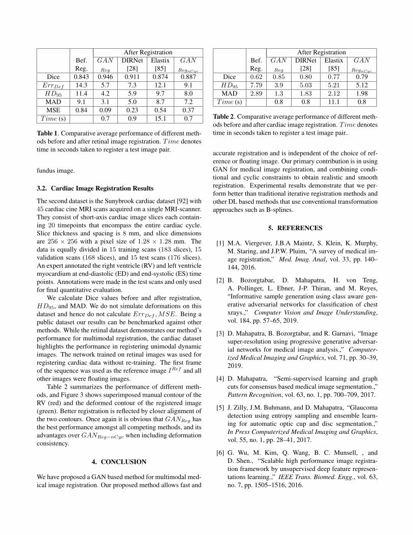
Dice 0.843 0.946 0.911 0.874 0.887ErrDef 14.3 5.7 7.3 12.1 9.1HD95 11.4 4.2 5.9 9.7 8.0MAD 9.1 3.1 5.0 8.7 7.2MSE 0.84 0.09 0.23 0.54 0.37
Time (s) 0.7 0.9 15.1 0.7
Table 1. Comparative average performance of different meth-ods before and after retinal image registration. Time denotestime in seconds taken to register a test image pair.
fundus image.
3.2. Cardiac Image Registration Results
The second dataset is the Sunybrook cardiac dataset [92] with45 cardiac cine MRI scans acquired on a single MRI-scanner.They consist of short-axis cardiac image slices each contain-ing 20 timepoints that encompass the entire cardiac cycle.Slice thickness and spacing is 8 mm, and slice dimensionsare 256 × 256 with a pixel size of 1.28 × 1.28 mm. Thedata is equally divided in 15 training scans (183 slices), 15validation scans (168 slices), and 15 test scans (176 slices).An expert annotated the right ventricle (RV) and left ventriclemyocardium at end-diastolic (ED) and end-systolic (ES) timepoints. Annotations were made in the test scans and only usedfor final quantitative evaluation.
We calculate Dice values before and after registration,HD95, and MAD. We do not simulate deformations on thisdataset and hence do not calculate ErrDef ,MSE. Being apublic dataset our results can be benchmarked against othermethods. While the retinal dataset demonstrates our method’sperformance for multimodal registration, the cardiac datasethighlights the performance in registering unimodal dynamicimages. The network trained on retinal images was used forregistering cardiac data without re-training. The first frameof the sequence was used as the reference image IRef and allother images were floating images.
Table 2 summarizes the performance of different meth-ods, and Figure 3 shows superimposed manual contour of theRV (red) and the deformed contour of the registered image(green). Better registration is reflected by closer alignment ofthe two contours. Once again it is obvious that GANReg hasthe best performance amongst all competing methods, and itsadvantages over GANReg−nCyc when including deformationconsistency.
4. CONCLUSION
We have proposed a GAN based method for multimodal med-ical image registration. Our proposed method allows fast and
After RegistrationBef. GAN DIRNet Elastix GANReg. Reg [28] [85] RegnCyc
Dice 0.62 0.85 0.80 0.77 0.79HD95 7.79 3.9 5.03 5.21 5.12MAD 2.89 1.3 1.83 2.12 1.98
Time (s) 0.8 0.8 11.1 0.8
Table 2. Comparative average performance of different meth-ods before and after cardiac image registration. Time denotestime in seconds taken to register a test image pair..
accurate registration and is independent of the choice of ref-erence or floating image. Our primary contribution is in usingGAN for medical image registration, and combining condi-tional and cyclic constraints to obtain realistic and smoothregistration. Experimental results demonstrate that we per-form better than traditional iterative registration methods andother DL based methods that use conventional transformationapproaches such as B-splines.
5. REFERENCES
[1] M.A. Viergever, J.B.A Maintz, S. Klein, K. Murphy,M. Staring, and J.P.W. Pluim, “A survey of medical im-age registration,” Med. Imag. Anal, vol. 33, pp. 140–144, 2016.
[2] B. Bozorgtabar, D. Mahapatra, H. von Teng,A. Pollinger, L. Ebner, J-P. Thiran, and M. Reyes,“Informative sample generation using class aware gen-erative adversarial networks for classification of chestxrays.,” Computer Vision and Image Understanding,vol. 184, pp. 57–65, 2019.
[3] D. Mahapatra, B. Bozorgtabar, and R. Garnavi, “Imagesuper-resolution using progressive generative adversar-ial networks for medical image analysis.,” Computer-ized Medical Imaging and Graphics, vol. 71, pp. 30–39,2019.
[4] D. Mahapatra, “Semi-supervised learning and graphcuts for consensus based medical image segmentation.,”Pattern Recognition, vol. 63, no. 1, pp. 700–709, 2017.
[5] J. Zilly, J.M. Buhmann, and D. Mahapatra, “Glaucomadetection using entropy sampling and ensemble learn-ing for automatic optic cup and disc segmentation.,”In Press Computerized Medical Imaging and Graphics,vol. 55, no. 1, pp. 28–41, 2017.
[6] G. Wu, M. Kim, Q. Wang, B. C. Munsell, , andD. Shen., “Scalable high performance image registra-tion framework by unsupervised deep feature represen-tations learning.,” IEEE Trans. Biomed. Engg., vol. 63,no. 7, pp. 1505–1516, 2016.
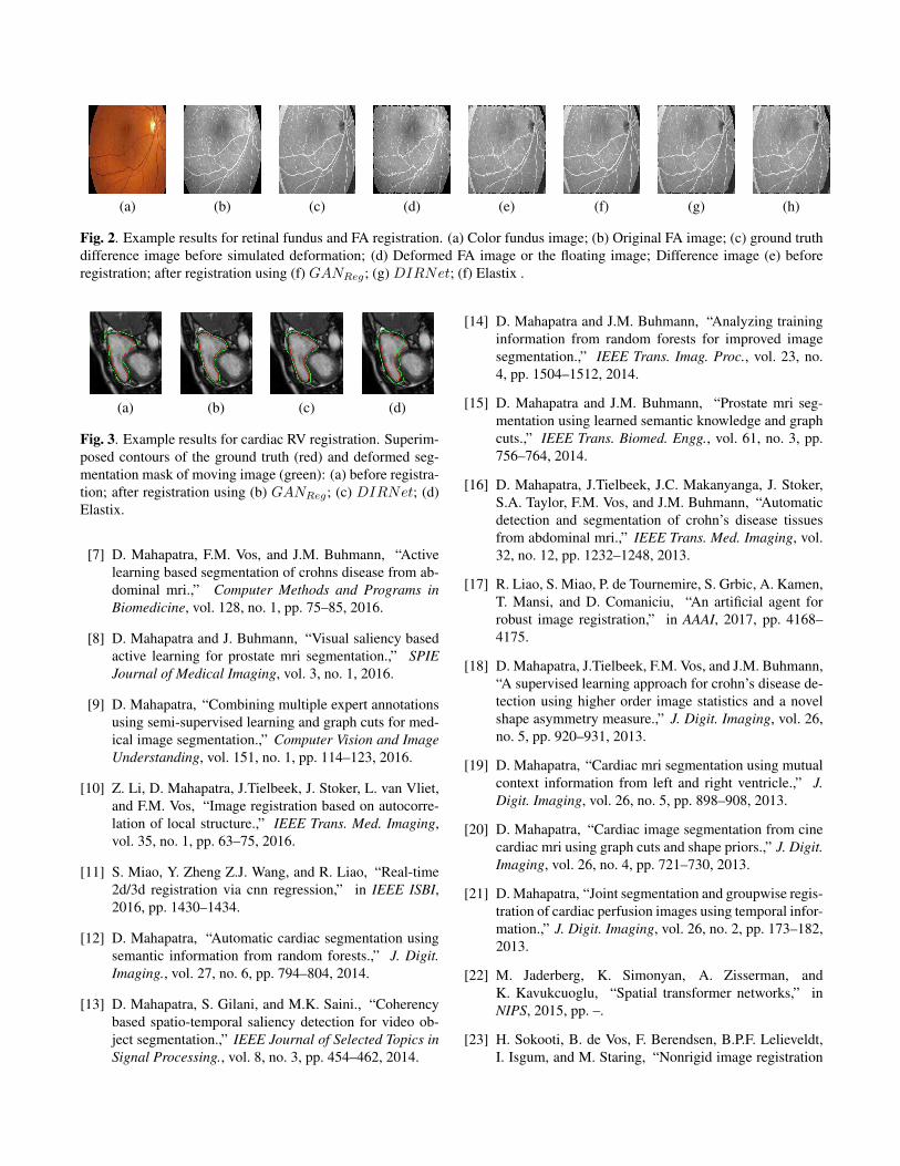
(a) (b) (c) (d) (e) (f) (g) (h)
Fig. 2. Example results for retinal fundus and FA registration. (a) Color fundus image; (b) Original FA image; (c) ground truthdifference image before simulated deformation; (d) Deformed FA image or the floating image; Difference image (e) beforeregistration; after registration using (f) GANReg; (g) DIRNet; (f) Elastix .
(a) (b) (c) (d)
Fig. 3. Example results for cardiac RV registration. Superim-posed contours of the ground truth (red) and deformed seg-mentation mask of moving image (green): (a) before registra-tion; after registration using (b) GANReg; (c) DIRNet; (d)Elastix.
[7] D. Mahapatra, F.M. Vos, and J.M. Buhmann, “Activelearning based segmentation of crohns disease from ab-dominal mri.,” Computer Methods and Programs inBiomedicine, vol. 128, no. 1, pp. 75–85, 2016.
[8] D. Mahapatra and J. Buhmann, “Visual saliency basedactive learning for prostate mri segmentation.,” SPIEJournal of Medical Imaging, vol. 3, no. 1, 2016.
[9] D. Mahapatra, “Combining multiple expert annotationsusing semi-supervised learning and graph cuts for med-ical image segmentation.,” Computer Vision and ImageUnderstanding, vol. 151, no. 1, pp. 114–123, 2016.
[10] Z. Li, D. Mahapatra, J.Tielbeek, J. Stoker, L. van Vliet,and F.M. Vos, “Image registration based on autocorre-lation of local structure.,” IEEE Trans. Med. Imaging,vol. 35, no. 1, pp. 63–75, 2016.
[11] S. Miao, Y. Zheng Z.J. Wang, and R. Liao, “Real-time2d/3d registration via cnn regression,” in IEEE ISBI,2016, pp. 1430–1434.
[12] D. Mahapatra, “Automatic cardiac segmentation usingsemantic information from random forests.,” J. Digit.Imaging., vol. 27, no. 6, pp. 794–804, 2014.
[13] D. Mahapatra, S. Gilani, and M.K. Saini., “Coherencybased spatio-temporal saliency detection for video ob-ject segmentation.,” IEEE Journal of Selected Topics inSignal Processing., vol. 8, no. 3, pp. 454–462, 2014.
[14] D. Mahapatra and J.M. Buhmann, “Analyzing traininginformation from random forests for improved imagesegmentation.,” IEEE Trans. Imag. Proc., vol. 23, no.4, pp. 1504–1512, 2014.
[15] D. Mahapatra and J.M. Buhmann, “Prostate mri seg-mentation using learned semantic knowledge and graphcuts.,” IEEE Trans. Biomed. Engg., vol. 61, no. 3, pp.756–764, 2014.
[16] D. Mahapatra, J.Tielbeek, J.C. Makanyanga, J. Stoker,S.A. Taylor, F.M. Vos, and J.M. Buhmann, “Automaticdetection and segmentation of crohn’s disease tissuesfrom abdominal mri.,” IEEE Trans. Med. Imaging, vol.32, no. 12, pp. 1232–1248, 2013.
[17] R. Liao, S. Miao, P. de Tournemire, S. Grbic, A. Kamen,T. Mansi, and D. Comaniciu, “An artificial agent forrobust image registration,” in AAAI, 2017, pp. 4168–4175.
[18] D. Mahapatra, J.Tielbeek, F.M. Vos, and J.M. Buhmann,“A supervised learning approach for crohn’s disease de-tection using higher order image statistics and a novelshape asymmetry measure.,” J. Digit. Imaging, vol. 26,no. 5, pp. 920–931, 2013.
[19] D. Mahapatra, “Cardiac mri segmentation using mutualcontext information from left and right ventricle.,” J.Digit. Imaging, vol. 26, no. 5, pp. 898–908, 2013.
[20] D. Mahapatra, “Cardiac image segmentation from cinecardiac mri using graph cuts and shape priors.,” J. Digit.Imaging, vol. 26, no. 4, pp. 721–730, 2013.
[21] D. Mahapatra, “Joint segmentation and groupwise regis-tration of cardiac perfusion images using temporal infor-mation.,” J. Digit. Imaging, vol. 26, no. 2, pp. 173–182,2013.
[22] M. Jaderberg, K. Simonyan, A. Zisserman, andK. Kavukcuoglu, “Spatial transformer networks,” inNIPS, 2015, pp. –.
[23] H. Sokooti, B. de Vos, F. Berendsen, B.P.F. Lelieveldt,I. Isgum, and M. Staring, “Nonrigid image registration
using multiscale 3d convolutional neural networks,” inMICCAI, 2017, pp. 232–239.
[24] D. Mahapatra, “Skull stripping of neonatal brain mri:Using prior shape information with graphcuts.,” J. Digit.Imaging, vol. 25, no. 6, pp. 802–814, 2012.
[25] D. Mahapatra and Y. Sun, “Integrating segmentationinformation for improved mrf-based elastic image reg-istration.,” IEEE Trans. Imag. Proc., vol. 21, no. 1, pp.170–183, 2012.
[26] D. Mahapatra and Y. Sun, “Mrf based intensity invari-ant elastic registration of cardiac perfusion images usingsaliency information,” IEEE Trans. Biomed. Engg., vol.58, no. 4, pp. 991–1000, 2011.
[27] D. Mahapatra and Y. Sun, “Rigid registration of re-nal perfusion images using a neurobiology based visualsaliency model,” EURASIP Journal on Image and VideoProcessing., pp. 1–16, 2010.
[28] B. de Vos, F. Berendsen, M.A. Viergever, M. Staring,and I. Isgum, “End-to-end unsupervised deformable im-age registration with a convolutional neural network,” inarXiv preprint arXiv:1704.06065, 2017.
[29] B. Bozorgtabar, M. Saeed Rad, D. Mahapatra, and J-P.Thiran, “Syndemo: Synergistic deep feature alignmentfor joint learning of depth and ego-motion,” in In Proc.IEEE ICCV, 2019.
[30] Y. Xing, Z. Ge, R. Zeng, D. Mahapatra, J. Seah, M. Law,and T. Drummond, “Adversarial pulmonary pathologytranslation for pairwise chest x-ray data augmentation,”in In Proc. MICCAI, 2019.
[31] D. Mahapatra and Z. Ge, “Training data independentimage registration with gans using transfer learning andsegmentation information,” in In Proc. IEEE ISBI, 2019,pp. 709–713.
[32] D. Mahapatra, S. Bozorgtabar, J.-P. Thiran, andM. Reyes, “Efficient active learning for image classi-fication and segmentation using a sample selection andconditional generative adversarial network,” in In Proc.MICCAI (2), 2018, pp. 580–588.
[33] D. Mahapatra, Z. Ge, S. Sedai, and R. Chakravorty.,“Joint registration and segmentation of xray imagesusing generative adversarial networks,” in In Proc.MICCAI-MLMI, 2018, pp. 73–80.
[34] C. Ledig and et. al., “Photo-realistic single im-age super-resolution using a generative adversarial net-work,” CoRR, vol. abs/1609.04802, 2016.
[35] D Mahapatra, B Bozorgtabar, S Hewavitharanage, andR Garnavi, “Image super resolution using generativeadversarial networks and local saliency maps for retinalimage analysis,” in MICCAI, 2017, pp. 382–390.
[36] S. Sedai, D. Mahapatra, B. Antony, and R. Garnavi,“Joint segmentation and uncertainty visualization ofretinal layers in optical coherence tomography imagesusing bayesian deep learning,” in In Proc. MICCAI-OMIA, 2018, pp. 219–227.
[37] S. Sedai, D. Mahapatra, Z. Ge, R. Chakravorty, andR. Garnavi, “Deep multiscale convolutional featurelearning for weakly supervised localization of chestpathologies in x-ray images,” in In Proc. MICCAI-MLMI, 2018, pp. 267–275.
[38] D. Mahapatra, B. Antony, S. Sedai, and R. Garnavi,“Deformable medical image registration using genera-tive adversarial networks,” in In Proc. IEEE ISBI, 2018,pp. 1449–1453.
[39] S. Sedai, D. Mahapatra, S. Hewavitharanage,S. Maetschke, and R. Garnavi, “Semi-supervisedsegmentation of optic cup in retinal fundus imagesusing variational autoencoder,,” in In Proc. MICCAI,2017, pp. 75–82.
[40] D. Mahapatra, S. Bozorgtabar, S. Hewavitahranage, andR. Garnavi, “Image super resolution using generativeadversarial networks and local saliencymaps for retinalimage analysis,,” in In Proc. MICCAI, 2017, pp. 382–390.
[41] P. Isola, J.Y. Zhu, T. Zhou, and A.A. Efros, “Image-to-image translation with conditional adversarial net-works,” in CVPR, 2017.
[42] P. Roy, R. Tennakoon, K. Cao, S. Sedai, D. Mahapa-tra, S. Maetschke, and R. Garnavi, “A novel hybrid ap-proach for severity assessment of diabetic retinopathy incolour fundus images,,” in In Proc. IEEE ISBI, 2017, pp.1078–1082.
[43] P. Roy, R. Chakravorty, S. Sedai, D. Mahapatra, andR. Garnavi, “Automatic eye type detection in retinal fun-dus image using fusion of transfer learning and anatom-ical features,” in In Proc. DICTA, 2016, pp. 1–7.
[44] R. Tennakoon, D. Mahapatra, P. Roy, S. Sedai, andR. Garnavi, “Image quality classification for dr screen-ing using convolutional neural networks,” in In Proc.MICCAI-OMIA, 2016, pp. 113–120.
[45] S. Sedai, P.K. Roy, D. Mahapatra, and R. Garnavi, “Seg-mentation of optic disc and optic cup in retinal imagesusing coupled shape regression,” in In Proc. MICCAI-OMIA, 2016, pp. 1–8.
[46] D. Mahapatra, P.K. Roy, S. Sedai, and R. Garnavi,“Retinal image quality classification using saliencymaps and cnns,” in In Proc. MICCAI-MLMI, 2016, pp.172–179.
[47] J.Y. Zhu, T.park, P. Isola, and A.A. Efros, “Un-paired image-to-image translation using cycle-consistent adversarial networks,” in arXiv preprintarXiv:1703.10593, 2017.
[48] S. Sedai, P.K. Roy, D. Mahapatra, and R. Garnavi, “Seg-mentation of optic disc and optic cup in retinal fundusimages using shape regression,” in In Proc. EMBC,2016, pp. 3260–3264.
[49] D. Mahapatra, P.K. Roy, S. Sedai, and R. Garnavi, “Acnn based neurobiology inspired approach for retinalimage quality assessment,” in In Proc. EMBC, 2016,pp. 1304–1307.
[50] J. Zilly, J. Buhmann, and D. Mahapatra, “Boosting con-volutional filters with entropy sampling for optic cupand disc image segmentation from fundus images,” inIn Proc. MLMI, 2015, pp. 136–143.
[51] D. Mahapatra and J. Buhmann, “Visual saliency basedactive learning for prostate mri segmentation,” in InProc. MLMI, 2015, pp. 9–16.
[52] D. Mahapatra and J. Buhmann, “Obtaining consensusannotations for retinal image segmentation using ran-dom forest and graph cuts,” in In Proc. OMIA, 2015,pp. 41–48.
[53] D. Rueckert, L.I Sonoda, C. Hayes, D.L.G Hill, M.OLeach, and D.J Hawkes., “Nonrigid registration usingfree-form deformations: application to breast mr im-ages.,” IEEE Trans. Med. Imag.., vol. 18, no. 8, pp.712–721, 1999.
[54] D. Mahapatra and J.M. Buhmann, “A field of expertsmodel for optic cup and disc segmentation from retinalfundus images,” in In Proc. IEEE ISBI, 2015, pp. 218–221.
[55] D. Mahapatra, Z. Li, F.M. Vos, and J.M. Buhmann,“Joint segmentation and groupwise registration of car-diac dce mri using sparse data representations,” in InProc. IEEE ISBI, 2015, pp. 1312–1315.
[56] D. Mahapatra, F.M. Vos, and J.M. Buhmann, “Crohn’sdisease segmentation from mri using learned image pri-ors,” in In Proc. IEEE ISBI, 2015, pp. 625–628.
[57] H. Kuang, B. Guthier, M. Saini, D. Mahapatra, and A. ElSaddik, “A real-time smart assistant for video surveil-lance through handheld devices.,” in In Proc: ACM Intl.Conf. Multimedia, 2014, pp. 917–920.
[58] D. Mahapatra, J.Tielbeek, J.C. Makanyanga, J. Stoker,S.A. Taylor, F.M. Vos, and J.M. Buhmann, “Combin-ing multiple expert annotations using semi-supervisedlearning and graph cuts for crohns disease segmenta-tion,” in In Proc: MICCAI-ABD, 2014.
[59] Z. Wang and et. al., “Image quality assessment: from er-ror visibility to structural similarity.,” IEEE Trans. Imag.Proc., vol. 13, no. 4, pp. 600–612, 2004.
[60] P. Schuffler, D. Mahapatra, J. Tielbeek, F.M. Vos,J. Makanyanga, D.A. Pends, C.Y. Nio, J. Stoker, S.A.Taylor, and J.M. Buhmann, “Semi automatic crohns dis-ease severity assessment on mr imaging,” in In Proc:MICCAI-ABD, 2014.
[61] D. Mahapatra, J.Tielbeek, J.C. Makanyanga, J. Stoker,S.A. Taylor, F.M. Vos, and J.M. Buhmann, “Activelearning based segmentation of crohn’s disease usingprinciples of visual saliency,” in Proc. IEEE ISBI, 2014,pp. 226–229.
[62] D. Mahapatra, P. Schuffler, J. Tielbeek, F.M. Vos, andJ.M. Buhmann, “Semi-supervised and active learningfor automatic segmentation of crohn’s disease,” in Proc.MICCAI, Part 2, 2013, pp. 214–221.
[63] P. Schuffler, D. Mahapatra, J. Tielbeek, F.M. Vos,J. Makanyanga, D.A. Pends, C.Y. Nio, J. Stoker, S.A.Taylor, and J.M. Buhmann, “A model developmentpipeline for crohns disease severity assessment frommagnetic resonance images,” in In Proc: MICCAI-ABD,2013.
[64] D. Mahapatra, “Graph cut based automatic prostate seg-mentation using learned semantic information,” in Proc.IEEE ISBI, 2013, pp. 1304–1307.
[65] K. Simonyan and A. Zisserman., “Very deep convo-lutional networks for large-scale image recognition,”CoRR, vol. abs/1409.1556, 2014.
[66] D. Mahapatra and J.M. Buhmann, “Automatic cardiacrv segmentation using semantic information with graphcuts,” in Proc. IEEE ISBI, 2013, pp. 1094–1097.
[67] D. Mahapatra, J. Tielbeek, F.M. Vos, and J.M. Buh-mann, “Weakly supervised semantic segmentation ofcrohn’s disease tissues from abdominal mri,” in Proc.IEEE ISBI, 2013, pp. 832–835.
[68] D. Mahapatra, J. Tielbeek, F.M. Vos, and J.M. Buhmann., “Crohn’s disease tissue segmentation from abdominalmri using semantic information and graph cuts,” in Proc.IEEE ISBI, 2013, pp. 358–361.
[69] D. Mahapatra, J. Tielbeek, F.M. Vos, and J.M. Buh-mann, “Localizing and segmenting crohn’s disease af-fected regions in abdominal mri using novel context fea-tures,” in Proc. SPIE Medical Imaging, 2013.
[70] D. Mahapatra, J. Tielbeek, J.M. Buhmann, and F.M.Vos, “A supervised learning based approach to detectcrohn’s disease in abdominal mr volumes,” in Proc.MICCAI workshop Computational and Clinical Appli-cations in Abdominal Imaging(MICCAI-ABD), 2012,pp. 97–106.
[71] D.P. Kingma and J. Ba, “Adam: A method for stochas-tic optimization,” in arXiv preprint arXiv:1412.6980,,2014.
[72] D. Mahapatra, “Cardiac lv and rv segmentation usingmutual context information,” in Proc. MICCAI-MLMI,2012, pp. 201–209.
[73] D. Mahapatra, “Landmark detection in cardiac mri us-ing learned local image statistics,” in Proc. MICCAI-Statistical Atlases and Computational Models of theHeart. Imaging and Modelling Challenges (STACOM),2012, pp. 115–124.
[74] F. M. Vos, J. Tielbeek, R. Naziroglu, Z. Li, P. Schuffler,D. Mahapatra, Alexander Wiebel, C. Lavini, J. Buh-mann, H. Hege, J. Stoker, and L. van Vliet, “Compu-tational modeling for assessment of IBD: to be or not tobe?,” in Proc. IEEE EMBC, 2012, pp. 3974–3977.
[75] D. Mahapatra, “Groupwise registration of dynamic car-diac perfusion images using temporal information andsegmentation information,” in In Proc: SPIE MedicalImaging, 2012.
[76] D. Mahapatra, “Neonatal brain mri skull stripping us-ing graph cuts and shape priors,” in In Proc: MICCAIworkshop on Image Analysis of Human Brain Develop-ment (IAHBD), 2011.
[77] S.A.M Hajeb, H. Rabbani, and M.R. Akhlaghi., “Dia-betic retinopathy grading by digital curvelet transform.,”Comput Math Methods Med., pp. 7619–01, 2012.
[78] A.F. Frangi, W.J. Niessen, K.L. Vincken, and M.A.Viergever, “Multiscale vessel enhancement filtering,” inMICCAI, 1998, pp. 130–137.
[79] D. Mahapatra and Y. Sun, “Orientation histograms asshape priors for left ventricle segmentation using graphcuts,” in In Proc: MICCAI, 2011, pp. 420–427.
[80] D. Mahapatra and Y. Sun, “Joint registration and seg-mentation of dynamic cardiac perfusion images usingmrfs.,” in Proc. MICCAI, 2010, pp. 493–501.
[81] D. Mahapatra and Y. Sun., “An mrf framework for jointregistration and segmentation of natural and perfusionimages,” in Proc. IEEE ICIP, 2010, pp. 1709–1712.
[82] D. Mahapatra and Y. Sun, “Retrieval of perfusion im-ages using cosegmentation and shape context informa-tion,” in Proc. APSIPA Annual Summit and Conference(ASC), 2010.
[83] D. Mahapatra and Y. Sun, “A saliency based mrf methodfor the joint registration and segmentation of dynamicrenal mr images,” in Proc. ICDIP, 2010.
[84] D. Mahapatra and Y. Sun, “Nonrigid registration of dy-namic renal MR images using a saliency based MRFmodel,” in Proc. MICCAI, 2008, pp. 771–779.
[85] S. Klein, M. Staring, K. Murphy, M.A. Viergever, andJ.P.W. Pluim., “Elastix: a toolbox for intensity basedmedical image registration.,” IEEE Trans. Med. Imag..,vol. 29, no. 1, pp. 196–205, 2010.
[86] D. Mahapatra and Y. Sun, “Registration of dynamic re-nal mr images using neurobiological model of saliency,”in Proc. ISBI, 2008, pp. 1119–1122.
[87] D. Mahapatra, M.K. Saini, and Y. Sun, “Illumina-tion invariant tracking in office environments usingneurobiology-saliency based particle filter,” in IEEEICME, 2008, pp. 953–956.
[88] D. Mahapatra, S. Roy, and Y. Sun, “Retrieval of mrkidney images by incorporating spatial information inhistogram of low level features,” in In 13th InternationalConference on Biomedical Engineering, 2008.
[89] D. Mahapatra and Y. Sun, “Using saliency features forgraphcut segmentation of perfusion kidney images,” inIn 13th International Conference on Biomedical Engi-neering, 2008.
[90] D. Mahapatra, S. Winkler, and S.C. Yen, “Mo-tion saliency outweighs other low-level features whilewatching videos,” in SPIE HVEI., 2008, pp. 1–10.
[91] D. Mahapatra, A. Routray, and C. Mishra, “An activesnake model for classification of extreme emotions,” inIEEE International Conference on Industrial Technol-ogy (ICIT), 2006, pp. 2195–2199.
[92] P. Radau, Y. Lu, K. Connelly, G. Paul, and et. al ., “val-uation framework for algorithms segmenting short axiscardiac MRI,” in The MIDAS Journal-Cardiac MR LeftVentricle Segmentation Challenge, 2009.