Embed Size (px)
Citation preview

Elective course of Anatomy
(# 2)
Applied Anatomy
Dr. Karim M. Khalil
Lecturer of Veterinary Anatomy and EmbryologyCairo University
/ Karim Khalil
1
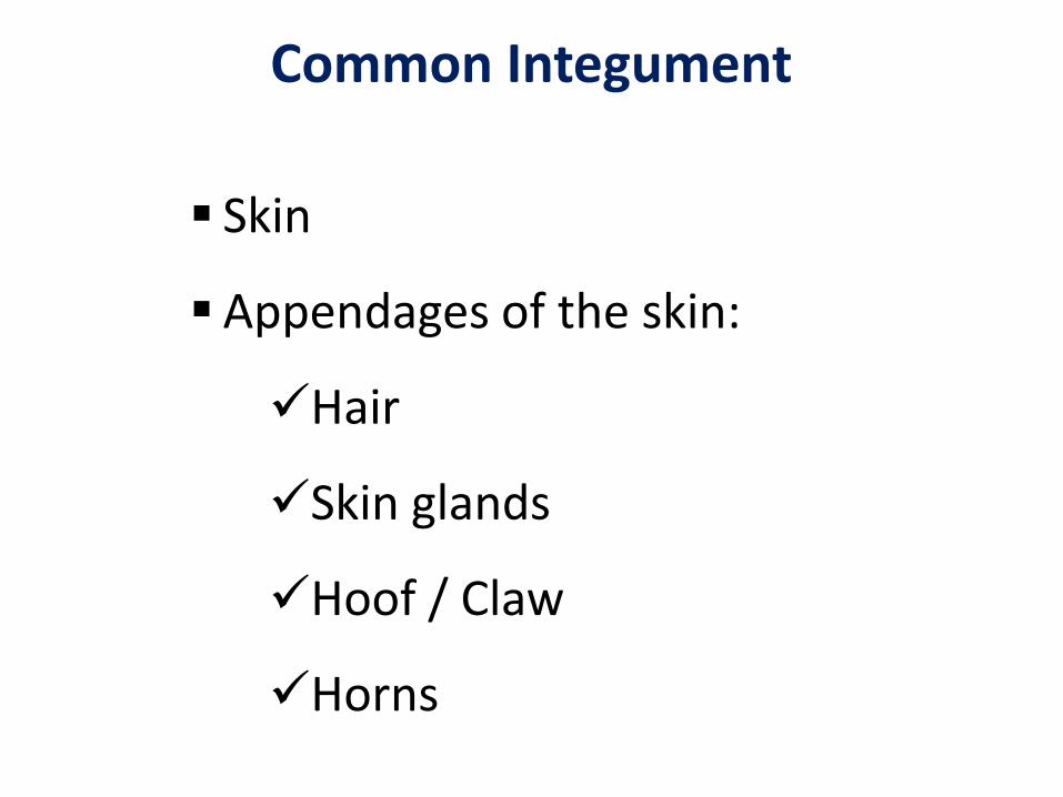
Common Integument
Skin
Appendages of the skin:
Hair
Skin glands
Hoof / Claw
Horns

SkinFunctions:
• Covering the body and protect underlying tissue.
• Regulation of body temperature.
• Excretes water and salts through sweat gland.
• Senses the environment.
• Synthesizes of Vit D3.
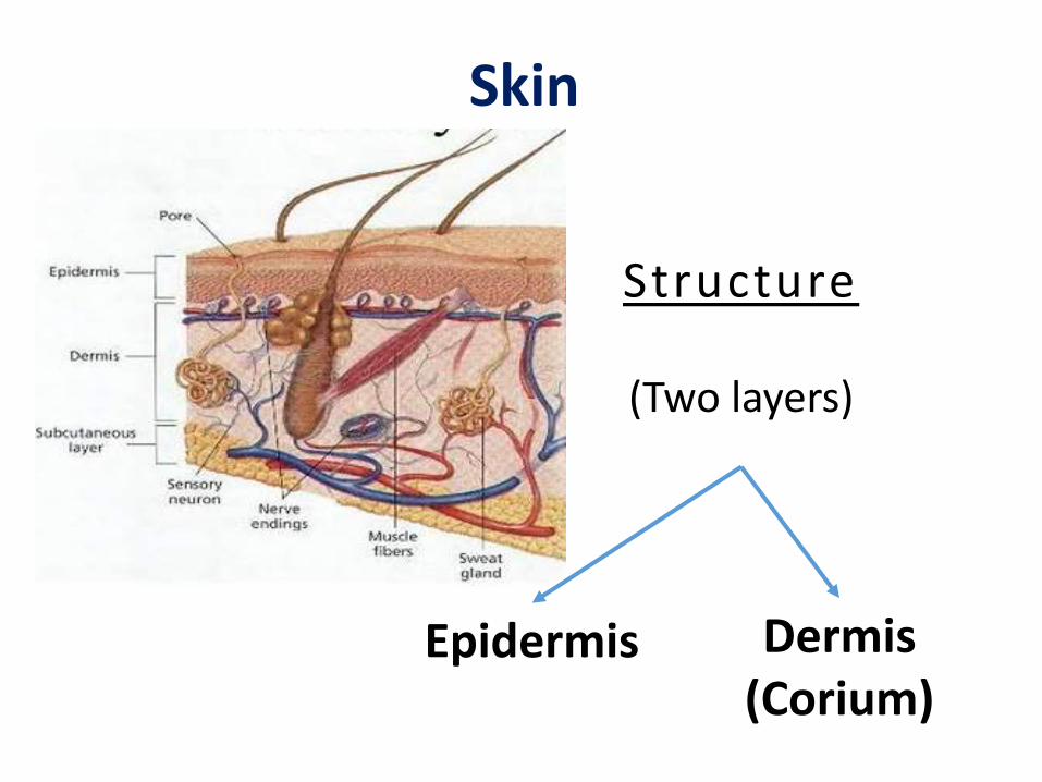
Structure
(Two layers)
Epidermis Dermis(Corium)
Skin
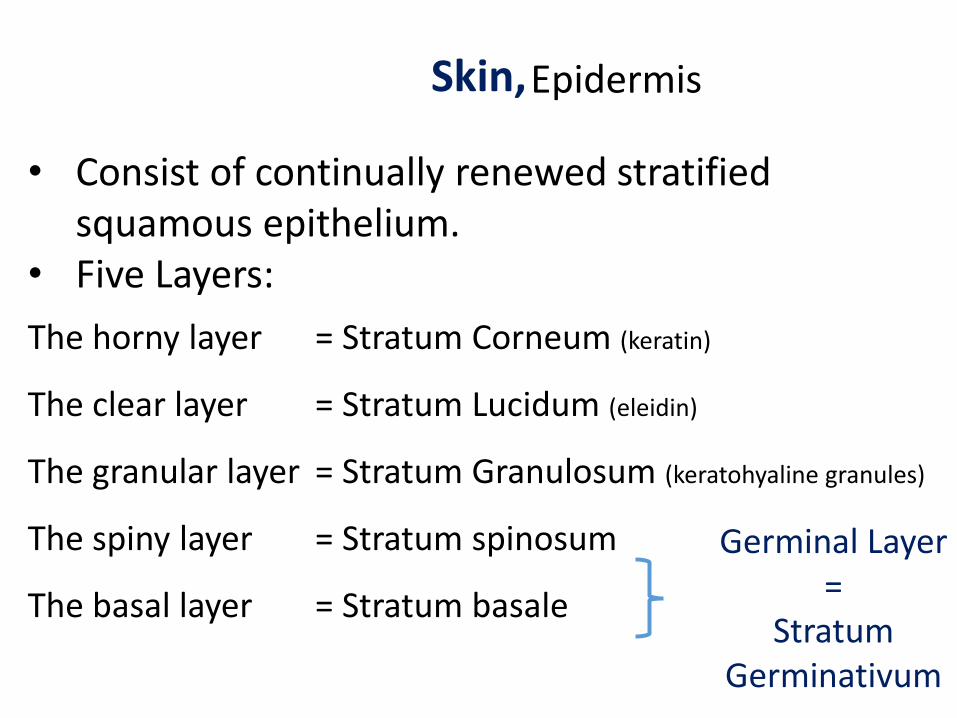
Skin,Epidermis
• Consist of continually renewed stratified squamous epithelium.
• Five Layers:
The horny layer = Stratum Corneum (keratin)
The clear layer = Stratum Lucidum (eleidin)
The granular layer = Stratum Granulosum (keratohyaline granules)
The spiny layer = Stratum spinosum
The basal layer = Stratum basale
Germinal Layer=
Stratum Germinativum


Skin, Dermis

Horny layer = Stratum Corneum
Clear layer = Stratum Lucideum
Granular layer = Stratum granulosum
Spiny layer = Stratum spinosum
basal layer = Stratum basale
Basement memberane
Papillary layer = Dermal papillae
Reticular layer = Reticulum
Subcutaneous = Superficial fascia = Hypodermis
Epid
erm
is
Dermis
Layers of the Skin

Common Integument
Skin
Appendages of the skin:
Hair
Skin glands
Hoof / Claw
Horns
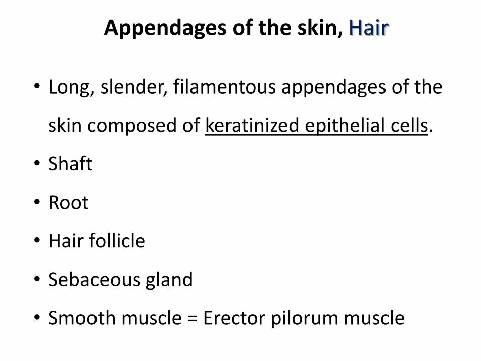
Appendages of the skin, Hair
• Long, slender, filamentous appendages of the
skin composed of keratinized epithelial cells.
• Shaft
• Root
• Hair follicle
• Sebaceous gland
• Smooth muscle = Erector pilorum muscle
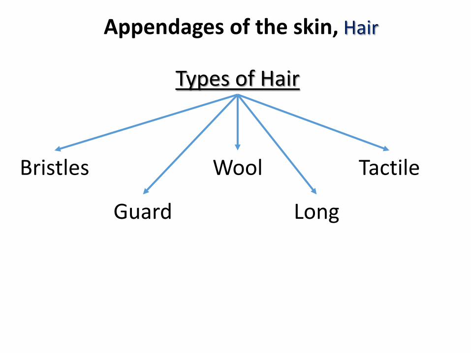
Appendages of the skin, Hair
Types of Hair
Bristles
Guard
Wool
Long
Tactile

Appendages of the skin, Hair
Species Hair follicles Hair Type
Horse and cattle
Single Guard and Long
DogCompound with smaller secondary hairs
Guard
CatSingle (guard) surrounded by compound follicles
Guard
Sheep Wool
PigSingle follicles grouped in clusters
Bristles

Appendages of the skin, Hair
Color of Hair
Black Brown Yellow
No pigments
AlbinoColored
Pigments
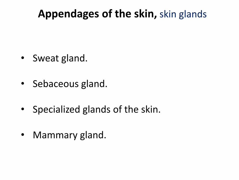
Appendages of the skin, skin glands
• Sweat gland.
• Sebaceous gland.
• Specialized glands of the skin.
• Mammary gland.

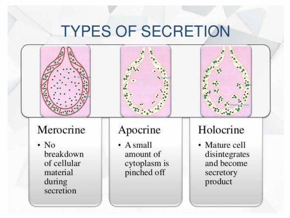
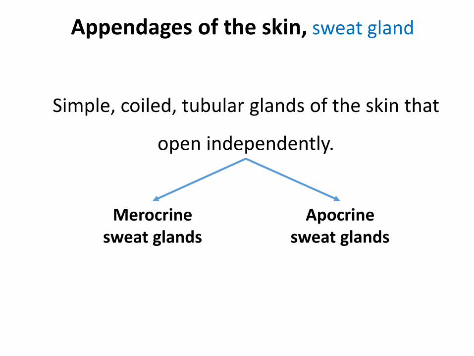
Appendages of the skin, sweat gland
Simple, coiled, tubular glands of the skin that
open independently.
Merocrinesweat glands
Apocrine sweat glands
Appendages of the skin, sweat gland
Simple, coiled, tubular glands of the skin that
open independently.
Merocrinesweat glands
Apocrine sweat glands

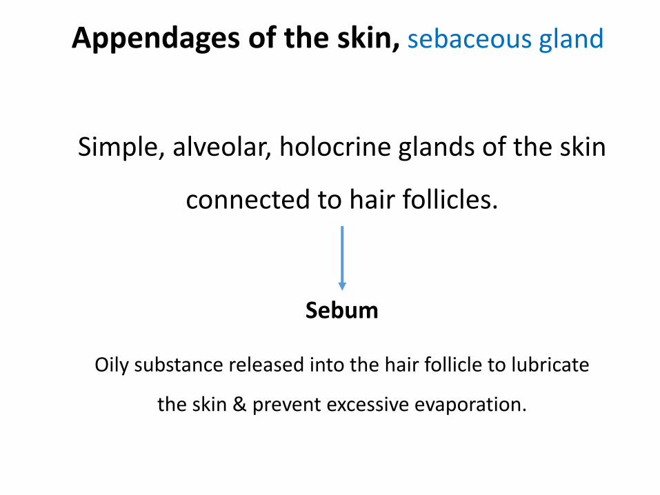
Appendages of the skin, sebaceous gland
Simple, alveolar, holocrine glands of the skin
connected to hair follicles.
Sebum
Oily substance released into the hair follicle to lubricate
the skin & prevent excessive evaporation.

Appendages of the skin, specialized glandSpecies Gland Site
Sheep
Infraorbital sinus gland Infraorbital sinus
Inguinal sinus gland On either side of the udder / scrotum
Interdigital sinus gland Between digits
Nasal skin gland Skin of nose
Sheep and goat Horn gland Base of horn
Goat Sub caudal gland Below tail (buck`s smell)
Dog
Circumanal gland Around the anus
Glands of the Anal sac Wall of anal sac
Coccygeal gland Dorsum of root of tail
Cat Circumoral (peri oral) gland Around the mouth
Pig Carpal glands (sexual pheromones) Caudo-medial, Proximal to carpus
Mental gland Chin
All domestic animals
Ciruminous gland External auditory canal (ear wax)
Preputeal gland Prepuce (smegma)
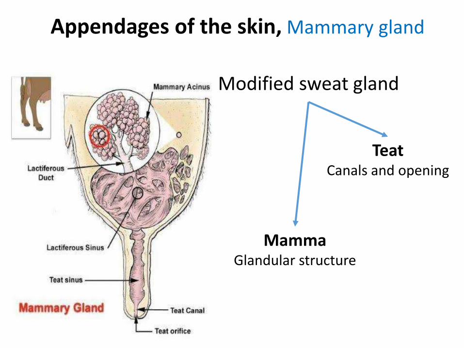
Appendages of the skin, Mammary gland
Modified sweat gland
MammaGlandular structure
TeatCanals and opening
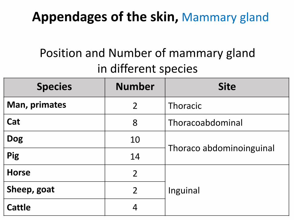
Appendages of the skin, Mammary gland
Species Number Site
Man, primates 2 Thoracic
Cat 8 Thoracoabdominal
Dog 10Thoraco abdominoinguinal
Pig 14
Horse 2
Inguinal Sheep, goat 2
Cattle 4
Position and Number of mammary gland in different species
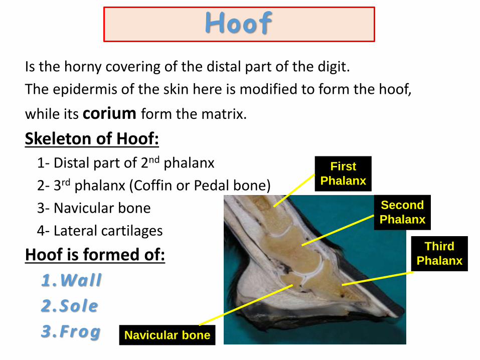
Is the horny covering of the distal part of the digit.
The epidermis of the skin here is modified to form the hoof,
while its corium form the matrix.
Skeleton of Hoof:
1- Distal part of 2nd phalanx
2- 3rd phalanx (Coffin or Pedal bone)
3- Navicular bone
4- Lateral cartilages
Hoof is formed of:
1.Wal l
2 .So le
3.Frog
First
Phalanx
Second
Phalanx
Third
Phalanx
Navicular bone
Hoof
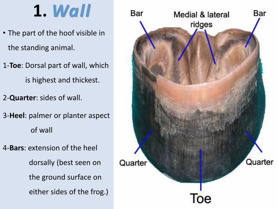
1. Wall• The part of the hoof visible in
the standing animal.
1-Toe: Dorsal part of wall, which
is highest and thickest.
2-Quarter: sides of wall.
3-Heel: palmer or planter aspect
of wall
4-Bars: extension of the heel
dorsally (best seen on
the ground surface on
either sides of the frog.)

2- Sole• Crescent in shape, form the greater part of the ground surface of hoof.
• Its convex border separated from the wall by a horny material of light color called the white line. While the part between The wall & bar called angle of the sole.
• White line (zona alba): the junction between the wall and sole on the ground surface of foot. It indication of the sensitive internal structures.
Sole
Palmar View
Angle of
Sole
White Line

3- frog (cuneus) • It is called the heart of the horse's foot as it pushes blood
from foot toward body.
• It is wedge shape and occupies the angle between the bars of wall and concave border of the sole.
The frog consists of:a) Apex:b) 2 crura (ridges): c) base:
form two caudal
prominences
called Bulbs of the heel.

The corium (matrix) Underlying Dermis = Corium = Matrix
• Highly vascular tissue, which nourishes the hoof capsule.
• Nerves are located in corium, so it is the sensitive part of the foot.
It is divided into:
1) Periople (Marginal)dermis
which produces thin, shiny external layer of the wall.
2) Coronary dermis raised band distal to the perioplic dermis.
3) Laminar (Lamellar) dermis
About 600 dermal laminae (sensitive, non pigmented) whichnourishes and interdigitate with the horny, non sensitiveepidermal laminae of the hoof capsule, Deeply it is connected tothe periostium of third phalanx.
4) Solar Dermis which nourishes the horny sole.
5) Dermis of the frogwhich nourishes the horny frog, Deeply it blends with digital cushion.

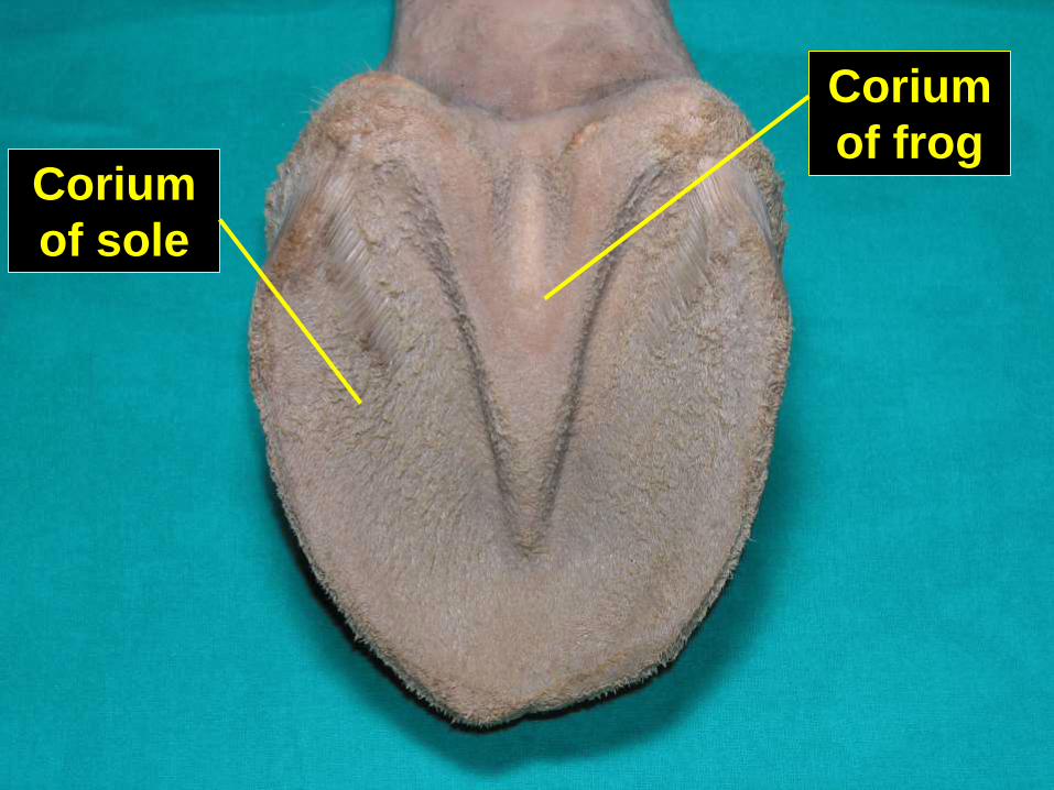
Corium
of frogCorium
of sole

Thanks