Embed Size (px)
Citation preview
SAGE-Hindawi Access to ResearchInternational Journal of ElectrochemistryVolume 2011, Article ID 863196, 17 pagesdoi:10.4061/2011/863196
Review Article
Electrochemical Aptamer-Based Biosensors:Recent Advances and Perspectives
Abd-Elgawad Radi
Department of Chemistry, Faculty of Science, Mansoura University, Dumyat 34517, Egypt
Correspondence should be addressed to Abd-Elgawad Radi, [email protected]
Received 17 February 2011; Revised 12 May 2011; Accepted 16 May 2011
Academic Editor: Farnoush Faridbod
Copyright © 2011 Abd-Elgawad Radi. This is an open access article distributed under the Creative Commons Attribution License,which permits unrestricted use, distribution, and reproduction in any medium, provided the original work is properly cited.
This paper reviews the advancements of a wide range of electrochemical aptamer-based biosensors, electrochemical aptasensors,for target analytes monitoring. Methods for immobilizing aptamers onto an electrode surface are discussed. Aptasensors arepresented according to their detection strategies. Many of these are simply electrochemical, aptamer-based equivalents oftraditional immunochemical approaches, sandwich and competition assays employing electroactive signaling moieties. Others,exploiting the unusual physical properties of aptamers, are signal-on (positive readout signal) and signal-off (negative readoutsignal) aptasensors based on target binding-induced conformational change of aptamers. Aptamer label-free devices are alsodiscussed.
1. Introduction
Biosensors are devices detecting the presence of a target byusing a particular recognition element and then monitoringthe mass, optical, electronic, or magnetic signal changes,which are induced by the interaction of the recognitionelement and the analyte of interest. Molecular recognition isconsequently the key for the sensor performance. The rec-ognition elements were initially isolated naturally from liv-ing systems; now, they are available by synthesis in the lab,including receptors, enzymes, antibodies, nucleic acids, mo-lecular imprints and lectins. The mostly used recognitioncomponents for clinic diagnostics and genomics/proteomicsstudies are antibodies and nucleic acids based on affinityassays owing to their special high sensitivity and selectivityin the affinity to target molecules. Antibodies are producedby immune system when it responds to antigens (i.e., toxins,chemicals, drugs, and virus particles, spores, bacterial toxins,and other foreign substrates). Antibodies were generatedby animal immunization, and now, cell clone technologycan produce poly/monoclonal antibodies in large quantities.The antibodies still encounter the challenges of pH andtemperature sensitivity, short shelf life, easily degradation,and consequently, its repeatable usage is also a problem.
Aptamers, first reported in 1990, are attracting interestin the areas of therapeutics and diagnostics [1–3]. Aptamersare specific oligonucleic acid sequences (ca. 30 to 100 nu-cleotides), which recognize specific ligands and bind to var-ious target molecules ranging from small ions to large pro-teins with high affinity and specificity. The term aptamerderives from aptus that means to fit. The RNA or DNAaptamers molecules are selected in vitro (selection evolutionof ligands by exponential enrichment, SELEX process) fromvast populations of random sequences. Aptamers are oftencalled synthetic antibodies and can mimic antibodies in anumber of applications. The selected aptamers bind theirtargets with affinities and specificities that can be comparableto those of antibodies. Aptamers present some advantagescompared to antibodies, especially accurate and reproduciblechemical production. Moreover, Aptamer offering chemicalstability under a wide range of buffer conditions, resistantto harsh treatments without losing its bioactivity, and thethermal denaturation is reversible for aptamer. While it isimportant to remain proteins in a moisturized environmentto maintain their bioactivity, and the physical or chemicaldenaturation is irreversible for antibodies. Aptamer has awide range of molecular and therapeutic targets, includingamino acids, any class of proteins (enzymes, membrane
2 International Journal of Electrochemistry
proteins, viral proteins, cytokines and growth factors, andimmunoglobulins), drugs, metal ions, other small bio-/or-ganic/inorganic small molecules, and even whole cells. How-ever, antibody is only employed for immunogenic com-pounds. Aptamers are small in size, cost effective, offeringremarkable flexibility and convenience in designing theirspecial structure. Moreover, combinatorial chemical synthe-sis offers a wide variety of methods for aptamer sequencemodifications such as the terminal tagging chemical groups.
Biosensors based on aptamers as biorecognition elementshave been coined aptasensors. The aptamers were initiallyused as therapeutic agents. For example, aptamer that selec-tively binds thrombin, a multifunctional serine protease thatplays an important role in procoagulant and anticoagulantfunctions, was developed with the purpose of applicationas an anticoagulant [4]. Only recently, the aptamers havebeen used as recognition elements in biosensing. The firstaptasensor was reported in 1996, with an optical biosensorbased on fluorescently labeled aptamers [5]. To date, thebest investigated aptamers are those for thrombin. The firstelectrochemical aptasensor with an amperometric sandwich-based biosensor based on glucose dehydrogenase-labeledsignaling aptamers was described in 2004 [6]. The field ofresearch is progressing so rapidly that new achievementshave appeared, especially those focused on electrochemicalmethods of detection. Electrochemical devices have receivedconsiderable recent attention in connection to the transduc-tion of aptamer interactions. Electrochemical transductionpresents considerable advantages over optical, piezoelectricor thermal detection. The electrochemical detection offerhigh sensitivity and selectivity, compatibility with novel mi-crofabrication technologies, inherent miniaturization, lowcost, disposability, minimal simple-to-operate, robust, powerrequirements, and independence of sample turbidity. Thispaper examines electrochemical aptasensor discussing sur-face immobilization techniques and different detectionschemes used to detect target analytes.
2. Immobilization of Aptamers
The crucial step in electrochemical aptasensors developmentis the immobilization of aptamers to an electrode surface,and it is important to develop strategies for reliable immo-bilization of aptamers so that they retain their biophysicalcharacteristics and binding abilities, as well as for mini-mizing nonspecific binding/adsorption events. In principlethese strategies are similar to those applied previously forthe immobilization of single- or double-stranded DNA ingenosensors or DNA biosensors for detection of DNA dam-age [7].
The methods of immobilization based on physical ad-sorption of DNA by means of electrostatic interactions are ingeneral not suitable due to low stability caused by aptamersdesorption from the surface. The common pathways forimmobilizing a stable, flexible and repeatable aptamer layersurface are chemical covalent attachment, via avidin-to-bi-otin conjugation [8], and self-assembling the thiolated ap-tamer onto gold substrate using a thiol-alkane linked to the
aptamer sequence [7]. streptavidin-polymer-coated indium-tin oxide electrode for immobilizing a DNA aptamer againstlysozyme have been also designed.
Mixed two-component alkanethiol self assembly mono-layers of recognition and shielding components, similar tothose used in DNA hybridization sensors, are extremelyattractive for achieving the desired balance between highloading, minimal nonspecific interactions, and preferred/accessible orientation. Such mixed coassembly monolayershave been widely used in DNA hybridization sensors and arebeing employed for the design of electrochemical aptasensors[9, 10]. Aptamers can be attached to the solid support at ei-ther the 5′-end or the 3′ end; both positions have been re-ported as being used for aptasensor development. However,there are very few studies looking at the effect of the two typesof end attachment. Recent work suggests that it depends onthe particular aptamer [11] although for biological targeting,it may be that the 3′ end is more suitable, since the 3′ end isthe primary target for exonucleases, and thus, its coupling tothe solid support would simultaneously confer resistance tonucleases.
It is also highly advantageous to explore the possibilityof immobilization of aptamers onto novel materials, espe-cially through the covalent linking approaches onto goldfilms/particles, silicates and silicon oxide surfaces, quantumdots, carbon fabricated nanotubes, and carbohydrates ordendrimers [12]. The advantage of dendrimers is their highstability and relatively large surface in comparison with flatelectrode [13, 14]. An aptasensor based on a polyamido-amine dendrimer modified gold electrode was developed forthe determination of thrombin. Amino-terminated polyami-doamine dendrimer was firstly covalently attached to thecysteine functionalized gold electrode through glutaralde-hyde coupling. Subsequently, the dendrimer was activatedwith glutaraldehyde, and amino-modified thrombin aptamerprobe was immobilized onto the activated dendrimer mono-layer film. Poly(amidoamine) dendrimers were also used foraptamer immobilization [14], using glutaraldehyde forcrosslinking of avidin to a dendrimer surface then immobi-lization of biotinylated aptamers.
Multiwalled carbon nanotubes (MWCNTs) were usedas modifiers of screen-printed carbon electrotransducers(SPCEs) to immobilize 5′ amino linked aptamer sequenceshowed improved characteristics compared to the bareSPCEs [15].
The nanotubes were pretreated with carbodiimidazole-activated Tween 20 and 3-end of thrombin aptamer wasmodified by groups, which allowed covalent binding ofthrombin aptamer [16] for detection of thrombin. single-walled carbon nanotubes SWCNTs allowed covalent attach-ment of the amino aptamers at the surface of field effecttransistor [17] for effective detection of IgE. A Nafion-multiwalled carbon nanotubes coated electrode modifiedwith electrochemical probe of methylene blue was designed[18], and gold-platinum alloy nanoparticles Au-PtNPs wereelectrodeposited onto the electrode surface for the immo-bilization of aptamer. For pathogen detection, an aptamerattached to an electrode coated with SWCNTs interactsselectively with bacteria [19] resulting highly accurate and
International Journal of Electrochemistry 3
reproducible electrochemical response at ultralow bacteriaconcentrations.
The effect of aptamer structure and immobilization plat-form on the efficiency of thrombin characteristics was inves-tigated with aptasensors based on glassy carbon electrodescovered with multiwalled carbon nanotubes (MWNTs) [20].Aptamers with one or two binding sequences GGTTG-GTGTGGTTGG specific for thrombin and poly(dA) andpoly(dT) tags able to form dimeric products (aptabodies)were used to establish significance of steric and electrostaticfactors in aptasensor performance. The electropolymeriza-tion of methylene blue onto MWNTs significantly improvedelectrochemical characteristics and sensitivity of thrombindetection against bare MWNTs. Amine-modified capturethrombin-binding aptamer probe (12-mer) was covalentlyconjugated to the MWCNTs-modified glassy carbon elec-trode (GCE) [21]. The target aptamer probe (21-mer) con-tains TBA (15-mer) labeled with ferrocene (Fc), which isdesigned to hybridize with capture probe and specificallyrecognize thrombin, is immobilized on the electrode surfaceby hybridization reaction.
Aptasensing layer-by-layer (LBL) strategy was developedfor protein detection using self-assembled multilayers withferrocene-appended poly(ethyleneimine) (Fc-PEI), carbonnanotubes (CNTs), and aptamer [22]. The Fc-PEI, CNTs,and DNA aptamer are LBL assembled on the electrode sur-face via electrostatic interaction. The single-walled carbonnanotube (SWNT) network-based biosensor using aptamersas a protein recognition site have been successfully demon-strated [23]. Aluminum was first patterned on the substratewith CVD-grown oxide. Then, gold was electrolessly platedon the Al electrodes and SWNTs were dip-coated on thesubstrate. Finally, aptamers were attached on the surface ofSWNTs and were used as a recognition site for human serumalbumin.
The biocatalytic growth of high-density gold agglomer-ates on a gold electrode surface to form a carrier for ap-tamer probe immobilization was described [24]. The ap-proach provides a simple strategy to promote the seed-mediated deposition of Au from AuCl4 onto surface-attachedAu nanoparticles (AuNPs) in the presence of reductive co-enzyme and surfactant. This nanostructured platform iseffective and prospective toward the aptamer probe immo-bilization.
3. Electrochemical AptasensorsDetection Schemes
The electrochemical aptasensors can be divided into threebroad classes depending on the assay format and the methodof detection. These detection schemes will be reviewed. Thefirst class of electrochemical aptasensors is sandwich-and competition-type assays. The electrochemical sandwichassays are reminiscent of the exceedingly well-establishedELISA (enzyme-linked immunosorbent assay) approach, inwhich an electrode-bound aptamer is used to bring a com-plex composed of the target and some redox-active species to
the electrode. Another commonly employed immunochem-ical approach is the competition or displacement assay inwhich unlabeled target molecules compete with exogenouslyadded, redox-labeled target molecules for a limited numberof binding sites on the sensing electrode. The second classof electrochemical aptasensors are based on detecting targetsadsorbed to an aptamer-modified electrode surface usingelectrochemical impedance spectroscopy. The third class ofelectrochemical aptasensor involves the use of electrochem-istry to monitor binding-specific conformational changes inan electrode-bound aptamer. This class of electrochemicalaptasensors, for which no antibody-based analogue has beenreported, appears to offer particular promise with regard torapid, reagentless detection under realistically complex, real-time conditions. The approach is relatively insensitive tononspecific binding of interferants and allows using them incomplex sample matrices. Nevertheless, these systemspresent a limited application, whereas target binding-in-duced strand displacement appears as a more generalizableprocedure. Sandwich structure strategy offers the advantagesof high sensitivity and simple operation for biosensor fab-rication when compared to the strategy by using only onerecognition element to capture and label the target mol-ecules. In a sandwich structured aptasensor, the target shouldhave two or more recognition elements including aptamer,one is utilized as capturing element to be immobilized onelectrode surface and catch target molecules, and the otherone serves as probing element to marker the target withelectroactive molecules or nanoparticles.
3.1. Sandwich or Competition (Displacement-) Type Elec-trochemical Detection. Sandwich or competition (displace-ment-) type electrochemical detection approaches haveadapted by several groups. An electrochemical assay based onthe aptamer and the signal of amplification of nanoparticles(NPs) was constructed for the determination of thrombin[25]. Aptamers immobilized on the electrode and AuNPscould be assembled with the target protein to form a sand-wich structure. Differential pulse voltammetry was employedto detect the CdS NPs loaded on the surface of the Au NPsthrough the linker DNA, which was related to the concen-tration of the target protein. The assay took advantage of theamplification ability of Au nanoparticles carrying multiplexCdS NPs and the specific affinity of aptamers. Thrombinwas detected in real samples with high sensitivity and goodselectivity.
A sandwich structure detection model by using antibodyas the capturing element, aptamer as the detecting element,and methylene blue as the electroactive marker intercalatinginto the aptamer bases was introduced [26]. An immobi-lization interface consisting of nanogold-chitosan compositefilm was used to improve the conductivity and performancecharacteristics of the electrode. The capturing antibody waslinked to the glassy carbon electrodes modified with com-posite film via a linker of glutaraldehyde. Au nanoparticles asthe electroactive labels tagged at probing aptamer was usedto measure thrombin on a screen-printed carbon electrodeusing differential pulse voltammetry signal. The signalwas further amplified by hybridizing the aptamer with its
4 International Journal of Electrochemistry
AuAuAuThrombin 2nd aptamer
et
Gluconolactone
Glucose
PQQGDH
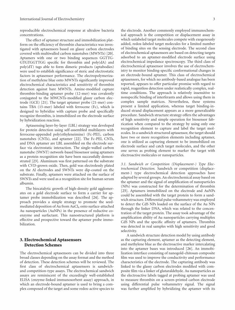
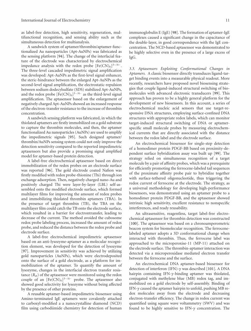
Figure 1: Sandwich assay: the immobilized aptamer capturesthrombin and aptamer labeled with pyrroquinoline quinone glu-cose dehydrogenase ((PQQ)GDH) generates electrical current uponglucose addition.
complementary DNA, which was also labeled with goldnanoparticles [21]. Consequently, more gold nanoparticleswere attached to each target protein, and thus, the signal wassignificantly enhanced.
A sandwich-type aptasensing system was constructed oftwo different aptamers which recognize different positions ofthrombin (Figure 1) [6, 27]. One aptamer was immobilizedonto the gold electrode for capturing thrombin onto theelectrode and the other was used for detection. The aptamerfor detection was labeled with pyrroquinoline quinone glu-cose dehydrogenase ((PQQ)GDH), and the electrical cur-rent, generated from glucose addition after the formationof the complex of thrombin, gold immobilized aptamer,and the (PQQ)GDH-labeled aptamer on the electrode, wasmeasured. The increase of the electric current generated by(PQQ)GDH was observed in dependent manner of the con-centration of thrombin.
A method utilized antibodies immobilized on the elec-trode surface to capture the protein target, the platelet-de-rived growth factor B-chain (PDGF-BB) as a model target,and the surface-captured protein was then sandwiched by anaptamer-primer complex was adapted for the detection ofthe amplified copies via enzymatic silver deposition thenallowed enormous sensitivity enhancement in the assay oftarget protein [28].
A sensitive electrochemical aptasensor was successfullyfabricated for the detection of adenosine triphosphate (ATP)by combining three-dimensionally ordered macroporous(3DOM) gold film and quantum dots (QDs) [29]. 5′-Thi-olated ATP-binding aptamer (ABA) was first assembled ontothe 3DOM gold film. Then, 5′-biotinated complementarystrand (BCS) was immobilized via hybridization reaction toform the DNA/DNA duplex. The tertiary structure of theaptamer was stabilized in the presence of target ATP, theduplex can be denatured to liberate BCS. The reaction wasmonitored by electrochemical stripping analysis of dissolvedQDs which were bound to the residual BCS through biotin-streptavidin system. The unique interconnected structure in3DOM gold film along with the built-in preconcentrationremarkably improved the sensitivity.
An ultrasensitive and highly specific electrochemicalaptasensor for thrombin based on amplification of aptamer-gold nanoparticles-horseradish peroxidase (aptamer-AuNPs-HRP) conjugates was successfully developed [30].
In this electrochemical protocol, aptamer1 (Apt1) was im-mobilized on core/shell Fe3O4/Au magnetic nanoparticles(AuMNPs) and served as capture probe. Aptamer2 (Apt2)was dual labeled with AuNPs and HRP and used as detectionprobe. Remarkable signal amplification was realized by tak-ing the advantage of AuNPs and catalytic reactions of HRP.The presence of proteins, such as human serum albumin,lysozyme, fibrinogen, and IgG did not show significantinterference with the assay for thrombin.
An ultrasensitive aptasensor for the electronic monitor-ing of proteins through a dual amplified strategy was pre-sented [31]. The target protein thrombin is sandwiched be-tween an electrode surface confined aptamer and an aptam-er-enzyme-carbon nanotube bioconjugate. The analyticalsignal amplification is achieved by coupling the signal ampli-fication nature of multiple enzymes with the biocatalyticsignal enhancement of redox recycling. This approach couldbe an attractive alternative to other common PCR-based sig-nal amplification in ultralow level of protein detection.
A sandwich format of magnetic nanoparticle/throm-bin/gold nanoparticle and thiocyanuric acid was presentedfor detection of thrombin [32]. An aptamer I was immobi-lized on the magnetic nanoparticles, aptamer II was labeledwith gold nanoparticles. The magnetic nanoparticle was usedfor separation and collection, and gold nanoparticle offeredexcellent electrochemical signal transduction. The significantsignal amplification was further implemented by formingnetwork-like thiocyanuric acid/gold nanoparticles. The pres-ence of other proteins such as BSA and lysozyme did notaffect the detection of thrombin.
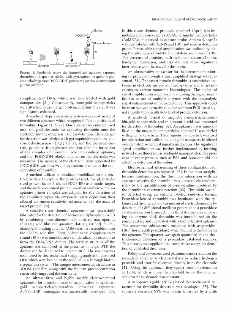
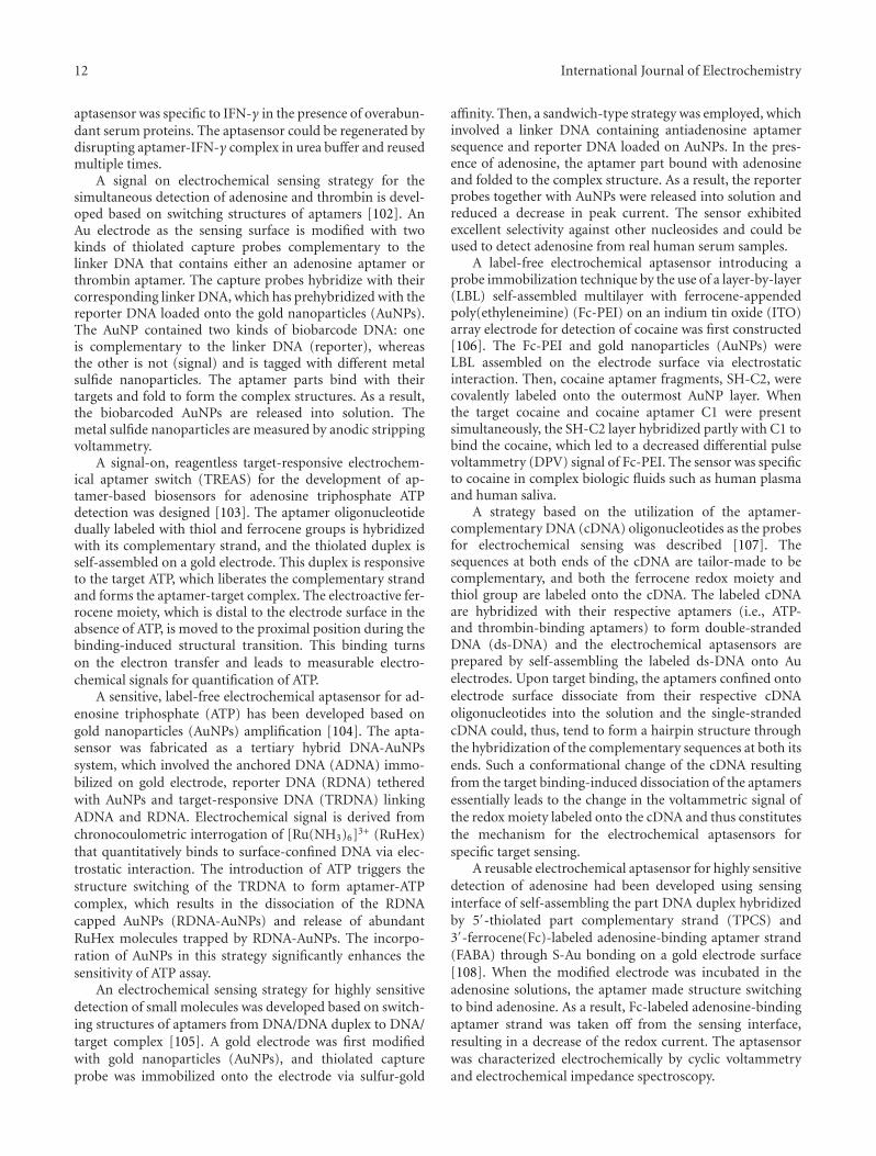
Electrochemical aptasensing of three configurations forthrombin detection was reported [33]. In the most straight-forward configuration, the thrombin interaction with anaptamer selective for thrombin was detected electrochem-ically by the quantification of p-nitroaniline produced bythe thrombin’s enzymatic reaction [33]. Thrombin was al-so detected using an enzyme labeled sandwich format.Peroxidase-labeled thrombin was incubated with the ap-tamer and the interaction was measured electrochemically bydetection of a diffusional mediator generated in a peroxidasecatalyzed reaction (Figure 2). In a third strategy also employ-ing an enzyme label, thrombin was immobilized on thesensor surface and incubated with a biotin labeled aptamer.The sensor was subsequently incubated with streptavidin-HRP (horseradish peroxidase), which bound to the biotin onthe aptamer. The aptamer was again quantified by the elec-trochemical detection of a peroxidase catalyzed reaction.This strategy was applicable to competitive assays for detec-tion of unlabeled thrombin.
Polsky and coworkers used platinum nanocrystals on thesecondary aptamer as electrocatalysts to reduce hydrogenperoxide and transfer electrons directly from the electrode[34]. Using this approach, they report thrombin detectionat 1 nM, which is more than 25-fold below the aptamersolution-phase dissociation constant.
A nanoporous gold- (NPG-) based electrochemical ap-tasensor for thrombin detection was developed [35]. Thesubstrate electrode NPG was in situ fabricated by a facile
International Journal of Electrochemistry 5
AuAuAuLabeledtarget
Target
et
Product
Substrate
HRP
Figure 2: Competition (or displacement) assays: target moleculesin a sample displace labeled-target molecules previously bound tothe sensor surface.
one-step square wave potential pulse treatment. The elec-trochemical aptasensor was fabricated using a layer-by-layerassembling strategy. The sandwich structure was formed viathrombin connecting the aptamer-modified NPG and theaptamer-modified Au nanoparticles (AuNPs). The AuNPswas modified with two kinds of single-strand DNA (ssDNA).One was aptamer of thrombin, but the other was not, reduc-ing the cross-reaction between thrombin and its aptamer onthe same AuNP. The electrochemical signal produced by the[Ru(NH3)6]+3 bound to ssDNA via electrostatic interactionwas measured by chronocoulometry [36]. This NPG-basedaptasensor also exhibited excellent sensitivity, due to theamplification effects of both NPG and AuNPs.
The advantages of aptamer, nanomaterial, and antibodyto design an electrochemical sandwich immunoassay for theultrasensitive detection of human immunoglobulin E (IgE)was combined by using methylene blue (MB) as electro-chemical indicator [36]. The sandwich structure was fab-ricated by using goat antihuman IgE as capturing probe.Aptamer-Au nanoparticles (NPs) conjugates were used bothas a sandwich amplification element as well as an accumu-lation reagent of MB. Once the aptamer-Au NPs conjugatesspecifically bind to electrode surface, MB molecules wereaccumulated on its surface by the specific interaction of MBwith G base of aptamer-Au NPs conjugates. Therefore, withthe increase of human IgE concentration, more aptamer-Au-NPs conjugates were bound, and thus, more MB moleculeswere accumulated. This sensing system showed excellentspecificity for the detection of human IgE against otherproteins: BSA, human IgA, and human IgM.
An electrochemical detection based on enzymatic sil-ver deposition has been proposed to detect thrombin[37]. The target protein, thrombin, was first captured bythrombin-binding thiolated aptamer self-assembled mono-layers (SAMs) on the gold electrode surface and then sand-wiched with another biotinylated thrombin-binding aptamerfor the association of alkaline phosphatase (Av-ALP). Theattached Av-ALP enzymatically converted the nonelectroac-tive substrate p-aminophenyl phosphate (p-APP) to p-ami-nophenol (p-AP) which could reduce silver ions in solutionleading to deposition of the metal onto the electrode surface.Linear sweep voltammetry was used to detect the amount
of deposited silver which reflected the amount of the targetprotein captured into the sandwich configuration.
A multifunctional electrochemical strategy based on adual-aptamer for the detection of adenosine and thrombin inone-pot was developed, based on biobarcode amplificationassay [38, 39]. The capture DNA aptamer I was immobilizedon the Au electrode. The functional Au nanoparticles (DNA-AuNPs) were loaded with barcode-binding DNA and aptam-er II. Through the specific recognition for thrombin, a sand-wich format of Au/aptamerI/thrombin/DNA-AuNPs wasfabricated. After hybridization with the PbSNPs-labeledbarcode DNA, the assembled sensor was obtained. The con-centration of thrombin was monitored based on the con-centration of lead ions dissolved through differential pulseanodic stripping voltammetry.
A disposable electrochemical assay involving mag-netic particles and carbon-based screen-printed electrodes(SPCEs) was developed for the detection of C-reactive pro-tein (CRP) [40]. The assay was based on a sandwich format inwhich a RNA aptamer was coupled to a monoclonal antibodyand alkaline phosphatase (AP) was used as enzymatic label.After the sandwich assay, the modified magnetic beads werecaptured by a magnet on the surface of a graphite workingelectrode and the electrochemical detection was thusachieved through the addition of the AP substrate (α-naph-thyl-phosphate) and α-naphthol produced during the enzy-matic reaction was detected using differential pulse voltam-metry. The assay was applied to the analysis of CRP freeserum and serum samples.
A sensitively amplified electrochemical aptasensor wasdesigned for adenosine triphosphate (ATP) detection [41].In the sensing process, duplexes consisting of partly comple-mentary strand (PCS1), ATP aptamer (ABA), and anotherpartly complementary strand (PCS2) were immobilized ontoAu electrode through the 5′-HS on the PCS1. Meanwhile,PCS2 was grafted with the Au nanoparticles (AuNPs) toamplify the detection signals. In the absence of ATP, probemethylene blue (MB) bound to the DNA duplexes and alsobound to guanine bases specifically to produce a strong dif-ferential pulse voltammetry signal. In the presence of ATP,the ABA-PCS2 or ABA-PCS1 part duplexes might be de-stroyed, which decreased the amount of MB on the electrodeand led to obviously decreased DPV signal. Therefore, suchPCS1-ABA-PCS2/AuNPs sensing system could provide apromising signal-amplified model for aptamer-based small-molecules detection.
The self-assembly of labeled aptamer subunits in the pre-sence of their substrates provides a method for the fluores-cence or electrochemical detection of the substrate [42]. Forelectrochemical detection of cocaine, the thiolated aptamersubunit is assembled on an Au electrode. The methyleneblue-labeled subunit binds to the surface-confined fragmentin the presence of cocaine. The amperometric response of thesystem allows the detection of cocaine.
An aptamer-based sandwich assay with electrochemicaldetection for thrombin analysis was proposed using Au na-noparticles [43]. The primary aptamer was immobilized onthe surface of a screen-printed carbon electrode (SPCE) and
6 International Journal of Electrochemistry
the secondary aptamer was immobilized on Au nanoparti-cles. The electrochemical reduction current response of Aunanoparticles was monitored for the quantitative detectionof thrombin. The effect of interfering proteins such as bovineserum albumin (BSA) was investigated. Control experimentsalso involved the use of an aptamer that has a binding affinityto immunoglobulin E (IgE).
An electrochemical method for the detection of throm-bin based on a gold-nanoparticles sensing platform and us-age of stripping voltammetry technique was developed [44].The aptamer was immobilized on a screen-printed electrodemodified with gold-nanoparticles by avidin-biotin technol-ogy. The oxidation of gold surface resulted in gold oxideformation upon polarization served as a basis for analyticalresponse. The cathodic peak area was found proportional tothrombin quantity specifically adsorbed onto electrode sur-face. Binding of thrombin to an aptamer has also been de-tected using the ferricyanide/ferrocyanide redox couple aselectrochemical indicator.
The Au nanoparticles-doped conducting polymer na-norods electrodes (AuNPs/CPNEs) were prepared by coatingAu nanorods (AuNRs) with a conducting polymer layer[45]. The AuNRs were prepared through an electroless dep-osition method using the polycarbonate membrane as atemplate. The AuNPs/CPNEs combining catalytic activity offerrocene to ascorbic acid were used for the fabrication ofan ultrasensitive aptamer sensor for thrombin detection.Sandwiched immunoassay for r-human thrombin with NH2-functionalized-thrombin-binding aptamer (Apt) immobi-lized on AuNPs/3D-CPNEs was studied through the elec-trocatalytic oxidation of ascorbic acid by the ferrocene moi-ety that was bound with an antithrombin antibody and at-tached with the Apt/3D-CPNEs probe through target bind-ing. The selectivity and the stability of the proposed throm-bin aptamer sensor were excellent, and it was tested in areal human serum sample for the detection of spiked con-centrations of thrombin.
A simple electrochemical approach for the detection ofthrombin, using aptamer-gold nanoparticles-modified elec-trodes was presented [46]. 1,6-Hexanedithiol was used as themedium to link Au nanoparticles to a bare gold electrode.Anti-thrombin aptamers were immobilized on the gold na-noparticles surfaces by self-assembly. The use of gold na-noparticles results in significant signal enhancement forsubsequent detection. The total amount of aptamer probesimmobilized on the gold nanoparticle surface is six-foldhigher than that on the bare electrode, leading to increasedsensitivity of the aptasensor.
The electrochemical thrombin detection system was de-veloped using two different aptamers recognizing differentparts of the protein in sandwich manner [47]. Aptamer 1-thrombin-ap2 glucose dehydrogenase complex was formedin the presence of thrombin, and a response current of theenzyme label was obtained.
An ultrasensitive label-free bioelectrochemical methodfor rapid determination of thrombin has been developed bydirectly detecting the redox activity of adenine (A) nucle-obases of anti-thrombin aptamer using a pyrolytic graphiteelectrode [48]. The bioelectrochemical protocol involves a
sandwich format Thrombin, captured by immobilized anti-thrombin antibody on microtiter plates, and was detectedby anti-thrombin aptamer-Au nanoparticle biobarcodes. Theadenine nucleobases were released by acid or nuclease fromAu nanoparticles bound on microtiter plates. Differentialpulse voltammetry was employed to investigate the electro-chemical behaviors of the purine nucleobases based on thewell-defined adenine signal. There was substantial amplifi-cation and thrombin can be detected at a very low levelof detection as the nanoparticle carries a large number ofaptamers per thrombin binding event. This method has beenused to detect thrombin in complex matrix such as fetal calfserum with minimum background interference.
A method for the determination of platelet-derivedgrowth factor BB (PDGF-BB) was developed using an elec-trochemical immunosensor with an aptamer-primed, long-strand circular detection probe [49]. Rabbit antihumanPDGF-B polyclonal antibody was immobilized on the elec-trode to serve as the capture antibody. The detection probewas synthesized via polymerase extension along a single-stranded circular plasmid DNA template with a primerheaded by the anti-PDGF-B aptamer. In the presence of theanalyte, the aptamer-primed circular probe was captured onthe electrode via the formation of an antibody/PDGF-BB/aptamer sandwiched complex. The electroactivity indica-tor methylene blue was adsorbed on the electrode surface viathe analyte-sandwiched complex with long-strand circularDNA, thus yielding a strong oxidation peak current of meth-ylene blue in square wave voltammetric signal for the quan-tification of PDGF-BB. This strategy allowed electrochemicaldetection with enormous signal amplification arising fromthe long-strand localized circular probe.
Immense effort has been placed on the realization ofimmunoassays exploiting displacement of redox-labeled tar-get molecules, due to the ease of use and applicability toimmunochromatographic strips and immunosensors. Mostof the efforts reported to date focus on the use of a redox-labeled target molecules target that is displaceable by theunlabeled target molecules toward which the antibody hashigher affinity. Limited success has been achieved due todifficulty in obtaining redox-labeled target molecules targetsto which the antibody has enough affinity to bind while atthe same time having lower levels of affinity in comparisonto the unlabeled target molecules to facilitate displacement.Aptamers, in contrast to antibodies, require the formationof a three-dimensional structure for target binding and can,thus, be anticipated to have a much higher affinity for bind-ing its target rather than a modified form of the target (e.g.,redox-labeled target). This phenomenon can be exploitedfor the development of a displacement assay, using enzyme-labeled target as a displaceable molecule.
In the first, Baldrich et al. measured thrombin at concen-trations down to 5 nM via competition between horseradishperoxidase (HRP) modified thrombin and unlabeled targetmolecules in the sample [50].
The coupling of aptamers with the coding and amplifica-tion features of inorganic nanocrystals offer a highly sensitiveand selective simultaneous bioelectronic detection of several
International Journal of Electrochemistry 7
Au AuThrombin
et No et
Fe(CN) 3−/4−6
Fe(CN) 3−/4−6
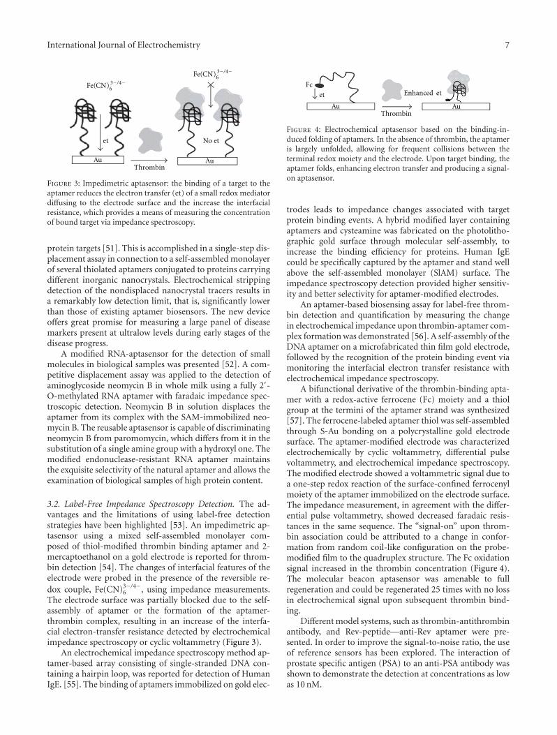
Figure 3: Impedimetric aptasensor: the binding of a target to theaptamer reduces the electron transfer (et) of a small redox mediatordiffusing to the electrode surface and the increase the interfacialresistance, which provides a means of measuring the concentrationof bound target via impedance spectroscopy.
protein targets [51]. This is accomplished in a single-step dis-placement assay in connection to a self-assembled monolayerof several thiolated aptamers conjugated to proteins carryingdifferent inorganic nanocrystals. Electrochemical strippingdetection of the nondisplaced nanocrystal tracers results ina remarkably low detection limit, that is, significantly lowerthan those of existing aptamer biosensors. The new deviceoffers great promise for measuring a large panel of diseasemarkers present at ultralow levels during early stages of thedisease progress.
A modified RNA-aptasensor for the detection of smallmolecules in biological samples was presented [52]. A com-petitive displacement assay was applied to the detection ofaminoglycoside neomycin B in whole milk using a fully 2′-O-methylated RNA aptamer with faradaic impedance spec-troscopic detection. Neomycin B in solution displaces theaptamer from its complex with the SAM-immobilized neo-mycin B. The reusable aptasensor is capable of discriminatingneomycin B from paromomycin, which differs from it in thesubstitution of a single amine group with a hydroxyl one. Themodified endonuclease-resistant RNA aptamer maintainsthe exquisite selectivity of the natural aptamer and allows theexamination of biological samples of high protein content.
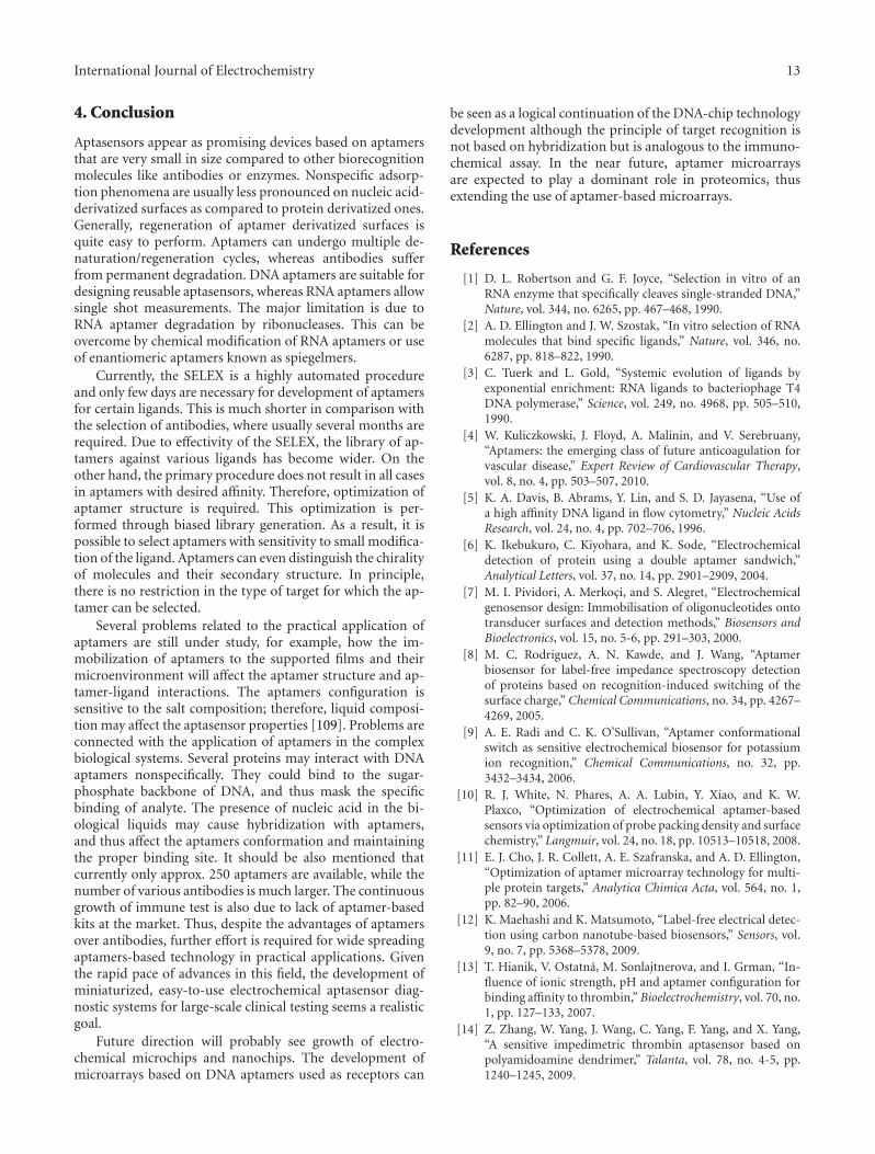
3.2. Label-Free Impedance Spectroscopy Detection. The ad-vantages and the limitations of using label-free detectionstrategies have been highlighted [53]. An impedimetric ap-tasensor using a mixed self-assembled monolayer com-posed of thiol-modified thrombin binding aptamer and 2-mercaptoethanol on a gold electrode is reported for throm-bin detection [54]. The changes of interfacial features of theelectrode were probed in the presence of the reversible re-dox couple, Fe(CN)3−/4−
6 , using impedance measurements.The electrode surface was partially blocked due to the self-assembly of aptamer or the formation of the aptamer-thrombin complex, resulting in an increase of the interfa-cial electron-transfer resistance detected by electrochemicalimpedance spectroscopy or cyclic voltammetry (Figure 3).
An electrochemical impedance spectroscopy method ap-tamer-based array consisting of single-stranded DNA con-taining a hairpin loop, was reported for detection of HumanIgE. [55]. The binding of aptamers immobilized on gold elec-
Au Au
Fc
et
Thrombin
Enhanced et
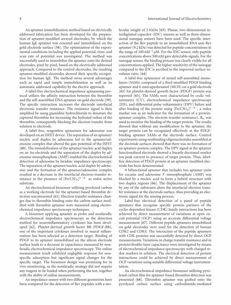
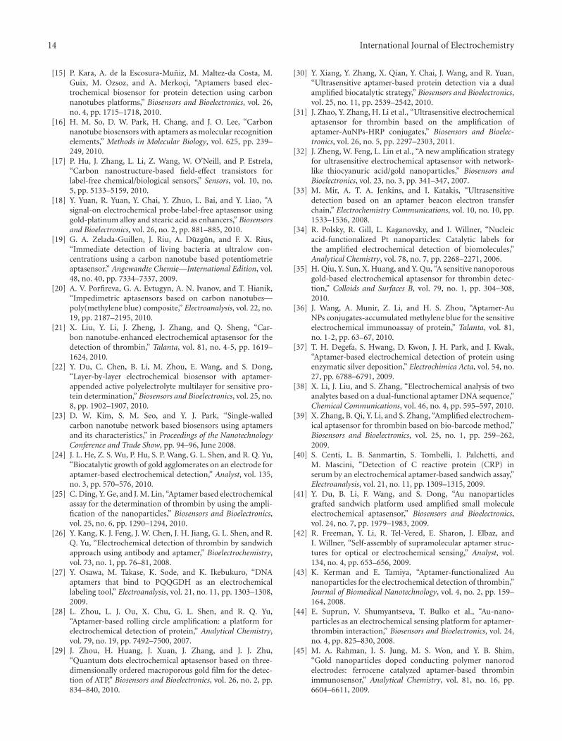
Figure 4: Electrochemical aptasensor based on the binding-in-duced folding of aptamers. In the absence of thrombin, the aptameris largely unfolded, allowing for frequent collisions between theterminal redox moiety and the electrode. Upon target binding, theaptamer folds, enhancing electron transfer and producing a signal-on aptasensor.
trodes leads to impedance changes associated with targetprotein binding events. A hybrid modified layer containingaptamers and cysteamine was fabricated on the photolitho-graphic gold surface through molecular self-assembly, toincrease the binding efficiency for proteins. Human IgEcould be specifically captured by the aptamer and stand wellabove the self-assembled monolayer (SlAM) surface. Theimpedance spectroscopy detection provided higher sensitiv-ity and better selectivity for aptamer-modified electrodes.
An aptamer-based biosensing assay for label-free throm-bin detection and quantification by measuring the changein electrochemical impedance upon thrombin-aptamer com-plex formation was demonstrated [56]. A self-assembly of theDNA aptamer on a microfabricated thin film gold electrode,followed by the recognition of the protein binding event viamonitoring the interfacial electron transfer resistance withelectrochemical impedance spectroscopy.
A bifunctional derivative of the thrombin-binding apta-mer with a redox-active ferrocene (Fc) moiety and a thiolgroup at the termini of the aptamer strand was synthesized[57]. The ferrocene-labeled aptamer thiol was self-assembledthrough S-Au bonding on a polycrystalline gold electrodesurface. The aptamer-modified electrode was characterizedelectrochemically by cyclic voltammetry, differential pulsevoltammetry, and electrochemical impedance spectroscopy.The modified electrode showed a voltammetric signal due toa one-step redox reaction of the surface-confined ferrocenylmoiety of the aptamer immobilized on the electrode surface.The impedance measurement, in agreement with the differ-ential pulse voltammetry, showed decreased faradaic resis-tances in the same sequence. The “signal-on” upon throm-bin association could be attributed to a change in confor-mation from random coil-like configuration on the probe-modified film to the quadruplex structure. The Fc oxidationsignal increased in the thrombin concentration (Figure 4).The molecular beacon aptasensor was amenable to fullregeneration and could be regenerated 25 times with no lossin electrochemical signal upon subsequent thrombin bind-ing.
Different model systems, such as thrombin-antithrombinantibody, and Rev-peptide—anti-Rev aptamer were pre-sented. In order to improve the signal-to-noise ratio, the useof reference sensors has been explored. The interaction ofprostate specific antigen (PSA) to an anti-PSA antibody wasshown to demonstrate the detection at concentrations as lowas 10 nM.
8 International Journal of Electrochemistry
An aptamer immobilization method based on electricallyaddressed fabrication has been developed for the prepara-tion of aptamer-modified arrayed electrodes, by which thehuman IgE aptamer was oriented and immobilized on thegold electrode surface [58]. The optimization of the experi-mental conditions including the applied potential, time, andscan rate of potential was investigated. The method wassuccessfully used to immobilize the aptamer onto the desiredelectrodes, pixel by pixel, based on the electrically addressedapproach. Compared to the control electrodes, the resultingaptamer-modified electrodes showed their specific recogni-tion for human IgE. The method owns several advantagessuch as rapid and simple immobilization as well as itsautomatic addressed capability by the electric approach.
A label-free electrochemical impedance aptasensing pro-tocol utilizes the affinity interaction between the thrombinand the self-assembled DNA aptamer on gold electrode [59].The specific interaction increases the electrode interfacialelectronic transfer resistance. The resistance signal is thenamplified by using guanidine hydrochloride to denature thecaptured thrombin for increasing the hydrated radius of thethrombin, consequently blocking the electron transfer fromsolution to electrode.
A label free, reagentless aptasensor for adenosine wasdeveloped on an ISFET device. The separation of an aptamer/nucleic acid duplex by adenosine led to the aptamer/ad-enosine complex that altered the gate potential of the ISFET[60]. The immobilization of the aptamer/nucleic acid duplexon an Au-electrode and the separation of the duplex by ad-enosine monophosphate (AMP) enabled the electrochemicaldetection of adenosine by faradaic impedance spectroscopy.The separation of the aptamer/nucleic acid duplex by adeno-sine and the formation of the aptamer/adenosine complexresulted in a decrease in the interfacial electron-transfer re-sistance in the presence of [Fe(CN)6]3−/4− as redox activesubstrate.
An electrochemical biosensor utilizing pyrolyzed carbonas a working electrode for the aptamer-based thrombin de-tection was presented [61]. Electron transfer resistance chan-ges due to thrombin binding onto the carbon surface mod-ified with thrombin aptamer were measured using electro-chemical impedance spectroscopy techniques.
A biosensor applying aptamer as probe and nonfaradicelectrochemical impedance spectroscopy as the detectionmethod for neuroinflammatory cytokines has been devel-oped [62]. Platelet-derived growth factor BB (PDGF-BB),one of the important cytokines involved in neural inflam-mation has been selected as a detection target. Binding ofPDGF to its aptamer immobilized on the silicon electrodesurface leads to a decrease in capacitance measured by non-faradic electrochemical impedance spectroscopy. The onlinemeasurement result exhibited negligible response for non-specific adsorption but significant signal changes for thespecific target. The biosensor design was promising for invivo monitoring, as the nonfaradic strategy did not requireany reagent to be loaded when performing the test, togetherwith the ability of online measurements.
An impedance-sensor with two different geometries havebeen compared for the detection of Rev peptides with a mo-
lecular weight of 2.4 kDa [63]. Planar, two-dimensional in-terdigitated capacitor (IDC) sensors as well as three-dimen-sional nanogap sensors have been used. The specific inter-action of the Rev peptide to an immobilized RNA anti-Revaptamer (9.2 kDa) was detected for peptide concentrations inthe range of 100 nM−2 μM. For the IDC sensor, only peptideconcentrations above 500 nM gave detectable signals. For thenanogap sensor, the binding process was clearly visible for allconcentrations applied. The higher sensitivity of the nanogapcompared to the IDC is ascribed to the improved surface-to-volume ratio. [64]
A label-free aptasensor of mixed self-assembled mono-layers (SAMs) composed of a thiol-modified PDGF bindingaptamer and 6-mercaptohexanol (MCH) on a gold electrode[65] for platelet-derived growth factor (PDGF) protein wasreported [65]. The SAMs were characterized by cyclic vol-tammetry (CV), electrochemical impedance spectroscopy(EIS), and differential pulse voltammetry (DPV) before andafter binding of the protein using [Fe(CN)6]3−/4−, a redoxmarker ion as an indicator for the formation of a protein-aptamer complex. The electron transfer resistance). Ret wasused to monitor the binding of the target protein. The resultsshowed that without any modification to the aptamer, thetarget protein can be recognized effectively at the PDGF-binding aptamer SAMs at the electrode surface. Controlexperiments using nonbinding oligonucleotides assembled atthe electrode surfaces showed that there was no formation ofan aptamer-protein complex. The DPV signal at the aptamerfunctionalized electrode showed a linearly decreased markerion peak current in presence of target protein. Thus, label-free detection of PDGF protein at an aptamer modified elec-trode has been demonstrated.
A bifunctional aptamer that includes two aptamer unitsfor cocaine and adenosine 5′-monophosphate (AMP) wasblocked by a nucleic acid to form a hybrid structure withtwo duplex regions [66]. The displacement of the aptamerby any of the substrates alters the interfacial electron trans-fer resistance at the electrode surface, thus providing an elec-tronic signal for the sensing process.
Label-free electrical detection of a panel of peptideaptamers that recognise specific protein partners of thecyclin-dependent kinase (CDK) family interactions has beenachieved by direct measurement of variations at open cir-cuit potential (OCP) using an accurate differential voltagemeasurement [67]. Different peptide aptamers immobilizedon gold electrodes were used for the detection of humanCDK2 and CDK4. The interaction of the peptide aptamerswith CDK proteins was successfully detected by direct OCPmeasurements. Variations in charge transfer resistance and inprotein/double-layer capacitance were investigated by meansof electrochemical impedance spectroscopy with charged re-dox markers in solution. The electrical detection of proteininteractions could be achieved by direct measurement ofOCP variations using suitable differential voltage instrumen-tation.
An electrochemical impedance biosensor utilizing pyro-lyzed carbon film for aptamer-based thrombin detection waspresented [68]. Thrombin aptamer was grafted onto thepyrolyzed carbon surface using carbodiimide-mediated
International Journal of Electrochemistry 9
chemistry, followed by Triton-X 100 and BSA treatment toreduce nonspecific binding of thrombin. Electron-transferresistance changes due to thrombin binding onto the carbonsurface were measured, using electrochemical impedancespectroscopy. Pyrolyzed carbon can provide a new approachfor miniaturization, integration, and low-cost fabrication inelectrochemical biosensors.
GNPs electrodeposited on GCE used as a platform forthe immobilization of the thiolated aptamer can improvethe sensitivity of an EIS biosensor for the determinationof thrombin [69]. In the measurement of thrombin, thechange in interfacial electron transfer resistance of the bio-sensor using a redox couple of [Fe(CN)6]3−/4− as the probewas monitored. The association and dissociation constants ofthree different immobilized aptamers binding with thrombinwere measured and the difference of the dissociation con-stants obtained was discussed.
Electrochemical protein biosensors using aptamers probedoped in polypyrrole and subsequent electrochemical im-pedance spectroscopy have been successfully developed [70,71]. Two targets, platelet-derived growth factor and im-munoglobulin E, have been also tested [70]. A sensitive andreal-time biosensor for inflammatory cytokine detection hasbeen successfully measured in both offline ElS characteriza-tion and real-time impedance monitoring [71].
An aptamer-based sensor development, utilizing a modelsystem of human alpha thrombin interacting with a thiolatedDNA aptamer, immobilized on gold electrodes [72]. EISmeasurements took place in the presence of iron ferrocy-anides.
A simple and highly sensitive electrochemical impedancespectroscopy (EIS) biosensor based on nano-MnO2 as a plat-form for the immobilization of the aptamer was developedfor the determination of adenosine [73]. In the measurementof adenosine, the change in interfacial electron transferresistance (Ret) of the biosensor using a redox couple of[Fe(CN)6]3/4 as the probe was monitored. The sensor wasshown to exhibit high sensitivity, desirable selectivity andgood stability.
An impedimetric electrochemical biosensor was devel-oped for the label-free and selective detection of leukemiacells based on aptamer-modified gold electrode using elec-trochemical impedance spectroscopy (EIS) technique [74].The thiol-terminated aptamer selected for acute leukemiacells was self-assembled onto the gold electrode surface asrecognition probe, which was characterized by cyclic voltam-metry (CV) and EIS using Fe(CN)3−/4−
6 as a redox probe. Theelectron-transfer resistance Ret of [Fe(CN)6]3−/4− on the sen-sor surface increased substantially upon incubation of ap-tamer-modified electrode in cell solution. The work provideda simple, convenient, low-cost, and label-free method forearly leukemia diagnosis.
A label-free and sensitive faradic impedance spectroscopy(FIS) aptasensor based on target-induced aptamer displace-ment was developed for the determination of lysozyme [75].The aptasensor was fabricated by self-assembling the par-tial complementary single-strand DNA (pcDNA)-lysozymebinding aptamer (LBA) duplex on the surface of a gold elec-
trode. The introduction of target lysozyme induced the dis-placement of the LBA from the pcDNA-LBA duplex on theelectrode into the solution, decreasing the electron transferresistance of the aptasensor. The fabricated aptasensor showsa high sensitivity, good selectivity, and satisfactory regener-ation. This work demonstrates that a high sensitivity of thefabricated aptasensor can be obtained using a relatively shortpcDNA.
Faradaic impedance spectroscopy and ion-sensitive field-effect transistor (ISFET) were applied to sense aptamer-substrate complexes [76]. The methods utilized anticocaineaptamer fragments that self-assembled, in the presence ofcocaine, to a supramolecular aptamer fragments/cocainecomplex on the electrode surface or ISFET gate. One of theaptamer fragments is assembled on a Au electrode or theISFET gate. The second thiolated aptamer fragment is used tomodify Au NPs that are used as amplifying labels for the twodetection schemes. The impedimetric and ISFET methodsenabled the analysis of cocaine.
A protein assay method based on a DNA array was de-veloped in which human immunoglobulin E (hIgE) and itsDNA aptamer were used as an analytical model [77]. Thetarget protein hIgE was captured by the aptamer in homoge-neous solution and then the resulting hIgE-aptamer complexwas hybridized onto probes self-assembled on the DNAarray. The charge transfer resistance (Rct) of electrodes beforeand after hybridization were measured by electrochemicalimpedance spectroscopy (EIS). To test the selectivity of themethod, four different probes with one-to-three mismatchedbases were immobilized on respective electrodes. The resultsshowed that the complex could be hybridized and detectedout on the electrodes modified with the fully complementarysequences. In addition, the DNA array could be employedto analyze multiple samples selectively with the matchedaptamer.
A reusable label-free electrochemical nucleic acid apta-sensor for the determination of cocaine by the immobili-zation of thiolated self-assembled DNA sequences on a goldnanoparticles-modified electrode was constructed [78].When cocaine was complexed specifically to the aptamer, theconfiguration of the nucleic acid aptamer switched to alocked structure and the interface of the biosensor changed,resulting in a variation of the corresponding peak currentof an electrochemical probe ([Fe(CN)6]3−/4 as monitored bycyclic voltammetry and electrochemical impedance spectro-scopy (EIS).
A sensitive aptamer-based electrochemical biosensor todetect human immunoglobulin E (IgE) was designed [79]. 5′
Biotin labeled 45 mer DNA aptamer sequence was immobi-lized onto streptavidin coated graphite surfaces. Interactionbetween human IgE and DNA aptamer was monitored byelectrochemical impedance spectrometry.
A multispecific electrochemical array with eight indi-vidually addressable gold working electrodes for rapid bio-sensing of 2.7 kb-long target Yersinia pestis DNA and for pro-tein sensing of ricin toxin chain A (RTA) in the presence ofredox agent were designed [80]. The array allowed to in-corporate multiple negative controls in the course of a singlebinding experiment as well as to perform parallel identical
10 International Journal of Electrochemistry
experiments to improve reliability of detection. Eight indi-vidual EIS measurements were completed in 15 min. Thearray is disposable, economical, and easy to use.
A dual RNA/peptide aptamer probe for simultaneousdetection of PSMA (+) and PSMA (−) prostate cancer cellsusing electrochemical impedance spectroscopy was reported[81]. This approach can be applied as a general tool for earlydiagnosis of prostate cancer.
Aptamer-based capacitive label-free biosensors for mon-itoring aptamer-protein recognition events, based on chargedistribution under the applied frequency by nonfaradaic im-pedance spectroscopy (NFIS) was reported [82, 83]. Thebiosensors based on gold interdigitated (GID) capacitor ar-rays functionalized with synthetic RNA aptamers. The RNAaptamers served as biorecognition elements for C-reactiveprotein (CRP). The signal is generated as a result of thechange in relative capacitance occurring as a result of theformation of an RNA-CRP complex on GID capacitors. TheRNA-protein complex on GID capacitors could be extendedto the development of electrical biosensor systems for theearly diagnosis.
Two modified aptamers, a partially (ATA) and a fully O-methylated aptamer (FATA), were proposed as recognitionelements for the detection of tobramycin at therapeutic rangein human serum [84]. A displacement assay was developedusing faradaic electrochemical impedance spectroscopy (F-EIS) as a detection technique. The affinity constant, KDb,for both aptamers was estimated, and the selectivity towardsother aminoglycosides was also tested.
Lysozyme has been detected selectively in a mixture con-taining a large excess of six proteins and amino acids (bothelectroactive and nonelectroactive) by combining aptamer-coated magnetic beads and chronopotentiometric strippingmeasurements of the captured protein (in connection tothe intrinsic electroactivity of the protein) [85]. The proteinmeasurement by adsorptive chronopotentiometric based onscanning the guanine bases of the guanine-rich secondaryaptamer. When involving PCR reaction to amplify theseguanine bases, fM level of detection limit has been obtained.The approach has also been employed for electrochemicallyinvestigating amino acid amides by using guanine-rich DNAaptamer as the electroactive marker.
The effect of aptamer structure and immobilization plat-form on the efficiency of thrombin binding and its detectionusing electrochemical impedance spectroscopy (EIS) charac-teristics was investigated with aptasensors based on glassycarbon electrodes covered with multiwalled carbon nan-otubes (MWNTs) [86]. Aptamers with one or two bindingsequences GGTTGGTGTGGTTGG specific for thrombinand poly(dA) and poly(dT) tags able to form dimericproducts (aptabodies) were used to establish significance ofsteric and electrostatic factors in aptasensor performance.The electropolymerization of methylene blue onto MWNTssignificantly improved electrochemical characteristics andsensitivity of thrombin detection against bare MWNTs.
The biosensors based on DNA aptamers immobilized byelectrostatic adsorption onto electropolymerized methyleneGreen imprinted with DNA have been developed and ex-amined for thrombin detection using electrochemical im-
pedance spectroscopy and potentiometry [87]. The additionof DNA at the electropolymerization stage followed by acidictreatment of the coating significantly improved the efficiencyof electrostatic adsorption of the DNA aptamer and providedsensitive detection of thrombin.
An amperometric aptasensor based on DNA aptamersimmobilized by avidin-biotin method or by electrostatic ad-sorption onto multiwalled carbon nanotube layer containedmethylene blue have been developed and examined forthrombin detection in buffer and in spiked blood serum [88].The presence of MB increases the binding capacity of thesurface layer and enhances the range of thrombin concen-trations to be determined.
An artificial receptor formed by hybridization of twoDNA aptamers for human thrombin (aptabody) was re-ported [89]. The aptasensor based on multiwalled carbonnanotubes allowed to detect thrombin with detection limit,3 times better in comparison with conventional aptamer.
A potentiometric detection of DNA-protein interactionshas been proposed [90]. The polymeric phenothiazine dyes,methylene blue and methylene green, were electrochemicallydeposited onto the glassy carbon electrode and covered withdouble stranded DNA (dsDNA) as a target for antibodies(DNA sensor) or DNA aptamer specific to human α-throm-bin. The developed potentiometric biosensors can be usedfor preliminary diagnostics of autoimmune diseases andthrombin detection with sensitivity comparable to tradi-tional methods.
Electrochemical indicator methylene blue and differen-tial pulse voltammetry allowed to determine charge transferfrom electrode surface to the thrombin bounded on a DNAaptamer with high selectivity in comparison with nonspecificbinding caused by human IgG or human serum albumin[91]. The method of detection thrombin-aptamer interac-tion based on measurement the charge consumption fromthe electrode covered by DNA aptamers to an electrochem-ical indicator methylene blue (MB), which is bounded to athrombin.
An electrochemical sensor to detect interferon (IFN)-γ,a selective marker for tuberculosis pleurisy, using its RNAor DNA 5′-thiol-modified aptamer probe immobilized onthe gold electrode [92]. Interaction between IFN-γ andthe aptamer was recorded using electrochemical impedancespectroscopy. IFN-γ was detected in fetal bovine serum, amimicked biological system, which has similar componentsto pleural fluid.
A multifunctional reusable label-free electrochemicalbiosensor based on an integrated aptamer for parallel de-tection of adenosine triphosphate (ATP) and α-thrombin,by using electrochemical impedance spectroscopy (EIS) andcyclic voltammetry (CV), was reported [93]. Au electrode asthe sensing surface was modified with a part DNA duplexwhich contained a 5′-thiolated partly complementary strand(PCS) and a mixed aptamer (MBA). The unimolecular MBAcontained small-molecule ATP-binding aptamer (ABA) andalso protein α-thrombin binding aptamer (TBA). Thus,the aptasensor could be used for detection of ATP andα-thrombin. The aptasensor held several advantages such
International Journal of Electrochemistry 11
as label-free detection, high sensitivity, regeneration, mul-tifunctional recognition, and sensing ability such as thesimultaneous detection for multianalysis.
A sandwich system of aptamer/thrombin/aptamer-func-tionalized Au nanoparticles (Apt-AuNPs) was fabricated asthe sensing platform [94]. The change of the interfacial fea-ture of the electrode was characterized by electrochemicalimpedance analysis with the redox probe [Fe(CN)6]3−/4−.The three-level cascaded impedimetric signal amplificationwas developed: Apt-AuNPs as the first-level signal enhancer,the steric-hindrance between the enlarged Apt-AuNPs as thesecond-level signal amplification, the electrostatic-repulsionbetween sodium dodecylsulfate (SDS) stabilized Apt-AuNPs,and the redox probe [Fe(CN)6]3−/4− as the third-level signalamplification. The aptasensor based on the enlargement ofnegatively charged Apt-AuNPs showed an increased responseof the electron-transfer resistance to the increase of thrombinconcentration.
A sandwich sensing platform was fabricated, in which thethiolated aptamers are firstly immobilized on a gold substrateto capture the thrombin molecules, and then, the aptamerfunctionalized Au nanoparticles (AuNPs) are used to amplifythe impedimetric signals [95]. Such designed aptamer/thrombin/AuNPs sensing system could not only improve thedetection sensitivity compared to the reported impedimetricaptasensors but also provide a promising signal amplifiedmodel for aptamer-based protein detection.
A label-free electrochemical aptasensor based on directimmobilization of the redox probes on an electrode surfacewas reported [96]. The gold electrode coated Nafion wasfirstly modified with redox probe-thionine (Thi) through ionexchange adsorption. Then, negatively charged nano-Au andpositively charged Thi were layer-by-layer (LBL) self-as-sembled onto the modified electrode surface, which formedmultilayer films for improving the amount of redox probesand immobilizing thiolated thrombin aptamers (TBA). Inthe presence of target thrombin (TB), the TBA on themultilayer film could catch the TB onto the electrode surface,which resulted in a barrier for electrontransfer, leading todecrease of the current. The method avoided the cubosomeredox probe labeling process, increased the amount of redoxprobe, and reduced the distance between the redox probe andelectrode surface.
A label-free electrochemical impedimetric aptasensorbased on an anti-lysozyme-aptamer as a molecular recogni-tion element, was developed for the detection of lysozyme[97]. Improvement in sensitivity was achieved by utilizinggold nanoparticles (AuNPs), which were electrodepositedonto the surface of a gold electrode, as a platform for im-mobilization of the aptamer. To quantify the amount oflysozyme, changes in the interfacial electron transfer resis-tance (Ret) of the aptasensor were monitored using the redoxcouple of an [Fe(CN)6]3−/4− probe. The aptasensor alsoshowed good selectivity for lysozyme without being affectedby the presence of other proteins.
A reusable aptamer-based impedimetric biosensor usingAmino-terminated IgE aptamers were covalently attachedto carboxyl-modified a a nanocrystalline diamond (NCD)film using carbodiimide chemistry for detection of human
immunoglobulin E (IgE) [98]. The formation of aptamer-IgEcomplexes caused a significant change in the capacitance ofthe double-layer, in good correspondence with the IgE con-centration. The NCD-based aptasensor was demonstrated tobe highly selective even in the presence of a large excess ofIgG.
3.3. Aptasensors Exploiting Conformational Changes inAptamers. A classic biosensor directly transduces ligand-tar-get binding events into a measurable physical readout. Morerecently, researchers have proposed novel biosensing strate-gies that couple ligand-induced structural switching of bio-molecules with advanced electronic transducers [99]. Thisapproach has proven to be a highly general platform for thedevelopment of new biosensors. In this account, a series ofelectrochemical nucleic acid sensors that use target-re-sponsive DNA structures, employing surface-confined DNAstructures with appropriate redox labels, which can monitortarget-induced structural switching of DNA or aptamer-specific small molecule probes by measuring electrochem-ical currents that are directly associated with the distancebetween the redox label and the electrode surface.
An electrochemical biosensor for single-step detectionof a homodimer protein PDGF-BB based on proximity-de-pendent surface hybridization assay was built up [99]. Thestrategy relied on simultaneous recognition of a targetmolecule by a pair of affinity probes, which was a prerequisitefor efficiently promoting the ferrocene-labeled tail sequencesof the proximate affinity probe pair to hybridize togetherwith surface-tethered oligonucleotide, thus triggering theredox current of ferrocene at the electrode. The strategy, asa universal methodology for developing high-performancebiosensors, was demonstrated using an aptamer probe to ahomodimer protein PDGF-BB, and the aptasensor showedintrinsic high sensitivity, excellent resistance to nonspecificinterferences, and ready reusability.
An ultrasensitive, reagentless, target label-free electro-chemical aptasensor for thrombin detection was constructed[100]. The aptasensor was based on a chronoamperometricbeacon system for biomolecular recognition. The ferrocene-labeled aptamer adopts a 3D conformational change wheninteracted with thrombin. Thus, the ferrocene label wasapproached to the microperoxise-11 (MP-11) attached onthe electrode surface. The thrombin-aptamer interaction wasdetected via a microperoxidase mediated electron transferbetween the ferrocene and the surface.
An electrochemical DNA aptamer-based biosensor fordetection of interferon (IFN)-γ was described [101]. A DNAhairpin containing IFN-γ-binding aptamer was thiolated,conjugated with methylene blue (MB) redox tag, and im-mobilized on a gold electrode by self-assembly. Binding ofIFN-γ caused the aptamer hairpin to unfold, pushing MB re-dox molecules away from the electrode and decreasingelectron-transfer efficiency. The change in redox current wasquantified using square wave voltammetry (SWV) and wasfound to be highly sensitive to IFN-γ concentration. The
12 International Journal of Electrochemistry
aptasensor was specific to IFN-γ in the presence of overabun-dant serum proteins. The aptasensor could be regenerated bydisrupting aptamer-IFN-γ complex in urea buffer and reusedmultiple times.
A signal on electrochemical sensing strategy for thesimultaneous detection of adenosine and thrombin is devel-oped based on switching structures of aptamers [102]. AnAu electrode as the sensing surface is modified with twokinds of thiolated capture probes complementary to thelinker DNA that contains either an adenosine aptamer orthrombin aptamer. The capture probes hybridize with theircorresponding linker DNA, which has prehybridized with thereporter DNA loaded onto the gold nanoparticles (AuNPs).The AuNP contained two kinds of biobarcode DNA: oneis complementary to the linker DNA (reporter), whereasthe other is not (signal) and is tagged with different metalsulfide nanoparticles. The aptamer parts bind with theirtargets and fold to form the complex structures. As a result,the biobarcoded AuNPs are released into solution. Themetal sulfide nanoparticles are measured by anodic strippingvoltammetry.
A signal-on, reagentless target-responsive electrochem-ical aptamer switch (TREAS) for the development of ap-tamer-based biosensors for adenosine triphosphate ATPdetection was designed [103]. The aptamer oligonucleotidedually labeled with thiol and ferrocene groups is hybridizedwith its complementary strand, and the thiolated duplex isself-assembled on a gold electrode. This duplex is responsiveto the target ATP, which liberates the complementary strandand forms the aptamer-target complex. The electroactive fer-rocene moiety, which is distal to the electrode surface in theabsence of ATP, is moved to the proximal position during thebinding-induced structural transition. This binding turnson the electron transfer and leads to measurable electro-chemical signals for quantification of ATP.
A sensitive, label-free electrochemical aptasensor for ad-enosine triphosphate (ATP) has been developed based ongold nanoparticles (AuNPs) amplification [104]. The apta-sensor was fabricated as a tertiary hybrid DNA-AuNPssystem, which involved the anchored DNA (ADNA) immo-bilized on gold electrode, reporter DNA (RDNA) tetheredwith AuNPs and target-responsive DNA (TRDNA) linkingADNA and RDNA. Electrochemical signal is derived fromchronocoulometric interrogation of [Ru(NH3)6]3+ (RuHex)that quantitatively binds to surface-confined DNA via elec-trostatic interaction. The introduction of ATP triggers thestructure switching of the TRDNA to form aptamer-ATPcomplex, which results in the dissociation of the RDNAcapped AuNPs (RDNA-AuNPs) and release of abundantRuHex molecules trapped by RDNA-AuNPs. The incorpo-ration of AuNPs in this strategy significantly enhances thesensitivity of ATP assay.
An electrochemical sensing strategy for highly sensitivedetection of small molecules was developed based on switch-ing structures of aptamers from DNA/DNA duplex to DNA/target complex [105]. A gold electrode was first modifiedwith gold nanoparticles (AuNPs), and thiolated captureprobe was immobilized onto the electrode via sulfur-gold
affinity. Then, a sandwich-type strategy was employed, whichinvolved a linker DNA containing antiadenosine aptamersequence and reporter DNA loaded on AuNPs. In the pres-ence of adenosine, the aptamer part bound with adenosineand folded to the complex structure. As a result, the reporterprobes together with AuNPs were released into solution andreduced a decrease in peak current. The sensor exhibitedexcellent selectivity against other nucleosides and could beused to detect adenosine from real human serum samples.
A label-free electrochemical aptasensor introducing aprobe immobilization technique by the use of a layer-by-layer(LBL) self-assembled multilayer with ferrocene-appendedpoly(ethyleneimine) (Fc-PEI) on an indium tin oxide (ITO)array electrode for detection of cocaine was first constructed[106]. The Fc-PEI and gold nanoparticles (AuNPs) wereLBL assembled on the electrode surface via electrostaticinteraction. Then, cocaine aptamer fragments, SH-C2, werecovalently labeled onto the outermost AuNP layer. Whenthe target cocaine and cocaine aptamer C1 were presentsimultaneously, the SH-C2 layer hybridized partly with C1 tobind the cocaine, which led to a decreased differential pulsevoltammetry (DPV) signal of Fc-PEI. The sensor was specificto cocaine in complex biologic fluids such as human plasmaand human saliva.
A strategy based on the utilization of the aptamer-complementary DNA (cDNA) oligonucleotides as the probesfor electrochemical sensing was described [107]. Thesequences at both ends of the cDNA are tailor-made to becomplementary, and both the ferrocene redox moiety andthiol group are labeled onto the cDNA. The labeled cDNAare hybridized with their respective aptamers (i.e., ATP-and thrombin-binding aptamers) to form double-strandedDNA (ds-DNA) and the electrochemical aptasensors areprepared by self-assembling the labeled ds-DNA onto Auelectrodes. Upon target binding, the aptamers confined ontoelectrode surface dissociate from their respective cDNAoligonucleotides into the solution and the single-strandedcDNA could, thus, tend to form a hairpin structure throughthe hybridization of the complementary sequences at both itsends. Such a conformational change of the cDNA resultingfrom the target binding-induced dissociation of the aptamersessentially leads to the change in the voltammetric signal ofthe redox moiety labeled onto the cDNA and thus constitutesthe mechanism for the electrochemical aptasensors forspecific target sensing.
A reusable electrochemical aptasensor for highly sensitivedetection of adenosine had been developed using sensinginterface of self-assembling the part DNA duplex hybridizedby 5′-thiolated part complementary strand (TPCS) and3′-ferrocene(Fc)-labeled adenosine-binding aptamer strand(FABA) through S-Au bonding on a gold electrode surface[108]. When the modified electrode was incubated in theadenosine solutions, the aptamer made structure switchingto bind adenosine. As a result, Fc-labeled adenosine-bindingaptamer strand was taken off from the sensing interface,resulting in a decrease of the redox current. The aptasensorwas characterized electrochemically by cyclic voltammetryand electrochemical impedance spectroscopy.
International Journal of Electrochemistry 13
4. Conclusion
Aptasensors appear as promising devices based on aptamersthat are very small in size compared to other biorecognitionmolecules like antibodies or enzymes. Nonspecific adsorp-tion phenomena are usually less pronounced on nucleic acid-derivatized surfaces as compared to protein derivatized ones.Generally, regeneration of aptamer derivatized surfaces isquite easy to perform. Aptamers can undergo multiple de-naturation/regeneration cycles, whereas antibodies sufferfrom permanent degradation. DNA aptamers are suitable fordesigning reusable aptasensors, whereas RNA aptamers allowsingle shot measurements. The major limitation is due toRNA aptamer degradation by ribonucleases. This can beovercome by chemical modification of RNA aptamers or useof enantiomeric aptamers known as spiegelmers.
Currently, the SELEX is a highly automated procedureand only few days are necessary for development of aptamersfor certain ligands. This is much shorter in comparison withthe selection of antibodies, where usually several months arerequired. Due to effectivity of the SELEX, the library of ap-tamers against various ligands has become wider. On theother hand, the primary procedure does not result in all casesin aptamers with desired affinity. Therefore, optimization ofaptamer structure is required. This optimization is per-formed through biased library generation. As a result, it ispossible to select aptamers with sensitivity to small modifica-tion of the ligand. Aptamers can even distinguish the chiralityof molecules and their secondary structure. In principle,there is no restriction in the type of target for which the ap-tamer can be selected.
Several problems related to the practical application ofaptamers are still under study, for example, how the im-mobilization of aptamers to the supported films and theirmicroenvironment will affect the aptamer structure and ap-tamer-ligand interactions. The aptamers configuration issensitive to the salt composition; therefore, liquid composi-tion may affect the aptasensor properties [109]. Problems areconnected with the application of aptamers in the complexbiological systems. Several proteins may interact with DNAaptamers nonspecifically. They could bind to the sugar-phosphate backbone of DNA, and thus mask the specificbinding of analyte. The presence of nucleic acid in the bi-ological liquids may cause hybridization with aptamers,and thus affect the aptamers conformation and maintainingthe proper binding site. It should be also mentioned thatcurrently only approx. 250 aptamers are available, while thenumber of various antibodies is much larger. The continuousgrowth of immune test is also due to lack of aptamer-basedkits at the market. Thus, despite the advantages of aptamersover antibodies, further effort is required for wide spreadingaptamers-based technology in practical applications. Giventhe rapid pace of advances in this field, the development ofminiaturized, easy-to-use electrochemical aptasensor diag-nostic systems for large-scale clinical testing seems a realisticgoal.
Future direction will probably see growth of electro-chemical microchips and nanochips. The development ofmicroarrays based on DNA aptamers used as receptors can
be seen as a logical continuation of the DNA-chip technologydevelopment although the principle of target recognition isnot based on hybridization but is analogous to the immuno-chemical assay. In the near future, aptamer microarraysare expected to play a dominant role in proteomics, thusextending the use of aptamer-based microarrays.
References
[1] D. L. Robertson and G. F. Joyce, “Selection in vitro of anRNA enzyme that specifically cleaves single-stranded DNA,”Nature, vol. 344, no. 6265, pp. 467–468, 1990.
[2] A. D. Ellington and J. W. Szostak, “In vitro selection of RNAmolecules that bind specific ligands,” Nature, vol. 346, no.6287, pp. 818–822, 1990.
[3] C. Tuerk and L. Gold, “Systemic evolution of ligands byexponential enrichment: RNA ligands to bacteriophage T4DNA polymerase,” Science, vol. 249, no. 4968, pp. 505–510,1990.
[4] W. Kuliczkowski, J. Floyd, A. Malinin, and V. Serebruany,“Aptamers: the emerging class of future anticoagulation forvascular disease,” Expert Review of Cardiovascular Therapy,vol. 8, no. 4, pp. 503–507, 2010.
[5] K. A. Davis, B. Abrams, Y. Lin, and S. D. Jayasena, “Use ofa high affinity DNA ligand in flow cytometry,” Nucleic AcidsResearch, vol. 24, no. 4, pp. 702–706, 1996.
[6] K. Ikebukuro, C. Kiyohara, and K. Sode, “Electrochemicaldetection of protein using a double aptamer sandwich,”Analytical Letters, vol. 37, no. 14, pp. 2901–2909, 2004.
[7] M. I. Pividori, A. Merkoci, and S. Alegret, “Electrochemicalgenosensor design: Immobilisation of oligonucleotides ontotransducer surfaces and detection methods,” Biosensors andBioelectronics, vol. 15, no. 5-6, pp. 291–303, 2000.
[8] M. C. Rodriguez, A. N. Kawde, and J. Wang, “Aptamerbiosensor for label-free impedance spectroscopy detectionof proteins based on recognition-induced switching of thesurface charge,” Chemical Communications, no. 34, pp. 4267–4269, 2005.
[9] A. E. Radi and C. K. O’Sullivan, “Aptamer conformationalswitch as sensitive electrochemical biosensor for potassiumion recognition,” Chemical Communications, no. 32, pp.3432–3434, 2006.
[10] R. J. White, N. Phares, A. A. Lubin, Y. Xiao, and K. W.Plaxco, “Optimization of electrochemical aptamer-basedsensors via optimization of probe packing density and surfacechemistry,” Langmuir, vol. 24, no. 18, pp. 10513–10518, 2008.
[11] E. J. Cho, J. R. Collett, A. E. Szafranska, and A. D. Ellington,“Optimization of aptamer microarray technology for multi-ple protein targets,” Analytica Chimica Acta, vol. 564, no. 1,pp. 82–90, 2006.
[12] K. Maehashi and K. Matsumoto, “Label-free electrical detec-tion using carbon nanotube-based biosensors,” Sensors, vol.9, no. 7, pp. 5368–5378, 2009.
[13] T. Hianik, V. Ostatna, M. Sonlajtnerova, and I. Grman, “In-fluence of ionic strength, pH and aptamer configuration forbinding affinity to thrombin,” Bioelectrochemistry, vol. 70, no.1, pp. 127–133, 2007.
[14] Z. Zhang, W. Yang, J. Wang, C. Yang, F. Yang, and X. Yang,“A sensitive impedimetric thrombin aptasensor based onpolyamidoamine dendrimer,” Talanta, vol. 78, no. 4-5, pp.1240–1245, 2009.
14 International Journal of Electrochemistry
[15] P. Kara, A. de la Escosura-Muniz, M. Maltez-da Costa, M.Guix, M. Ozsoz, and A. Merkoci, “Aptamers based elec-trochemical biosensor for protein detection using carbonnanotubes platforms,” Biosensors and Bioelectronics, vol. 26,no. 4, pp. 1715–1718, 2010.
[16] H. M. So, D. W. Park, H. Chang, and J. O. Lee, “Carbonnanotube biosensors with aptamers as molecular recognitionelements,” Methods in Molecular Biology, vol. 625, pp. 239–249, 2010.
[17] P. Hu, J. Zhang, L. Li, Z. Wang, W. O’Neill, and P. Estrela,“Carbon nanostructure-based field-effect transistors forlabel-free chemical/biological sensors,” Sensors, vol. 10, no.5, pp. 5133–5159, 2010.
[18] Y. Yuan, R. Yuan, Y. Chai, Y. Zhuo, L. Bai, and Y. Liao, “Asignal-on electrochemical probe-label-free aptasensor usinggold-platinum alloy and stearic acid as enhancers,” Biosensorsand Bioelectronics, vol. 26, no. 2, pp. 881–885, 2010.
[19] G. A. Zelada-Guillen, J. Riu, A. Duzgun, and F. X. Rius,“Immediate detection of living bacteria at ultralow con-centrations using a carbon nanotube based potentiometrieaptasensor,” Angewandte Chemie—International Edition, vol.48, no. 40, pp. 7334–7337, 2009.
[20] A. V. Porfireva, G. A. Evtugyn, A. N. Ivanov, and T. Hianik,“Impedimetric aptasensors based on carbon nanotubes—poly(methylene blue) composite,” Electroanalysis, vol. 22, no.19, pp. 2187–2195, 2010.
[21] X. Liu, Y. Li, J. Zheng, J. Zhang, and Q. Sheng, “Car-bon nanotube-enhanced electrochemical aptasensor for thedetection of thrombin,” Talanta, vol. 81, no. 4-5, pp. 1619–1624, 2010.
[22] Y. Du, C. Chen, B. Li, M. Zhou, E. Wang, and S. Dong,“Layer-by-layer electrochemical biosensor with aptamer-appended active polyelectrolyte multilayer for sensitive pro-tein determination,” Biosensors and Bioelectronics, vol. 25, no.8, pp. 1902–1907, 2010.
[23] D. W. Kim, S. M. Seo, and Y. J. Park, “Single-walledcarbon nanotube network based biosensors using aptamersand its characteristics,” in Proceedings of the NanotechnologyConference and Trade Show, pp. 94–96, June 2008.
[24] J. L. He, Z. S. Wu, P. Hu, S. P. Wang, G. L. Shen, and R. Q. Yu,“Biocatalytic growth of gold agglomerates on an electrode foraptamer-based electrochemical detection,” Analyst, vol. 135,no. 3, pp. 570–576, 2010.
[25] C. Ding, Y. Ge, and J. M. Lin, “Aptamer based electrochemicalassay for the determination of thrombin by using the ampli-fication of the nanoparticles,” Biosensors and Bioelectronics,vol. 25, no. 6, pp. 1290–1294, 2010.
[26] Y. Kang, K. J. Feng, J. W. Chen, J. H. Jiang, G. L. Shen, and R.Q. Yu, “Electrochemical detection of thrombin by sandwichapproach using antibody and aptamer,” Bioelectrochemistry,vol. 73, no. 1, pp. 76–81, 2008.
[27] Y. Osawa, M. Takase, K. Sode, and K. Ikebukuro, “DNAaptamers that bind to PQQGDH as an electrochemicallabeling tool,” Electroanalysis, vol. 21, no. 11, pp. 1303–1308,2009.
[28] L. Zhou, L. J. Ou, X. Chu, G. L. Shen, and R. Q. Yu,“Aptamer-based rolling circle amplification: a platform forelectrochemical detection of protein,” Analytical Chemistry,vol. 79, no. 19, pp. 7492–7500, 2007.
[29] J. Zhou, H. Huang, J. Xuan, J. Zhang, and J. J. Zhu,“Quantum dots electrochemical aptasensor based on three-dimensionally ordered macroporous gold film for the detec-tion of ATP,” Biosensors and Bioelectronics, vol. 26, no. 2, pp.834–840, 2010.
[30] Y. Xiang, Y. Zhang, X. Qian, Y. Chai, J. Wang, and R. Yuan,“Ultrasensitive aptamer-based protein detection via a dualamplified biocatalytic strategy,” Biosensors and Bioelectronics,vol. 25, no. 11, pp. 2539–2542, 2010.
[31] J. Zhao, Y. Zhang, H. Li et al., “Ultrasensitive electrochemicalaptasensor for thrombin based on the amplification ofaptamer-AuNPs-HRP conjugates,” Biosensors and Bioelec-tronics, vol. 26, no. 5, pp. 2297–2303, 2011.
[32] J. Zheng, W. Feng, L. Lin et al., “A new amplification strategyfor ultrasensitive electrochemical aptasensor with network-like thiocyanuric acid/gold nanoparticles,” Biosensors andBioelectronics, vol. 23, no. 3, pp. 341–347, 2007.
[33] M. Mir, A. T. A. Jenkins, and I. Katakis, “Ultrasensitivedetection based on an aptamer beacon electron transferchain,” Electrochemistry Communications, vol. 10, no. 10, pp.1533–1536, 2008.
[34] R. Polsky, R. Gill, L. Kaganovsky, and I. Willner, “Nucleicacid-functionalized Pt nanoparticles: Catalytic labels forthe amplified electrochemical detection of biomolecules,”Analytical Chemistry, vol. 78, no. 7, pp. 2268–2271, 2006.
[35] H. Qiu, Y. Sun, X. Huang, and Y. Qu, “A sensitive nanoporousgold-based electrochemical aptasensor for thrombin detec-tion,” Colloids and Surfaces B, vol. 79, no. 1, pp. 304–308,2010.
[36] J. Wang, A. Munir, Z. Li, and H. S. Zhou, “Aptamer-AuNPs conjugates-accumulated methylene blue for the sensitiveelectrochemical immunoassay of protein,” Talanta, vol. 81,no. 1-2, pp. 63–67, 2010.
[37] T. H. Degefa, S. Hwang, D. Kwon, J. H. Park, and J. Kwak,“Aptamer-based electrochemical detection of protein usingenzymatic silver deposition,” Electrochimica Acta, vol. 54, no.27, pp. 6788–6791, 2009.
[38] X. Li, J. Liu, and S. Zhang, “Electrochemical analysis of twoanalytes based on a dual-functional aptamer DNA sequence,”Chemical Communications, vol. 46, no. 4, pp. 595–597, 2010.
[39] X. Zhang, B. Qi, Y. Li, and S. Zhang, “Amplified electrochem-ical aptasensor for thrombin based on bio-barcode method,”Biosensors and Bioelectronics, vol. 25, no. 1, pp. 259–262,2009.
[40] S. Centi, L. B. Sanmartin, S. Tombelli, I. Palchetti, andM. Mascini, “Detection of C reactive protein (CRP) inserum by an electrochemical aptamer-based sandwich assay,”Electroanalysis, vol. 21, no. 11, pp. 1309–1315, 2009.
[41] Y. Du, B. Li, F. Wang, and S. Dong, “Au nanoparticlesgrafted sandwich platform used amplified small moleculeelectrochemical aptasensor,” Biosensors and Bioelectronics,vol. 24, no. 7, pp. 1979–1983, 2009.
[42] R. Freeman, Y. Li, R. Tel-Vered, E. Sharon, J. Elbaz, andI. Willner, “Self-assembly of supramolecular aptamer struc-tures for optical or electrochemical sensing,” Analyst, vol.134, no. 4, pp. 653–656, 2009.
[43] K. Kerman and E. Tamiya, “Aptamer-functionalized Aunanoparticles for the electrochemical detection of thrombin,”Journal of Biomedical Nanotechnology, vol. 4, no. 2, pp. 159–164, 2008.
[44] E. Suprun, V. Shumyantseva, T. Bulko et al., “Au-nano-particles as an electrochemical sensing platform for aptamer-thrombin interaction,” Biosensors and Bioelectronics, vol. 24,no. 4, pp. 825–830, 2008.
[45] M. A. Rahman, I. S. Jung, M. S. Won, and Y. B. Shim,“Gold nanoparticles doped conducting polymer nanorodelectrodes: ferrocene catalyzed aptamer-based thrombinimmunosensor,” Analytical Chemistry, vol. 81, no. 16, pp.6604–6611, 2009.
International Journal of Electrochemistry 15
[46] L. Li, H. Zhao, Z. Chen, X. Mu, and L. Guo, “Aptamer-basedelectrochemical approach to the detection of thrombin bymodification of gold nanoparticles,” Analytical and Bioana-lytical Chemistry, vol. 398, no. 1, pp. 563–570, 2010.
[47] K. Ikebukuro, C. Kiyohara, and K. Sode, ElectrochemicalSensing of Protein Using Two Aptamers in Sandwich Manner,2004.
[48] P. He, L. Shen, Y. Cao, and D. Li, “Ultrasensitive electro-chemical detection of proteins by amplification of aptamer-nanoparticle bio bar codes,” Analytical Chemistry, vol. 79, no.21, pp. 8024–8029, 2007.
[49] Y. Huang, X. M. Nie, S. L. Gan, J. H. Jiang, G. L. Shen, andR. Q. Yu, “Electrochemical immunosensor of platelet-derivedgrowth factor with aptamer-primed polymerase amplifica-tion,” Analytical Biochemistry, vol. 382, no. 1, pp. 16–22,2008.
[50] E. Baldrich, J. L. Acero, G. Reekmans, W. Laureyn, and C.K. O’Sullivan, “Displacement enzyme linked aptamer assay,”Analytical Chemistry, vol. 77, no. 15, pp. 4774–4784, 2005.
[51] J. A. Hansen, J. Wang, A. N. Kawde, Y. Xiang, K. V. Gothelf,and G. Collins, “Quantum-dot/aptamer-based ultrasensi-tive multi-analyte electrochemical biosensor,” Journal of theAmerican Chemical Society, vol. 128, no. 7, pp. 2228–2229,2006.
[52] N. De-los-Santos-Alvarez, M. J. Lobo-Castanon, A. J.Miranda-Ordieres, and P. Tunon-Blanco, “Modified-RNAaptamer-based sensor for competitive impedimetric assay ofneomycin B,” Journal of the American Chemical Society, vol.129, no. 13, pp. 3808–3809, 2007.
[53] N. de-los-Santos-Alvarez, M. J. Lobo-Castanon, A. J.Miranda-Ordieres, and P. Tunon-Blanco, “Aptamers asrecognition elements for label-free analytical devices,”TrAC—Trends in Analytical Chemistry, vol. 27, no. 5, pp. 437–446, 2008.
[54] A. E. Radi, J. L. A. Sanchez, E. Baldrich, and C. K. O’Sullivan,“Reusable impedimetric aptasensor,” Analytical Chemistry,vol. 77, no. 19, pp. 6320–6323, 2005.
[55] D. Xu, D. Xu, X. Yu, Z. Liu, W. He, and Z. Ma, “Label-freeelectrochemical detection for aptamer-based array elec-trodes,” Analytical Chemistry, vol. 77, no. 16, pp. 5107–5113,2005.
[56] H. Cai, T. M. H. Lee, and I. M. Hsing, “Label-free proteinrecognition using an aptamer-based impedance measure-ment assay,” Sensors and Actuators B, vol. 114, no. 1, pp. 433–437, 2006.
[57] A. E. Radi, J. L. Acero Sanchez, E. Baldrich, and C. K.O’Sullivan, “Reagentless, reusable, ultrasensitive electro-chemical molecular beacon aptasensor,” Journal of the Amer-ican Chemical Society, vol. 128, no. 1, pp. 117–124, 2006.
[58] D. Xu, H. Han, W. He, Z. Liu, D. Xu, and X. Liu, “Electricallyaddressed fabrication of aptamer-based array electrodes,”Electroanalysis, vol. 18, no. 18, pp. 1815–1820, 2006.
[59] Y. Xu, L. Yang, X. Ye, P. He, and Y. Fang, “An aptamer-basedprotein biosensor by detecting the amplified impedancesignal,” Electroanalysis, vol. 18, no. 15, pp. 1449–1456, 2006.
[60] M. Zayats, Y. Huang, R. Gill, C. A. Ma, and I. Willner,“Label-free and reagentless aptamer-based sensors for smallmolecules,” Journal of the American Chemical Society, vol.128, no. 42, pp. 13666–13667, 2006.
[61] J. A. Lee, S. Hwang, K.-C. Lee et al., Pyrolyzed CarbonBiosenosor for Aptamer-Protein Interactions Using Electro-chemical Impedance Spectroscopy, 2007.
[62] W. Liao and X. T. Cui, “Reagentless aptamer based im-pedance biosensor for monitoring a neuro-inflammatory cy-
tokine PDGF,” Biosensors and Bioelectronics, vol. 23, no. 2, pp.218–224, 2007.
[63] U. Schlecht, A. Malave, T. M. A. Gronewold, M. Tewes, andM. Lohndorf, “Detection of Rev peptides with impedance-sensors—comparison of device-geometries,” Biosensors andBioelectronics, vol. 22, no. 9-10, pp. 2337–2340, 2007.
[64] J. Bai, H. Wei, B. Li et al., “[Ru(bpy)2(dcbpy)NHS] labeling/aptamer-based biosensor for the detection of lysozyme byincreasing sensitivity with gold nanoparticle amplification,”Chemistry—An Asian Journal, vol. 3, no. 11, pp. 1935–1941,2008.
[65] T. H. Degefa and J. Kwak, “Label-free aptasensor for platelet-derived growth factor (PDGF) protein,” Analytica ChimicaActa, vol. 613, no. 2, pp. 163–168, 2008.
[66] J. Elbaz, B. Shlyahovsky, D. Li, and I. Willner, “Parallelanalysis of two analytes in solutions or on surfaces by usinga bifunctional aptamer: applications for biosensing and logicgate operations,” ChemBioChem, vol. 9, no. 2, pp. 232–239,2008.
[67] P. Estrela, D. Paul, P. Li et al., “Label-free detection of proteininteractions with peptide aptamers by open circuit potentialmeasurement,” Electrochimica Acta, vol. 53, no. 22, pp. 6489–6496, 2008.
[68] J. A. Lee, S. Hwang, J. Kwak, S. I. Park, S. S. Lee, and K. C.Lee, “An electrochemical impedance biosensor with aptamer-modified pyrolyzed carbon electrode for label-free proteindetection,” Sensors and Actuators B, vol. 129, no. 1, pp. 372–379, 2008.
[69] X. Li, L. Shen, D. Zhang et al., “Electrochemical impedancespectroscopy for study of aptamer-thrombin interfacial inter-actions,” Biosensors and Bioelectronics, vol. 23, no. 11, pp.1624–1630, 2008.
[70] W. Liao, B. A. Randall, N. A. Alba, and X. T. Cui, “Conductingpolymer-based impedimetric aptamer biosensor for in situdetection,” Analytical and Bioanalytical Chemistry, vol. 392,no. 5, pp. 861–864, 2008.
[71] W. Liao, B. Randall, N. Alba, and X. T. Cui, ConductingPolymer-Based Aptamer Biosensor for in Situ Monitoring ofCytokine, 2008.
[72] A. Bogomolova, E. Komarova, K. Reber et al., “Challenges ofelectrochemical impedance spectroscopy in protein biosens-ing,” Analytical Chemistry, vol. 81, no. 10, pp. 3944–3949,2009.
[73] Z. Liu, Z. Li, G. Shen, and R. Yu, “Reagentless aptamerbased impedance biosensor for monitoring adenosine,” Elec-troanalysis, vol. 21, no. 16, pp. 1781–1785, 2009.
[74] C. Pan, M. Guo, Z. Nie, X. Xiao, and S. Yao, “Aptamer-based electrochemical sensor for label-free recognition anddetection of cancer cells,” Electroanalysis, vol. 21, no. 11, pp.1321–1326, 2009.
[75] Y. Peng, D. Zhang, Y. Li, H. Qi, Q. Gao, and C. Zhang, “Label-free and sensitive faradic impedance aptasensor for thedetermination of lysozyme based on target-induced aptamerdisplacement,” Biosensors and Bioelectronics, vol. 25, no. 1,pp. 94–99, 2009.
[76] E. Sharon, R. Freeman, T. V. Ran, and I. Willner, “Im-pedimetric or ion-sensitive field-effect transistor (ISFET)aptasensors based on the self-assembly of au nanoparticle-functionalized supramolecular aptamer nanostructures,”Electroanalysis, vol. 21, no. 11, pp. 1291–1296, 2009.
[77] J. Wang, D. Xu, and H. Y. Chen, “A novel protein analyticalmethod based on hybrid-aptamer and DNA-arrayed elec-trodes,” Electrochemistry Communications, vol. 11, no. 8, pp.1627–1630, 2009.
16 International Journal of Electrochemistry
[78] M. Hua, M. Tao, P. Wang et al., “Label-free electrochemicalcocaine aptasensor based on a target-inducing aptamerswitching conformation,” Analytical Sciences, vol. 26, no. 12,pp. 1265–1270, 2010.
[79] P. Kara, B. Meric, and M. Ozsoz, “Development of a label freeIgE sensitive aptasensor based on electrochemical ImpedanceSpectrometry,” Combinatorial Chemistry and High Through-put Screening, vol. 13, no. 7, pp. 578–581, 2010.
[80] E. Komarova, K. Reber, M. Aldissi, and A. Bogomolova,“New multispecific array as a tool for electrochemicalimpedance spectroscopy-based biosensing,” Biosensors andBioelectronics, vol. 25, no. 6, pp. 1389–1394, 2010.
[81] K. Min, K. M. Song, M. Cho et al., “Simultaneous electro-chemical detection of both PSMA (+) and PSMA (-) prostatecancer cells using an RNA/peptide dual-aptamer probe,”Chemical Communications, vol. 46, no. 30, pp. 5566–5568,2010.
[82] M. S. Mannoor, T. James, D. V. Ivanov, L. Beadling, and W.Braunlin, “Nanogap dielectric spectroscopy for aptamer-based protein detection,” Biophysical Journal, vol. 98, no. 4,pp. 724–732, 2010.
[83] A. Qureshi, Y. Gurbuz, S. Kallempudi, and J. H. Niazi,“Label-free RNA aptamer-based capacitive biosensor for thedetection of C-reactive protein,” Physical Chemistry ChemicalPhysics, vol. 12, no. 32, pp. 9176–9182, 2010.
[84] E. Gonzalez-Fernandez, N. de-los-Santos-Alvarez, M. J.Lobo-Castanon, A. J. Miranda-Ordieres, and P. Tunon-Blanco, “Impedimetric aptasensor for tobramycin detectionin human serum,” Biosensors and Bioelectronics, vol. 26, no.5, pp. 2354–2360, 2011.
[85] A. N. Kawde, M. C. Rodriguez, T. M. H. Lee, and J. Wang,“Label-free bioelectronic detection of aptamer-protein inter-actions,” Electrochemistry Communications, vol. 7, no. 5, pp.537–540, 2005.
[86] G. Evtugyn, A. Porfireva, A. Ivanov, O. Konovalov, andT. Hianik, “Molecularly imprinted polymerized methylenegreen as a platform for electrochemical sensing of aptamer-thrombin interactions,” Electroanalysis, vol. 21, no. 11, pp.1272–1277, 2009.
[87] G. Evtugyn, A. Porfireva, M. Ryabova, and T. Hianik, “Ap-tasensor for thrombin based on carbon nanotubes-meth-ylene blue composites,” Electroanalysis, vol. 20, no. 21, pp.2310–2316, 2008.
[88] G. A. Evtugyn, A. V. Porfireva, T. Hianik, M. S. Cheburova,and H. C. Budnikov, “Potentiometric DNA sensor based onelectropolymerized phenothiazines for protein detection,”Electroanalysis, vol. 20, no. 12, pp. 1300–1308, 2008.
[89] T. Hianik, V. Ostatna, Z. Zajacova, E. Stoikova, and G.Evtugyn, “Detection of aptamer-protein interactions usingQCM and electrochemical indicator methods,” Bioorganicand Medicinal Chemistry Letters, vol. 15, no. 2, pp. 291–295,2005.
[90] T. Hianik, A. Porfireva, I. Grman, and G. Evtugyn, “Apta-bodies—new type of artificial receptors for detection pro-teins,” Protein and Peptide Letters, vol. 15, no. 8, pp. 799–805,2008.
[91] A. Porfirieva, G. Evtugyn, and T. Hianik, “Polyphenothiazinemodified electrochemical aptasensor for detection of humanα-thrombin,” Electroanalysis, vol. 19, no. 18, pp. 1915–1920,2007.
[92] K. Min, M. Cho, S. Y. Han, Y. B. Shim, J. Ku, and C.Ban, “A simple and direct electrochemical detection ofinterferon-γ using its RNA and DNA aptamers,” Biosensorsand Bioelectronics, vol. 23, no. 12, pp. 1819–1824, 2008.
[93] Y. Du, B. Li, H. Wei, Y. Wang, and E. Wang, “Multifunctionallabel-free electrochemical biosensor based on an integratedaptamer,” Analytical Chemistry, vol. 80, no. 13, pp. 5110–5117, 2008.
[94] C. Deng, J. Chen, Z. Nie et al., “Impedimetric aptasensor withfemtomolar sensitivity based on the enlargement of surface-charged gold nanoparticles,” Analytical Chemistry, vol. 81,no. 2, pp. 739–745, 2009.
[95] B. Li, Y. Wang, H. Wei, and S. Dong, “Amplified electro-chemical aptasensor taking AuNPs based sandwich sensingplatform as a model,” Biosensors and Bioelectronics, vol. 23,no. 7, pp. 965–970, 2008.
[96] Y. Yuan, R. Yuan Ruo, Y. Chai et al., “A novel label-freeelectrochemical aptasensor for thrombin based on the nano-Au/thioninen multilayer films as redox probes,” AnalyticaChimica Acta, vol. 668, no. 2, pp. 171–176, 2010.
[97] Z. Chen, L. Li, H. Zhao, L. Guo, and X. Mu, “Electrochemicalimpedance spectroscopy detection of lysozyme based onelectrodeposited gold nanoparticles,” Talanta, vol. 83, no. 5,pp. 1501–1506, 2011.
[98] D. T. Tran, V. Vermeeren, L. Grieten et al., “Nanocrystallinediamond impedimetric aptasensor for the label-free detec-tion of human IgE,” Biosensors and Bioelectronics, vol. 26, no.6, pp. 2987–2993, 2011.
[99] Y. L. Zhang, Y. Huang, J. H. Jiang, G. L. Shen, and R. Q. Yu,“Electrochemical aptasensor based on proximity-dependentsurface hybridization assay for single-step, reusable, sensitiveprotein detection,” Journal of the American Chemical Society,vol. 129, no. 50, pp. 15448–15449, 2007.
[100] M. Mir, M. Vreeke, and I. Katakis, “Different strategies todevelop an electrochemical thrombin aptasensor,” Electro-chemistry Communications, vol. 8, no. 3, pp. 505–511, 2006.
[101] Y. Liu, N. Tuleouva, E. Ramanculov, and A. Revzin,“Aptamer-based electrochemical biosensor for interferongamma detection,” Analytical Chemistry, vol. 82, no. 19, pp.8131–8136, 2010.
[102] X. Li, J. Xia, W. Li, and S. Zhang, “Multianalyte electrochemi-cal biosensor based on aptamer- and nanoparticle-integratedbio-barcode amplification,” Chemistry—An Asian Journal,vol. 5, no. 2, pp. 294–300, 2010.
[103] X. Zuo, S. Song, J. Zhang, D. Pan, L. Wang, and C. Fan, “Atarget-responsive electrochemical aptamer switch (TREAS)for reagentless detection of nanomolar ATP,” Journal of theAmerican Chemical Society, vol. 129, no. 5, pp. 1042–1043,2007.
[104] W. Li, Z. Nie, X. Xu et al., “A sensitive, label free electrochem-ical aptasensor for ATP detection,” Talanta, vol. 78, no. 3, pp.954–958, 2009.
[105] S. Zhang, J. Xia, and X. Li, “Electrochemical biosensor fordetection of adenosine based on structure-switching aptamerand amplification with reporter probe DNA modified Aunanoparticles,” Analytical Chemistry, vol. 80, no. 22, pp.8382–8388, 2008.
[106] Y. Du, C. Chen, J. Yin et al., “Solid-state probe based elec-trochemical aptasensor for cocaine: a potentially convenient,sensitive, repeatable, and integrated sensing platform fordrugs,” Analytical Chemistry, vol. 82, no. 4, pp. 1556–1563,2010.
[107] Y. Lu, X. Li, L. Zhang, P. Yu, L. Su, and L. Mao, “Aptamer-based electrochemical sensors with aptamer-complementaryDNA oligonucleotides as probe,” Analytical Chemistry, vol.80, no. 6, pp. 1883–1890, 2008.

International Journal of Electrochemistry 17
[108] Z. Liu, R. Yuan, Y. Chai et al., “Highly sensitive, reusableelectrochemical aptasensor for adenosine,” ElectrochimicaActa, vol. 54, no. 26, pp. 6207–6211, 2009.
[109] M. Cho, Y. Kim, S. Y. Han et al., “Detection for folding of thethrombin binding aptamer using label-free electrochemicalmethods,” Journal of Biochemistry and Molecular Biology, vol.41, no. 2, pp. 126–131, 2008.

Submit your manuscripts athttp://www.hindawi.com
Hindawi Publishing Corporationhttp://www.hindawi.com Volume 2014
Inorganic ChemistryInternational Journal of
Hindawi Publishing Corporation http://www.hindawi.com Volume 2014
International Journal ofPhotoenergy
Hindawi Publishing Corporationhttp://www.hindawi.com Volume 2014
Carbohydrate Chemistry
International Journal of
Hindawi Publishing Corporationhttp://www.hindawi.com Volume 2014
Journal of
Chemistry
Hindawi Publishing Corporationhttp://www.hindawi.com Volume 2014
Advances in
Physical Chemistry
Hindawi Publishing Corporationhttp://www.hindawi.com
Analytical Methods in Chemistry
Journal of
Volume 2014
Bioinorganic Chemistry and ApplicationsHindawi Publishing Corporationhttp://www.hindawi.com Volume 2014
SpectroscopyInternational Journal of
Hindawi Publishing Corporationhttp://www.hindawi.com Volume 2014
The Scientific World JournalHindawi Publishing Corporation http://www.hindawi.com Volume 2014
Medicinal ChemistryInternational Journal of
Hindawi Publishing Corporationhttp://www.hindawi.com Volume 2014
Chromatography Research International
Hindawi Publishing Corporationhttp://www.hindawi.com Volume 2014
Applied ChemistryJournal of
Hindawi Publishing Corporationhttp://www.hindawi.com Volume 2014
Hindawi Publishing Corporationhttp://www.hindawi.com Volume 2014
Theoretical ChemistryJournal of
Hindawi Publishing Corporationhttp://www.hindawi.com Volume 2014
Journal of
Spectroscopy
Analytical ChemistryInternational Journal of
Hindawi Publishing Corporationhttp://www.hindawi.com Volume 2014
Journal of
Hindawi Publishing Corporationhttp://www.hindawi.com Volume 2014
Quantum Chemistry
Hindawi Publishing Corporationhttp://www.hindawi.com Volume 2014
Organic Chemistry International
ElectrochemistryInternational Journal of
Hindawi Publishing Corporation http://www.hindawi.com Volume 2014
Hindawi Publishing Corporationhttp://www.hindawi.com Volume 2014
CatalystsJournal of
![Electrochemical miRNA Biosensors: The Benefits of ...€¦ · electrochemical nanobiosensors [6, 7]. The electrochemical nanobiosensors are pulling together the advantages of electrochemical](https://img.pdfslide.net/doc/110x75/5f5dab2fa5702b13b4580399/electrochemical-mirna-biosensors-the-benefits-of-electrochemical-nanobiosensors.jpg)