Embed Size (px)
Citation preview

Open Research OnlineThe Open University’s repository of research publicationsand other research outputs
New trends in aptamer-based electrochemicalbiosensorsJournal ItemHow to cite:
Velasco-Garcia, Maria and Missailidis, Sotiris (2009). New trends in aptamer-based electrochemical biosensors.Gene Therapy and Molecular Biology, 13(1) pp. 1–10.
For guidance on citations see FAQs.
c© 2009 Gene Therapy Press
Version: Version of Record
Link(s) to article on publisher’s website:http://web.ebscohost.com.libezproxy.open.ac.uk/ehost/detail?sid=cb883070-dae6-422d-85cf-47a6993126fb%40sessionmgr110&vid=1&hid=113&bdata=JnNpdGU9ZWhvc3QtbGl2ZSZzY29wZT1zaXRl#db=a9h&AN=37599829
Copyright and Moral Rights for the articles on this site are retained by the individual authors and/or other copyrightowners. For more information on Open Research Online’s data policy on reuse of materials please consult the policiespage.
oro.open.ac.uk
brought to you by COREView metadata, citation and similar papers at core.ac.uk
provided by Open Research Online
Gene Therapy and Molecular Biology Vol 13, page 1
1
Gene Ther Mol Biol Vol 13, 01-10, 2009
New trends in aptamer-based electrochemical
biosensors Review Article
Maria N. Velasco-Garcia*, Sotiris Missailidis Department of Chemistry and Analytical Sciences, Faculty of Science, The Open University, Walton Hall, Milton Keynes,
United Kingdom, MK7 6AA
__________________________________________________________________________________
*Correspondence: Maria N. Velasco-Garcia, Department of Chemistry and Analytical Sciences, Faculty of Science, The Open
University, Walton Hall, Milton Keynes, United Kingdom, MK7 6AA; e-mail: [email protected]
Sotiris Missailidis, Department of Chemistry and Analytical Sciences, Faculty of Science, The Open University, Walton Hall, Milton
Keynes, United Kingdom, MK7 6AA; e-mail: [email protected]
Key words: Aptamer, Biosensor, Aptasensor, Electrochemical detection, SELEX
Abbreviations: Platelet-derived growth factor BB (PDGF-BB); reverse-transcription PCR (RT PCR); self-assembled monolayers
(SAMs); Systematic Evolution of Ligands by EXponential enrichment, (SELEX)
Received: 28 January 2009; Revised: 6 February 2009
Accepted: 6 February 2009; electronically published: February 2009
Summary The analytical characteristics of aptamers are comparable with those of antibodies for the development of biosensor
technology. However, aptamers offer some crucial advantages over antibodies such as selection capability for a
variety of targets, easy synthesis, improved reproducibility and stability, simple modification for immobilization to
solid supports and enhanced selectivity. This article reviews aptamer technology as well as aptamer-based assay
configurations and goes on to explore reported applications in electrochemical aptasensors.
I. Introduction Biosensor technology holds a great promise for the
healthcare market, the security sector, the food industry,
environmental and veterinary diagnostic; harnessing the
specificity and sensitivity of biological-based assays
packaged into portable and low cost devices which allow
for rapid analysis of complex samples in out-of-laboratory
environments. However the application of biosensors lags
far behind the fundamental research; the challenges facing
this basic technology are associated with sensitive
detection of specific molecules in samples, stability issues,
quality assurance, instrumentation design and cost
considerations (Velasco-Garcia and Tottram, 2003).
The main biological sensing materials used in
biosensor development are the couples enzyme/substrate
and antibody/antigen. These are limited by temperature,
sensitivity, stability, batch-to-batch variation, large size
and difficulty in production. Recent advances and
developments in the aptamer area offer a powerful
alternative approach involving the use of small RNA or
DNA molecules that bind to specific targets with very high
affinity and specificity. Aptamer receptors are a novel
entity of undeniable potential in analytical applications
and can complement or substitute antibodies or offer
applications where the later are not compatible (Tombelli
et al, 2005, 2007).
Despite the fact that development of aptasensors has
been boosted by using optical and acoustic transducers,
this review summarizes the recent developments in the
design of electrochemical aptamer-based affinity sensors.
In comparison with other detection systems, the
electrochemical detection combines a high sensitivity,
direct electronic signal production, fast response,
robustness, low cost, the possibility of miniaturization and
simultaneous multianalyte detection.
II. Aptamers As aptamers approach 20 years since they were
originally described (Ellington and Szostak, 1990; Tuerk
and Gold, 1990), they are currently receiving a wider
recognition in the literature as research reagents,
inhibitors, imaging or diagnostic agents (Luzi et al, 2003;
Hamula et al, 2006). Aptamers are short, single stranded
oligonucleotides, which inherently adopt stable three
dimensional sequence-dependent structures. This intrinsic
property makes them efficient binding molecules, capable
of binding to an array of molecular targets ranging from
small ions and organic molecules to large glycoproteins
Velasco-Garcia and Missailidis: New trends in aptamer-based electrochemical biosensors
2
and mucins (Ferreira et al, 2006). Aptamers are a novel
and particularly interesting targeting modality, with the
ability to bind to a variety of targets including proteins,
peptides, enzymes, antibodies and cell surface receptors,
as well as small molecules ranging from glucose and
caffeine, to steroids to TNT. Aptamers are single stranded
oligonucleotides that vary in size between 25-90 bases
long and adopt complex secondary and tertiary structures,
which facilitate specific interactions with other molecules.
They are derived from vast combinatorial libraries through
selective targeting and competitive binding. There are two
different configurations of aptamers: (i) linear and (ii)
molecular beacon. Aptamers with a linear configuration
maintain in certain physicochemical conditions a typical 3-
D conformation with specific binding sites for the target
molecule. On the other hand aptamers with a molecular
beacon configuration initially form a loop that changes
conformation following binding to the analyte of interest.
Aptamers offer unique benefits compared to other
targeting agents; not only they bind specific ligands with
high affinity and selectivity, but aptamers can be easily
selected using in vitro techniques and are chemically
synthesized, overcoming the use of animal for their
production. In comparison to antibodies, aptamers are
purified to a very high degree of purity, which eliminates
the batch-to-batch variation found in antibodies. Aptamers
have higher temperature stability (stable at room
temperature) and because of their small size, denser
receptor layers could be generated. The animal-free
production of aptamers is especially advantageous in cases
where the immune response can fail when the target
molecule (e.g. a protein) has a structure similar to
endogenous proteins or when the antigen consists of toxic
or non-immunogenic compounds. Aptamers are relatively
stable under a wide range of buffer conditions and
resistant to chemical degradation, although, due to their
DNA or RNA constitution, they are sensitive to hydrolytic
digestion by nucleases. Aptamers have been modified into
nuclease-resistant moieties by modification of the ribose
ring at the 2’-position or by the specific modification of
the pyrimidine nucleotide (Pieken et al, 1991; Heidenreich
and Eckstein, 1992; Kusser, 2000). It is also possible to
chemically modify aptamers to facilitate covalent
conjugation to reporters and nanoparticles with 5’ or 3’
amino, biotin or thiol groups. These characteristics make
them extremely attractive as alternatives to antibodies and
peptides for use in assays, or as diagnostic agents.
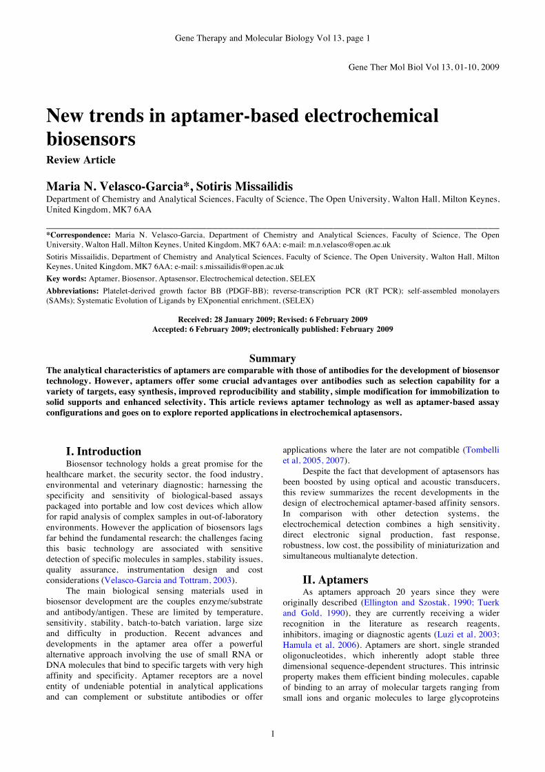
A. The SELEX process Aptamers are typically isolated from combinatorial
libraries by a process of in vitro evolution, termed SELEX
(Systematic Evolution of Ligands by EXponential
enrichment). This procedure is an in vitro evolutionary
selection process that allows the isolation of aptamer(s),
with unique binding properties, from a large library of
oligonucleotides through iterative cycles of (i) interaction
of a large library of aptamers with the target molecule, (ii)
separation of bound from unbound aptamer species, (iii)
elution of bound aptamers and (iv) PCR amplification of
the binding aptamers for further selection rounds (Figure
1 for an example of the process).
Library is incubated with target
under desired selection conditions
Unbound or non-specifically
bound species are washed
away and discarded
Unbound or non-specifically
bound species are washed
away and discarded
Aptamers are interacting
with and separated from
target using various separation
techniques, including affinity
chromatography columns,
magnetic bids or membranes
Desalted aptamers
are lyophilised and
PCR reagents added
for amplification
Amplified aptamers
are used in successive
selection rounds
Aptamers from the
final selection round
are double stranded
and cloned in E. coli
Clones are sequenced
for identification of
high binding aptamer
species to be used
subsequently for
further development
Bound aptamers are
eluted from their
target using high
salt or chaotropic
agents and desalted
for further PCR
amplification steps
Positive clones are analysedPositive clones are analysed
Figure 1. The SELEX process.
Gene Therapy and Molecular Biology Vol 13, page 3
3
An aptamer library usually consists of a variable
region (20-40 nucleotides) flanked by known primer
sequences on either end for the amplification during the
SELEX procedure. The variable region makes up to 1015
different sequences which, combined with the innate
ability of oligonucleotides to form stable sequence-
dependent structures, provide an array of molecular shapes
available for the selection process (Khan and Missailidis,
2008). In the selection steps, the library is incubated with
the immobilised target. Unbound or weak-binding species
are removed and bound aptamers are eluted using high
salt, temperature, chaotropic agents or other such
conditions that would affect molecular structure or disrupt
molecular interactions. Eluted aptamers are subsequently
amplified by PCR (DNA) or reverse-transcription PCR
(RT PCR) using primers complementary to the flanking
sequences in the aptamer library. The enriched pool of
binding species forms the pool for the next round of
selection. Repeated selection and amplification steps allow
identification of the highest binding species, through
competitive binding. The selection and amplification step
constitutes one round or cycle in a typical SELEX
procedure, with anything between 1 and 15 cycles often
described in the literature. Counter- or negative selection
steps can ensure that the finally selected aptamers are very
specific for their target and do not interact with
homologous proteins or chemically closely-related
molecular targets (Missailidis, 2008).
Selected aptamers are subsequently cloned and
sequenced to identify the sequence of the binding species
and their interactions are usually characterised by a variety
of analytical methodologies, prior to move into the various
applications they were originally destined for. Selected
aptamer can be easily produced by solid phase synthesis
and appropriate modifications can be introduced at this
stage to confer additional properties to the selected
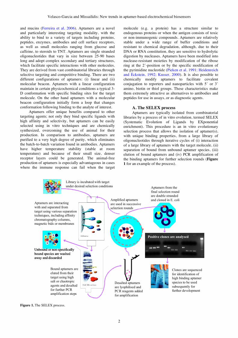
aptamers, such as nuclease resistance (Figure 2), cross-
linking ability or improved pharmacokinetic properties.
Although SELEX has been the initial methodology
associated with aptamer selection and has remained a
robust and powerful technique, which has been adapted to
various systems and targets, a number of other
methodologies have also emerged for the selection of
aptamers. Such “non-SELEX” based methods for the
selection of aptamers include capillary electrophoresis
methodologies (Berezovski et al, 2005; Drabovich et al,
2005), isolation of aptamers with predefined kinetic and
thermodynamic properties of their interaction with the
target, without the need for amplification, allowing the use
of libraries which are difficult or cannot be amplified, or
computational methods, which are particularly important
in selecting aptamers with inhibitory activities or
sequences that undergo ligand dependent conformational
changes (Ikebukuro et al, 2005).
The SELEX procedure and subsequent technologies for
aptamer selection have offered the tools for the designing
of aptamers that have found a range of diagnostic
applications (Khan and Missailidis, 2008). Such
applications include Photo-SELEX (www.somalogic.com)
and SELEX NADIR (Winters-Hilt, 2006) using optical
probe reporting or nanopore reporting mechanisms
respectively, aptamer microarrays (Cho et al, 2005),
currently in the market by LC Sciences
(www.lcsciences.com), fluorescent aptamers in chips and
microspheres (Potyrailo et al, 1998; Kirby et al, 2004),
fluorescent sensors for small molecule recognition
(Yamana et al, 2003; Ozaki et al, 2006), quantum dots
(Levy et al, 2005; Choi et al, 2006; Ivanovic et al, 2007;
Liu et al, 2007), colorimetric detection (Liu and Lu, 2004;
Cho et al, 2006; Liu and Lu, 2006), electrochemical
detection (Xiao et al, 2005; Mir et al, 2006; Lai et al,
2007; Papamichael et al, 2007) and piezoelectric quartz
crystal sensors (Bini et al, 2007).
The above methods, fluorescent, electrochemical and
colorimetric detection, have also been used in molecular
switch type sensors or modular sensor assemblies, where
the aptamers usually change conformation upon binding to
either emit a fluorescent signal based on an aptamer
beacon on sensor, or through non-covalent interaction with
the fluorescent label, triggering an electrochemical sensor
or leading to change of colour (Frauendorf and Jaschke,
2001; Stojanovic et al, 2001; Stojanovic and Landry,
2002; Stojanovic and Kolpashchikov, 2004; Baker et al,
2006; Zuo et al, 2007), with particular sensitivities in the
recognition of small analytes.
Aptamers have also been used in enzymatic sensing,
without the use of any label or signal related directly to the
aptamer. These applications remain based on changes in
the conformation of bifunctional aptamers that recognise
the target ligand and an enzyme or ribosome. The binding
of the aptamer to the ligand results in conformational
changes that affect enzymatic activity or protein
expression, and it is the later that is subsequently
measured (Ogawa and Maeda, 2007; Yoshida et al, 2006;
Yoshida et al, 2006) or utilises an enzyme to ligate
N
NN
N
NH2
O
HO
HH
HH
PO
O
O
O-
N
NH2
ON
O
NH2 or FOH
HH
HH
Figure 2. An amino or fluoro modification at the 2’ position of
the sugar can confer the oligonucleotide aptamer stability against
nuclease degradation. An alternative to using modifications at the
2’ of the sugar (whether at the 3’ or 5’ end of the aptamer, or
Velasco-Garcia and Missailidis: New trends in aptamer-based electrochemical biosensors
4
both) for nuclease resistance is to use a flipped base added to the
end of the aptamer.
proximally bound aptamers to large protein targets and
allow their subsequent PCR amplification (Fredriksson et
al, 2002).
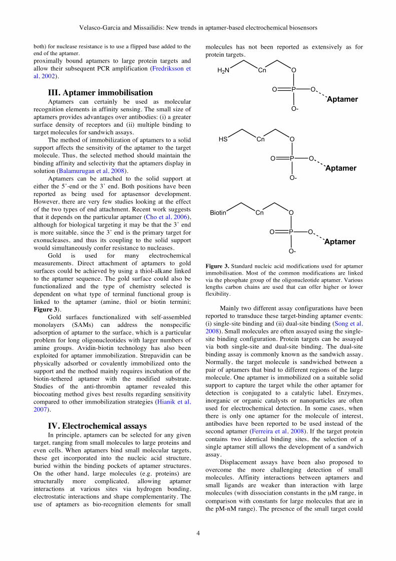
III. Aptamer immobilisation Aptamers can certainly be used as molecular
recognition elements in affinity sensing. The small size of
aptamers provides advantages over antibodies: (i) a greater
surface density of receptors and (ii) multiple binding to
target molecules for sandwich assays.
The method of immobilization of aptamers to a solid
support affects the sensitivity of the aptamer to the target
molecule. Thus, the selected method should maintain the
binding affinity and selectivity that the aptamers display in
solution (Balamurugan et al, 2008).
Aptamers can be attached to the solid support at
either the 5’-end or the 3’ end. Both positions have been
reported as being used for aptasensor development.
However, there are very few studies looking at the effect
of the two types of end attachment. Recent work suggests
that it depends on the particular aptamer (Cho et al, 2006),
although for biological targeting it may be that the 3’ end
is more suitable, since the 3’ end is the primary target for
exonucleases, and thus its coupling to the solid support
would simultaneously confer resistance to nucleases.
Gold is used for many electrochemical
measurements. Direct attachment of aptamers to gold
surfaces could be achieved by using a thiol-alkane linked
to the aptamer sequence. The gold surface could also be
functionalized and the type of chemistry selected is
dependent on what type of terminal functional group is
linked to the aptamer (amine, thiol or biotin termini;
Figure 3).
Gold surfaces functionalized with self-assembled
monolayers (SAMs) can address the nonspecific
adsorption of aptamer to the surface, which is a particular
problem for long oligonucleotides with larger numbers of
amine groups. Avidin-biotin technology has also been
exploited for aptamer immobilization. Strepavidin can be
physically adsorbed or covalently immobilized onto the
support and the method mainly requires incubation of the
biotin-tethered aptamer with the modified substrate.
Studies of the anti-thrombin aptamer revealed this
biocoating method gives best results regarding sensitivity
compared to other immobilization strategies (Hianik et al,
2007).
IV. Electrochemical assays In principle, aptamers can be selected for any given
target, ranging from small molecules to large proteins and
even cells. When aptamers bind small molecular targets,
these get incorporated into the nucleic acid structure,
buried within the binding pockets of aptamer structures.
On the other hand, large molecules (e.g. proteins) are
structurally more complicated, allowing aptamer
interactions at various sites via hydrogen bonding,
electrostatic interactions and shape complementarity. The
use of aptamers as bio-recognition elements for small
molecules has not been reported as extensively as for
protein targets.
O
PO O
O-
CnHS
Aptamer
O
PO O
O-
CnBiotin
Aptamer
O
PO O
O-
CnH2N
Aptamer
Figure 3. Standard nucleic acid modifications used for aptamer
immobilisation. Most of the common modifications are linked
via the phosphate group of the oligonucleotide aptamer. Various
lengths carbon chains are used that can offer higher or lower
flexibility.
Mainly two different assay configurations have been
reported to transduce these target-binding aptamer events:
(i) single-site binding and (ii) dual-site binding (Song et al,
2008). Small molecules are often assayed using the single-
site binding configuration. Protein targets can be assayed
via both single-site and dual-site binding. The dual-site
binding assay is commonly known as the sandwich assay.
Normally, the target molecule is sandwiched between a
pair of aptamers that bind to different regions of the large
molecule. One aptamer is immobilized on a suitable solid
support to capture the target while the other aptamer for
detection is conjugated to a catalytic label. Enzymes,
inorganic or organic catalysts or nanoparticles are often
used for electrochemical detection. In some cases, when
there is only one aptamer for the molecule of interest,
antibodies have been reported to be used instead of the
second aptamer (Ferreira et al, 2008). If the target protein
contains two identical binding sites, the selection of a
single aptamer still allows the development of a sandwich
assay.
Displacement assays have been also proposed to
overcome the more challenging detection of small
molecules. Affinity interactions between aptamers and
small ligands are weaker than interaction with large
molecules (with dissociation constants in the µM range, in
comparison with constants for large molecules that are in
the pM-nM range). The presence of the small target could
Gene Therapy and Molecular Biology Vol 13, page 5
5
induce the separation of two strands of a duplex nucleic
acid (one strand being the aptamer immobilised to a solid
support). Another strategy could rely on the displacement
of the aptamer from its complex with the immobilised
target molecule when the molecule is present in solution
(De-los-Santos-Alvarez et al, 2008).
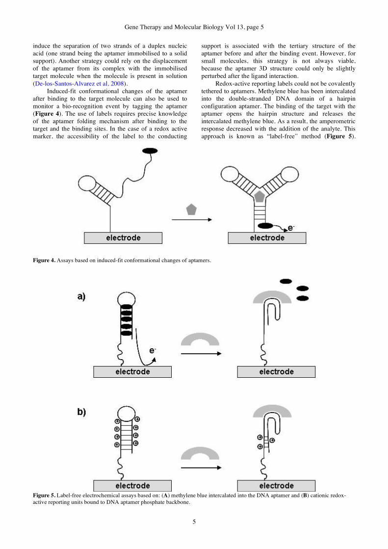
Induced-fit conformational changes of the aptamer
after binding to the target molecule can also be used to
monitor a bio-recognition event by tagging the aptamer
(Figure 4). The use of labels requires precise knowledge
of the aptamer folding mechanism after binding to the
target and the binding sites. In the case of a redox active
marker, the accessibility of the label to the conducting
support is associated with the tertiary structure of the
aptamer before and after the binding event. However, for
small molecules, this strategy is not always viable,
because the aptamer 3D structure could only be slightly
perturbed after the ligand interaction.
Redox-active reporting labels could not be covalently
tethered to aptamers. Methylene blue has been intercalated
into the double-stranded DNA domain of a hairpin
configuration aptamer. The binding of the target with the
aptamer opens the hairpin structure and releases the
intercalated methylene blue. As a result, the amperometric
response decreased with the addition of the analyte. This
approach is known as “label-free” method (Figure 5).
Figure 4. Assays based on induced-fit conformational changes of aptamers.
Figure 5. Label-free electrochemical assays based on: (A) methylene blue intercalated into the DNA aptamer and (B) cationic redox-
active reporting units bound to DNA aptamer phosphate backbone.
Velasco-Garcia and Missailidis: New trends in aptamer-based electrochemical biosensors
6
Related approaches use cationic redox-active reporting
units bound to the electrode via electrostatic interactions
with the DNA aptamer phosphate backbone. The binding
of the target molecule with the aptamer blocked the
binding of the cationic reporting units and the
electrochemical response decreased. The main
disadvantage of these latter approaches is a negative
detection signal.
Recently, nanomaterials are also providing novel
electrochemical sensing approaches. Single-walled carbon
nanotube field-effect transistor sensors were developed to
monitor aptamer-protein binding studies. Aptamers are
well suited for FET sensing due to their small size (1-2
nm) and recognition occurs inside the electrical double-
layer associated with the gate (within the Debye length).
The single-walled carbon nanotubes were assembled
between source and drain electrodes and the aptamers
were immobilized to these nanomaterials. In this label-free
approach, the binding of the target molecule to the
aptamers altered conductance through the device. The ease
of miniaturization of these sensing devices opens up the
feasibility of high-throughput assays in microarrays.
Nanoparticles have also been reported as catalytic
labels, instead of enzymes, and carriers for ultrasensitive
electrochemical detection; because one nanoparticle
contains a large number of aptamers, the target binding
process is amplified.
Impedance spectroscopy has been the most
frequently used electrochemical method in the
development of electrochemical aptasensors and has
shown excellent sensitivity, achieving limit of detection of
fM. However, despite the fact that the analytical technique
is simple to perform, the data fitting remains a bit
complicated. Easier data processing and faster response
could be achieved with chonoamperometry, but the limit
of detection will be higher and in the nM range.
IV. Applications of electrochemical
aptasensors Aptamer publications have now appeared in the
literature using most of the electrochemical transducers.
The majority of aptamer work on electrochemical sensors
is focused on amperometric transducers, but there have
been references on aptamers used in impedimetric, FET
and recently potentiometric sensors. Furthermore, a lot of
the work on the aptamers in electrochemical sensors has
been on the model protein, thrombin, which is one of the
best characterised complexes in the aptamer literature.
These have provided proof of principle concepts as to how
aptamers could be developed in novel sensors. However, a
number of other systems have also now been described,
which will be presented in this review.
A. Electrochemical aptasensors for the model
protein The thrombin-binding aptamer (15-mer, 5’-
GGTTGGTGTGGTTGG-3’) was the first one selected in
1992 by Block and colleagues and its structure has been
well characterized and studied. The folded structure in
solution is composed of two guanine quartets connected
by two T-T loops spanning the narrow grooves at one end
and a T-G-T loop spanning a wide groove at the other end
(known as the G-quartet structure). This anti-thrombin
aptamer has been extensively used as the model
oligonucleotide by many researchers to demonstrate the
wide applicability of aptamers as bio-recognition elements
in biosensors.
In the literature, many different electrochemical
aptasensors for thrombin detection have been reported.
The most straightforward configuration is based on the
immobilization of a thiol terminated aptamer on a gold
electrode. The aptamer-thrombin interaction is transduced
by the electrochemical quantification of p-nitroaniline
produced by the thrombin’s enzymatic reaction. Thrombin
has two electropositive exosites both capable of binding
the aptamer, allowing the development of an
electrochemical sensor system in a sandwich manner. The
thiolated aptamer was immobilized on a gold electrode
and, after incubation with the thrombin, a second
incubation step with an HRP labelled aptamer took place.
Electrochemical detection of HRP was performed using
H2O2 and a diffusional osmium based mediator. A similar
aptasensor system in sandwich manner for thrombin was
developed based on the aptamer for detection, labelled
with pyrroquinoline quinine glucose dehydrogenase, and
the electric current generated from glucose addition after
the formation of the complex on a gold electrode
(Ikebukuro et al, 2005). Another strategy for the thrombin
sensing is the direct immobilization of the protein on the
electrode surface. After the incubation with biotin-labelled
aptamer and then with streptavidin-HRP, the
electrochemical detection is performed using H2O2 and a
diffusional osmium-based mediator. The latter approach
achieved the lower limit of detection, 3.5 nM (Mir et al,
2006).
Mir and colleagues also developed in 2008 a
chronoamperometric beacon biosensor based on a
ferrocene-labelled thiol-aptamer. The aptamer adopts a 3-
D conformational change after binding the thrombin,
allowing the ferrocene label to approach to the gold
electrode. The interaction is detected via a
microperoxidase mediated electron transfer between the
label and the electrode surface. The system was
demonstrated with impedance spectroscopy and
chronoamperometry measurements, achieving a limit of
detection of 30 fM with the impedance spectroscopy (Mir
et al, 2008).
Methylene blue has also been used as an
electrochemical marker. The beacon aptamer surface was
prepared following formation of 11-mercaptoundecanoic
acid self-assembled monolayer on gold electrode.
Methylene blue was intercalated on the aptamer by the
interaction with two guanine bases. Binding of the
thrombin is correlated with the decrease in electrical current intensity in voltammetry. The estimated detection
limit of the target thrombin was 11 nM (Bang et al, 2005).
The modification of antibodies is difficult, costly and
time consuming; however researchers have been using
conventional polyclonal antibodies as a capturing probe
and labelled-aptamers as the detection probe in new
sandwich approaches for protein detection. Kang and
Gene Therapy and Molecular Biology Vol 13, page 7
7
colleagues reported in 2008 a modified electrochemical
sandwich model for thrombin, based on capturing
antibody immobilized onto glassy carbon electrodes with
nanogold-chitosan composite film and Methylene blue
labelled aptamer as the electrochemical detection probe.
Lu and colleagues described in 2008 an
electrochemical aptasensor for thrombin that is not based
on the target binding-induced conformational change of
aptamers. The thrombin-binding aptamer is first assembled
onto a gold electrode and then hybridized with a ferrocene
labelled short aptamer-complementary DNA
oligonucleotide. The binding of the thrombin to the
aptamer destroys the double-stranded DNA
oligonucleotide and leads to the dissociation of the label
short complementary DNA oligonucleotide from the
electrode surface, resulting in a decrease in the differential
pulse voltammetry responses at the electrode (Lu et al,
2008). This strategy is based on the stronger binding
affinity of the aptamers towards their targets rather than to
the short aptamer-complementary DNA oligonucleotide
labelled with electroactive moieties.
The majority of the work performed on aptamer-
based electrochemical biosensors is based on aptamers
labelled using redox compounds, such as methylene blue,
and catalysts such as horseradish peroxidase. However,
nanoparticle-based materials offer excellent prospects for
a new signal amplification strategy for ultrasensitive
electrochemical aptasensing. Platinum nanoparticles have
been reported as catalytic labels when linked to a thiolated
aptamer. The nanoparticles catalysed the electrochemical
reduction of H2O2 and the resulting current enabled the
amplified detection of thrombin sandwiched between the
aptamer on the electrode surface and the aptamer labelled
with the nanoparticles (Polsky et al, 2006). Gold
nanoparticles offer several advantages such as electrical
conductivity, biocompatibility, ease of self-assembly
through a thiol group, increase electrode surface area and
amount of immobilized capturing probe. Gold
nanoparticles have been used as an electrochemical
sensing platform for direct detection of thrombine. The
aptamer was immobilised on a screen-printed electrode
modified with gold-nanoparticles by avidin-biotin
technology. The gold-nanoparticles surface status is
evaluated by the Au/Au oxide film formation with cyclic
and stripping voltammetry. Gold nanoparticles signal
changed with the deposition of biolayers due to
differences in electron transfer efficacy and availability of
buffer oxygen. Aptamers prefer to adopt the G-quarter
structure when binding with thrombin and the
conformational changes made double strand DNA zones
appear and facilitated the electron transfer from solution to
the electrode surface, based on the double stranded DNA’s
ability to transport charge along the nucleotide stacking
(Suprun et al, 2008). The detection limit of this novel
approach is in the nM range. However, the aptasensor
measured directly binding events and opened 4 orders of
magnitude the operating range of protein concentration.
Assays coupling aptamers with magnetic beads for
the aptamer or target immobilisation before the
electrochemical transduction have also been proposed
(Centi et al, 2008). The use of magnetic beads improved
the assay kinetics due to the beads being in suspension and
also minimized matrix effect because of better washing
and separation steps.
An ultrasensitive electrochemical aptasensor for
thrombin in a sandwich format of magnetic nanoparicle-
immobilized aptamer, thrombin and gold nanoparticle-
labelled aptamer was reported by Zheng and colleagues in
2007. The magnetic nanoparticle-immobilized aptamer
was used for capturing and separating the target protein.
The gold nanoparticle-labelled aptamer offered the
electrochemical signal transduction. The signal was
amplified by forming a network like thiocyanuric acid/
gold nanoparticles to cap more nanoparticles per assay,
lowering the detection limit to the aM range
B. Other targets Aptamer have been selected against a wide range of
targets with typical binding affinities in the nanomolar to
picomolar range. Recently, electrochemical aptasensors
have been reported to detect proteins, hormones and drugs.
Papamichael and colleagues described in 2007 a
disposable electrochemical aptasensor for
Immunoglobulin E, a key marker of atopic diseases (such
as asthma, dermatitis and pollenosis). The sensor
incorporates a competitive format for IgE detection using
a biotinylated form of the aptamer. A standard, indirect
method was used where competition between surface-
bound IgE and IgE in solution proceeded for the aptamer.
The electrochemical detection is achieved by the use of an
extravidin-alkaline phosphatase label. After careful
optimization of conditions (buffer pH, ionic strength,
additional ions and proteins), the aptasensor was
performing at levels suitable for human testing (>300ng
ml-1).
Platelet-derived growth factor BB (PDGF-BB) is one
important cytokine involved in neural inflammation and
was selected as target for the development of an
electrochemical aptasensor based on capacitance change
induced by aptamer-protein specific binding, measured by
non-faradic impedance spectroscopy. The biosensor
detection limit was 40 nM. Electrochemical impedance
spectroscopy is a very attractive method for in vivo
diagnostics, due to its high sensitivity and label free
characteristics (Liao and Cui, 2007). A similar
electrochemical detection was also reported to a
tuberculosis-related cytokine, the interferon-!. The
aptamer-based electrochemical impedance biosensor
successfully detected interferon-! to a level of 100 fM
with an RNA aptamer and 1 pM with a DNA aptamer
probe (Min et al, 2008).
Electrochemical aptasensors for 17-" estradiol have
also been reported. The selected biotinylated DNA
aptamer was immobilized on a streptavidin-modified gold
electrode. The chemical binding of the hormone to the
aptamer was monitored by cyclic and square wave
voltammetry. When the 17-" estradiol interacted with the
aptamer, the current decreased due to the interference of
the bound target molecule with the electron flow produced
by a redox reaction between ferrocyanide (the mediator)
and ferricyanide. The linear range of this aptasensing
device was 1-0.01 nM of 17-" estradiol (Kim et al, 2007).
Velasco-Garcia and Missailidis: New trends in aptamer-based electrochemical biosensors
8
Cocaine has been detected by an electrochemical
aptasensor incorporating gold nanoparticles onto the
surface of a gold electrode. The thiol-derivative aptamer
was self-assembled onto the gold nanoparticles. The
aptamer was also functionalized at the other termini of the
strand with a redox-active ferrocene moiety. The cocaine
binding to the aptamer induces the conformational change
of the aptamer, bringing the redox tag in close proximity
to the electrode, leading to an increase in the current (Li et
al, 2008). Methylene blue tagged aptamer has been also
explored for the detection of cocaine (Baker et al, 2006).
A novel adenosine aptasensor was reported based on
the structure change of an aptamer probe immobilized on a
gold electrode. After the binding aptamer-target
nucleoside, a higher surface charge density and an
increasing steric hindrance were obtained that reduce the
diffusion of [Fe(CN)6]3-/[Fe(CN)6]
4- towards the electrode
surface, resulting in a decrease of the current. The
biosensing surface was easily regenerated and the
aptasensor limit of detection was 10 nM (Zheng et al,
2008).
C. Aptasensor arrays Some of the aptamer-based biosensor technology
described in this review could be transferred from single-
analyte devices to electrochemical methods offering the
possibility of simultaneous measurements of a panel of
targets. Wang reviewed the use of metal nanoparticles as
tracers for the analysis of nucleic acid hybridization.
Magnetic nanoparticles were linked to different probe
DNAs and incubated with samples containing different
DNA targets. Semiconductor quantum dots were
functionalized each with different nucleic acids
complementary to the free chain of the target DNA. After
dissolution of the metal nanoparticles, the identification of
the metal ions by stripping voltammetry enabled the
analysis of the different DNA targets (Wang, 2003).
Thrombin and lysozyme were detected in parallel
using a competitive assay in which thrombin and
lysozyme were modified with different semiconductor
quantum dots (Hansen et al, 2006). Specific aptamers were
immobilized on a gold electrode and bound to the
respective labelled protein. In the presence of unlabelled
protein in the sample, the quantum-dot functionalized
protein is displaced from the electrode into solution. The
dissolution of the remaining metal ions on the surface and
the electrochemical detection of the released ions enabled
the quantitative detection of the proteins.
IV. Conclusions Aptamers have been widely used in a variety of
diagnostic and sensor applications, offering a variety of
possibilities for aptamer-based sensors in early disease
diagnosis and prognosis, substance control, environmental
measurements or national security applications on
measurements of explosives or potential infectious agents.
Yet, despite the advances and the huge body of literature
documenting the success of the technology, the
commercial application of aptamers in the field of
diagnostics remains relatively undeveloped, not least due
to the exclusive IP portfolio, and the fact that there is a
vast antibody-based diagnostic market and a certain degree
of hesitation to move to a new type of product, unless
aptamers offer verifiably significant improvements on
current technologies that warrant substitution of antibodies
in some current assay formats. In this review, different
types of electrochemical aptamer-based biosensors have
been discussed. Although the optical and mass-sensitive aptasensors have been the most commonly described in
the literature, electrochemical transducers have enormous
potential and offer simple, rapid, cost-effective and easy to
miniaturize sensing in many diagnostic fields. Emerging
nanomaterials have also brought new possibilities for
developing novel ultrasensitive electrochemical
aptasensors.
References Baker BR, Lai RY, Wood MCS, Doctor EH, Heeger AJ, Plaxco
KW (2006) An electronic, aptamer-based small-molecule
sensor for the rapid label-free detection of cocaine in
adulterated samples and biological fluids. J Am Chem Soc
128, 3138-3139.
Balamurugan S, Obubuafo A, Soper SA, Spivak DA (2008)
Surface immobilization methods for aptamer diagnostic
applications. Anal Bioanal Chem 390, 1009-1021.
Bang GS, Cho S, Kim BG (2005) A novel electrochemical
detection method for aptamer biosensors. Biosens Bioelec
21, 863-870.
Berezovski M, Drabovich A, Krylova SM, Musheev M, Okhonin
V, Petrov A, Krylov SN (2005) Nonequilibrium Capillary
Electrophoresis of Equilibrium Mixtures: A Universal Tool
for Development of Aptamers. J Am Chem Soc 127, 3165-
3171.
Bini A, Minunni M, Tombelli S, Centi S, Mascini M (2007)
Analytical performances of aptamer-based sensing for
thrombin detection. Anal Chem 79, 3016-3019.
Centi S, Messina G, Tombelli S, Palchetti I, Mascini M (2008)
Different approaches for the detection of thrombin by an
electrochemical aptamer-based assay coupled to magnetic
beads. Biosens Bioelec 23, 1602-1609.
Cho EJ, Collett JR, Szafranska AE, Ellington AD (2006)
Optimization of aptamer microarray technology for multiple
protein targets. Anal Chim Acta 564, 82-90.
Cho S, Kim JE, Lee BR, Kim JH, Kim BG (2005) Bis-aptazyme
sensors for hepatitis C virus replicase and helicase without
blank signal. Nucleic Acids Res 33, e177.
Choi JH, Chen KH, Strano MS (2006) Aptamer-Capped
Nanocrystal Quantum Dots: A New Method for Label-Free
Protein Detection. J Am Chem Soc 128, 15584-15585.
De-los-Santos-Alvarez N, Lobo-Castañón MJ, Miranda-Ordieres
AJ, Tuñón-Blanco P (2008) Aptamers as recognition
elements for label-free analytical devices. Trends Anal
Chem 27, 437-446.
Drabovich A, Berezovski M, Krylov SN (2005) Selection of
Smart Aptamers by Equilibrium Capillary Electrophoresis of
Equilibrium Mixtures (ECEEM). J Am Chem Soc 127,
11224-11225.
Ellington AD, Szostak JW (1990) In vitro selection of RNA
molecules that bind specific ligands. Nature 346, 818-822.
Ferreira CSM, Matthews CS, Missailidis S (2006) DNA
aptamers that bind to MUC1 tumour marker: Design and
characterization of MUC1-binding single stranded DNA
aptamers. Tumor Biol 27, 289-301.
Ferreira CSM, Papamichael K, Guilbault G, Schwarzacher T,
Gariepy J, Missailidis S (2008) Design of aptamer-antibody
Gene Therapy and Molecular Biology Vol 13, page 9
9
sandwich ELISA for early tumour diagnosis. Anal Bioanal
Chem 390, 1039-1050.
Frauendorf C, Jaschke A (2001) Detection of small organic
analytes by fluorescing molecular switches. Bioorg Med
Chem 9, 2521-2524.
Fredriksson S, Gullberg M, Jarvius J, Olsson C, Pietras K,
Gustafsdottir SM, Ostman A, Landegren U (2002) Protein
detection using proximity-dependent DNA ligation assays.
Nature Biotechnol 20, 473-477.
Hamula CLA, Guthrie JW, Zhang H, Li XF, Le XC (2006)
Selection and analytical applications of aptamers. Trends
Anal Chem 25, 681-691.
Hansen JA, Wang J, Kawde AN, Xiang Y, Gothelf KV, Collins
G (2006) Quantum-dot/aptamer-based ultrasensitive multi-
analyte electrochemical biosensor. J Am Chem Soc 128,
2228-2229.
Heidenreich O, Eckstein F (1992) Hammerhead ribozyme-
mediated cleavage of the long terminal repeat RNA of
human immunodeficiency virus type 1. J Biol Chem 267,
1904-1909.
Hianik T, Ostatna V, Sonlajtnerova M, Grman I (2007) Influence
of ionic strength, pH and aptamer configuration for binding
affinity to thrombin. Bioelectrochemistry 70, 127-133.
Ikanovic M, Rudzinski WE, Bruno JG, Allman A, Carrillo MP,
Dwarakanath S, Bhahdigadi S, Rao P, Kiel JL, Andrews CJ
(2007) Fluorescence Assay Based on Aptamer-Quantum Dot
Binding to Bacillus thuringiensis Spores. J Fluoresc 17, 193-
199.
Ikebukuro K, Kiyohara C, Sode K (2005) Novel electrochemical
sensor system for protein using the aptamers in sandwich
manner. Biosens Bioelec 20, 2168-2172.
Ikebukuro K, Okumura Y, Sumikura K, Karube I (2005) A novel
method of screening thrombin-inhibiting DNA aptamers
using an evolution-mimicking algorithm. Nucleic Acids Res
33, e108.
Kang Y, Feng KJ, Chen JW, Jiang JH, Shen GL, Yu RQ (2008)
Electrochemical detection of thrombin by sandwich approach
using antibody and aptamer. Bioelectrochemistry 73, 76-81.
Khan H, Missailidis S (2008) Aptamers in oncology: a diagnostic
perspective. Gene Ther Mol Biol 12, 111-128.
Kim YS, Jung HS, Matsuura T, Lee HY, Kawai T, Gu MB
(2007) Electrochemical detection of 17beta-estradiol using
DNA aptamer immobilized gold electrode chip. Biosens
Bioelec 22, 2525-2531.
Kirby R, Cho EJ, Gehrke B, Bayer T, Park YS, Neikirk DP,
McDevitt JT, Ellington AD (2004) Aptamer-based sensor
arrays for the detection and quantification of proteins. Anal
Chem 76, 4066-4075.
Kusser W (2000) Chemically modified nucleic acid aptamers for
in vitro selections: evolving evolution. Rev Mol Biotechnol
74, 27-38.
Lai RY, Plaxco KW, Heeger AJ (2007) Aptamer-based
electrochemical detection of picomolar platelet-derived
growth factor directly in blood serum. Anal Chem 79, 229-
233.
Levy M, Cater SF, Ellington AD (2005) Quantum-Dot Aptamer
Beacons for the Detection of Proteins. ChemBioChem 6,
2163-2166.
Li X, Qi H, Shen L, Gao Q, Zhang C (2008) Electrochemical
Aptasensor for the Determination of Cocaine Incorporating
Gold Nanoparticles Modification. Electroanalysis 20, 1475-
1482.
Liao W, Cui XT (2007) Reagentless aptamer based impedance
biosensor for monitoring a neuro-inflammatory cytokine
PDGF. Biosens Bioelec 23, 218-224.
Liu J, Lee JH, Lu Y (2007) Quantum Dot Encoding of Aptamer-
Linked Nanostructures for One-Pot Simultaneous Detection
of Multiple Analytes. Anal Chem 79, 4120-4125.
Liu J, Lu Y (2004) Adenosine-dependent assembly of aptazyme-
functionalized gold nanoparticles and its application as a
colorimetric biosensor. Anal Chem 76, 1627-1632.
Liu J, Lu Y (2006) Fast colorimetric sensing of adenosine and
cocaine based on a general sensor design involving aptamers
and nanoparticles. Angew Chem Int Ed 45, 90-94.
Lu Y, Zhu N, Yu P, Mao L (2008) Aptamer-based
electrochemical sensors that are not based on the target
binding-induced conformational change of aptamers.
Analyst 133, 1256-1260.
Luzi E, Minunni M, Tombelli S, Mascini M (2003) New trends
in affinity sensing: aptamers for ligand binding. Trends
Anal Chem 22, 810-818.
Min K, Cho M, Han SY, Shim YB, Ku J, Ban C (2008) A simple
and direct electrochemical detection of interfero-gamma
using its RNA and DNA aptamers. Biosens Bioelec 23,
1819-1824.
Mir M, Vreeke M, Katakis I (2006) Different strategies to
develop an electrochemical thrombin aptasensor.
Electrochem Comm 8, 505-511.
Mir M, Vreeke M, Katakis I (2008) Ultrasensitive detection
based on an aptamer beacon electron transfer chain.
Electrochem Comm 10, 1533-1536.
Missailidis S (2008) Combinatorial Approach to Anticancer
Drug Design, In-Anticancer Therapeutics (Eds: S.
Missailidis), Wiley & Sons Ltd.
Ogawa A, Maeda M (2007) Aptazyme-based riboswitches as
label-free and detector-free sensors for cofactors. Bioorg
Med Chem Letters 17, 3156-3160.
Ozaki H, Nishihira A, Wakabayashi M, Kuwahara M, Sawai H
(2006) Biomolecular sensor based on fluorescence-labeled
aptamer. Bioorg Med Chem Letters 16, 4381-4384.
Papamichael KI, Kreuzer MP, Guilbault GG (2007) Viability of
allergy (IgE) detection using an alternative aptamer receptor
and electrochemical means. Sens Actuat B-Chem 121, 178-
186.
Pieken W, Olsen DB, Benseler F, Aurup H, Eckstein HF (1991)
Kinetic characterization of ribonuclease-resistant 2’-modified
hammerhead ribozymes. Science 253, 314-317.
Polsky R, Gill R, Willner I (2006) Nucleic Acid-Functionalized
Pt Nanoparticles: Catalytic Labels for the Amplified
Electrochemical Detection of Biomolecules. Anal Chem 78,
2268-2271.
Potyrailo RA, Conrad RC, Ellington AD, Hieftje GM (1998)
Adapting selected nucleic acid ligands (aptamers) to
biosensors. Anal Chem 70, 3419-3425.
Song S, Wang L, Li J, Zhao J, Fan C (2008) Aptamer-based
biosensors. Trends Anal Chem 27, 108-117.
Stojanovic MN, Kolpashchikov DM (2004) Modular aptameric
sensors. J Am Chem Soc 126, 9266-9270.
Stojanovic MN, Landry DW (2002) Aptamer-based colorimetric
probe for cocaine. J Am Chem Soc 124, 9678-9679.
Stojanovic MN, Prada PD, Landry DW (2001) Aptamer-based
folding fluorescent sensor for cocaine. J Am Chem Soc 123,
4928-4931.
Suprun E, Shumyantseva V, Bulko T, Rachmetova S, Rad’ko S,
Bodeoev N, Archakov A (2008) Au-nanoparticles as an
electrochemical sensing platform for aptamer-thrombin
interaction. Biosens Bioelec 24, 831-836.
Tombelli S, Minunni M, Mascini M (2005) Analytical
applications of aptamers. Biosens Bioelec 20, 2424-2434.
Tombelli S, Minunni M, Mascini M (2007) Aptamers- based
assays for diagnostics, environmental and food analysis.
Biomol Eng 24, 191-200.
Tuerk C, Gold L (1990) Systematic evolution of ligands by
exponential enrichment - RNA ligands to bacteriophage-T4
DNA-polymerase. Science 249, 505-510.

Velasco-Garcia and Missailidis: New trends in aptamer-based electrochemical biosensors
10
Velasco-Garcia MN, Mottram TT (2003) Biosensor Technology
addressing Agricultural Problems Biosys Eng 84, 1-12.
Wang J (2003) Nanoparticle-based electrochemical DNA
detection. Anal Chim Acta 500, 247-257.
Winters-Hilt S (2006) Nanopore Detector based analysis of
single-molecule conformational kinetics and binding
interactions. BMC Bioinformatics 7, S21.
www.lcsciences.com , accessed January 2009.
www.somalogic.com, accessed January 2009.
Xiao Y, Lubin AA, Heeger AJ, Plaxco KW (2005) Label-free
electronic detection of thrombin in blood serum by using an
aptamer-based sensor. Angew Chem Int Ed 44, 5456-5459.
Yamana K, Ohtani Y, Nakano H, Saito I (2003) Bis-pyrene
labeled DNA aptamer as an intelligent fluorescent biosensor.
Bioorg Med Chem Letters 13, 4329-4331.
Yoshida W, Sode K, Idebukuro K (2006b) Homogeneous DNA
sensing using enzyme-inhibiting DNA aptamers. Biochem
Biophys Res Comm 348, 245-252.
Yoshida W, Sode K, Ikebukuro K (2006a) Aptameric Enzyme
Subunit for Biosensing Based on Enzymatic Activity
Measurement. Anal Chem 78, 3296-3303.
Zheng F, Wu ZS, Zhang SB, Guo MM, Chen CR, Shen GL, Yu
RQ (2008) Aptamer-based Electrochemical Biosensors for
Highly Selective and Quantitative Detection of Adenosine.
Chem Res Chinese Universities 24, 138-142.
Zheng J, Feng W, Lin L, Zhang F, Cheng G, He P, Fang Y
(2007) A new amplification strategy for ultrasensitive
electrochemical aptasensor with network-like thiocyanuric
acid/gold nanoparticles. Biosens Bioelec 23, 341-347.
Zuo X, Song S, Zhang J, Pan D, Wang L, Fan C (2007) A target-
responsive electrochemical aptamer switch (TREAS) for
reagentless detection of nanomolar ATP. J Am Chem Soc
129, 1042-1043.
Maria N. Velasco-Garcia and Sotiris Missailidis


![Electrochemical miRNA Biosensors: The Benefits of ...€¦ · electrochemical nanobiosensors [6, 7]. The electrochemical nanobiosensors are pulling together the advantages of electrochemical](https://img.pdfslide.net/doc/110x75/5f5dab2fa5702b13b4580399/electrochemical-mirna-biosensors-the-benefits-of-electrochemical-nanobiosensors.jpg)