Embed Size (px)
Citation preview
SECTION 18
Electromagnetic Field Exposure Effects
(ELF-EMF and RFR)
on Fertility and Reproduction
Prof. Jitendra Behari, PhD
Bioelectromagnetics Laboratory
School of Environmental Sciences
Jawaharlal Nehru University
New Delhi, India
Dr. Paulraj Rajamani, PhD
Bioelectromagnetics Laboratory
School of Environmental Sciences
Jawaharlal Nehru University
New Delhi, India
Prepared for the BioInitiative Working Group
November 2012
2
I. INTRODUCTION
Electromagnetic fields and radiofrequency radiation (RFR) interact with human tissues and
may have adverse effects on fertility and reproduction. This review presents evidence for
ELF-EMF and RFR effects on many parameters of male sperm function; leading to questions
about the genotoxicity and carcinogenicity of such exposures on fertility and reproduction in
men. Much of the evidence comes from human and animal studies on sperm and male
fertility factors, but there are also studies showing adverse effects on fertility and miscarriage
in women.
During the last four decades or so there has been a growing concern on the effects of
electromagnetic radiations on biological systems in general. This is because of the global
introduction of electronic devices on a massive level for communications and data
transmission, personal wireless devices, air surveillance systems, industry applications,
medical/diagnostic and therapeutic purposes that are now new sources of electromagnetic
fields (ELF-EMF) and radiofrequency microwave radiation (RFR). This has added another
layer of pollutant (electropollution) to a growing list of environmental contaminants in air,
water, soil and from noise pollution which can adversely affect human health.
There are many sources of EMF in our environment and this non-ionizing radiation interacts
with the human body. Use of electronic household items and cell phones are reported to
decrease fertility potential in men by decreasing sperm count, motility, viability, inducing
pathological changes in sperm and testes morphology, and so on (Erogul et al. 2006). In
accordance with this, several authors (Agarwal et al. 2008, 2009; Kumar et al. 2010, 2011a;
Pourlis 2009; Kesari et al. 2010, 2011, 2012) focused mainly on the male reproduction
patterns. It involves the development from undifferentiated diploid stem cells to highly
differentiated haploid stem cells. Spermatogenesis is a complex process and it is influenced
by many genes and hormones. It takes place in the testis, which may be exposed to various
microwave frequencies which are currently in use (Behari and Kesari 2006). Among various
factors of infertility, oxidative stress has become the main focus of interest as a potential
cause of male infertility (Agarwal and Said 2003; Aitken and Roman, 2008; Kumar et al,
2010, 2011a). Male infertility is commonly associated with high rates of DNA
(deoxyribonucleic acid) damage in the spermatozoa and such damage is correlated with a
wide range of adverse clinical outcomes. Several studies, especially at power frequency 50/60
3
Hz magnetic field have found an association of exposure to human health, with emphasis on a
range of clinical conditions including childhood leukaemia, brain tumours, genotoxicity and
neurodegenerative disease, infertility, birth defects, increased risk of miscarriage, childhood
morbidity and de novo mutations (Hardell and Sage 2008; Gharagozloo and Aitken 2011;
Garcia et al. 2008; Huss et al. 2008; O’Carroll and Henshaw 2008; International Agency for
Research on Cancer (IARC) Monographs of the Evaluation of Carcinogenic Risks to Human
2002; California Health Department Services (CHDS) Report 2002). Sperm DNA damage is
therefore regarded as a potential risk factor to the development of normal human embryos
leading to impaired embryonic development.
II. THE BIOPHYSICS OF EXTREMELY LOW FREQUENCY FIELDS
Whenever a body having finite conductivity (biological body) is intercepted by EMF it
induces electric fields and circulating electric currents, which in turn competes with
endogenous current and voltages, thus disturbing normal physiological balance. The depth of
penetration within the body depends upon its frequency and the electric properties of the
exposed portion in the body. If the current density exceeds a certain threshold value,
excitation of muscles and nerves due to membrane depolarization is possible. The mode of
interaction of non-ionizing radiation with biological systems can be broadly divided into two
parts: extremely low frequency and radiofrequency/microwaves.
Whenever an electric field interacts with a biological body the incident field will be distorted,
such that the external field will be nearly perpendicular to the boundary surface. At 60 Hz
Einternal / Eexternal ≈ 4(10-8
). (1)
Thus a 60 Hz external field of 100 kV/m will produce an average internal E field of the order
of 4mV/m.
As far as the magnetic components of the extremely low frequency fields are concerned,
magnetic permeability of most biological materials is practically equal to that of free space
(4.10-7
) H/m. This signifies that ELF H field ‘inside’ will be practically equal to the H field
‘outside’. Only exceptions could be those biological materials that have magnetic particles
inside. A time varying magnetic field (also electric field) can also induce electric currents
into stationary conducting objects. Thus, all modes of interaction of time varying E fields
with living matter may be triggered by time-varying (not by static) magnetic field. According
to Faraday’s law of electromagnetic induction time varying magnetic flux will induce E fields
with resulting electrical potential differences and “eddy” currents through available
4
conducting paths. Sources generating low frequency electric and magnetic fields are more
likely to produce physiologically significant internal E fields through the mechanism of
magnetic induction. If an erect person is targeted by a vertical electric field it will be
considerably “enhanced” at the top of the person’s head and shoulder, and one would predict
therefore that the field in the tissue would also be enhanced above that of a flat slice exposed
to the same field (Deon, 1982). In a 60 Hz electric field of 1kV/m in air, the current densities
(Am/m2) in neck, waist and ankle turn out to be 0.591x10
-3, 0.427 x
-3 and 3.35x10
-3
respectively (Polk 1986).
III. THE BIOPHYSICS OF RADIOFREQUENCY AND MICROWAVE FIELDS
The biological bodies are inhomogeneous, having tissue-specific dielectric properties and the
complexity of the shape; which make the computations of the induced field difficult. The
fields induced inside the body act differently depending upon the frequency and more
particularly on (L/λ), (where L is the length of the biological body and λ the wavelength of
the incident field) upon, but are not limited to the following parameters:
(i) The location of the field with respect to the surroundings, e.g. if there are metallic
objects around, the person is grounded or otherwise.
(ii) Polarisation of the incident wave with respect to the orientation of the human
body.
(iii) Size of the human body (L) with respect to the wavelength (λ) of the incident
radiations (L/λ).
(iv) The portion of the human body.
(v) The electrical properties of the tissue in question.
In free space propagation of electromagnetic field the power density is given by
Power density = E2/1200 Π mW/cm
2 (1)
Where, E is the electric field strength.
The frequency in the radio frequency-microwave region are somewhat penetrated inside the
biological body interacting with the tissues inside.
5
From simple biophysical considerations, it follows that each body has a characteristic
resonant frequency depending upon the length of the long axis. Correspondingly, for the
same level of incident exposure the average value of power absorbed is dependent upon the
length of the body, the degree of decoupling decreasing the average value of SAR by more
than an order of magnitude. It is suggestive that absorbed RF energy can be converted into
other form of energy and can cause interference with the functioning of the biological
systems. A significant portion of this energy is converted into heat (absorption). The
biological effects are frequency dependent. Well below 100 KHz, the induced fields can even
stimulate nervous tissue.
IV. FERTILITY AND REPRODUCTION EFFECTS: ELF-EMF FIELD EXPOSURE
Since the biological body is diamagnetic it is transparent to the static magnetic field. It can
therefore interact with the motional activity of paramagnetic materials. Amara et al (2006)
has shown that adult male rats exposed to such fields (128 mT, 1hr/day for 30 days) show a
decrease in testosterone levels and induced DNA oxidation. Subchronic exposure failed to
alter spermatogenesis in rat testis. In a similar study Hong et al (2005) also concluded that 50
Hz EMFs (0.2 mT or 6.4 mT, exposed for a period of 4 weeks) may have the potential to
induce DNA strand breakage in testicular cells and sperm chromatin condensation in mice.
Al-Akhras et al (2006) also treated male adult rats to 50 Hz sinusoidal magnetic field (25T
or 250 mg) for 18 consecutive weeks. They reported no significant effects on the absolute
body weight and the weight of the testis of the exposed rats. However the weight of the
seminal vesicles and preputial glands were significantly reduced in the exposed male rats,
along with significant reduction in sperm count of the exposed rats. There was no significant
effect on the serum levels of male follicle stimulating hormone (FSH) during the 18 weeks of
exposure period. On the other hand there was a significant increase in the serum levels of
male luteinizing hormone (LH) after 18 weeks of exposure (p<0.005) while testosterone
levels were significantly decreased after 18 weeks of exposure period. These results suggest
that long term exposure of ELF could have adverse effects on mammalian fertility and
reproduction.
Different results have been presented by Chung et al (2005) where animals exposed in-utero
and subsequent neonatal exposure to a 60 Hz EMF(field strength 500 T or 5000 mG) from
6
day 6 of gestation to day 21 of lactation, did not produce any detectable alteration in
offspring spermatogenesis and fertility.
Akdag et al (2006) examined the effects of ELF magnetic fields (1.35 mT) on sperm count,
malondialdehyde concentration, the histology of organs as: testes, brain, liver, and kidney
tissues, p53 immunoreactivity of bone marrow and the serum concentrations of Cu2+
,
Zn2+
,Mn2+
and Fe3+
in rats. These authors found no statistically significant alteration except in
Mn2+
concentrations (p<0.001).
Influence of ultrasound (frequency 2,4 and 8 MHz) and constant magnetic field (7T) on
gametes, zygotes and embryos of the sea urchin were studied by Drozdov et al (2008).
Magnetic field exposure interrupts the process of the gamete fusion but did not influence
gametes, embryos, or embryonic development. The nature of these two stimuli is of different
type. Ultrasound may heat up the water if is of sufficient power, by way of increase in water
temperature and cavitation temperature, which may also break the cellular structure. The
effect of magnetic field is connected to the response of the cortical cytoskeleton, which
consists of bundles of actin microfilaments. The rearrangement of the cortical cytoskeleton
occurs during the first 20 minutes after the contact of sperm with the egg.
Kim et al (2009) examined the effect of a 16-week continuous exposure to ELF magnetic
field (MF) of 14 or 200 T (140 or 2000 mG) on testicular germ cell apoptosis in mice. They
reported no significant adverse effects of MF on body weight and testosterone levels in mice.
In TUNEL staining (in situ terminal deoxynucleotidyl transferase-mediated deoxy-UTP nick
end labelling), germ cells show a significantly higher apoptotic rate in exposed mice than in
sham controls (P<0.001). TUNEL-positive cells were mainly spermatogonia. In an electron
microscope study, degenerating spermatogonia showed condensation of nuclear chromatin
similar to apoptosis. These results indicate that apoptosis may be induced in spermatogenic
cells in mice by continuous exposure to 60 Hz of 14 MF T (140 mG).
Roychoudhury et al (2009) examined the effects of 50 Hz extremely low frequency
electromagnetic field on in vitro rabbit spermatozoa motility. These authors also studied the
effects after insemination. Pooled semen samples and a control were exposed to 50 Hz ELF
EMF. The difference of the test groups G1 and G2 with the control group CG (75.56%) for
spermatozoa motility were found to be significant (P<0.01). Differences were significant
(P<0.01) for curvilinear velocity (VCL) between the test group G3 (122.38 µ/s). Hormonally
simulated adult (9-12 months) females (n=140) were inseminated with semen samples from
G1, G2, G3 and G4 (0.88 x10⁹ spermatozoa /0.5 ml average insemination portion)
7
immediately after ELF EMF exposure and fertilization (kindling) rates were calculated. For
the G2 it was 54.28% data indicate 50 Hz ELF EMF induced alterations of spermatozoa
motility and kindling rate in rabbits, therefore influencing fertility.
Cao et al (2009) also reported that magnetic fields at 1000 Hz or 2000 Hz may damage the
testis by inducing injury to seminiferous tubules and Leydig cells, thickening the basal
membrane, derangement, exfoliation, massive apoptosis and necrosis of spermatogenic cells
in the lumen, epididymis, and consequently result in the absence of sperm.
Bernabo et al (2010) assessed the effect of acute (1hr) exposure of boar spermatozoa to an
extremely low frequency electromagnetic field (ELF-EMF) (50 Hz, MF 0-2 mT) on early
fertility outcome. They examined morpho-functional integrity of capacitated spermatozoa in
vitro and reported in vitro ELF-EMF >0.5 mT induced a progressive acrosome damage, thus
compromising the ability of spermatozoa to undergo acrosomal reaction after zona-pellucida
stimulation and reducing the in vitro fertilization outcome. These effects became evident at
0.75 mT and reached the plateau at 1 mT. Under in vivo conditions, ELF-EMF intensity of 1
mT was able to compromise sperm function, significantly reducing the fertilization rate. In
addition, the exposure of oviducts field 0.75 mT in the absence of spermatozoa was able to
negatively affect early embryo development. In fact it was found to cause a slowdown in the
embryo cleavage. It is apparent that at mentioned intensities the fields has negative effect on
early fertility outcome in a predictive animal model.
Earlier these authors (Bernabo et al 2007) reported that MF-ELF influence negatively by
dramatically effecting sperm morphology and function.
The blood-testis barrier is sensitive to environmental stimulation, which can affect its
permeability and then result in antisperm antibody (AsAb) generation, which is a key step in
male immune fertility. Wang et al (2010) reported the results of male mice exposed to
electromagnetic pulse (EMP) by measuring the expression of tight-junction of associated
proteins(ZO-1 and Occludin), vimentin microfilaments, and mice were sham exposed or
exposed to EMP at two different intensities (200 kV/m and 400 kV/m) for 200 pulses. The
testes were collected at different points after EMP exposure. Immunofluorescence
histochemistry, western blot, laser confocal microscopy and RT-PCR were used in this study.
Compared with sham group, the expression of ZO-1 and TGF-beta3 were significantly
decreased accompanied with unevenly stained vimentin microfilaments and increased serum
AsAb levels in EMP-exposed mice. These results are indicative of a potential BTB injury and
immune infertility in male mice exposed to certain intensity of EMP.
8
Lorio et al (2011) studied the functional relationship between the energy metabolism and the
enhancement of human sperm motility induced by ELF-EMF was investigated. Sperm
exposure to ELF-EMF resulted in a progressive and significant increase of mitochondrial
membrane potential and levels of ATP, ADP, and NAD(+) associated with sperm kinetic
parameters. However no significant effects were detected on other parameters such as
ATP/ADP ratio and energy change. When carbamoyl cyanide m-chlorophenyllhydrazone
(CICCP) was applied to inhibit the oxidative phosphorylation in the mitochondria, the values
of energy parameters and motility in the sperm incubated in the presence of glucose and
exposed ELF-EMF did not change, thus indicating that the glycolysis was not involved in
mediating ELF-EMF stimulatory effect on motility. By contrast, when pyruvate and lactate
were provided instead of glucose, the energy status and motility increased significantly in
ELF-EMF-treated sperm. Under these culture conditions, the inhibition of glycolytic
metabolism by 2-deoxy-D-glucose (DOG) again resulted in increased values of energy and
kinematic parameters, indicating that gluconeogenesis was not involved in producing glucose
for use in glycolysis. These authors concluded that the key role in mediating the stimulatory
effects exerted by ELF-EMF on human sperm motility is played by mitochondrial oxidative
phosphorylation rather than glycolysis. Earlier these authors (Lorio et al 2007) reported that
ELF-EMF exposure can improve spermatozoa motility and that this effect depends on the
field characteristics. ELF-EMF with 50 Hz and square wave shape (amplitude 5 mT),while
that of a sine wave of the same amplitude (also of 2.5 mT) and the same frequency had no
such effect. Further a three hour exposure in the first case had the effect on sperm motility
persisting for 21 hours.
People connected to local area networks wirelessly (Wi-Fi) were examined for human
spermatozoa. These authors (Avendano et al 2012) selected sperms from 29 healthy donors
for their capability to swim. This study using a laptop as a source contributed both ELF-EMF
and RFR to the exposure conditions. Each sperm suspension was divided into two aliquots.
One sperm aliquot (experimental) from each patient was exposed to an internet connected lap
top by Wi-Fi for 4 hours, whereas the second aliquot (unexposed) was used as control and
incubated under identical conditions without being exposed to the laptop. These authors
evaluated sperm motility, viability, and DNA. These authors reported that normozoospermic,
exposed ex vivo during 4 hour to a wireless internet –connected laptop showed a significant
decrease in progressive sperm motility and an increase in DNA fragmentation. Level of dead
sperm showed no significant differences between the two groups. They concluded that the
effect (which is non-thermal) decreased motility and induced DNA fragmentation. It is
9
therefore speculated that keeping a laptop connected wirelessly to the internet on the lap near
the testes may result in decreased male fertility.
Sage et al (2007) reported that personal and occupational use of personal digital assistants
(PDAs or palm-held wireless units) produce high intensity bursts of ELF-EMF exposure in
persons that carry a PDA close to the body (i.e., in a pocket or in a belt); or held to the head
for cell phone conversations. ELF-EMF emissions of 10T (100 mG) were recorded on
PDAs during normal office use over a 24 hr test period. Results of ELF-EMF measurements
show that email transmit and receive functions produce rapid, short duration ELF-EMF
spikes in the 2-10T (20 to 100 mG) range, each lasting several seconds to over a minute,
depending on the download size. Switching the PDAs produced continuously elevated ELF-
EMF pulses of over 90 T on two units. Thus the user who wears the PDA may be receiving
high-intensity ELF-EMF pulses throughout the day and night.
Avendano et al (2012) investigated the effect of laptop computers connected to internet
through Wi-Fi on human sperm motility. Donor sperm samples, mostly normozoospermic,
exposed ex vivo during 4 hours connection showed a significant decrease in progressive
sperm motility and an increase in sperm DNA fragmentation due to nonthermal effect, thus
showing potential risks to male fertility.
Bellieni et al (2012) has investigated a much wider issue of reproduction relating to that of
fetal growth and the effect of emissions from lap top computers (LTC). Such wireless and
ELF-EMF exposures may have adverse effects on the offspring. They measured magnetic
field in the range 1 Hz -400 kHz range as emitted from LTC. These field have the advantage
that being quasi static can penetrate inside the body and thereby induce voltage and induce
currents. The authors reported that the magnetic field at dominant frequencies ranged from
1.8-6 T (18 to 60 mG), where from the power supply ranges from 0.7 to 29.5 T (7 to 295
mG). They found that the power supply produces strong intracorporal electric current in the
fetus and in the mother, higher than ICNIRP (1998) basic restriction recommend to prevent
adverse health effects. The field emissions from video terminals are reported to be low
(0.1T or 1 mG) and the effect of higher exposures needs to be investigated (Bellieni et al
2012)
10
Sun et al. (2005) investigated the effects of EMR emitted by computers on human sperm
quality and did not find any adverse effect.
An observation that women who use video display terminals suffers miscarriages has led to
the beginning of diagnosing the possible adverse effects of electric and magnetic fields
Extremely low frequency electromagnetic fields are likely to produce greater damage to the
body systems for several reasons. One that these frequencies are close to those of
physiological range and hence any overlap of these can perturb on-going biological
processes. When in close contact with the body the generation of eddy currents and
accompanied heating are added parameters. To differentiate their respective contributions on
biological system is an impossible demand.
Extremely low frequency EMF effects induced due to electric(E) blankets generate eddy
currents in the body.60 Hz magnetic field exposure generate about 3-4 mG for waterbeds (W)
and about 15 mG for E (Electric Blankets),as reported by (Wertheimer and Leeper 1986).
They have estimated that electric fields are of the magnitude 100 V/m. E and W both have the
potential for providing excessive body heating, which may have adverse effect on sperm
(Van Demark and Free 1970), leading to adverse effect on the process of embryogenesis
(Edwards et al 1974,Lacy et al 1981). This high temperature could also be teratogenic in
humans too (Miller et al 1978, Fraser and Skelton 1978).It is obvious that either the heat or
the electromagnetic fields produced by electric or bed heating might affect the fetus. These
authors concluded that E or W use has a direct effect on fetal development. It is argued that
heat or electromagnetic field exposure is he seasonal. Both prolonged gestation and fetal loss
have been shown to be associated with high blanket settings used by the mother, but not those
used by the father. Earlier workers have also pointed out that electromagnetic exposure may
cause abnormal fetal development (Delgado et al 1982).Marx (1981) pointed out that current
and field distribution in embryos, responsible for normal fetal development are disturbed due
to the presence of externally imposed fields .
Li et al (1995) studied the effect of prenatal electromagnetic field exposure on the risk of
congenital urinary tract anomalies (CUTAs) among women with a history of subfertility as
well as in general population. These authors found no consistent relation between the risk of
CUTAs and prenatal exposure to electromagnetic fields from E,W ,and video display
terminals among all cases of controls. The risk appeared to increase with increasing duration
of use and was greatest among women who used Es during the first trimester .CUTA cases
11
exposed to Es prenatally appeared more likely to have anomalies of the ureter, bladder than
unexposed cases. However there is an absence of association with the risk of electrically
heated water beds and video display terminals and demands further investigations. They
further pointed out that only women with a history of subfertility were subject to said
exposure ,since the positive association between potential E use and risk of CUTAs was
observed in this group. They concluded that out of the three E,W and video terminals, E has
the maximum capacity,keeping in view the proximity with all parts of the body and duration
of exposure. Women with subfertility history are more prone to adverse pregnancy outcome.
Juutilainen et al (1993) carried out case control study, although on a small number ,on
women .They measured magnetic field at the front door and reported a five-fold increase in
preclinical miscarriage. Lee et al (2001) conducted a case control study nested in a
miscarriage study. They defined cases as women who had a clinical miscarriage before 20
weeks of gestation and controls as women who had a live birth. They observed a gradient in
miscarriage risk as the number of environmental parameters increased above the 50th
percentile. Their findings are not consistent with the results of mechanistic and mammalian
studies (Portiere and Wolfe 1987) ,while some laboratory results supports alterations in the
development of chick embryos exposed to EMF.(Farrell et al 1997). While numerous data
have been generated but are inconclusive and the possibility of more funding seems remote.
In summary the possibility of immediate abortion has not found favour with the researchers.
However a weak link is possible. A temperature rise causing adverse effect on sperm is
possible and certainly avoidance is recommended more so for pregnant women. Another
point of interest would be to see if any adverse effects are reversible.
The area certainly demands more investigations.
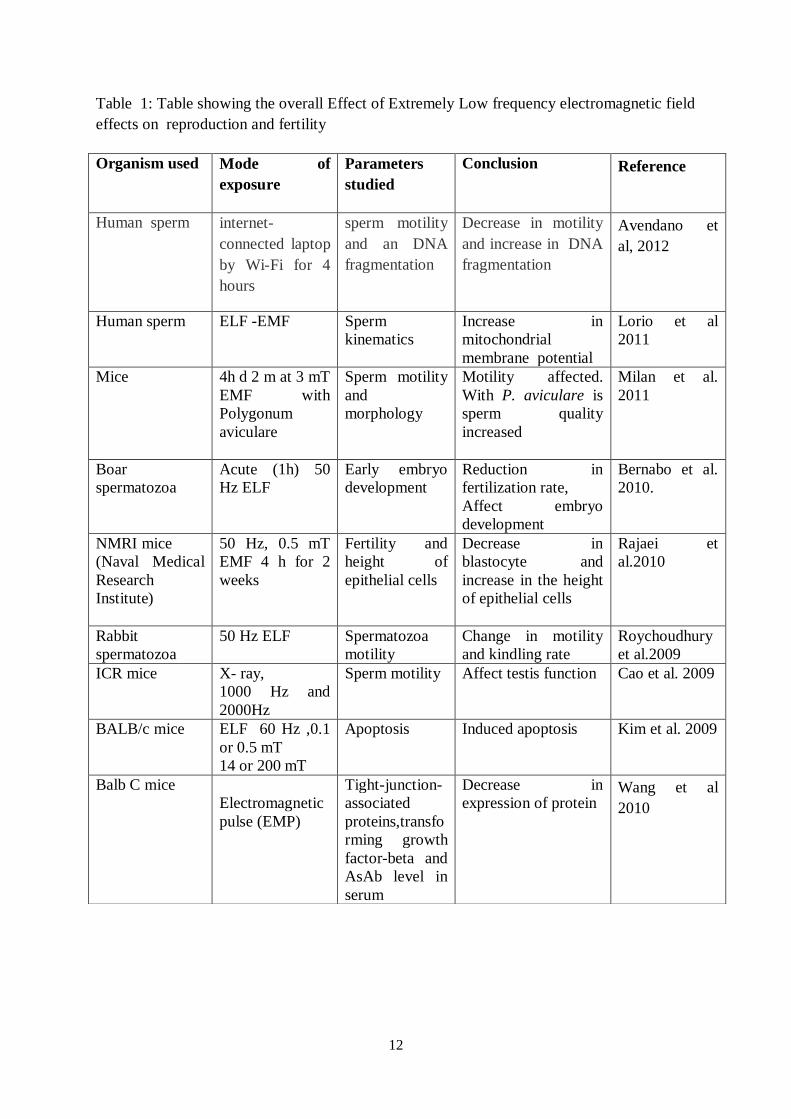
A summary of these data is presented in Table 1 (Studies on Effects of ELF-EMF on Fertility
and Reproduction).
12
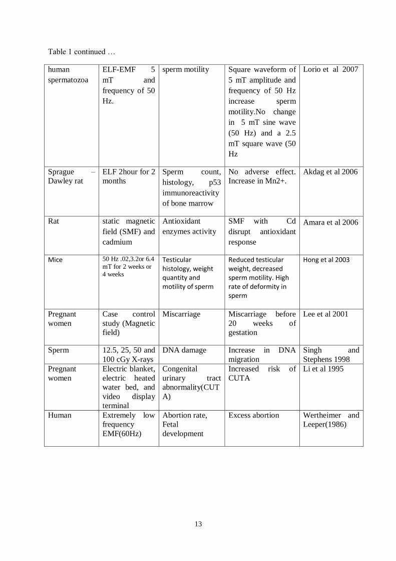
Table 1: Table showing the overall Effect of Extremely Low frequency electromagnetic field
effects on reproduction and fertility
Organism used Mode of
exposure
Parameters
studied
Conclusion Reference
Human sperm internet-
connected laptop
by Wi-Fi for 4
hours
sperm motility
and an DNA
fragmentation
Decrease in motility
and increase in DNA
fragmentation
Avendano et
al, 2012
Human sperm ELF -EMF Sperm
kinematics
Increase in
mitochondrial
membrane potential
Lorio et al
2011
Mice
4h d 2 m at 3 mT
EMF with
Polygonum
aviculare
Sperm motility
and
morphology
Motility affected.
With P. aviculare is
sperm quality
increased
Milan et al.
2011
Boar
spermatozoa
Acute (1h) 50
Hz ELF
Early embryo
development
Reduction in
fertilization rate,
Affect embryo
development
Bernabo et al.
2010.
NMRI mice (Naval Medical
Research
Institute)
50 Hz, 0.5 mT
EMF 4 h for 2
weeks
Fertility and
height of
epithelial cells
Decrease in
blastocyte and
increase in the height
of epithelial cells
Rajaei et
al.2010
Rabbit
spermatozoa
50 Hz ELF
Spermatozoa
motility
Change in motility
and kindling rate
Roychoudhury
et al.2009
ICR mice
X- ray,
1000 Hz and
2000Hz
Sperm motility
Affect testis function
Cao et al. 2009
BALB/c mice
ELF 60 Hz ,0.1
or 0.5 mT
14 or 200 mT
Apoptosis
Induced apoptosis
Kim et al. 2009
Balb C mice
Electromagnetic
pulse (EMP)
Tight-junction-
associated
proteins,transfo
rming growth
factor-beta and
AsAb level in
serum
Decrease in
expression of protein
Wang et al
2010
13
Table 1 continued …
human
spermatozoa
ELF-EMF 5
mT and
frequency of 50
Hz.
sperm motility Square waveform of
5 mT amplitude and
frequency of 50 Hz
increase sperm
motility.No change
in 5 mT sine wave
(50 Hz) and a 2.5
mT square wave (50
Hz
Lorio et al 2007
Sprague –
Dawley rat
ELF 2hour for 2
months
Sperm count,
histology, p53
immunoreactivity
of bone marrow
No adverse effect.
Increase in Mn2+.
Akdag et al 2006
Rat
static magnetic
field (SMF) and
cadmium
Antioxidant
enzymes activity
SMF with Cd
disrupt antioxidant
response
Amara et al 2006
Mice
50 Hz .02,3.2or 6.4
mT for 2 weeks or
4 weeks
Testicular histology, weight quantity and motility of sperm
Reduced testicular weight, decreased sperm motility. High rate of deformity in sperm
Hong et al 2003
Pregnant
women
Case control
study (Magnetic
field)
Miscarriage
Miscarriage before
20 weeks of
gestation
Lee et al 2001
Sperm 12.5, 25, 50 and
100 cGy X-rays
DNA damage Increase in DNA
migration
Singh and
Stephens 1998
Pregnant
women
Electric blanket,
electric heated
water bed, and
video display
terminal
Congenital
urinary tract
abnormality(CUT
A)
Increased risk of
CUTA
Li et al 1995
Human
Extremely low
frequency
EMF(60Hz)
Abortion rate,
Fetal
development
Excess abortion
Wertheimer and
Leeper(1986)
14
V. FERTILITY AND REPRODUCTION EFFECTS REPORTED FOR RADIO-
FREQUENCY AND MICROWAVE EXPOSURE
Nakamura et al. (2000) found that exposure to 2.45 GHz continuous wave (CW) microwave
at 2mW/cm2 power density for 90 min decreased uteroplacental blood flow, increased
progesterone and PGF2 in pregnant rats. Dasdag et al. (2003) reported the decrease in
seminiferous tubule diameter in male rat testes after exposure. They used commercially
available 890-915 MHz GSM (global signal module) with 0.141 W/kg whole body SAR.
More recently, Aitken et al. (2005) found significant damage to mitochondrial and nuclear
genome in epididymal spermatozoa of mice, when exposed to RF 900 MHz EMW, 12 hr a
day for 7 days. Several authors (Fejes et al. 2005; Ji-Geng et al. 2007; Kesari and Behari,
2008) have also observed that carrying the mobile phones near reproductive organs for longer
time may have negative effects on the sperm motility and male fertility.
Aitken et al (2005) exposed mice to 900 MHz radiofrequency electromagnetic radiation at a
SAR of 90 mW/kg inside a waveguide for 7 days (12 hr/day). Following exposure DNA
damage to caudal epididymal spermatozoa was assessed. These authors reported no gross
evidence of single-or double strand DNA breakage in spermatozoa taken from treated
animals. However an analysis of DNA integrity revealed significant damage to both the
mitochondrial genome (P<0.05) and the nuclear beta-globin locus (P<0.01). This study
suggests that while RF EMR does not have a dramatic impact on male germ cell
development, a significant genotoxic effect on epididymal spermatozoa is seen.
Kilgalton and Simmons (2005) report decreased semen quality with prolonged use of cell
phones with negative effects on sperm motility characteristics (Fejes et al, 2005). It has been
shown that sperm DNA damage is not repaired, because of chromatin structure (Singh and
Stephens 1998).
Yan et al (2007) studied the effects of cellular phone emissions on sperm motility in rats.
Rats were exposed to two 3-hr periods of daily cellular phone emissions for 18 weeks, sperm
samples were then collected for evaluation. These authors concluded that exposed group of
15
rats exhibited a significantly higher incidence of sperm cell death than control group rats. In
addition, abnormal clumping of sperm cells was present in rats exposed to cellular phone
emissions and absent from control group rats. A study carried out in Poland (Wdowiak et al
2007) on the population using mobile phone (GSM equipment), spread over a period (1-2
years) indicates sperm quality is lowered. The authors report a decrease in the percentage of
sperm cells with normal motility in the semen. The decrease in motility correlates with the
frequency of using mobile phones. These two finding seem to be mutually supportive.
However there are also reports indicating no effects (Panagopoulos and Margaritis 2008,
2009, 2010).
Overall, the evidence from various laboratories studying fertility and reproduction effects
over the last ten years is important enough raise questions about possible public health
consequences of chronic, long-term exposure to mobile phone use, and when carried on the
body close to the reproductive organs. While assessing the biological implications of mobile
phone radiofrequency exposures, field based experiments are not possible. Sham exposure
controls cannot be obtained. Therefore it is imperative to fall back upon laboratory
experiments performed in a variety of situations (e.g. animals at different distances from the
mobile phone and head) while also simulated variable distances and angles for the mobile
phone variation while in actual use.
Gutschi et al (2011) studied human sperm obtained from 2110 patients attending clinics from
1993 to 2007. Semen analysis was performed in all patients. Serum free testosterone (T),
follicle stimulating hormone (FSH), luteinising hormone (LH) and prolactin (PRL) were
collected from all patients. Information on cell phone use from each patient was collected and
the subjects were divided into two groups according to their cell phone use. Group A: cell
phone use (n=991), Group B: no use (n=1119). Patients with cell phone use showed a
significant higher T and lower LH levels than those who did not use a cell phone. However
no significant difference was observed regarding FSH and PRL values. These authors
concluded that cell phone use had a negative effect on sperm quality in men.
Kesari et al (2011) assessed free radical formation due to mobile phone exposure (2 hr a day
for 35 days) and examined fertility patterns in 70-days old male Wistar rats. The specific
absorption rate of the mobile phone was 0.9 W/kg. An analysis of anti-oxidant enzymes
glutathione peroxidise(p<0.001) and superoxide dismutase (p<o.007) showed a decline, while
16
an increase in catalase (p<0.005) was observed. Malondialdehyde (p< 0.003) showed an
increase and histone kinase (p=0.006) showed a significant decrease in the exposed group.
Correspondingly, micronuclei also showed a significant decrease (p<0.002). A change in
sperm cell cycle of G0 –G1 (p=0.42) and G2/M (p=0.022) was recorded. These authors
concluded that changes occurred due to overproduction of ROS and oxidative damage,
leading to infertility.
Yan et al (2007) studied the effects of cellular phone emissions on sperm motility in rats.
Rats were exposed to two 3-hr periods of daily cellular phone emissions for 18 weeks. After
the exposure period, sperm samples were collected for evaluation. The authors concluded that
exposed group of rats exhibited a significantly higher incidence of sperm cell death than
control group rats. In addition, abnormal clumping of sperm cells was present in rats exposed
to cellular phone emissions and absent from control group rats.
A related issue is the corresponding effect on male infertility.
Sommer et al (2009) undertook a very exhaustive study where male and female mice were
chronically exposed (life-long, 24 hr/day) to mobile phone frequency EMF at 1966 MHz
(UMTS). They studied their development and fertility patterns over four generations by
investigating histological, physiological, behavioural and reproductive functions. They tested
SAR from the time of mating at 0 (sham), 0.08, 0.4 and 1.3 W/kg. Power densities were kept
constant for each group (0, 1.35, 6.8 and 22 W/m2), resulting in varying SARs due to
different number of adults and pups. The results show no harmful effects of exposure on the
fertility and development of the animals. The number and the development of the pups were
not affected by the exposure. These authors concluded no harmful effects occurred with long-
term exposure of mice to UMTS mobile phone frequency radiation over several generations.
DeIuliis et al (2009) used purified human spermatozoa for exposure to electromagnetic
radiation at 1.8 GHz with specific absorption rates varying from 0.4 to 2.75 W/kg. These
investigators reported that motility and vitality were significantly reduced after RFR
exposure, while the mitochondrial generation of reactive oxygen species and DNA
fragmentation was significantly elevated (P<0.001). They also found a highly significant
relationship between SAR, the oxidative DNA damage biomarker 8-OH-dG, and DNA
fragmentation after exposure. These results have bearing on safety of people of reproductive
age, and wellbeing of their offspring. Erogul et al (2006) also support these finding by
showing effect on sperm motility and that long-term exposure may lead to behavioural or
17
structural changes of the male germ cell. These may appear later in life and need
investigation on a longer term basis.
As a follow up of the above, Otitoloju et al (2010) exposed male mice to radiofrequency
radiations at mobile phone (GSM) base station-level RFR. Sperm head abnormalities
occurred in 39% to 46% of exposed mice, but in only 2% of the controls (P<0.005). The
major abnormalities observed were knobbed hook, pin head and banana-shaped sperm head.
The abnormalities were also found to be dose-dependent. This may have severe consequences
for the off spring.
Gul et al (2009) investigated toxicity of microwaves (as emitted by cellular phones on ovaries
in rats. In this study 82 female rats of aged 21 days (43 in the study group and 39 in the
control group) were used. Pregnant rats exposed to mobile phones that were kept underneath
the cages during the whole period of pregnancy. A mobile phone in a standby position for 11
hr and 45 min was turned on to speech position for 15 min every 12 hr and the battery was
charged continuously. On the 21st day after the delivery , the female rat pups were killed and
the right ovaries were removed. The volumes of the ovaries were measured and the number
of follicles in every tenth section was counted. These authors found that the number of
follicles in pups exposed to mobile phone microwaves suggest that intrauterine exposure has
toxic effects on ovaries.
Salama et al (2010) examined the accumulating effects of exposure to electromagnetic
radiation emitted by a conventional mobile phone (800 MHz, standby position, kept opposite
to the testis) on the testicular function and structure. The animals were exposed 8 hr daily for
a period of 12 weeks in a specially designed cage. Semen analysis and sperm function tests
were conducted weekly. Other parameters examined were histological testicular sections and
serum total testosterone. When compared with other two groups (stress control and ordinary),
the exposed animals showed a drop in sperm concentration at week 6, which became
significant at week 8. Mobile sperm population showed similarity amongst the three study
groups until week 10 when it declined significantly, and thereafter in phone and stress control
groups, with more significant decline in the exposed animals (50.6% and 72.4%,
respectively). Histological examination showed a significant decrease in the diameter of
seminiferous tubules in the exposed group vs the stress and ordinary controls (191 m vs.
206 and 226 m, respectively). The authors concluded that the pulsed radiofrequency emitted
by a conventional mobile phone kept in the standby position could affect the testicular
function and structure in the adult rabbit.
18
Falzone et al (2011) evaluated the effect of RF-EMF on sperm characteristics to assess the
fertilizing potential of sperm. They exposed highly motile human spermatozoa to 900 MHz
for an hour (SAR =2.0 W/kg) and examined effects at various time after exposure. The
acrosome reaction was evaluated using flow cytometry. They did not find any effect on
sperm propensity for the acrosome reaction. They obtained significant reduction in sperm
head area (21.5±4% vs 35.5±11.4%) was obtained when compared among exposed and
unexposed samples. Sperm zona binding was assessed directly after exposure. The mean
number of zona-bound sperm of the test hemizona and controls was 22.8±12.4 and 31.8±12.8
(p<0.05) respectively. They concluded that though the radiation exposure did not adversely
affect the acrosome reaction, it had a significant effect on sperm morphometry. They also
observed a significant decrease in sperm binding to the hemizona. These data point toward
sperm fertilization potential. These studies are in contradiction that fertility impairment was
not caused by the induction of apoptosis in spermatozoa (Falzone et al 2010).
In a study undertaken by Ribeiro et al (2007), while experimenting with male Wistar rats,
they exposed testis in the frequency and in the range of intensity (1835-1856 MHz, 0.04-1.4
mW/cm2). The authors reported that the total body weight and absolute and relative testicular
and epididymal weight did not change significantly, nor did the epididymal sperm count.
Human spermatozoa are known to be known to be vulnerable to oxidative stress because of
abundant availability of substrates for free radical attack, and the lack of cytoplasmic space to
accommodate antioxidant enzymes. The ROS generation does DNA damage, besides
reducing fertility. The former has been linked with poor fertility, incidence of miscarriage
and possible morbidity in the offspring, including childhood cancer.
There are other reports showing lack of effect on testicular function in experimental animals
in the non-thermal range. They concluded that the responses are identical to those produced
by hyperthermia caused by mere heating(Ribeiro et al 2007, Sommer et al 2009).
Comparison between non-modulated (DTX) and Modulated (Talk Signal) GSM
Radiation
In an experimentation with insects, Panagopoulos (2011) divided these into two groups: a)the
exposed (E) and b) the sham exposed (control) group (SE). Each of the two groups consisted
of ten female and ten male newly emerged adult flies. The sham exposed groups had identical
treatment as the exposed ones, except that the mobile phone during the “exposures” was
turned off. The duration of exposure was 6 min per day in one dose extending over a period
of 5 days.
19
In the first part of the exposure (1A) the insects were exposed in non-modulated GSM 900
MHz radiation (TDX-discontinuous transmission mode –signal ) while in the second part
(1B) they were exposed to modulated GSM 900 MHz radiation (or GSM talk signal). In both
cases, the exposures were performed with the antenna of the mobile phone in contact with the
walls of the glass vials containing the insects.
The difference between the modulated and the corresponding non-modulated GSM radiation
is that the intensity of the modulated radiation is about ten times higher than the intensity of
the corresponding non-modulated from the same handset (mobile phone) and additionally
that the modulated radiation includes more and larger variations in its intensity within the
same time interval, than the corresponding non-modulated one (Panagopoulos and Margaritis
2008). The power level of exposure for the modulated signal was 0.436±0.060 mW/cm2
and
the corresponding mean value for the non-modulated emission was (0.041±0.006) mW/cm2.
The measured ELF mean values of electric field intensity of the GSM signals excluding the
ambient fields of 50 Hz were 6.05±1.02 V/m for modulated signal and 3.18±1.10 V/m for the
non-modulated signal.
Experiments with the non-modulated GSM 900 MHz radiation (non-speaking mode of
transmission) showed that this radiation decreased insect reproduction by an average of
18.24%. Correspondingly experiments with modulated GSM at 900 MHz (GSM “talk”
signal) exposure shows that the radiation decreases reproduction by an average of 53.01 %.
Above results indicate that the decrease in population is linked with intensity of the radiation.
These authors concluded that between 900 MHz and 1800 MHz, the former is more bioactive
owing to the difference in radiation intensity. Performing experiments at various distances (0
to 100cm) from mobile phone, Panagopoulos (2011) reported that the distance dependence is
not linear. At the distances at 0 and 30 cm (intensity 378 W/cm2
and 10 W/cm2
respectively ) show a maximum of decrease in reproductive capacity (window of maximum
bioactivity). Correspondingly for GSM 1800 MHz at 0 and 20 cm (intensity 252W/cm 2
and
11W/cm2
respectively) bioactivity is maximum (decrease in reproduction, window of
maximum bioactivity) i.e. in the vicinity of free space wavelength of the corresponding
radiation. For distances greater than 20 cm (up to 80 cm) the effect decreases rapidly and
becomes very small for distances longer than 40 cm, but it is still evident for distances up to
80 cm (intensity down to 1.1W/2
). These authors have further pointed out that it is the
intensity which is primarily important rather than the frequency or the distance as such.
20
These distances (30 and 20 cm from GSM 900 MHz and GSM 1800 MHz correspond to the
same RF intensity (10W/cm2) and also to the same electric field intensity of about 0.6-0.7
V/m. Maximum bioactivity is attributed to a distance of 0 cm or at approximately the two
nodes of the wavelength, after which the effect declines. These authors reported no
temperature increase inside any of the vials. They further concluded that the ELF components
of digital mobile telephony signals that play a key role in their bioactivity, alone or in
combination with the RF carrier signal . This also suggest that low frequency signals are
more bioactive than higher frequency ones. Accordingly, electric field of the order of 10-3
V/m are able to disrupt cell function, perhaps by irregular gating of electrosensitive ion
channels on the cell membranes. We conclude that both the GSM signal at 900 MHz and
1800 MHz fields appear to possess sufficient intensity for this for distances up to 50 cm from
the antenna of a mobile phone (or about 50 m from a corresponding base station antenna).
Therefore the restrictions being imposed on emission standards are with respect to continuous
wave frequencies, but not with respect to a pulsed type, the latter being important in
transmitting any intelligent information. Moreover real GSM signals are not constant in
frequency and intensity. This distance of 20-30cm from the mobile phone corresponds to a
distance of 20 to 30 m from a base station antenna. Panagopoulos et al (2010) showed that the
bioactivity of GSM radiation in regard to short-term exposure is evident for radiation
intensities down to 1W/cm2. This value of radiation intensity is encountered at about 1m
distance from a cell phone or about 100 m distance from a corresponding base station
antenna. This radiation intensity is 450 times and 900 times lower than the ICNIRP limits for
900 and 1800 MHz respectively (ICNIRP,1998). It has been estimated by Panagopoulos
(2011) that people may be exposed to this level of radiation for long distances so, a factor of
ten could be added as a safety factor, thereby bringing down the above figure to 0.1 W/cm2
,
suggesting a limit for public exposure. These results support the findings that GSM radiation
caused increased permeability of the blood –brain barrier in rat nerve cells and the strongest
effect was produced by the SAR values which correspond to the weakest radiation intensity
(Eberhardt et al.2008). The concept of window has earlier been described by Bawin et al
(1978), Blackman et al (1980,1989). They have reported that the reproductive capacity
decreases as the duration of exposure (1-21 minutes) increases(almost proportionally), for
either of the two radiation types. Using statistical analysis they have confirmed that this
variation is not because of the randomness of the subject, but because of the radiation
exposure.
21
Several other authors have echoed a wide range of damaging effects on the male reproductive
system and sperm parameters and cause significant changes in the sperm cell cycle (Derias et
al 2006; Ji-Geng. 2007; Gutschi et al, 2011).
Non-genotoxic effects of Radiofrequency Radiation
Several studies reported no effect of RF fields on cell cycle kinetics (Vijayalaxmi et al 2001,
Higashikubo et al 2001; Zeni et al, 2003; Miyakoshi et al, 2005; Lantow et al, 2006c).
Alteration in cell proliferation was described only in a few reports (Pacini et al, 2002, Capri
et al, 2004b).
Apoptosis is an important mechanism of protection against cancer. Several studies have
reported RF field effects on human peripheral blood mononuclear cells (Capri et al, 2004a),
lymphoblastoid cells (Marinelli et al, 2004), epidermis cancer cells (Caraglia et al 2005), and
human Mono Mac 6 cells (Lantow et al, 2006c) and in Molts4 cells (Hook et al, 2004). No
difference in apoptosis induction was detected between sham exposed and RF field exposed
cells by Hook et al (2004). On the other hand, Marinelli et al (2004) have reported better
survival rate of T lymphoblastoid leukaemia cells exposed to 900 MHz non-modulated RF
fields and Carglia et al (2005) found apoptosis induction in human epidermoid cancer cells
after exposure to 1.95 GHz fields. The European REFLEX study (Nikolova et al, 2005)
reported no effects of RF fields on cell cycle, cell proliferation, cell differentiation, apoptosis
induction, DNA synthesis and immune cell functionality. These authors described some
findings after RF exposure on the transcript level of genes related to apoptosis and cell cycle
control; however these responses were not associated with detectable changes of cell
physiology. Analysis on whole genome cDNA arrays show alterations in gene expression
after various RF exposure conditions using different cell types, but no consistent RF-
signature such as stress response could be identified (Remondini et al, 2006).
Heat shock proteins act primarily as molecular chaperones to eliminate unfolded proteins,
which can also appear from cellular stress. This stress response can be induced by many
different external factors, including temperature, chemicals, oxidative stress, heavy metals,
ionizing and non-ionizing radiation and ultrafine carbon black particles. Hsp70 has been
shown to interfere with post mitochondrial events to prevent free radical mediated apoptosis
(Gotoh et al 2001). An increased expression level of Hsp70 can thus offer protection against
stress. Heat shock proteins are also involved in oncogenic processes (Jolly et al, 2000; Inoue
et al, 1999; French et al, 2001).Some investigators have described increased heat shock
22
protein level after RF exposure (Leszczynski et al, 2002; Kwee et al, 2001). However, these
results are controversial, because there are negative findings also (Cotgreave 2005).
Nikolova et al (2005) described modulation in gene regulation after RF field’s exposure at a
SAR of 1.5 W/kg in p53-deficient embryonic stem cells. Proteomic analyses of human
endothelial cell lines showed RF fields induced changes in this expression and
phosphorylation state of numerous proteins including the hsp27.
Mitochondrial generation of ROS : DNA fragmentation and Effects
Free radical formation and their interaction with biological system is a matter of major
concern for it has health implications. There is evidence of free radical generation after RF-
microwave exposures (Phillips et al 2009; De lullis et al 2009;Kesari and Behari 2012,Kesari
et al 2012).
Mitochondrial respiratory chain is the major site for the generation of superoxide radicals (O2
and H2O2). It is possible that EMF may affect the mitochondrial membranes to produce large
amount of radicals ROS under experimental conditions. EMF may disturb ROS metabolism
by increasing the production of ROS or by decreasing the activity of antioxidant enzymes.
From the data presented here it is obvious that such a change in testes that is highly
dependent on oxygen to drive spermatogenesis and yet highly susceptible to the toxic effects
of reactive oxygen metabolites, activity of anti-oxidant enzymes, and increases in ROS
production. Reactive oxygen species (ROS) such as superoxide anions (O¯), hydroxyl
radicals (OH¯) and hydrogen peroxide (H2 O2) may influence the structural integrity and
function of sperm, such as motility, capacitation, and sperm-oocyte fusion (Griveau et al
1995). Spermatozoa are particularly vulnerable to oxidative stress because their plasma
membrane is rich in polyunsaturated fatty acids (PUFAS) and membrane bound NADPH
oxidase. Increased ROS production has been shown to correlate with reduced male fertility
(Iwasaki and Gagnon 1992), to cause perioxidative damage to the sperm plasma membrane
(Hughes et al 1996), and induce both DNA strand breakages and oxidative base damage in
human sperm (Kodama et al 1997). A decrease in total antioxidant capacity of seminal
plasma has been correlated with a reduction in sperm quality, such as concentration, motility
and morphology (Smith et al 1996).
Since the most abundant molecule in biological cells is that of water (H2O) microwave
radiation can generate free radicals like OH-, O
-2, H, and H
-. These molecules are extremely
reactive, having a tendency to react with different biomolecules including DNA, because of
an unpaired electron that they comprise, which try to give up this extra charge and go into the
23
paired mode. Also hydrogen peroxide (H2O2), a product of oxidative respiration in the
mitochondria, which can be converted by electromagnetic radiation(EMR)into hydroxyl free
radical via the Fenton reaction catalyzed by iron within the cells:
H2O2 + (EMR)-----OH-+OH
-
ROS generated by mobile phone exposure if not scavenged may lead to widespread lipid,
protein, and DNA damage (Jajte et al 2002).
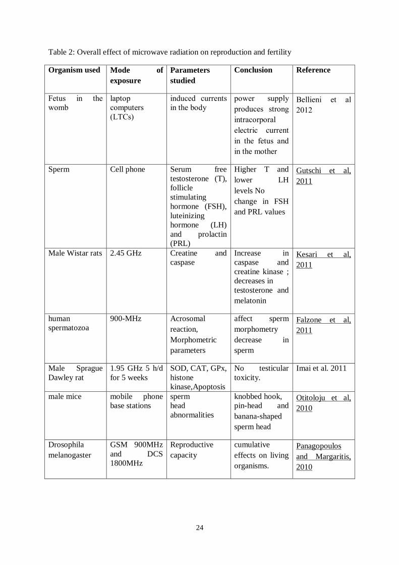
A summary of these results on Effects of Radiofrequency Microwave Radiation on Fertility
and Reproduction is presented in Table 2.
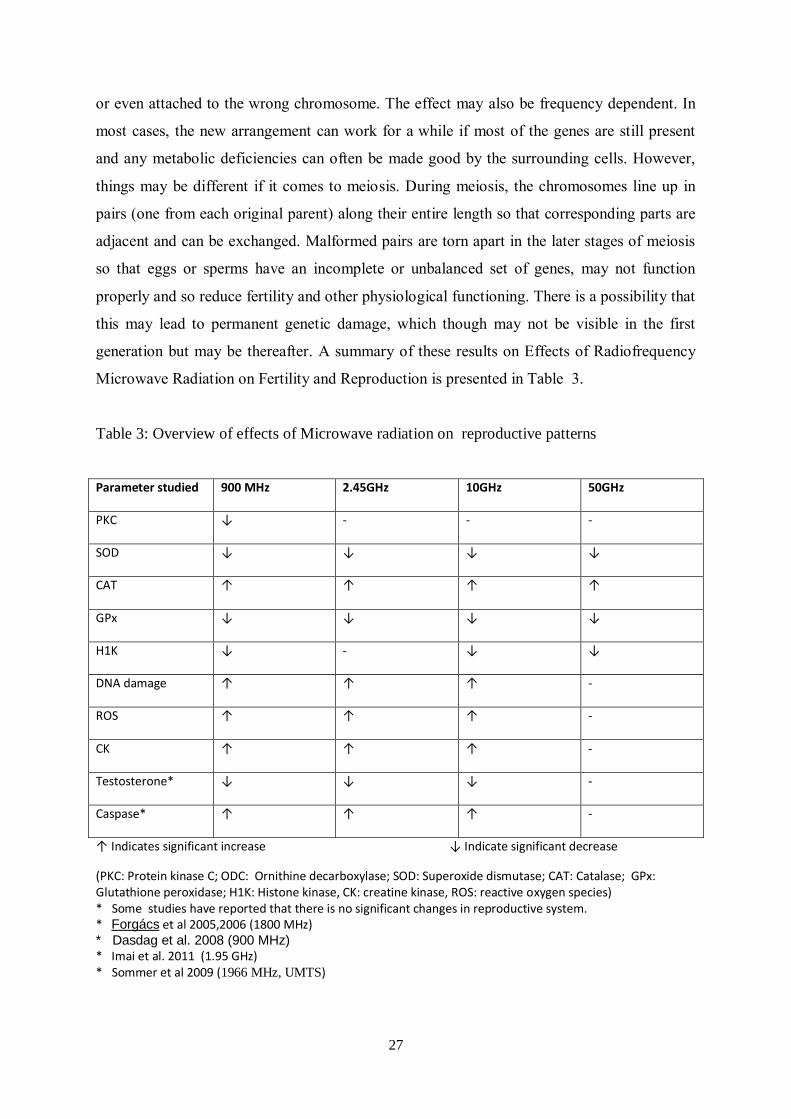
The sequence of events leading toward infertility
A wide range of studies extending up to 50 GHz (Kesari and Behari 2009)) suggest that the
DNA interaction with EMF is similar in nature across wide frequency ranges. DNA appears
to possess the two structural characteristics of fractal antennas, electronic conduction and
self- symmetry (Blank and Goodman 2011). These properties contribute to greater reactivity
of DNA with EMF in the environment. The DNA damage could account for cancer
promotion.
While damage to DNA has been confirmed in numerous scientific studies, it is argued that
DNA repair is an on-going process and the damaged chromosomes can be reconstituted.
However, this proposition is not without risk. There is no guarantee that these will replicate
in the manner they were originally present. Pieces may be left out (deletions), joined in the
backwards (inversions), swapped between different parts of the chromosomal (translocations)
24
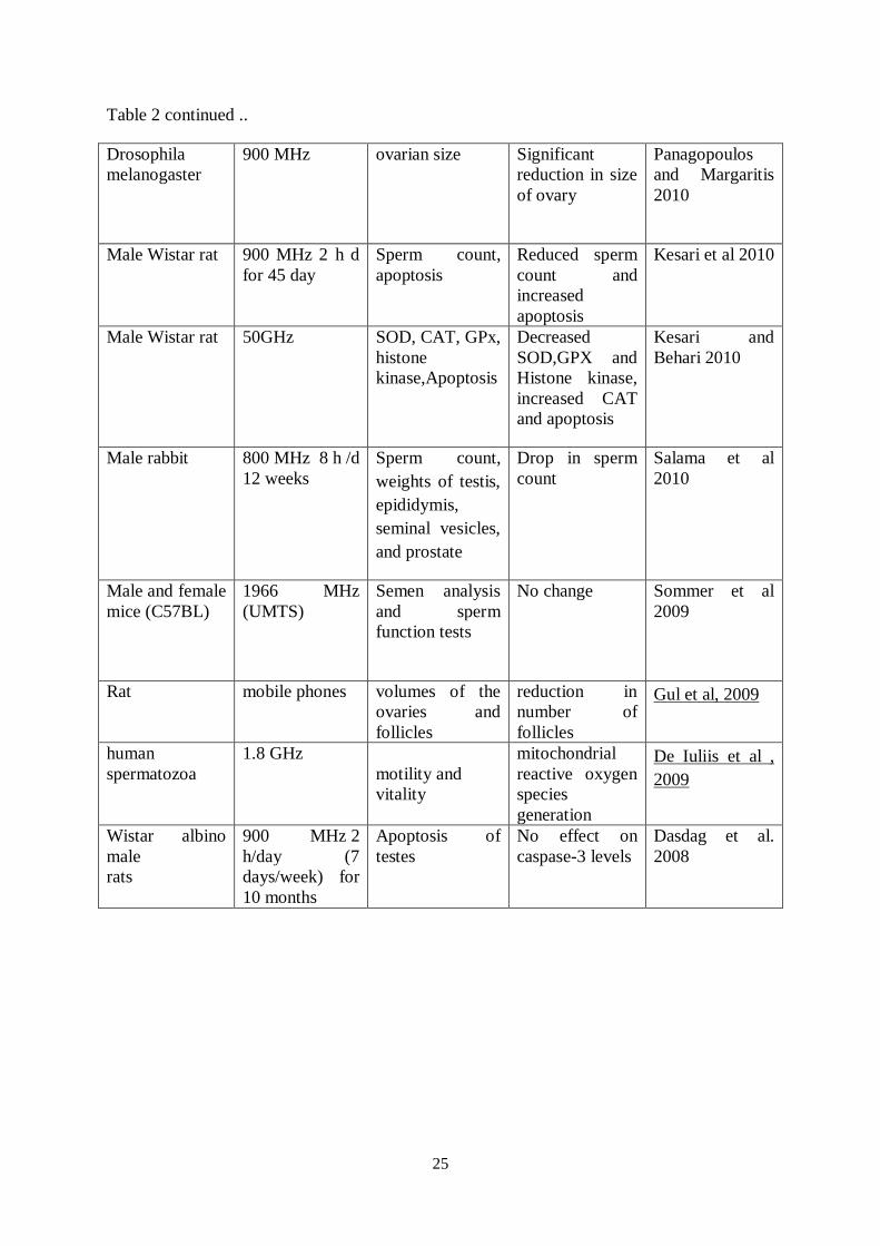
Table 2: Overall effect of microwave radiation on reproduction and fertility
Organism used Mode of
exposure
Parameters
studied
Conclusion Reference
Fetus in the
womb
laptop
computers
(LTCs)
induced currents
in the body
power supply
produces strong
intracorporal
electric current
in the fetus and
in the mother
Bellieni et al
2012
Sperm Cell phone
Serum free
testosterone (T),
follicle
stimulating
hormone (FSH),
luteinizing
hormone (LH)
and prolactin
(PRL)
Higher T and
lower LH
levels No
change in FSH
and PRL values
Gutschi et al,
2011
Male Wistar rats
2.45 GHz
Creatine and
caspase
Increase in
caspase and
creatine kinase ;
decreases in
testosterone and
melatonin
Kesari et al,
2011
human
spermatozoa
900-MHz
Acrosomal
reaction,
Morphometric
parameters
affect sperm
morphometry
decrease in
sperm
Falzone et al,
2011
Male Sprague
Dawley rat
1.95 GHz 5 h/d
for 5 weeks
SOD, CAT, GPx,
histone
kinase,Apoptosis
No testicular
toxicity.
Imai et al. 2011
male mice
mobile phone
base stations
sperm
head
abnormalities
knobbed hook,
pin-head and
banana-shaped
sperm head
Otitoloju et al,
2010
Drosophila
melanogaster
GSM 900MHz
and DCS
1800MHz
Reproductive
capacity
cumulative
effects on living
organisms.
Panagopoulos
and Margaritis,
2010
25
Table 2 continued ..
Drosophila
melanogaster
900 MHz
ovarian size
Significant
reduction in size
of ovary
Panagopoulos
and Margaritis
2010
Male Wistar rat
900 MHz 2 h d
for 45 day
Sperm count,
apoptosis
Reduced sperm
count and
increased
apoptosis
Kesari et al 2010
Male Wistar rat
50GHz
SOD, CAT, GPx,
histone
kinase,Apoptosis
Decreased
SOD,GPX and
Histone kinase,
increased CAT
and apoptosis
Kesari and
Behari 2010
Male rabbit
800 MHz 8 h /d
12 weeks
Sperm count,
weights of testis,
epididymis,
seminal vesicles,
and prostate
Drop in sperm
count
Salama et al
2010
Male and female
mice (C57BL)
1966 MHz
(UMTS)
Semen analysis
and sperm
function tests
No change
Sommer et al
2009
Rat
mobile phones
volumes of the
ovaries and
follicles
reduction in
number of
follicles
Gul et al, 2009
human
spermatozoa
1.8 GHz
motility and
vitality
mitochondrial
reactive oxygen
species
generation
De Iuliis et al ,
2009
Wistar albino
male
rats
900 MHz 2
h/day (7
days/week) for
10 months
Apoptosis of
testes
No effect on
caspase-3 levels
Dasdag et al.
2008
26
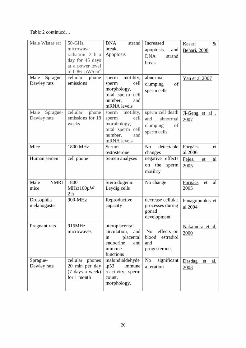
Table 2 continued…
Male Wistar rat
50-GHz
microwave
radiation 2 h a
day for 45 days
at a power level
of 0.86 µW/cm2
DNA strand
break,
Apoptosis
Increased
apoptosis and
DNA strand
break
Kesari &
Behari, 2008
Male Sprague-
Dawley rats
cellular phone
emissions
sperm motility,
sperm cell
morphology,
total sperm cell
number, and
mRNA levels
abnormal
clumping of
sperm cells
Yan et al 2007
Male Sprague-
Dawley rats
cellular phone
emissions for 18
weeks
sperm motility,
sperm cell
morphology,
total sperm cell
number, and
mRNA levels
sperm cell death
and , abnormal
clumping of
sperm cells
Ji-Geng et al ,
2007
Mice
1800 MHz
Serum
testosterone
No detectable
changes
Forgács et
al.2006
Human semen
cell phone
Semen analyses negative effects
on the sperm
motility
Fejes, et al
2005
Male NMRI
mice
1800
MHz(100µW
2 h
Steroidogenic
Leydig cells
No change
Forgács et al
2005
Drosophila
melanogaster
900-MHz Reproductive
capacity
decrease cellular
processes during
gonad
development
Panagopoulos et
al 2004
Pregnant rats
915MHz
microwaves
uteroplacental
circulation, and
in placental
endocrine and
immune
functions
No effects on
blood estradiol
and
progesterone,
Nakamura et al,
2000
Sprague-
Dawley rats
cellular phones
20 min per day
(7 days a week)
for 1 month
malondialdehyde
,p53 immune
reactivity, sperm
count,
morphology,
No significant
alteration
Dasdag et al,
2003
27
or even attached to the wrong chromosome. The effect may also be frequency dependent. In
most cases, the new arrangement can work for a while if most of the genes are still present
and any metabolic deficiencies can often be made good by the surrounding cells. However,
things may be different if it comes to meiosis. During meiosis, the chromosomes line up in
pairs (one from each original parent) along their entire length so that corresponding parts are
adjacent and can be exchanged. Malformed pairs are torn apart in the later stages of meiosis
so that eggs or sperms have an incomplete or unbalanced set of genes, may not function
properly and so reduce fertility and other physiological functioning. There is a possibility that
this may lead to permanent genetic damage, which though may not be visible in the first
generation but may be thereafter. A summary of these results on Effects of Radiofrequency
Microwave Radiation on Fertility and Reproduction is presented in Table 3.
Table 3: Overview of effects of Microwave radiation on reproductive patterns
↑ Indicates significant increase ↓ Indicate significant decrease
(PKC: Protein kinase C; ODC: Ornithine decarboxylase; SOD: Superoxide dismutase; CAT: Catalase; GPx: Glutathione peroxidase; H1K: Histone kinase, CK: creatine kinase, ROS: reactive oxygen species) * Some studies have reported that there is no significant changes in reproductive system. * Forgács et al 2005,2006 (1800 MHz) * Dasdag et al. 2008 (900 MHz) * Imai et al. 2011 (1.95 GHz) * Sommer et al 2009 (1966 MHz, UMTS)
Parameter studied 900 MHz 2.45GHz 10GHz 50GHz
PKC ↓ - - -
SOD ↓ ↓ ↓ ↓
CAT ↑ ↑ ↑ ↑
GPx ↓ ↓ ↓ ↓
H1K ↓ - ↓ ↓
DNA damage ↑ ↑ ↑ -
ROS ↑ ↑ ↑ -
CK ↑ ↑ ↑ -
Testosterone* ↓ ↓ ↓ -
Caspase* ↑ ↑ ↑ -
28
VI. PRUDENT AVOIDANCE AND GUIDANCE FOR SAFETY LIMITS
While it appears to have been convincingly established that electromagnetic fields have
adverse biological effects on fertility and reproduction, the emphasis is on ‘use with caution’
rather than no use at all. Children in the age 12 years and younger are more prone to the
damage because of their developing nervous system. Senior citizens and persons who are ill
should also exercise caution and use wireless devices only in a most demanding situation.
Mobile phones should thus be carried in close proximity of the body only in an OFF position
(not ON and transmitting on standby). This is so because in an “standby” mode the phone
emits signal intermittently - every few minutes they emit a periodic signal lasting a few
seconds long - to maintain connection with the nearest base station antenna. These periodic
signals are as powerful as the usual “talk signal” during a conversation. The user must make
use of mobile phone speaker mode and keep the handset at least 40 cm away from their heads
and other most sensitive organ like the head, heart and reproductive organs. Another method
of protection (e.g. wired ear phones) are less effective, because of the existence of intensity
window. The base station antennas should not be located within or near residential areas or
near heavily populated areas. If antenna placement in the vicinity of residential zones is
essential, they should be made to operate at substantially lowered power. Powerful wireless
antennas should be placed on the hilltops and far from populated areas . The focus thus then
shifts to prudent avoidance i.e. on to reduce the frequency and length of phone calls and keep
away from these devices when not in use.
Bellieni et al (2012) have quoted that levels of exposure from “laptop” computers are higher
than exposures that can be found in the proximity of high-voltage power lines and
transformers or the domestic video screens .It has been observed that the magnetic field
strength from power supplies is higher than that recommended by ICNIRP (1998) guidelines
but that from LTC are within safe limits. It is thus suggested that use of LTC in an inclined
position below the table level be avoided because it may cause increase in genital temperature
,besides causing back pain and fatigue. Moreover ‘laptop’ is a misnomer for its use in close
proximity to the body is harmful.
29
Guidelines for Safety Limits
While considering the far field exposures, there are two sources: one is the microwave
exposure from the base stations. While mobile phone exposure is localized, intermittent and
is under voluntary control of the user, radiation from base towers is involuntary, whole-body
and occurs 24 hours a day. While both the exposures may involve the same carrier frequency,
the exposures are basically different in type and duration. On the whole it can be concluded
that long term exposure near base stations can affect well-being of populations around them.
Symptoms mostly associated with such exposures are headaches, tremor, restlessness and
sleeping disorders.
The question of laying down the criteria for safe exposure is a problematic one, because the
dose needs to be assessed not just as external field frequency (and spectrum), intensity, but
also as cumulative exposure, as well as SAR, for whole body and specific anatomical sites.
Accurate knowledge of RF exposure in a given scenario is needed for several parameters.
The effect is not immediately visible but acts as silent killer. Any epidemiological studies for
a long period (ten years or more) are difficult to carry under controllable situation, and few
unexposed populations can serve as controls (non-exposed). Moreover the basic restrictions
are expressed in quantities that are internal to the body and are not measured such as SAR.
On the other hand, the reference levels are expressed (measured) in the free space situation,
such as electric field. It is evident that SAR-concept alone is insufficient to define the safety
guidelines for chronic exposure from mobile communications.
VI. CONCLUSIONS
Though causal evidence of one or more mechanism(s) are not yet fully refined, it is generally
accepted that oxidative stress and free radical action may be responsible for the recorded
genotoxic effects of EMFs which may lead to impairments in fertility and reproduction. Free
radical action and/or hydrolytic enzymes like DNAase induced by exposure to EMFs may
constitute the biochemical actions leading to adverse changes in hormones essential in males
and female reproduction, DNA damage, which in turn causes damage to sperm motility,
viability, and sperm morphology. Such exposures are now common in men who use and who
wear wireless devices on their body, or use wireless-mode laptop computers. It may also
account for damage to ovarian cells and female fertility, and miscarriage in women (ELF-
EMF at 16 mG intermittent exposure).

30
VIII. REFERENCES
Agarwal A, Tamer M. Said TM. Role of sperm chromatin abnormalities and DNA damage in male
infertility Human Reproduction Update 2003;9:331-345.
Agarwal A, Deepinder F, Sharma RK, Ranga G, Li J. Effect of cell phone usage on semen analysis in
men attending infertility clinic: an observational study. Fertil Steril. 2008;89(1):124-8.
Agarwal A, Desai NR, Makker K, Varghese A, Mouradi R, Sabanegh E, et al. Effect of
radiofrequency electromagnetic waves (RF-EMF) from cellular phones on human ejaculated semen:
an in vitro study. Fertility Sterility 2009;92(4):1318-1325.
Aitken RJ, Bennetts LE, Sawyer D, Wiklendt AM, King BV. Impact of radio frequency
electromagnetic radiation on DNA integrity in the male germline. Int J Androl. 2005 Jun;28(3):171-9.
Aitken RJ, Roman SD. Antioxidant systems and oxidant stress in the testes. Review. Oxidative Med.
Cell Longevity. 2008;1:15-24
Akdag MZ, Dasdag S, Aksen F, Isik B, Yilmaz F. Effect of ELF magnetic fields on lipid
peroxidation, sperm count, p53, and trace elements. Med Sci Monit. 2006;12 (11):BR366-71.
Al-Akhras MA, Darmani H, Elbetieha A. Influence of 50 Hz magnetic field on sex hormones and
other fertility parameters of adult male rats. Bioelectromagnetics 2006; 27(2):127-131.
Amara S, Abdelmelek H, Garrel C, Guiraud P, Douki Travant JL, et al. Effects of subchronic
exposure to static magnetic field on testicular function in rats. Arch Med Res. 2006;37(8):947-52.
Avendano C, Mata A, Sanchez Sarmiento CA, Doncel GF. Use of laptop computers connected to
internet through Wi-Fi decreases human sperm motility and increases sperm DNA fragmentation.
Fertility Sterlity 2012;97(1):39-45.
Bawin S, Adey W, Sabbot I. Ionic factors in release of 45 Ca2+ from chicken cerebral tissues by
electromagnetic fields, In Proc. Natl. Acad. Sci. 1978;75(12):6314-6318.
Behari J, Kesari KK. Effects of microwave radiations on reproductive system of male rats. Embryo
Talk 2006;1 (Suppl.1):81-5.
Bellieni CV, Pinto I, Bogi A, Zoppetti N, Andreuccetti D, Buonocore G. Exposure to electromagnetic
fields from laptop use of “laptop” computers, Arch Environ Occup Health, 2012;67:1:31-36
Bernabo N, Tettamant E, Pistilli MG, Nardinocchi D, Beradinelli P, Mattioli M, Barboni B. Effects of
50 Hz extremely low frequency magnetic field on the morphology and function of boar spermatozoa
capacitated in vitro. Theriogenology. 2007;67(4):801-815.
Bernabo N, Tettamant E, Pistilli MG, Nardinocchi D, Beradinelli P, Mattioli M, et al. Extremely low
frequency electromagnetic field exposure affects fertilization outcome in swine animal model.
Theriogenology. 2010;73(9):1293-1305.
Blackman CF, Benane SG, Elder JA, House DE, Lampe JA, Faulk JM.Induction of calcium-ion influx
from tissue by radiofrequency radiation : Effect of sample number and modulation frequency on the
power-density window. Bioelectromagnetics 1980;1:35-43.
31
Blackman CF, Kinney LS, House DE, Joines WT. Multiple power density windows and their origin.
Bioelectromagnetics 1989;10(2):115-128.
Blank M, Goodman R. DNA is a fractal antenna in electromagnetic fields. Int J Radiation Biol
201l;87:409-415.
Cao XW, Zhao TD, Wang CH, Zhou Q, Li LQ, Yao HG, Zhang SQ, Tang, JT, Wei W. Alternating
magnetic field damages the reproductive function of murine testes. Zhonghua Nan Ke Xue.
2009;15(6):530-533.
Capri M, Scarcella E, Fumelli C. Bianchi E, Salvioli S, Mesirca P. et al. In vitro exposure of human
lymphocytes to 900 MHz CW and GSM modulated radiofrequency: studies of proliferation, apoptosis
and mitochondrial membrane potential. Radiat Res. 2004a;162, 211-218.
Capri M, Scarcella E, Bianchi E, Fumelli C, Mesirca P, Agostini C, et al. 1800 MHz radiofrequency
(mobile phones, different Global System for Mobile communication modulations) does not affect
apoptosis and heat shock protein 70 level in peripheral blood mononuclear cells from young and old
donors. Int J Radiat Biol. 2004b;80:389-397.
Caraglia M, Marra M, Mancinelli F, D'Ambrosio G, Massa R, Giordano A. et al. Electromagnetic
fields at mobile phone frequency induce apoptosis and inactivation of the multi-chaperone complex in
human epidermoid cancer cells. J Cell Physiol. 2005; 204:539-548.
Roychoudhury S, Massanyi P, Slamecka J, Chlebec I, Trandzik J, et al. In vitro gossypol induced
spermatozoa motility alterations in rabbits. J Environ Sci Health B. 2009 Sep;44(7):730-41.
Chung MK, Lee SJ, Kim YB, Park SC, Shin DH, Kim SH, Kim JC. Evaluation of spermatogenesis
and fertility in F1 male rats after in utero and neonatal exposure to extremely low frequency
electromagnetic fields. Asian J Androl. 2005, 7(2):189-94.
Cotgreave IA. Biological stress responses to radio frequency electromagnetic radiation: are mobile
phones really so (heat) shocking?, Arch Biochem Biophys. 2005;435:227–240.
Dasdag S, Akdag MZ, Aksen F, Yilmaz F, Bashan M, Dasdag M, Salih Celik M. Whole body
exposure of rats to microwaves emitted from a cell phone does not affect the testes,
Bioelectromagnetics 2003;24(3):182-188.
Dasdag S, Akdag MZ, Ulukaya E, Uzunlar AK, Yegin D. Mobile phone exposure does not induce
apoptosis on spermatogenesis in rats. Arch Med Res. 2008 Jan;39(1):40-4.
Delgado JMR, Leal J, Monteagudo JL, Gracia MG. Embryological changes induced by weak,
extremely low frequency electromagnetic fields. J Anat (Lond) 1982;134:533–552.
DeIullis GN, Newey RJ, King BV, Aitken RJ. Mobile phone radiation induces reactive oxygen
species production and DNA damage in human spermatozoa in vitro. PLos One 2009;4(7):e6446.
Deno DW, Zaffanella LE. Field effects of overhead transmission lines and stations, In Transmission
Line Reference Book. 345 kV and above, 2nd edition , J J Ed. Project UHV, Technical Resource
Operations. Large Transformer Division. General Electric Company, Pinsfield Mass. 1982;329/625.
Derias EM, Stefanis P, Drakeley, A, Gazvani R, Lewis_Jones DI. Growing concern over the safety of
using mobile phones and male fertility. Arch. Androl. 2006;521:9-14.
32
Drozdov KA, Khlistun OA, Drozdov AL. The influence of ultrasound and constant magnetic field on
gametes, zygotes, and embryos of the sea urchin. Biofizika. 2008; 53(3):513-518.
Eberhardt JL, Persson BR, Brun AE, Salford LG, Malmgren LO. Blood-brain barrier permeability and
nerve cell damage in rat brain 14 and 28 days after exposure to microwaves from GSM mobile
phones. Electromagn Biol Med. 2008;27(3):215-29.
Edwards MJ , Mulley R, Ring S, Warmer RA. Mitotic cell death and delay of mitotic activity in
guinea pig embryos following brief material hyperthermia. J Embryol Exp Morphol 1974;32:593-602.
Erogul O, Oztas E, Yildirim I, Kir T, Aydur E, Komesli G, Irkilata HC, IrmakMK, Peker AF. Effects
of electromagnetic radiation from a cellular phone on human sperm motility:an vitro study. Arch Med
Res 2006;37(7):840-3.
Falzone N, Huyser C, Franken DR, Leszezynski D. Mobile phone radiation does not induce pro-
apoptosis effects in human spermatozoa. Radiation Res 2010;174(2):169-76.
Falzone N, Huyser C, Becker P, Leszezynski DR, Franken DR. The effect of pulsed 900 MHz GSM
mobile phone radiation on the acrosome reaction, head morphometry and zona binding of human
spermatozoa. Int J Androl 2011;34(1):20-6.
Farrell JM Litovitz TL, Penafiel M, Montrose CJ, Doinov P, Barber M, et al. The effect of pulsed and
sinusoidal magnetic fields on the morphology. Bioelectromagnetics. 1997;18:431-438.
Fraser FC, Skelton J (1978) Possible tetragenicity of maternal fever. Lancet 2:634.
Fejes I, Zavacki Z, Szollosi J, Koloszar Daru J, Kovacs L, Pal A. Is there a relationship between cell
phone use and semen quality ? Arch Androl. 2005;51, 385-393.
Forgács Z, Kubinyi G, Sinay G, Bakos J, Hudák A, Surján A, Révész C, Thuróczy G. Effects of 1800
MHz GSM-like exposure on the gonadal function and hematological parameters of male mice. Magy
Onkol. 2005;49(2):149-51. [Article in Hungarian]
Forgács Z, Somosy Z, Kubinyi G, Bakos J, Hudák A, Surján A, Thuróczy G. Effect of whole-body
1800 MHz GSM-like microwave exposure on testicular steroidogenesis and histology in mice. Reprod
Toxicol. 2006; Jul;22(1):111-7.
French PW, PennyR, Laurence JA, McKenzie DR. Mobile phones, heat shock proteins and cancer.
Differentiation 2001;67, 93-97.
García AM, Sisternas A, Hoyos SP. Occupational exposure to extremely low frequency electric and
magnetic fields and Alzheimer disease: a meta-analysis. Int J Epidemiol. 2008;37(2):329-40
Gharagozloo P, Aitken RJ. The role of sperm oxidative stress in male infertility and the significance
of oral antioxidant therapy. Hum Reprod 2011 Jul;26(7):1628-40. Epub 2011 May 5.
Gotoh T, Terada K, Mori M. hsp70-DnaJ chaperone pairs prevent nitric oxide-mediated apoptosis in
RAW 264. 7 macrophages. Cell Death Differ. 2001; 8, 357-366.
Gul A, Celebi H, Ugras S. The effects of microwaves emitted by cellular phones on ovarian follicles
in rats. Archives of Gynecology and Obstetrics 2009;280(5): 729-33.
33
Gutschi T, AI-Ali BM, Shamloul R, Pummer K, Trummer H. Impact of cell phone use on men’s
semen parameters. Andrologia. 2011;43, 5, 312–316.
Heredia-Rojas JA, Caballero-Hernandez DE, Rodriguez-de la Fuente AO, Ramos-Alfano G,
Rodriguez-Flores LE. Lack of alterations on meiotic chromosomes and morphological characteristics
of male germ cells in mice exposed to a 60 Hz and 2. 0 mT magnetic field. Bioelectromagnetics.
2004;25(1):63-8.
Hardell L, Sage C. Biological effects from electromagnetic field exposure and public exposure
standards. Biomed Pharmacother. 2008;62(2):104-9.
Higashikubo R, Ragouzis M, Moros EG, Straube WL, Roti Roti JL. Radiofrequency electromagnetic
fields do not alter the cell cycle progression of C3H 10T and U87MG cells. Radiat Res. 2001; 786–
795.
Hong R, Liu Y, Yu YM, Hu K, Weng EQ. Effects of extremely low frequency electro magnetic fields
on male reproduction in mice. Zhonghua Lao dong Wei Sheng, Zhi Ye Bing Za Zhi. 2003;21(5):342-
345.
Hong R, Zhang V, Liu Y, Weng EQ. Effects of extremely low frequency electromagnetic fields on
DNA of testicular cells and sperm chromatin structure in mice. Zhonghua Lao Dong Wei Sheng Zhi
Ye Bing Za Zhi. 2005;23(6):414-417.
Hook GJ, Zhang P, Lagroye I, Li L, Higashikubo R, Moros EG, et al. Measurement of DNA damage
and apoptosis in Molt-4 cells after in vitro exposure to radiofrequency radiation. Radiat Res. 2004;
161:193-200.
Hughes CM, Lewis SE, Mckelvey-Martin VJ, Thompson W. A comparison of baseline and induced
DNA damage in human spermatozoa from fertile and infertile men, using a modified comet assay.
Mol Hum Reprod. 1996; 13, 1240-1247.
Huss A, Spoerri A, Egger M, Röösli M and for the Swiss National Cohort Study. Residence near
power lines and mortality from neurodegenerative diseases: longitudinal study of the Swiss
Population. Am J Epidemiol. 2008;15, 169, 167-175.
ICNIRP. Guidelines for limiting exposure to time varying electric, magnetic, and electromagnetic
fields (upto 300 GHZ) 1998. Health Phys. 1998;74:494-522.
Imai N, Kawabe M, Hikage T, Nojima T, Takahashi S, Shirai T. Effects on rat testis of 1. 95-GHz W-
CDMA for IMT-2000 cellular phones. Syst Biol Reprod Med. 2011; Aug;57(4):204-9.
Inoue Y, Sato Y, Nishimura M, Seguchi M, Zaitsu Y, Yamada K. et al. Heat-induced drug resistance
is associated with increased expression of Bcl-2 in HL60. Anticancer Res. 1999;19:3989-3992.
Iwasaki A, Gagon C. Formation of reactive oxygen species in spermatozoa of infertile patients. Fertil
Steril. 1992; 57:409-416.
Jajte J, Grzegorczyk J, Zmyslony M, Rajkowska E. Effect of 7 mT static magnetic field and iron ions
on rat lymphocytes: apoptosis, necrosis and free radical processes. Bioelectrochemistry. 2002;57:107-
111.
34
Yan JG, Agresti M, Bruce T, Yan YH, Granlund A, Matloub HS. Effects of cellular phone emissions
on sperm motility in rats. Fertility Sterility, 2007;88(4):957-964.
Jolly C, Morimoto RI. Role of the heat shock response and molecular chaperones in oncogenesis and
cell death. J Natl Cancer Inst. 2000;92:1564 -1572.
Juutilainen J, Matilainen P, Saarikoski S, Läärä E, Suonio S. et al. Early pregnancy loss and exposure
to 50 Hz magnetic fields. Bioelectromagnetics 1993;14:220-236.
Kesari KK, Behari J. Comparative study of 900MHz and 2. 45 GHz radiation effect on reproductive
system of male rats. In: Recent Advances and Challenges in Reproductive Health Research. (RS
Sharma, A Rajanna, M Rajalakshmi. Proceedings of the conference on “Recent Advances and
Challenges in Reproductive Health Research (Feb 19-21, 2007 New Delhi) ICMR Publication, 2008.
Kesari KK, Behari J. Fifty gigahertz microwave exposure effect of radiation on rat brain. Appl
Biochem Biotechnol 2009;158:126-139.
Kesari KK, Behari J. Microwave exposure affecting reproductive system in male rats. Appl Biochem
Biotechnol. 2010;31(6):495-498.
Kesari KK, Behari J. Evidence for mobile phone radiation exposure effects on reproductive pattern of
male rats: Role of ROS. Electromagnetics Biology Medicine. 2012;31(3):213-222.
Kesari KK, Kumar S, Nirala J, Siddiqui MH, Behari J. Biophysical evaluation of radiofrequency
electromagnetic field effects on male reproductive pattern. Cell Biochem Biophys 2012;Aug 29;DOI
10. 1007/s12013-012-9414-6
Kesari KK, Kumar S, Behari J. Effects of radiofrequency electromagnetic wave exposure from
cellular phones on the reproductive pattern in male Wistar rats. Appl Biochem Biotechnol
2011;164(4):546-59.
Kim YW, Kim HS, Lee JS, Kim YJ, Lee SK, Seo JN, Jung KC, Kim N, Gimm YM. Effects of 60 Hz
14 T magnetic field on the apopotosis of testicular cell in mice. Bioelectromagnetics 2009;30(1):66-
72.
Kilgalton SJ, Simmons LW. Image content influences men’s semen quality. Biol Lett. 2005; 1, 385-
393.
Kodama H, Yamaguchi R, Fukada J, Kasai H, Tanaka T. Increased oxidative deoxyribonucleic acid
damage in the spermatozoa of infertile male patients. Fertil Steril. 1997;68, 519-524.
Kumar S, Kesari KK, Behari J. Evaluation of genotoxic effect in male wistar rats following
microwave exposure. Ind J. Exp Biology 2010;48, 586-592.
Kumar S, Kesari KK, Behari J. The therapeutic effect of a pulsed electromagnetic field on the
reproductive pattern of male wistar rats exposed to a 2. 45 GHz microwave field. Clinics
2011;66(7)1237-1245.
Kumar S, Kesari KK, Behari J. The influence of microwave exposure on male fertility. fertility and
sterility. 2011a;95 (4); 1500-1502.
35
Kwee S, Raskmark P, Velizarov S. Changes in cellular proteins due to environmental nonionizing
radiation. 1. Heat shock proteins. Electro- and Magnetobiol. 2001;20, 141-152.
Lacy KK, DeSesso JM, Lary JM. Early histological changes observed in the neural folds of day 9 rat
embryos subsequent to radio frequency radiation or water bath induced hyperthermia. Teratology
1981;23:48A.
Lantow M, Viergutz T, Weiss DG, Simkó M. Comparative study of cell cycle kinetics and induction
of apoptosis or necrosis after exposure to radiofrequency radiation in human Mono Mac 6 cells.
Radiat Res. 2006c;166, 539-543.
Lee GM, Neutra RR, Hristova L, Yost M, Hatt RA. A nested case-control study of residential and
personal magnetic field measures and miscarriages. Epidemiology 2001;13:21-31.
Leszczynski D, Joenväärä S, Reivinen J, Kuokka R. Non-thermal activation of the hsp27/p38MAPK
stress pathway by mobile phone radiation in human endothelial cells: molecular mechanism for cancer
and blood-brain barrier-related effects. Differentiation 2002;2–3:120.
Li De-Kun, Checkoway H, Muller A. Electric blanket use during pregnancy in relation to the risk of
congenital urinary tract anomalies among women with a history of subfertility. Epidemiology.
1995;6(5):485-489.
Lorio R, Scrimaglio R. Rantucci E, Delle Monache S, Di Gateano A, Finetti N, et al. A preliminary
study of oscillating electromagnetic field effects on human spermatozoon motility.
Bioelectromagnetics 2007;28(1): 72-75.
Lorio R, Delle Monache S, Bennato F, Di Bartolomeo C, Scrimaglio R, Cinque B, et al. Involvement
of mitochondrial activity in mediating ELF-EMF stimulatory effect on human sperm motility.
Bioelectromagnetics 2011;32 (1):15-27
Milan PB, Nejad DM, Ghanbari AA, Rad JS, Nasrabadi HT, Roudkenar MH, et al. Effects of
Polygonum aviculare herbal extract on sperm parameters after EMF exposure in mouse. Pak J Biol
Sci. 2011;1;14(13):720-4.
Marinelli F, La Sala D, Cicciotti G, Cattini L, Trimarchi C, Putti S, et al. Exposure to 900 MHz
electromagnetic field induces an unbalance between pro-apoptotic and pro-survival signals in T-
lymphoblastoid leukaemia CCRF-CEM cells. J Cell Physiol. 2004;198, 324-332.
Marx JL. Electric currents may guide development. Science 1981;211:1147-1149.
Miller P, Smith DW, Shepard TH. Material Hyperthermia as a possible cause of anencphaly. Lancet
1978;i:519-520.
Miyakoshi J, Takemasa K, Takashima Y, Ding GR, Hirose H, Koyama S. Effects of exposure to a
1950 MHz radio frequency field on expression of Hsp70 and Hsp27 in human glioma cells.
Bioelectromagnetics 2005;26:251-257.
Nakamura H, Nagase H, Ogino K, Hatta K, Matsuzaki I. Uteroplacental circulatory disturbance
mediated by prostaglandin f2alpha in rats exposed to microwaves. Reprod Toxicol. 2000;14(3):235-
40.
36
Nikolova T, Czyz J, Rolletschek A, Blyszczuk P, Fuchs J, Jovtchev G, et al. Electromagnetic fields
affect transcript levels of apoptosis-related genes in embryonic stem cell-derived neural progenitor
cells. FASEB J. 2005;19:1686-1688.
O'Carroll MJ, Henshaw DL. Aggregating disparate epidemiological evidence: comparing two seminal
EMF reviews. Risk Anal. 2008;28(1):225-34.
Otitoloju AA, Obe IA, Adewale OA, Otubanjo OA, Osunkalu VO. Preliminary study on the reduction
of sperm head abnormalities in mice , Mus musculus, exposed to radiofrequency radiations from
global system for mobile communication base stations. Bull Environ Contamin Toxicol
2010;84(1):51-4.
Pacini S, RuggieroM, Sardi I, Aterini S, Gulisano F, Gulisano M. Exposure to global system for
mobile communication (GSM) cellular phone radiofrequency alters gene expression, proliferation,
and morphology of human skin fibroblasts. Oncol Res. 2002; 1, 19–24.
Panagopoulos DJ, Karabarbounis A, Margaritis LH. Effect of GSM 900 MHz mobile phone radiation
on the reproductive capacity of Drosophila melanogaster. Electromagnetic Biology and Medicine.
2004;23(1):29-43.
Panagopoulos DJ, Margaritis LH. Mobile Telephony radiation Efects on Living Organisms. In Harper
A C and Buress R V (Eds) “Mobile Telephones Networks, Applications and Performance”. Nova
Science Publishers. 2008;107-149.
Panagopoulos DJ, Margaritis LH. Mobile telephony radiations. International Journal of Medical and
Biological Frontiers. 2009;15(1-2), 33-76.
Panagopoulos DJ, Margaritis LH. The effects of exposure duration on the biological activity of
mobile telephony radiation. International Journal of Radiation Biology. 2010;86(5):358-366.
Panagopoulos D J (2011)Analyzing the Health Impacts oF Modern Telecommunications Microwaves.
Advances in Medicine and Biology. 17:1-54.
Phillips JL, Singh NP Lai H. Electromagnetic fields and DNA damage. Pathophysiology.
2009;16(23):79-88.
Polk C. Introduction. In: CRC Handbook of Biological Efects of Electromagnetic Fields (Polk C and
Postow E) CRC Press, Inc Boca Raton, Florida. 1986;1-24.
Portier CJ, Wolfe MS, eds. EMF Science Review Symposium Breakout Group Reports for
Theoretical Mechanisms and In Vitro Research Findings. Research Triangle Park: National Institute
of Environmental Health Sciences, 1997.
Rajaei F, Borhani N, Sabbagh-Ziarani F, Mashayekhi F. Effects of extremely low-
frequency electromagnetic field on fertility and heights of epithelial cells in pre-implantation stage
endometrium and fallopian tube in mice. Zhong Xi Yi Jie He Xue Bao. 2010;8(1):56-60.
Remondini D, Nylund R, Reivinen J, Poulletier de Gannes F, Veyret B, et al. Gene expression
changes in human cells after exposure to mobile phone microwaves. 2006;Proteomics, 6(17), 4745-
4754.
37
Ribeiro EP, Rhoden EL, Horn MM, Rhoden C, Lima LP, Toniolo L. Effects of subchronic exposure
to radiofrequency frequency from a conventional cellular telephone on testicular function in adult rats.
J Urol 2007;177(1):395-9.
Roychoudhury S, Jedicka S, Parkanyl V, Rafay J, Ondruska L, Massanyl P, et al. Influence of a 50 Hz
extremely low frequency electromagnetic field on spermatozoa motility and fertilization rats in
rabbits. J Environ Sci Health A Tox Hazard subst Environ Eng. 2009;44(10):1041-1047.
Sage C, Johansson O, Sage SA. Personal digital assistant (PDA) cell phone units produce elevated
extremely-low frequency electromagnetic field emissions. Bioelectromagnetics. 2007;28(5):386-392.
Salama N, Kishimoto T, Kanayama HO. Effects of exposure to a mobile phone on testicular function
and structure in adult rabbit. International Journal of Andrology 2010;33(1):88-94.
Singh NP, Stephens RE. X-ray induced DNA double strand breaks in human sperm. Mutagenesis
1998;13:75-79.
Smith R, Vantman D, Ponce J, Escobar J, Lissi E. Total antioxidant capacity of human seminal
plasma. Hum Reprod 1996;11:1655–60.
Sommer AM, Grote K, Reinhardt T, Streckert J, Hansen V, Lerchl A. Effects of radiofrequency
electromagnetic fields (UMTS) on reproduction and development of mice: a multi-generation study.
Radiation Research 2009;171(1):89-95.
Sun YL, Zhou WJ, Wu JQ, Gao ES. Does exposure to computers affect the routine parameters of
semen quality? Asian J Androl 2005;; 7:263-266.
VanDemark NL, Free MJ. Temperature effects. IN Johnson AD, Gomes WR, VanDemark NL(eds):
“The Testis, ” Vol III. New York: Academic, 1970;233-312.
Vijayalaxmi, Bisht KS, Pickard WF, Meltz ML, Roti JL, Moros EG. Chromosome damage and
micronucleus formation in human blood lymphocytes exposed in vitro to radiofrequency radiation at a
cellular telephone frequency 1847-74 MHz CDMA. radiation Research. 2001;156:430-432.
Wang XW, Ding GR, Shi CH, Zeng, LH, Liu JY, Li J, et al. Mechanism involved in the blood-testis
barrier increased permeability induced by EMP. Toxicology 2010;276:58-63.
Wdowiak A, Wdowiak L, Wiktor H. Evaluation of the effect of using mobile phones on male fertility.
Annals Agriculture Environmental Medicine: AAEM 2007;14(1):169-72.
Wertheimer N, Leeper E. Possible effects of electric blankets and heated waterbeds on fetal
development. Bioelectromagnetics 1986;7:13-22.
Yan JG, Agresti M, Bruce T, Yan YH, Granlund A, Metaloub HS. Effects of cellular phone emissions
on sperm motility in rats. Fertility Sterility 2007;88(4): 957-64.
Zeni O, Chiavoni AS, Sannino A, Antolini A, Forigo D, Bersani F, et al. Lack of genotoxic effects
(micronucleus induction) in human lymphocytes exposed in vitro to 900 electromagnetic fields.
Radiat Res. 2003;160:152-158.