Embed Size (px)
Citation preview
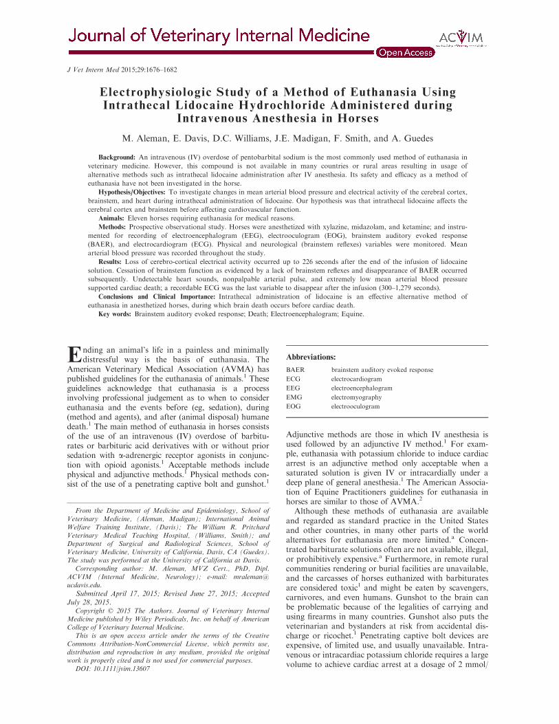
Electrophysiologic Study of a Method of Euthanasia UsingIntrathecal Lidocaine Hydrochloride Administered during
Intravenous Anesthesia in Horses
M. Aleman, E. Davis, D.C. Williams, J.E. Madigan, F. Smith, and A. Guedes
Background: An intravenous (IV) overdose of pentobarbital sodium is the most commonly used method of euthanasia inveterinary medicine. However, this compound is not available in many countries or rural areas resulting in usage ofalternative methods such as intrathecal lidocaine administration after IV anesthesia. Its safety and efficacy as a method ofeuthanasia have not been investigated in the horse.
Hypothesis/Objectives: To investigate changes in mean arterial blood pressure and electrical activity of the cerebral cortex,brainstem, and heart during intrathecal administration of lidocaine. Our hypothesis was that intrathecal lidocaine affects thecerebral cortex and brainstem before affecting cardiovascular function.
Animals: Eleven horses requiring euthanasia for medical reasons.Methods: Prospective observational study. Horses were anesthetized with xylazine, midazolam, and ketamine; and instru-
mented for recording of electroencephalogram (EEG), electrooculogram (EOG), brainstem auditory evoked response(BAER), and electrocardiogram (ECG). Physical and neurological (brainstem reflexes) variables were monitored. Meanarterial blood pressure was recorded throughout the study.
Results: Loss of cerebro-cortical electrical activity occurred up to 226 seconds after the end of the infusion of lidocainesolution. Cessation of brainstem function as evidenced by a lack of brainstem reflexes and disappearance of BAER occurredsubsequently. Undetectable heart sounds, nonpalpable arterial pulse, and extremely low mean arterial blood pressuresupported cardiac death; a recordable ECG was the last variable to disappear after the infusion (300–1,279 seconds).
Conclusions and Clinical Importance: Intrathecal administration of lidocaine is an effective alternative method ofeuthanasia in anesthetized horses, during which brain death occurs before cardiac death.
Key words: Brainstem auditory evoked response; Death; Electroencephalogram; Equine.
Ending an animal’s life in a painless and minimallydistressful way is the basis of euthanasia. The
American Veterinary Medical Association (AVMA) haspublished guidelines for the euthanasia of animals.1 Theseguidelines acknowledge that euthanasia is a processinvolving professional judgement as to when to considereuthanasia and the events before (eg, sedation), during(method and agents), and after (animal disposal) humanedeath.1 The main method of euthanasia in horses consistsof the use of an intravenous (IV) overdose of barbitu-rates or barbituric acid derivatives with or without priorsedation with a-adrenergic receptor agonists in conjunc-tion with opioid agonists.1 Acceptable methods includephysical and adjunctive methods.1 Physical methods con-sist of the use of a penetrating captive bolt and gunshot.1
Adjunctive methods are those in which IV anesthesia isused followed by an adjunctive IV method.1 For exam-ple, euthanasia with potassium chloride to induce cardiacarrest is an adjunctive method only acceptable when asaturated solution is given IV or intracardially under adeep plane of general anesthesia.1 The American Associa-tion of Equine Practitioners guidelines for euthanasia inhorses are similar to those of AVMA.2
Although these methods of euthanasia are availableand regarded as standard practice in the United Statesand other countries, in many other parts of the worldalternatives for euthanasia are more limited.a Concen-trated barbiturate solutions often are not available, illegal,or prohibitively expensive.a Furthermore, in remote ruralcommunities rendering or burial facilities are unavailable,and the carcasses of horses euthanized with barbituratesare considered toxic1 and might be eaten by scavengers,carnivores, and even humans. Gunshot to the brain canbe problematic because of the legalities of carrying andusing firearms in many countries. Gunshot also puts theveterinarian and bystanders at risk from accidental dis-charge or ricochet.1 Penetrating captive bolt devices areexpensive, of limited use, and usually unavailable. Intra-venous or intracardiac potassium chloride requires a largevolume to achieve cardiac arrest at a dosage of 2 mmol/
From the Department of Medicine and Epidemiology, School ofVeterinary Medicine, (Aleman, Madigan); International AnimalWelfare Training Institute, (Davis); The William R. PritchardVeterinary Medical Teaching Hospital, (Williams, Smith); andDepartment of Surgical and Radiological Sciences, School ofVeterinary Medicine, University of California, Davis, CA (Guedes).The study was performed at the University of California at Davis.
Corresponding author: M. Aleman, MVZ Cert., PhD, Dipl.ACVIM (Internal Medicine, Neurology); e-mail: [email protected].
Submitted April 17, 2015; Revised June 27, 2015; AcceptedJuly 28, 2015.
Copyright © 2015 The Authors. Journal of Veterinary InternalMedicine published by Wiley Periodicals, Inc. on behalf of AmericanCollege of Veterinary Internal Medicine.
This is an open access article under the terms of the CreativeCommons Attribution-NonCommercial License, which permits use,distribution and reproduction in any medium, provided the originalwork is properly cited and is not used for commercial purposes.
DOI: 10.1111/jvim.13607
Abbreviations:
BAER brainstem auditory evoked response
ECG electrocardiogram
EEG electroencephalogram
EMG electromyography
EOG electrooculogram
J Vet Intern Med 2015;29:1676–1682

kg.1 Furthermore, its preparation is time consuming, andintracardiac administration requires training and can betechnically challenging. Other methods of euthanasia havebeen used when concentrated barbiturate solution is notavailable. Anecdotally, 2% lidocaine hydrochloride hasbeen administered into the intrathecal space with thehorse under IV anesthesia as an alternative method ofeuthanasia.b Lidocaine hydrochloride is inexpensive andmay be more readily available in some countries. Addi-tionally, the supplies used for this procedure have a vari-ety of other uses for routine and emergency veterinarymedicine in the field. These considerations become impor-tant when working in remote communities where the effi-cient use of packing space is required for transportation.However, the lack of availability of drugs needed for theinduction of anesthesia before intrathecal administrationin some parts of the world might defeat the purpose ofusing lidocaine as an alternative method. Furthermore,intrathecal infusion of lidocaine could be technically chal-lenging without proper training.
A recent study of cerebral and brainstem electrophys-iologic activity during euthanasia with pentobarbitalsodium in horses demonstrated that this method ofeuthanasia is effective and rapid.3 In that study, respira-tory arrest occurred during the infusion, followed byloss of cerebro-cortical activity within 52 seconds aftercompletion of the infusion (supported by lack of aware-ness), and then loss of auscultable heart sounds andextremely low mean arterial blood pressure (lack of car-diac output), loss of brainstem activity (supportive ofbrainstem death), and lastly lack of ECG activity.3 Theefficacy of intrathecal lidocaine hydrochloride under IVanesthesia as a method to induce loss of consciousness,cardiac and respiratory arrest is unknown. Thisapproach might represent a major welfare concern thatshould be investigated because this method is already inpractice without previous objective assessment.b There-fore, the purpose of the study was to investigatewhether changes in respiratory, cardiovascular, andneurologic variables in support of respiratory and car-diac arrest and brain death occur with intrathecaladministration of lidocaine hydrochloride. Our hypothe-sis was that intrathecal lidocaine affects cerebro-corticaland brainstem activity before cardiovascular function.
Materials and Methods
Animals and Study Design
This observational prospective study included 11 horses forwhich euthanasia was elected based on published guidelines duringa study period from September to December 2014.1 Reasons foreuthanasia included poor quality of life, intractable pain, orchronic progressive debilitating or incapacitating disease with apoor prognosis. Horses were sourced from a research herd fromthe University of California at Davis. The study was approved bythe institutional animal care and use committee.
Pre-Euthanasia Protocol: Intravenous Anesthesia
All horses had an IV catheter placed in the jugular vein for theadministration of sedatives and injectable anesthetics. Intravenous
anesthesia consisted of xylazine hydrochloride at a dosage of1 mg/kg followed 5 minutes later by midazolam hydrochloride at0.05 mg/kg and ketamine hydrochloride at 2.2 mg/kg. Xylazine(0.3 mg/kg) and ketamine (0.7 mg/kg) were repeated as neededduring instrumentation that included catheterization of the facialartery for monitoring arterial blood pressure and needle electrodesfor electrophysiologic studies.
Physical and Neurological Variables
Physical variables included audible heart rate and rhythm, andthe presence and quality of the arterial pulse. The neurologicvariables consisted of presence or absence of brainstem reflexessuch as direct pupillary light, corneal, and palpebral reflexes. Thesubcortical dazzle reflex also was monitored. These variables weremonitored as follows: (1) before receiving any IV medication, (2)baseline once anesthetized, (3) after instrumentation (arterialcatheterization, electroencephalogram [EEG], electrooculogram[EOG], electrocardiogram [ECG], and brainstem auditory evokedresponse [BAER]), (4) immediately after centesis and withdrawalof CSF from the atlanto-occipital cistern, (5) within 20 secondsafter initiation of the infusion, (6) immediately after the end ofthe infusion, and (7) every 30 seconds thereafter until thesevariables were undetectable. Personnel assistance was used formonitoring physical (1st assistant) and neurologic (2nd assis-tant) variables. Mean arterial blood pressure (MAP) was recordedcontinuously.
Electrophysiologic Examination
The examination consisted of EEG, EOG, BAER, and ECGperformed as described elsewhere.4,5 The equipment used forEEG, EOG, and ECG was a wireless (telemetry) digital EEG sys-temc with integrated video monitoring. Instrumentation for theseprocedures has been described elsewhere.4 Needle electrodes wereplaced SC in the scalp of the horse for the recording of EEG.4
Baseline recordings were performed before euthanasia, and contin-uous recordings were made throughout the procedure in allhorses.
An evoked potential systemd was used for recording BAER.One set of baseline recordings (signal average of 200 responsesusing 2 derivations [vertex to mastoid, vertex to C2]5 run simulta-neously) with a single duplicate recorded for each ear were madebefore euthanasia. Immediately after the initial BAER recording,infusion of intrathecal lidocaine began and additional recordingswere obtained. Each complete recording took 90 seconds. Thesewere repeated continuously until BAER was absent (ie, no peakscould be detected). The sound intensity applied to the ear underexamination was 90 dB normal hearing level (nHL) with a mask-ing sound of 60 dB nHL applied to the contralateral ear.5 Peakswere labeled from I to V; these were consistent with the presenceof auditory function.5
Euthanasia Protocol
An area over the atlanto-occipital space was clipped andcleaned aseptically with betadine solution for cerebrospinal fluid(CSF) centesis as described previously.6 An 18-gauge 10.62-cm (3inches) needle was used for the collection of 60 mL of CSF (only30 mL in an Arabian colt) and administration of 2% lidocainehydrochloride at a dosage of 4 mg/kg administered within 30 sec-onds. The collection volume of 60 mL of CSF was selected basedon current anecdotal practices and the convenience of widely avail-able 60 mL syringes.b At the end of the infusion, the needle wasremoved and physical, neurologic, and electrophysiologic variablesrecorded.
Euthanasia with Intrathecal Lidocaine 1677

Statistical Analysis
Data were summarized as mean, standard deviation (SD),median and range values.
Results
Eleven horses of Thoroughbred (n = 10) and Arabian(n = 1) breeds were included in the study. There were 4males (castrated = 3, intact = 1), and 7 females. Themean age was 13.2 years (median, 10; range, 10 monthsto 24 years). Seven horses had multiple chronic ortho-pedic disease, 2 had neurologic disease (epilepsy = 1,progressive multifocal spinal cord disease = 1), and 2had chronic progressive systemic disease (metastaticmelanoma = 1, weight loss = 1).
Respiratory and Cardiovascular Variables
Heart rate increased during and immediately after theadministration of lidocaine. Visible and audible breathswere not evident 120 seconds (2 minutes) after the end ofthe infusion (Table 1). Heart sounds were not audible614 seconds (10.2 minutes) after the end of the infusion(Table 1). Mean arterial pressure decreased from a meanof 121 mmHg before the infusion to 42 mmHg 5 minutesafter the end of the infusion, and decreased to itslowest point within 688 seconds (11.5 minutes; mean,35 mmHg) after the infusion (Table 1). Despite unde-tectable heart sounds and the absence of a palpable arte-rial pulse, ECG monitoring showed ongoing activity untila mean time of 746.6 seconds (median 705; range,300–1,279 seconds [up to 21.3 minutes]) from termina-tion of the infusion (Table 1). Before complete loss ofECG activity in all horses, ECG waves became irregularin shape, size, and rhythm.
Neurologic Variables
All horses had intact brainstem reflexes beforeintrathecal infusion of lidocaine. The EEG recordingswere of continuous activity before and during the
infusion (Fig 1A). All 5 BAER peaks were readilyidentified on baseline recordings. Lack of detection ofEEG (a continuous isoelectric pattern) occurred at amean time of 144.7 seconds (median, 131; range,44–226 seconds) after the end of the infusion (Table 1).Lack of brainstem reflexes occurred a mean of170.5 seconds (median, 207; range, 44–284 seconds)after the end of the infusion. A breath-like movement(perceived as an agonal breath) concurrent with unde-tectable brainstem reflexes was observed in 2 horses(Table 1, Fig 1B). Decreased amplitudes of all BAERpeaks were noted seconds after termination of the infu-sion, with peak I being the last to disappear (Fig 2).Loss of a detectable BAER was seen at a mean timeof 329.5 seconds (median, 316; range, 172–444 seconds)after completion of the infusion (Fig 2). At the timeof undetectable BAER, the EEG was isoelectric (Fig 3).
Discussion
This study showed that euthanasia with intrathecallidocaine hydrochloride administration under IV anes-thesia is an effective method to end life. However, thistechnique results in longer time to respiratory, cerebral,brainstem, and cardiovascular arrest when compared tothe most common practice of an overdose of IV barbi-turates.3 The sequential order of events in this studyconsisted of loss of respiration, cerebro-cortical activity,brainstem (first reflexes, then BAER), and cardiovascu-lar activity (heart sounds, and lastly ECG). These find-ings are in contrast to euthanasia with barbituratesduring which the sequence of events consisted of loss ofrespiration, cerebro-cortical activity, heart sounds,brainstem (first reflexes, then BAER), and lastly ECGactivity.3
Intrathecal lidocaine administration has been usedwidely in human medicine for spinal anesthesia becauseof its local anesthetic effects as a sodium channelblocker.7,8 Other mechanisms of action affecting neu-ronal transmission include the inhibition of G-proteincoupled receptor and N-methyl-D-aspartate receptors.9
Table 1. Cardiovascular and neurologic variables. Values for intrathecal (IT) lidocaine and intravenous (IV) barbi-turates infusion are presented. Times at which respiration, heart sounds (HS), ECG, EEG, brainstem reflexes (BS),and BAER were undetectable after completion of the infusion. Time is in seconds (sec) for all variables. Negativenumbers represent time of undetectable variable before completion of the infusion. The same variables are shown forthe barbiturate study (n = 15 horses, except if marked* = 8 horses).3 NA = Not applicable because of being unde-tectable early during the infusion.
Protocol
Respiratory Cardiovascular Neurologic
Undetectable(sec)
Inaudible HS(sec)
Flat ECG(sec)
Flat EEG(sec)
No BS(sec)
Flat BAER(sec)
IT Lidocaine(N = 11)
Mean (SD) 33.9(36.2)
373.7(120.5)
746.6(355.3)
144.7(64.1)
170.5(96.4)
329.5(86.4)
Median (range) 25(!30 to 120)
400(221 to 614)
705(300 to 1,279)
131(44 to 226)
207(44 to 284)
316(172 to 444)
IV Barbiturate(N = 15)
Mean (SD) NA 43.2(12.1)
559.1(217.9)
23.7(21.3)
81.1(39)
122.6*(69.6)
Median (range) NA 38(25 to 60)
501(330 to 979)
18(!90 to 52)
80(36 to 169)
88*(73 to 261)
1678 Aleman et al

A
B
Fig 1. (A) This EEG recording is depicting the end of the intrathecal infusion with 100 mL of lidocaine (line labeled as lido 100 marksthe end of the infusion). Muscle (black arrows) and eye (black oval) movement can be seen. (B) EEG becoming isoelectric. Note the arti-fact of an agonal breath (oval) concurrently with loss of brainstem reflexes. Muscle (motor unit) artifact is apparent. For all figures show-ing electroencephalogram: Even numbers = right side, odd numbers = left side, z = midline. Fp = frontopolar, F = frontal, C = central,P = parietal, O = occipital, A = aural, EOG: right eye or left eye, ECG. Calibration bar shown is for EEG and EOG = 1 second (second),50 µV (microvolts). Calibration bar for ECG is not shown.
Euthanasia with Intrathecal Lidocaine 1679

Therefore, a possible explanation for the observedsequence of events during euthanasia with intrathecallidocaine could have been from a direct anesthetic effecton neural structures associated with the route of admin-istration. Furthermore, the dosage used here wassubstantially higher than any epidural dosage used inequine practice (0.35–0.4 mg/kg versus 4 mg/kg in thisstudy).10 Although lidocaine is known to affect thecardiovascular system,11 the route used here mighthave prolonged the time it took to affect this system.Lidocaine delivered directly into the subarachnoidspace close to the brain could have resulted in neural
anesthesia initially, as demonstrated by the loss of cere-bro-cortical activity and brainstem function relative tothe cardiovascular variables, whereas IV barbituratesdistribute rapidly to the cardiovascular system becauseof the route of administration with subsequent rapiddistribution to the brain.3
Similar to the barbiturate study, loss of respiratoryvariables (observable and auscultable breaths)occurred first.3 Loss of respiratory variables occurredwhile lidocaine was being administered and up to 2 min-utes after the end of the infusion, whereas undetectablebreaths occurred in all horses by the end of barbiturate
A B C D
Fig 2. Brainstem auditory evoked response (BAER). For all figures: Top tracing is vertex to mastoid (V-M) and bottom tracing is vertexto C2 (V-C2) recorded simultaneously. This figure represents stimulation of the left ear only. Calibration indicated for all figures(0.5 µV = microvolts, 1 ms = 1 millisecond per division). (A) Baseline BAER before euthanasia infusion. (B) BAER done at the time ofabsent brainstem reflexes. (C) BAER just before becoming absent. Note only wave I is visible on V-M but with decreased amplitude andslower latency than baseline. (D) Absent BAER.
Fig 3. Isoelectric EEG pattern recorded at the time when brainstem reflexes were absent. Brainstem auditory evoked response (BAER)loss is depicted by vertical line (flat BAER). Note irregular heart rate and altered complex morphology. For abbreviations see Fig 1.
1680 Aleman et al

infusion.3 Similarly, a few breath-like movementsoccurred at a time when EEG activity and brainstemreflexes were absent, and therefore were consideredreflexive (agonal breaths and not true breaths).3
Absence of detectable cerebro-cortical electrical activ-ity occurred up to 3.8 minutes after the end of lidocaineinfusion. This lack of EEG activity appeared to beirreversible based on continuous recording for severalminutes with no recovery of EEG activity. In contrast,lack of EEG activity occurred while administering anoverdose of barbiturates IV up to 52 seconds after theend of the infusion.3 Electroencephalography primarilyrepresents cerebro-cortical activity and an isoelectricpattern (absent EEG) has been used to aid in the deter-mination of brain death in human medicine.12 Also, anisoelectric recording is consistent with a lack of con-sciousness, and therefore a lack of awareness of theevents related to euthanasia. As is the case with otherdiagnostic electrophysiologic techniques, numerous fac-tors such as states of disease, body temperature, patientmovement, medications, and electrical interference canimpact the quality of the recordings obtained and makeinterpretation difficult.13,14 With 1 exception, none ofthe 11 horses had an illness that would have influencedEEG activity. However, the horse with epilepsy hadcontinuous EEG activity on the baseline recording andthis activity did not interfere with the identification ofthe onset of the isoelectric pattern. Movement artifactswere avoided in this study because the horses wereanesthetized. The chemical agents used for IV anesthe-sia in this study (xylazine hydrochloride, ketaminehydrochloride, and midazolam) have not been reportedto induce burst suppression or isoelectric patterns.4
Interference from electrical and medical equipmentwas unlikely because the only other machine presentduring the study was a device to measure arterialblood pressure, in addition to the EEG and BAERequipment.
Brainstem function was lost after the loss of cerebro-cortical activity based on absent brainstem reflexes andBAER. Brainstem reflexes were undetectable up to4.7 minutes after the end of the lidocaine infusion, andoccurred before loss of BAER. Agonal breaths (Fig 1B)were observed in 1 horse and happened concurrentlywith the loss of brainstem reflexes. In the euthanasiastudy with barbiturates, brainstem reflexes were lost upto 2.8 minutes from the end of the infusion.3
Brainstem auditory evoked response assesses theauditory pathway from the cochlear nerve to the caudaland cranial brainstem.15 Therefore, BAER could beused to evaluate the presence or absence of brainstemfunction and serve as a diagnostic aid in the determina-tion of brain death.15 Part of the criteria of brain deathin humans includes the absence of waves after wave Ion a BAER.16 All 11 of these horses had normal base-line BAERs before the infusion of lidocaine. All BAERpeaks were present in all horses and considered to bewithin published reference ranges.17,18 Similar to theeuthanasia study with IV barbiturates, the amplitude ofall peaks decreased and interpeak intervals increasedwithin seconds after termination of lidocaine infusion.3
Similarly, loss of peaks II to V (brainstem) occurredfirst, and peak I was the last wave to become unde-tectable (Fig 2). Complete absence of BAER in supportof brain death in the absence of brainstem disease inthese 11 horses was observed from 2.9 to 7.4 minutesafter the end of the infusion of lidocaine. SubsequentBAER testing up to 15 minutes after the first observa-tion of absent BAER showed no return of BAER. Incontrast, barbiturate infusion resulted in absent BAERfrom 1.2 to 4.4 minutes after the end of the infusion(Table 1).3 Despite high doses of barbiturates, BAERcan persist in people and animals.19–22 There have beenno studies of the influence of lidocaine by any route ofadministration on BAER in horses or other species.Body temperature should be noted when using BAERas an aid to determine brain death because hypothermiacan alter BAER by increasing interpeak intervals inpeople.23 The effect of temperature on the BAER inthese horses was not investigated but was similar toresults observed in the barbiturate study in horses.3
Absence and lack of recovery of any detectable brainelectrical activity, based on EEG and BAER, supportedthe diagnosis of brain death in these horses.
Cardiovascular variables were the last to be lost.These included audible heart sounds, mean arterial pres-sure, and ECG activity. Heart sounds were undetectablefrom 3.7 to 10.2 minutes after the end of the infusion.At that time, brain death had already occurred. Despiteundetectable heart sounds, palpable arterial pulse, andextremely low MAP, ECG activity was the last variableto be lost (after up to 21.3 minutes; Table 1). Electro-cardiographic activity was lost up to 16.3 minutes afterbarbiturate infusion (Table 1).3 This apparently pro-longed ECG activity represents electrical-mechanicaldissociation during which no effective cardiac contrac-tion and output occur as evidenced by undetectableheart sounds and extremely low pulse pressure. Meanarterial pressure decreased to a nadir by 5 minutespostinfusion, indicating severe progressive loss of car-diac function. In the barbiturate study, 2 horses withthe longest infusion times took the longest time to loseall ECG activity (16 minutes) postinfusion.3 Removingthese 2 horses, ECG activity was lost up to 12 minutesafter barbiturate infusion.3 In our lidocaine study, infu-sion time was set at ≤ 30 seconds to avoid time as aconfounding factor in the sequence and timing of respi-ratory, cardiovascular, cerebral, and brainstem events.
Euthanasia means ending life in a painless and mini-mally distressful humane way.1 Horses in this studywere anesthetized before CSF removal and infusion oflidocaine to minimize pain and distress while euthanasiawas being performed. Although longer times to respira-tory, cardiac and brainstem death were observed in thisstudy compared to the barbiturate study,3 this methodof euthanasia is already in practice in some parts of theworld and was effective and resulted in relative rapidloss of consciousness (up to 3.8 minutes) followed bybrainstem death and ultimately cardiac arrest. Based onour previous study, euthanasia by an overdose of barbi-turate solution administered IV must be considered thefirst option in equine practice if available.3 However,
Euthanasia with Intrathecal Lidocaine 1681

results of our study showed that intrathecaladministration of lidocaine hydrochloride under IVanesthesia could be used as an alternative method ofeuthanasia in situations in which other routine andapproved methods are not available.
In conclusion, intrathecal lidocaine hydrochlorideadministration under IV anesthesia is an effective methodof euthanasia, although it takes longer to induce respira-tory, cardiovascular and brain death than does an IVoverdose of barbiturates. We do not advocate its useinstead of approved methods of euthanasia but ratherprovide objective information about a method already inuse in other parts of the world where barbituratesolutions are not available or are cost prohibitive.Firearms are subject to strict regulations or are consid-ered illegal, and captive bolt devices are expensive andoften only approved for slaughter practices.1 It is not rec-ommended to perform the method of euthanasia we havedescribed without proper training because the procedureis technically challenging for the untrained practitioner.One major human health concern with this practicewould be the risk of exposure to CSF taken from animalswith an unknown disease, especially in countries whereviral (eg, rabies) and other encephalitides are commonand testing is not performed routinely. It is up to the pro-fessional judgment of the practicing veterinarian to con-sider the risks and benefits of this alternative method andto be cautious if it is elected. Furthermore, if this methodis elected in the field, every effort to confirm death of theanimal must be made.
Footnotesa Monger S. International Veterinary Consultants. Personal com-munication, 2013.
b Cardenas J. Brazil: Personal communication, 2013.c Neurofax Wireless Input 1000A, Nihon Kohden America Inc.,Foothill Ranch, CA
d VikingQuest, Nicolet Biomedical Inc., Madison, WI
Acknowledgments
The authors thank Dr Jairo Cardenas from Brazil forsharing his experience with this method of euthanasia,and Mrs. Cindy Davis for technical assistance.
Grant support: The study was supported by gifts fromanonymous donors to the Comparative NeurologyResearch Group at UCD.
Conflict of Interest Declaration: Authors disclose noconflict of interest.
Off-label Antimicrobial Declaration: Authors declareno off-label use of antimicrobials.
References
1. Leary S, Underwood W, Anthony R, et al. AVMA Guideli-nes for the euthanasia of animals: 2013 Edition. J Am Vet MedAssoc 2013:1–102.
2. Leary S, Underwood W, Raymond A, et al. AAEP Guide-lines for Euthanasia. Lexington, KY: American Association ofEquine Practitioners; 2011.
3. Aleman M, Williams DC, Guedes A, et al. Cerebral andbrainstem electrophysiologic activity during euthanasia with pento-barbital sodium in horses. J Vet Intern Med 2015;29:663–672.doi:10.1111/jvim.12570:1-10.
4. Williams DC, Aleman M, Holliday TA, et al. Quantitativeand qualitative characteristics of the electroencephalogram in nor-mal horses following sedative administration. J Vet Intern Med2012;26:645–653.
5. Aleman M, Puchalski SM, Williams DC, et al. Brainstemauditory evoked responses in horses with temporohyoidosteoarthropathy. J Vet Intern Med 2008;22:1196–1202.
6. Mayhew IG, de Lahunta A, Whitlock RH. Collection ofcerebrospinal fluid from the horse. Cornell Vet 1975;65:500–511.
7. Pratt S, Hess P, Vasudevan A. A prospective randomizedtrial of lidocaine 30 mg versus 45 mg for epidural test dose forintrathecal injection in the obstretic population. Anesth Analg2013;116:125–132.
8. Zhao G, Ding X, Guo Y, et al. Intrathecal lidocaine neuro-toxicity: Combination with bupivacaine and ropivacaine and effectof nerve growth factor. Life Sci 2014;112:10–21.
9. Tauzin-Fin P, Bernard O, Sesay M, et al. Benefits of intra-venous lidocaine on post-operative pain and acute rehabilitationafter laparoscopic nephrectomy. J Anaesthesiol Clin Pharmacol2014;30:366–372.
10. Fikes LW, Lin HC, Thurmon JC. A preliminary compar-ison of lidocaine and xylazine as epidural analgesics in ponies. VetSurg 1989;18:85–86.
11. Borowicz KK, Banach M. Antiarrhythmic drugs andepilepsy. Pharmacol Rep 2014;66:545–551.
12. Yingying S, Qinglin Y, Gang L, et al. Diagnosis of braindeath: Confirmatory tests after clinical test. Chin Med J (Engl)2014;127:1272–1277.
13. Monteiro LM, Bollen CW, van Huffelen AC, et al.Transcranial doppler ultrasonography to confirm brain death: Ameta-analysis. Intensive Care Med 2006;32:1937–1944.
14. Sloan TB. Anesthetic effects on electrophysiologic record-ings. J Clin Neurophysiol 1998;15:217–226.
15. Spehlmann R. The normal BAEP. In: Spehlmann R, ed.Evoked Potential Primer. Stoneham, MA: Butterworth Publishers;1985:204–216.
16. Spehlmann R. Coma. In: Spehlmann R, ed. Evoked PotentialPrimer. Stoneham,MA: Butterworth Publishers; 1985:217–235.
17. Aleman M, Holliday TA, Nieto JE, et al. Brainstem audi-tory evoked responses in an equine patient population. Part I:Adult horses. J Vet Intern Med 2014;28:1310–1317.
18. Aleman M, Madigan JE, Williams DC, et al. Brainstemauditory evoked responses in an equine patient population. PartII: Foals. J Vet Intern Med 2014;28:1318–1324.
19. Stockard JJ, Stockard JE, Sharbrough FW. Nonpathologi-cal factors influencing brainstem auditory evoked potentials. Am JEEG Technol 1978;18:177.
20. Bobbin RP, May JG, Lemoine RL. Effects of pentobarbitaland ketamine on brain stem auditory potentials: Latency andamplitude intensity functions after intraperitoneal administration.Arch Otolaryngol 1979;105:467–470.
21. Sutton LN, Frewen T, Marsh R, et al. The effects of deepbarbiturate coma on multimodality evoked potentials. J Neurosurg1982;57:178–185.
22. Marsh RR, Frewen TC, Sutton LN, et al. Resistane of theauditory brain stem response to high barbiturate levels. Otolaryn-gol Head Neck Surg 1984;92:685–688.
23. Stockard JJ, Sharbrough FW, Tinker JA. Effects ofhypothermia on the human brainstem auditory response. AnnNeurol 1978;3:368–370.
1682 Aleman et al