Embed Size (px)
Citation preview

—Original—
Electroporation-mediated genome editing in vitrified/warmed mouse zygotes created by IVF via ultra-superovulation
Yoshiko Nakagawa1)*, Tetsushi Sakuma2)*, Toru Takeo1), Naomi NakagaTa1), and Takashi YamamoTo2)
1) Center for Animal Resources and Development (CARD), Kumamoto University, 2-2-1 Honjo, Kumamoto 860-0811, Japan
2) Department of Mathematical and Life Sciences, Graduate School of Science, Hiroshima University, 1-3-1 Kagamiyama, Higashi-Hiroshima, Hiroshima 739-8526, Japan
Abstract: Recently, genome editing in mouse zygotes has become convenient and scalable, in association with various technological developments and improvements such as novel nuclease tools, alternative delivery methods, and contemporary reproductive engineering techniques. We have so far demonstrated the applicability of ultra-superovulation, in vitro fertilization (IVF), and vitrification/warming of zygotes in microinjection-mediated mouse genome editing. Moreover, an electroporation-mediated method has rapidly become established for simple gene knockout and small precise modifications including single amino acid substitutions. Here, we present an updated example of an application coupling the following three latest technologies: 1) CRISPR–Cas9 ribonucleoprotein as the most convenient genome-editing reagent, 2) electroporation as the most effortless delivery method, and 3) cryopreserved oocytes created by IVF via ultra-superovulation as the most animal welfare- and user-friendly strategy. We successfully created gene knockout and knock-in mice carrying insertion/deletion mutations and single amino acid substitutions, respectively, using the streamlined production system of mouse genome editing described above, referred to as the CREATRE (CARD-based Reproductive Engineering-Assisted Technology for RNP Electroporation) system. Owing to its accessibility, robustness, and high efficiency, we believe that our CREATRE protocol will become widely used globally for the production of genome-edited mice.Key words: CRISPR–Cas9, electroporation, genome editing, ultra-superovulation
Introduction
In recent years, genome-editing systems and their delivery technologies have progressed remarkably [29]. application of such advanced systems and technologies to mouse zygotes enabled simple and quick generation
of genetically modified (GM) mice. To generate GM mice, a microinjection technique has mainly been used with the clustered regularly interspaced short palin-dromic repeats (CRISPR)–CRISPR-associated protein 9 (Cas9) system. Although microinjection is a reliable and well-established method, it is time-consuming and
(Received 10 April 2018 / Accepted 19 June 2018 / Published online in J-STAGE 16 July 2018)*These two authors contributed equally to this work.Address corresponding: T. Sakuma, Department of Mathematical and Life Sciences, Graduate School of Science, Hiroshima University, 1-3-1 Kagamiyama, Higashi-Hiroshima, Hiroshima 739-8526, JapanSupplementary Figures and Tables: refer to J-STAGE: https://www.jstage.jst.go.jp/browse/expanim
This is an open-access article distributed under the terms of the Creative Commons Attribution Non-Commercial No Derivatives (by-nc-nd) License <http://creativecommons.org/licenses/by-nc-nd/4.0/>.
Exp. Anim. 67(4), 535–543, 2018
©2018 Japanese association for Laboratory animal Science
Y. NAKAGAWA, ET AL.536
difficult to master. To resolve this problem, various al-ternative delivery methods have been developed [23]. In particular, electroporation methods have often been used recently because of their easy handling and speed. Ini-tially, single guide RNA (gRNA) and Cas9 mRNA were used for electroporation; however, efficient production of gm mice was subsequently reported by many groups using gRNA and Cas9 recombinant protein with or with-out a single-stranded oligodeoxynucleotide (ssODN) donor [2, 4, 8, 22, 28]. The gRNA and Cas9 protein form a complex, and then introduce DNA double-strand breaks (DSBs) at the targeted locus. DSBs are mainly repaired by the nonhomologous end-joining (NHEJ) pathway, and errors in this repair lead to mutations. Furthermore, by using ssODN carrying objective base arrangements such as single amino acid modifications with Cas9 ribonu-cleoprotein (RNP) consisting of gRNA and Cas9 protein, mice with knock-in at a particular position can be gener-ated.
To date, fresh zygotes created by in vitro fertilization (IVF) or mating have typically been used for electro-poration [2, 4–6, 8, 22, 28]. Fresh zygotes are easy to use, but they cannot be obtained at any time without exact scheduling to prepare female mice and administer hormones for superovulation. To achieve flexible sched-uling and beneficial work efficiency, we previously re-ported the generation of various genome-edited mice by microinjection into vitrified/warmed zygotes. Efficient production of GM mice was confirmed by combining genome-editing technology with reproductive engineer-ing techniques [17, 19–21], such as our IVF method, providing high fertility using methyl-beta-cyclodextrin (MBCD)-treated sperm and reduced glutathione (GSH)-treated oocytes (CARD method) [26]; ultra-superovula-tion treatment of female mice, by which approximately 2–3 times as many oocytes can be collected per female mouse compared with the conventional superovulation method [27]; and vitrification/warming of fertilized oo-cytes [16]. However, the applicability of vitrified/warmed zygotes created by such advanced reproductive engineering technologies in electroporation-mediated genome editing has not yet been fully investigated.
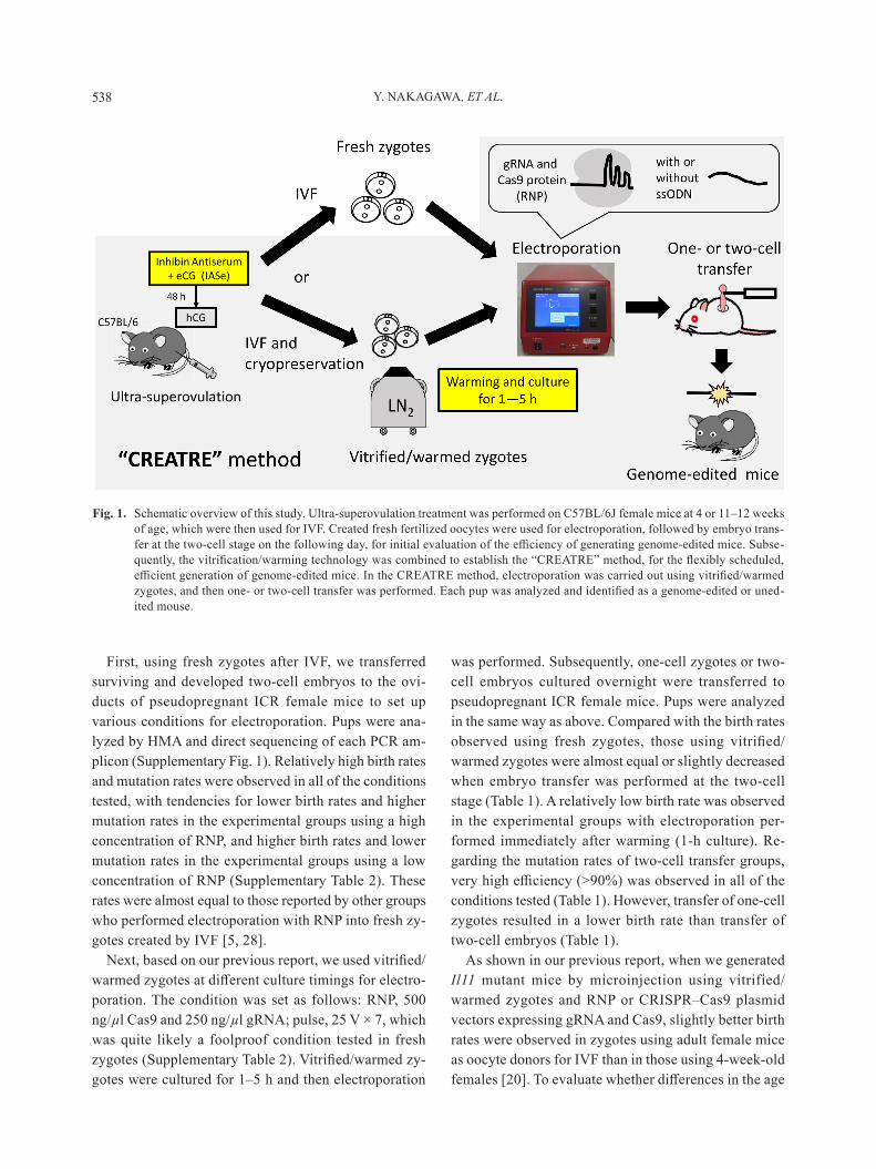
Here, to create an updated, streamlined pipeline of generating gm mice by combining electroporation of Cas9 RNP with reproductive engineering techniques, we used fresh or vitrified/warmed zygotes created by IVF (CARD method) via ultra-superovulation for electro-poration (Fig. 1). Fresh zygotes were used for electro-
poration after 6.5–7.5 h from insemination (around E0.3). Vitrified/warmed zygotes were cultured for 1–5 h and then used for electroporation at different timings (E0.3–E0.5), similar to the approach using microinjec-tion as described in our previous report [21]. after elec-troporation, the surviving one-cell zygotes or two-cell embryos were transferred to pseudopregnant female mice after culture for about 1 h or overnight, to examine whether different birth rates occurred.
Materials and Methods
gRNA synthesis and preparation of Cas9 protein and ssODN
In vitro-transcribed gRNAs were prepared in accor-dance with a previous report [1]. Briefly, template DNA fragments were generated using PCR amplification from CRISPR–Cas9 vectors with primers containing a T7 promoter sequence, in accordance with a previously described protocol [20]. Subsequently, the gRNAs were synthesized using a MEGAshortscript T7 Kit (Thermo Fisher Scientific, Tokyo, Japan), and then purified with a MEGAclear Kit (Thermo Fisher Scientific). The gRNA designs of interleukin-11 (Il11), tyrosinase (Tyr), and secreted phosphoprotein 1 (Spp1) genes were as de-scribed previously [20, 21]. In the generation of Il11 mutant mice, gRNA B was synthesized and used as shown in a previous report. Recombinant Cas9 protein was obtained from Integrated DNA Technologies Japan (Alt-RTm S.p. Cas9 Nuclease 3NLS; Tokyo, Japan). The sequence of ssODN for three-base substitution at the Spp1 locus was the same as in previous reports [20, 21]. ssODN was synthesized by Integrated DNA Technolo-gies (Coralville, IA, USA).
AnimalsC57BL/6J mice were purchased from CLEA Japan
(Tokyo, Japan). After breeding, C57BL/6J female mice were used as oocyte donors at 4 or 11–12 weeks of age. C57BL/6J male mice over 10 weeks of age were used as sperm donors for IVF. ICR mice at 8–20 weeks of age were used as recipients of zygotes. all animals were housed under a 12-h dark–light cycle (light from 07:00 to 19:00) at 22 ± 1°C with ad libitum access to food and water. all animal experiments were approved by the Animal Care and Experimentation Committee of the Center for Animal Resources and Development, Kuma-moto university, and were carried out in accordance with
ULTRA-SUPEROVULATION FOR ELECTROPORATION 537
the approved guidelines.
IVF and vitrification/warming of fertilized oocytesThe IVF and vitrification/warming procedures were
as described previously [19–21]. Cauda epididymides were obtained from C57BL/6J male mice and used as a source of sperm for IVF. C57BL/6J female mice were ultra-superovulated by the intraperitoneal administration of IASe (0.1 ml of IAS and 3.75 IU eCG; CARD Hy-perova®; kyudo, Saga, Japan), followed 48 h later by the intraperitoneal administration of hCG (7.5 IU; Go-natropin; ASKA Pharmaceutical, Tokyo, Japan) [27]. The cumulus–oocyte complexes were collected in CARD MEDIUM® (Kyudo), which contained 0.15 mM GSH. They were inseminated with sperm after preincubation in FERTIUP® Mouse Sperm Preincubation Medium (Kyudo) for 1–1.5 h, and then were incubated at 37°C in 5% CO2 and 95% humidified air. After 2.5 h of incu-bation, the inseminated oocytes were rinsed three times with modified human tubal fluid (mHTF) medium con-taining a high level of calcium. The generated fertilized oocytes were used for electroporation or cryopreserved by a simple vitrification method 6.5 h after insemination [15, 16]. at later time points, the cryopreserved oocytes were warmed, cultured in potassium simplex optimized medium with amino acids (KSOM-AA) for 1–5 h at 37°C in 5% CO2 and 95% humidified air, and used for elec-troporation. Descriptions of IVF and vitrification proce-dures are available as online manuals on our website (http://card.medic.kumamoto-u.ac.jp/card/english/sigen/manual/onlinemanual.html).
Electroporation and transferelectroporation was performed based on a previous
report [4]. Zygotes were rinsed with Opti-MEM I (Ther-mo Fisher Scientific) and then placed in the electrode gap filled with 5 µl of opti-mem I solution containing Cas9 protein and gRNA with or without ssODN. Using electrode (LF501PT1-10; BEX, Tokyo, Japan) and Ge-nome Editor (GEB15, BEX), electroporation was per-formed seven times under conditions of 25 or 30 V (3 ms oN + 97 ms oFF). The zygotes were rinsed with m2 medium (Sigma, Tokyo, Japan) and cultured in KSOM-AA at 37°C in 5% CO2 and 95% humidified air until transfer for about 1 h or overnight. Surviving one-cell zygotes or two-cell embryos were transferred to the ovi-ducts of pseudopregnant ICR female mice.
Analysis of pupsPup tail lysates were prepared by an alkaline lysis
method and PCR was performed using KOD FX (Toyo-bo, osaka, Japan) with each primer set. For the analysis of Il11-modified mice, the IL11 F and R primers listed in Supplementary Table 1 were used. Each PCR product was subjected to automatic electrophoresis using mul-tiNA (Shimadzu Corporation, Kyoto, Japan) and ana-lyzed by a heteroduplex mobility assay (HMA) [18]. The PCR products identified as negative or weakly positive by HMA were analyzed by direct sequencing using an ABI 3130 Genetic Analyzer (Thermo Fisher Scientific) with a BigDye Terminator v3.1 Cycle Sequencing Kit (Thermo Fisher Scientific). For the analysis of the Tyr gene, the eye color of each pup was confirmed (i.e., black eyes or albinism of both eyes) and then tail lysates of pups harboring black eyes were analyzed by direct se-quencing of each PCR product amplified with the Tyr F and R primers listed in Supplementary Table 1. In the Spp1 modified mice, the Spp1 F and R primers listed in Supplementary Table 1 were used, and then each PCR product was subjected to restriction fragment length polymorphism (RFLP) analysis and direct sequencing, in accordance with previous reports [20, 21]. we did not perform off-target analysis of each pup, because no off-target mutations were detected previously in Il11- and Tyr-targeted founders with the same gRNA design [10, 19], and the target sequence of Spp1-gRNA was care-fully selected using the COSMID web tool [3].
Results
Electroporation-mediated generation of Il11 mutant mice using fresh or vitrified/warmed zygotes
To examine whether reproductive engineering tech-niques such as IVF (CARD method) via ultra-superovu-lation and cryopreservation of zygotes are applicable for the generation of mutant mice using electroporation, we used Cas9 RNP with fresh or vitrified/warmed zygotes created by IVF via ultra-superovulation for the genera-tion of Il11 mutant mice, which were previously gener-ated by the microinjection method and validated well [19, 20]. C57BL/6J female mice were ultra-superovu-lated for IVF at 4 weeks of age in the initial trials. In general, approximately 2–3 times as many oocytes can be collected per immature female mouse than per adult female mouse. The number of oocytes collected per fe-male mouse peaks at 4 weeks of age (data not shown).
Y. NAKAGAWA, ET AL.538
First, using fresh zygotes after IVF, we transferred surviving and developed two-cell embryos to the ovi-ducts of pseudopregnant ICR female mice to set up various conditions for electroporation. Pups were ana-lyzed by HMA and direct sequencing of each PCR am-plicon (Supplementary Fig. 1). Relatively high birth rates and mutation rates were observed in all of the conditions tested, with tendencies for lower birth rates and higher mutation rates in the experimental groups using a high concentration of RNP, and higher birth rates and lower mutation rates in the experimental groups using a low concentration of RNP (Supplementary Table 2). These rates were almost equal to those reported by other groups who performed electroporation with RNP into fresh zy-gotes created by IVF [5, 28].
Next, based on our previous report, we used vitrified/warmed zygotes at different culture timings for electro-poration. The condition was set as follows: RNP, 500 ng/µl Cas9 and 250 ng/µl gRNA; pulse, 25 V × 7, which was quite likely a foolproof condition tested in fresh zygotes (Supplementary Table 2). Vitrified/warmed zy-gotes were cultured for 1–5 h and then electroporation
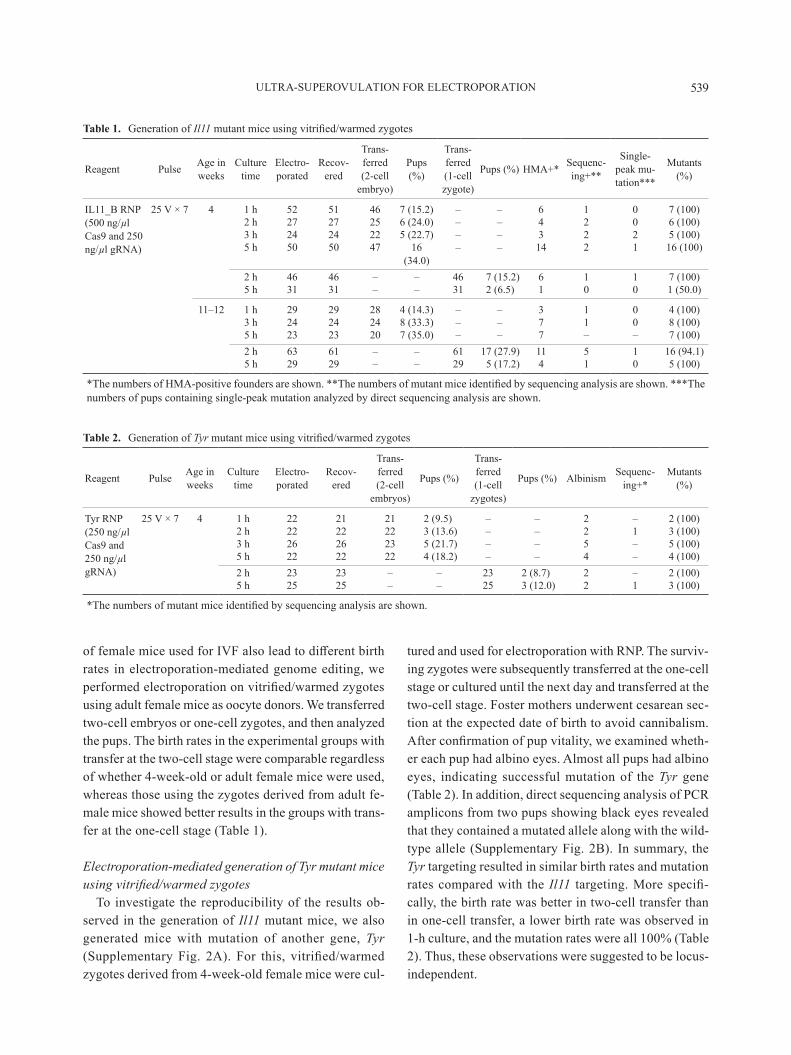
was performed. Subsequently, one-cell zygotes or two-cell embryos cultured overnight were transferred to pseudopregnant ICR female mice. Pups were analyzed in the same way as above. Compared with the birth rates observed using fresh zygotes, those using vitrified/warmed zygotes were almost equal or slightly decreased when embryo transfer was performed at the two-cell stage (Table 1). a relatively low birth rate was observed in the experimental groups with electroporation per-formed immediately after warming (1-h culture). Re-garding the mutation rates of two-cell transfer groups, very high efficiency (>90%) was observed in all of the conditions tested (Table 1). However, transfer of one-cell zygotes resulted in a lower birth rate than transfer of two-cell embryos (Table 1).
as shown in our previous report, when we generated Il11 mutant mice by microinjection using vitrified/warmed zygotes and RNP or CRISPR–Cas9 plasmid vectors expressing gRNA and Cas9, slightly better birth rates were observed in zygotes using adult female mice as oocyte donors for IVF than in those using 4-week-old females [20]. To evaluate whether differences in the age
Fig. 1. Schematic overview of this study. Ultra-superovulation treatment was performed on C57BL/6J female mice at 4 or 11–12 weeks of age, which were then used for IVF. Created fresh fertilized oocytes were used for electroporation, followed by embryo trans-fer at the two-cell stage on the following day, for initial evaluation of the efficiency of generating genome-edited mice. Subse-quently, the vitrification/warming technology was combined to establish the “CREATRE” method, for the flexibly scheduled, efficient generation of genome-edited mice. In the CREATRE method, electroporation was carried out using vitrified/warmed zygotes, and then one- or two-cell transfer was performed. Each pup was analyzed and identified as a genome-edited or uned-ited mouse.
ULTRA-SUPEROVULATION FOR ELECTROPORATION 539
of female mice used for IVF also lead to different birth rates in electroporation-mediated genome editing, we performed electroporation on vitrified/warmed zygotes using adult female mice as oocyte donors. we transferred two-cell embryos or one-cell zygotes, and then analyzed the pups. The birth rates in the experimental groups with transfer at the two-cell stage were comparable regardless of whether 4-week-old or adult female mice were used, whereas those using the zygotes derived from adult fe-male mice showed better results in the groups with trans-fer at the one-cell stage (Table 1).
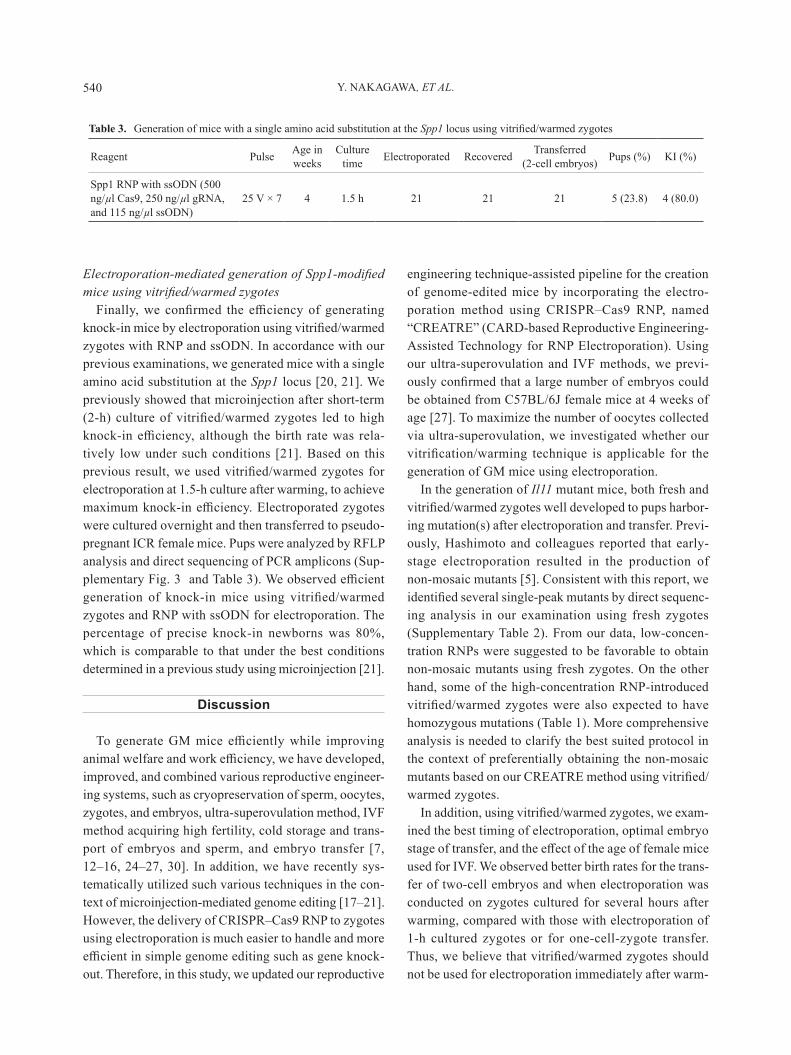
Electroporation-mediated generation of Tyr mutant mice using vitrified/warmed zygotes
To investigate the reproducibility of the results ob-served in the generation of Il11 mutant mice, we also generated mice with mutation of another gene, Tyr (Supplementary Fig. 2A). For this, vitrified/warmed zygotes derived from 4-week-old female mice were cul-
tured and used for electroporation with RNP. The surviv-ing zygotes were subsequently transferred at the one-cell stage or cultured until the next day and transferred at the two-cell stage. Foster mothers underwent cesarean sec-tion at the expected date of birth to avoid cannibalism. After confirmation of pup vitality, we examined wheth-er each pup had albino eyes. almost all pups had albino eyes, indicating successful mutation of the Tyr gene (Table 2). In addition, direct sequencing analysis of PCR amplicons from two pups showing black eyes revealed that they contained a mutated allele along with the wild-type allele (Supplementary Fig. 2B). In summary, the Tyr targeting resulted in similar birth rates and mutation rates compared with the Il11 targeting. More specifi-cally, the birth rate was better in two-cell transfer than in one-cell transfer, a lower birth rate was observed in 1-h culture, and the mutation rates were all 100% (Table 2). Thus, these observations were suggested to be locus-independent.
Table 1. generation of Il11 mutant mice using vitrified/warmed zygotes
Reagent Pulse age in weeks
Culture time
electro-porated
Recov-ered
Trans-ferred (2-cell
embryo)
Pups (%)
Trans-ferred (1-cell zygote)
Pups (%) HMA+* Sequenc-ing+**
Single-peak mu-tation***
mutants (%)
IL11_B RNP (500 ng/µl Cas9 and 250 ng/µl gRNA)
25 V × 7 4 1 h 52 51 46 7 (15.2) – – 6 1 0 7 (100)2 h 27 27 25 6 (24.0) – – 4 2 0 6 (100)3 h 24 24 22 5 (22.7) – – 3 2 2 5 (100)5 h 50 50 47 16
(34.0)– – 14 2 1 16 (100)
2 h 46 46 – – 46 7 (15.2) 6 1 1 7 (100)5 h 31 31 – – 31 2 (6.5) 1 0 0 1 (50.0)
11–12 1 h 29 29 28 4 (14.3) – – 3 1 0 4 (100)3 h 24 24 24 8 (33.3) – – 7 1 0 8 (100)5 h 23 23 20 7 (35.0) – – 7 – – 7 (100)2 h 63 61 – – 61 17 (27.9) 11 5 1 16 (94.1)5 h 29 29 – – 29 5 (17.2) 4 1 0 5 (100)
*The numbers of HMA-positive founders are shown. **The numbers of mutant mice identified by sequencing analysis are shown. ***The numbers of pups containing single-peak mutation analyzed by direct sequencing analysis are shown.
Table 2. generation of Tyr mutant mice using vitrified/warmed zygotes
Reagent Pulse age in weeks
Culture time
electro-porated
Recov-ered
Trans-ferred (2-cell
embryos)
Pups (%)
Trans-ferred (1-cell
zygotes)
Pups (%) albinism Sequenc-ing+*
mutants (%)
Tyr RNP (250 ng/µl Cas9 and 250 ng/µl gRNA)
25 V × 7 4 1 h 22 21 21 2 (9.5) – – 2 – 2 (100)2 h 22 22 22 3 (13.6) – – 2 1 3 (100)3 h 26 26 23 5 (21.7) – – 5 – 5 (100)5 h 22 22 22 4 (18.2) – – 4 – 4 (100)2 h 23 23 – – 23 2 (8.7) 2 – 2 (100)5 h 25 25 – – 25 3 (12.0) 2 1 3 (100)
*The numbers of mutant mice identified by sequencing analysis are shown.
Y. NAKAGAWA, ET AL.540
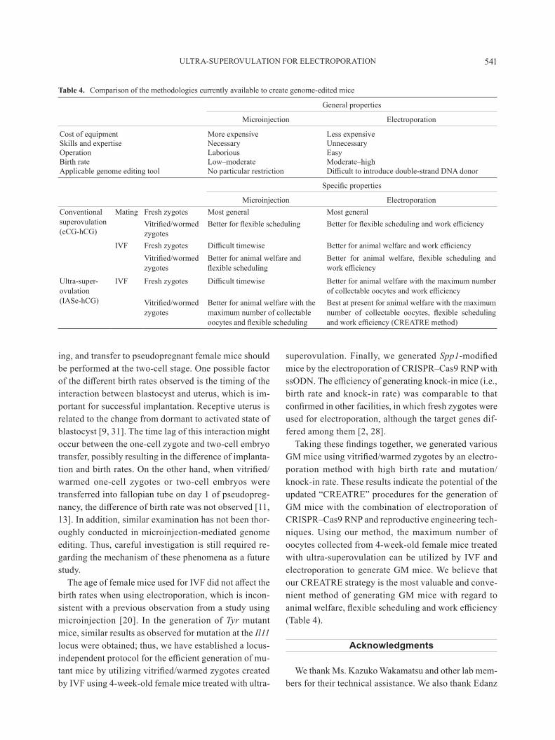
Electroporation-mediated generation of Spp1-modified mice using vitrified/warmed zygotes
Finally, we confirmed the efficiency of generating knock-in mice by electroporation using vitrified/warmed zygotes with RNP and ssODN. In accordance with our previous examinations, we generated mice with a single amino acid substitution at the Spp1 locus [20, 21]. we previously showed that microinjection after short-term (2-h) culture of vitrified/warmed zygotes led to high knock-in efficiency, although the birth rate was rela-tively low under such conditions [21]. Based on this previous result, we used vitrified/warmed zygotes for electroporation at 1.5-h culture after warming, to achieve maximum knock-in efficiency. Electroporated zygotes were cultured overnight and then transferred to pseudo-pregnant ICR female mice. Pups were analyzed by RFLP analysis and direct sequencing of PCR amplicons (Sup-plementary Fig. 3 and Table 3). We observed efficient generation of knock-in mice using vitrified/warmed zygotes and RNP with ssODN for electroporation. The percentage of precise knock-in newborns was 80%, which is comparable to that under the best conditions determined in a previous study using microinjection [21].
Discussion
To generate GM mice efficiently while improving animal welfare and work efficiency, we have developed, improved, and combined various reproductive engineer-ing systems, such as cryopreservation of sperm, oocytes, zygotes, and embryos, ultra-superovulation method, IVF method acquiring high fertility, cold storage and trans-port of embryos and sperm, and embryo transfer [7, 12–16, 24–27, 30]. In addition, we have recently sys-tematically utilized such various techniques in the con-text of microinjection-mediated genome editing [17–21]. However, the delivery of CRISPR–Cas9 RNP to zygotes using electroporation is much easier to handle and more efficient in simple genome editing such as gene knock-out. Therefore, in this study, we updated our reproductive
engineering technique-assisted pipeline for the creation of genome-edited mice by incorporating the electro-poration method using CRISPR–Cas9 RNP, named “CREATRE” (CARD-based Reproductive Engineering-Assisted Technology for RNP Electroporation). Using our ultra-superovulation and IVF methods, we previ-ously confirmed that a large number of embryos could be obtained from C57BL/6J female mice at 4 weeks of age [27]. To maximize the number of oocytes collected via ultra-superovulation, we investigated whether our vitrification/warming technique is applicable for the generation of gm mice using electroporation.
In the generation of Il11 mutant mice, both fresh and vitrified/warmed zygotes well developed to pups harbor-ing mutation(s) after electroporation and transfer. Previ-ously, Hashimoto and colleagues reported that early-stage electroporation resulted in the production of non-mosaic mutants [5]. Consistent with this report, we identified several single-peak mutants by direct sequenc-ing analysis in our examination using fresh zygotes (Supplementary Table 2). From our data, low-concen-tration RNPs were suggested to be favorable to obtain non-mosaic mutants using fresh zygotes. on the other hand, some of the high-concentration RNP-introduced vitrified/warmed zygotes were also expected to have homozygous mutations (Table 1). More comprehensive analysis is needed to clarify the best suited protocol in the context of preferentially obtaining the non-mosaic mutants based on our CREATRE method using vitrified/warmed zygotes.
In addition, using vitrified/warmed zygotes, we exam-ined the best timing of electroporation, optimal embryo stage of transfer, and the effect of the age of female mice used for IVF. We observed better birth rates for the trans-fer of two-cell embryos and when electroporation was conducted on zygotes cultured for several hours after warming, compared with those with electroporation of 1-h cultured zygotes or for one-cell-zygote transfer. Thus, we believe that vitrified/warmed zygotes should not be used for electroporation immediately after warm-
Table 3. generation of mice with a single amino acid substitution at the Spp1 locus using vitrified/warmed zygotes
Reagent Pulse age in weeks
Culture time electroporated Recovered Transferred
(2-cell embryos) Pups (%) KI (%)
Spp1 RNP with ssODN (500 ng/µl Cas9, 250 ng/µl gRNA, and 115 ng/µl ssODN)
25 V × 7 4 1.5 h 21 21 21 5 (23.8) 4 (80.0)
ULTRA-SUPEROVULATION FOR ELECTROPORATION 541
ing, and transfer to pseudopregnant female mice should be performed at the two-cell stage. one possible factor of the different birth rates observed is the timing of the interaction between blastocyst and uterus, which is im-portant for successful implantation. Receptive uterus is related to the change from dormant to activated state of blastocyst [9, 31]. The time lag of this interaction might occur between the one-cell zygote and two-cell embryo transfer, possibly resulting in the difference of implanta-tion and birth rates. On the other hand, when vitrified/warmed one-cell zygotes or two-cell embryos were transferred into fallopian tube on day 1 of pseudopreg-nancy, the difference of birth rate was not observed [11, 13]. In addition, similar examination has not been thor-oughly conducted in microinjection-mediated genome editing. Thus, careful investigation is still required re-garding the mechanism of these phenomena as a future study.
The age of female mice used for IVF did not affect the birth rates when using electroporation, which is incon-sistent with a previous observation from a study using microinjection [20]. In the generation of Tyr mutant mice, similar results as observed for mutation at the Il11 locus were obtained; thus, we have established a locus-independent protocol for the efficient generation of mu-tant mice by utilizing vitrified/warmed zygotes created by IVF using 4-week-old female mice treated with ultra-
superovulation. Finally, we generated Spp1-modified mice by the electroporation of CRISPR–Cas9 RNP with ssODN. The efficiency of generating knock-in mice (i.e., birth rate and knock-in rate) was comparable to that confirmed in other facilities, in which fresh zygotes were used for electroporation, although the target genes dif-fered among them [2, 28].
Taking these findings together, we generated various GM mice using vitrified/warmed zygotes by an electro-poration method with high birth rate and mutation/knock-in rate. These results indicate the potential of the updated “CREATRE” procedures for the generation of gm mice with the combination of electroporation of CRISPR–Cas9 RNP and reproductive engineering tech-niques. using our method, the maximum number of oocytes collected from 4-week-old female mice treated with ultra-superovulation can be utilized by IVF and electroporation to generate gm mice. we believe that our CREATRE strategy is the most valuable and conve-nient method of generating gm mice with regard to animal welfare, flexible scheduling and work efficiency (Table 4).
Acknowledgments
we thank ms. kazuko wakamatsu and other lab mem-bers for their technical assistance. we also thank edanz
Table 4. Comparison of the methodologies currently available to create genome-edited mice
general properties
microinjection electroporation
Cost of equipment more expensive Less expensiveSkills and expertise Necessary unnecessaryoperation Laborious easyBirth rate Low–moderate Moderate–highapplicable genome editing tool No particular restriction Difficult to introduce double-strand DNA donor
Specific properties
microinjection electroporationConventional superovulation (eCG-hCG)
mating Fresh zygotes most general most generalVitrified/wormed zygotes
Better for flexible scheduling Better for flexible scheduling and work efficiency
IVF Fresh zygotes Difficult timewise Better for animal welfare and work efficiencyVitrified/wormed zygotes
Better for animal welfare and flexible scheduling
Better for animal welfare, flexible scheduling and work efficiency
ultra-super-ovulation (IASe-hCG)
IVF Fresh zygotes Difficult timewise Better for animal welfare with the maximum number of collectable oocytes and work efficiency
Vitrified/wormed zygotes
Better for animal welfare with the maximum number of collectable oocytes and flexible scheduling
Best at present for animal welfare with the maximum number of collectable oocytes, flexible scheduling and work efficiency (CREATRE method)
Y. NAKAGAWA, ET AL.542
Group (www.edanzediting.com/ac) for editing a draft of this manuscript. This work was supported by a grant-in-Aid for Scientific Research B, Grant Number 15H04606 (to N.N.), from the Japan Society for the Promotion of Science (JSPS), and by a grant for Re-search on Development of New Drugs (Project ID: 16769865, to T.T.), from the Japan agency for medical Research and Development (AMED).
References
1. Aida, T., Chiyo, K., Usami, T., Ishikubo, H., Imahashi, R., wada, Y., Tanaka, k.F., Sakuma, T., Yamamoto, T., and Tanaka, K. 2015. Cloning-free CRISPR/Cas system facili-tates functional cassette knock-in in mice. Genome Biol. 16: 87. [medline] [CrossRef]
2. Chen, S., Lee, B., Lee, A.Y., Modzelewski, A.J., and He, L. 2016. Highly Efficient Mouse Genome Editing by CRIS-PR Ribonucleoprotein Electroporation of Zygotes. J. Biol. Chem. 291: 14457–14467. [medline] [CrossRef]
3. Cradick, T.J., Qiu, P., Lee, C.M., Fine, E.J., and Bao, G. 2014. COSMID: A Web-based Tool for Identifying and Vali-dating CRISPR/Cas Off-target Sites. Mol. Ther. Nucleic Ac-ids 3: e214. [medline] [CrossRef]
4. Hashimoto, M. and Takemoto, T. 2015. Electroporation en-ables the efficient mRNA delivery into the mouse zygotes and facilitates CRISPR/Cas9-based genome editing. Sci. Rep. 5: 11315. [medline] [CrossRef]
5. Hashimoto, M., Yamashita, Y., and Takemoto, T. 2016. Elec-troporation of Cas9 protein/sgRNA into early pronuclear zy-gotes generates non-mosaic mutants in the mouse. Dev. Biol. 418: 1–9. [medline] [CrossRef]
6. Horii, T., Morita, S., Kimura, M., Terawaki, N., Shibutani, M., and Hatada, I. 2017. Efficient generation of conditional knockout mice via sequential introduction of lox sites. Sci. Rep. 7: 7891. [medline] [CrossRef]
7. Horikoshi, Y., Takeo, T., and Nakagata, N. 2016. N-acetyl cysteine prolonged the developmental ability of mouse two-cell embryos against oxidative stress at refrigerated tempera-tures. Cryobiology 72: 198–204. [medline] [CrossRef]
8. kaneko, T. and mashimo, T. 2015. Simple genome editing of Rodent Intact Embryos by Electroporation. PLoS One 10: e0142755. [medline] [CrossRef]
9. Matsumoto, H. 2017. Molecular and cellular events during blastocyst implantation in the receptive uterus: clues from mouse models. J. Reprod. Dev. 63: 445–454. [medline] [CrossRef]
10. Mizuno, S., Dinh, T.T., Kato, K., Mizuno-Iijima, S., Tani-moto, Y., Daitoku, Y., Hoshino, Y., Ikawa, M., Takahashi, S., Sugiyama, F., and Yagami, k. 2014. Simple generation of albino C57BL/6J mice with G291T mutation in the tyrosi-nase gene by the CRISPR/Cas9 system. Mamm. Genome 25: 327–334. [medline] [CrossRef]
11. Nakagata, N. 1989. High survival rate of pronuclear mouse oocytes derived from in vitro fertilization following ultrar-apid freezing and thawing. Jpn. J. Fertil. Steril. 34: 757–760
(in Japanese with English abstract). 12. Nakagata, N. 1992. [embryo transfer through the wall of
the fallopian tube in mice]. Jikken Dobutsu 41: 387–388. (in Japanese) [medline]
13. Nakagata, N. 1995. Studies on cryopreservation of embryos and gametes in mice. Exp. Anim. 44: 1–8. [medline] [Cross-Ref]
14. Nakagata, N. 2000. Cryopreservation of mouse spermato-zoa. Mamm. Genome 11: 572–576. [medline] [CrossRef]
15. Nakagata, N., Takeo, T., Fukumoto, K., Kondo, T., Harugu-chi, Y., Takeshita, Y., Nakamuta, Y., Matsunaga, H., Tsuchi-yama, S., Ishizuka, Y., and araki, k. 2013. applications of cryopreserved unfertilized mouse oocytes for in vitro fertil-ization. Cryobiology 67: 188–192. [medline] [CrossRef]
16. Nakao, k., Nakagata, N., and katsuki, m. 1997. Simple and efficient vitrification procedure for cryopreservation of mouse embryos. Exp. Anim. 46: 231–234. [medline] [Cross-Ref]
17. Nakagawa, Y., Sakuma, T., Nakagata, N., Yamasaki, S., Takeda, N., ohmuraya, m., and Yamamoto, T. 2014. ap-plication of oocyte cryopreservation technology in TaLeN-mediated mouse genome editing. Exp. Anim. 63: 349–355. [medline] [CrossRef]
18. Nakagawa, Y., Yamamoto, T., Suzuki, k., araki, k., Takeda, N., ohmuraya, m., and Sakuma, T. 2014. Screening methods to identify TaLeN-mediated knockout mice. Exp. Anim. 63: 79–84. [medline] [CrossRef]
19. Nakagawa, Y., Sakuma, T., Sakamoto, T., ohmuraya, m., Nakagata, N., and Yamamoto, T. 2015. Production of knock-out mice by DNA microinjection of various CRISPR/Cas9 vectors into freeze-thawed fertilized oocytes. BMC Biotech-nol. 15: 33. [medline] [CrossRef]
20. Nakagawa, Y., Sakuma, T., Nishimichi, N., Yokosaki, Y., Yanaka, N., Takeo, T., Nakagata, N., and Yamamoto, T. 2016. Ultra-superovulation for the CRISPR-Cas9-mediated production of gene-knockout, single-amino-acid-substitut-ed, and floxed mice. Biol. Open 5: 1142–1148. [medline] [CrossRef]
21. Nakagawa, Y., Sakuma, T., Nishimichi, N., Yokosaki, Y., Takeo, T., Nakagata, N., and Yamamoto, T. 2017. Culture time of vitrified/warmed zygotes before microinjection af-fects the production efficiency of CRISPR-Cas9-mediated knock-in mice. Biol. Open 6: 706–713. [medline] [Cross-Ref]
22. Qin, W., Dion, S.L., Kutny, P.M., Zhang, Y., Cheng, A.W., Jillette, N.L., Malhotra, A., Geurts, A.M., Chen, Y.G., and Wang, H. 2015. Efficient CRISPR/Cas9-Mediated Genome editing in mice by Zygote electroporation of Nuclease. Ge-netics 200: 423–430. [medline] [CrossRef]
23. Sato, M., Ohtsuka, M., Watanabe, S., and Gurumurthy, C.B. 2016. Nucleic acids delivery methods for genome editing in zygotes and embryos: the old, the new, and the old-new. Biol. Direct 11: 16. [medline] [CrossRef]
24. Takeo, T., Hoshii, T., Kondo, Y., Toyodome, H., Arima, H., Yamamura, k., Irie, T., and Nakagata, N. 2008. methyl-beta-cyclodextrin improves fertilizing ability of C57BL/6 mouse sperm after freezing and thawing by facilitating cholesterol efflux from the cells. Biol. Reprod. 78: 546–551. [medline]
ULTRA-SUPEROVULATION FOR ELECTROPORATION 543
[CrossRef] 25. Takeo, T. and Nakagata, N. 2010. Combination medium of
cryoprotective agents containing L-glutamine and methyl-beta-cyclodextrin in a preincubation medium yields a high fertilization rate for cryopreserved C57BL/6J mouse sperm. Lab. Anim. 44: 132–137. [medline] [CrossRef]
26. Takeo, T. and Nakagata, N. 2011. Reduced glutathione en-hances fertility of frozen/thawed C57BL/6 mouse sperm af-ter exposure to methyl-beta-cyclodextrin. Biol. Reprod. 85: 1066–1072. [medline] [CrossRef]
27. Takeo, T. and Nakagata, N. 2015. Superovulation using the combined administration of inhibin antiserum and equine chorionic gonadotropin increases the number of ovulated oocytes in C57BL/6 female mice. PLoS One 10: e0128330. [medline] [CrossRef]
28. Wang, W., Kutny, P.M., Byers, S.L., Longstaff, C.J., DaCos-
ta, M.J., Pang, C., Zhang, Y., Taft, R.A., Buaas, F.W., and Wang, H. 2016. Delivery of Cas9 Protein into Mouse Zy-gotes through a Series of Electroporation Dramatically In-creases the Efficiency of Model Creation. J. Genet. Genom-ics 43: 319–327. [medline] [CrossRef]
29. Yin, H., Kauffman, K.J., and Anderson, D.G. 2017. Delivery technologies for genome editing. Nat. Rev. Drug Discov. 16: 387–399. [medline] [CrossRef]
30. Yoshimoto, H., Takeo, T., and Nakagata, N. 2017. Dimethyl sulfoxide and quercetin prolong the survival, motility, and fertility of cold-stored mouse sperm for 10 days. Biol. Re-prod. 97: 883–891. [medline] [CrossRef]
31. Zhang, S., Lin, H., Kong, S., Wang, S., Wang, H., Wang, H., and Armant, D.R. 2013. Physiological and molecular de-terminants of embryo implantation. Mol. Aspects Med. 34: 939–980. [medline] [CrossRef]