Embed Size (px)
Citation preview

Thermal Dissociation of Streptavidin Homotetramer in the Gas Phase.
Subunit Loss versus Backbone Fragmentation
Elena N. Kitova, Igor Sinelnikov, Lu Deng and John S. Klassen*
Department of Chemistry, University of Alberta, Edmonton, Alberta, Canada T6G 2G2

2
Abstract
The results of time-resolved blackbody infrared radiative dissociation experiments
performed on gaseous protonated ions, at charge states +15, +16 and +17, of the
homotetramer streptavidin (S4) are reported. Evidence is found for three dissociation
pathways involving (i) the loss of a single subunit, (ii) covalent cleavage of the backbone
of one of the subunits and ejection of the resultant b212+
ion (followed by loss of
complementary y106 ions), and (iii) the direct loss of one or more water molecules. The
contribution of the different dissociation channels was found to be dependent on
temperature, with the loss of subunit dominating at higher reaction temperatures and
backbone fragmentation dominating at lower temperatures, and reaction times, with
longer times favouring covalent fragmentation. Analysis of the dissociation kinetics and
the influence of reaction time on the relative abundance of product ions indicate that
backbone fragmentation and subunit loss are not produced via parallel pathways from a
single reactant. Instead, the results suggest the presence of multiple, non-interconverting
structures, which contribute differentially to the backbone fragmentation and subunit loss
pathways. The results of molecular dynamics simulations performed on S4+16
ions with
different charge configurations suggest that unfolding of the N-terminus of the subunit
may be associated with the backbone fragmentation pathway.

3
1. Introduction
Electrospray ionization mass spectrometry (ESI-MS) is an important tool for
characterizing noncovalent protein assemblies and is increasingly used to establish
subunit composition and topology [1-5]. These structural insights are often obtained by
heating (activating) protein complexes in the gas phase in order to induce structural
changes, usually dissociation reactions. Activation of the gas phase ions of protein
complexes is most commonly achieved through energetic collisions with neutral gases
(collision-induced dissociation, CID) [6-8]. However, other activation methods can also
be used, such as surface-induced dissociation (SID) [9], the blackbody infrared radiative
dissociation (BIRD) technique, which involves absorption of blackbody radiation in a
near collision-free environment [10-14], or electron mediated methods, such as electron
capture dissociation (ECD) [15].
The loss of one or more subunits from the protein complex is usually the major
dissociation pathway observed with CID, SID and BIRD. With CID, which is a relatively
slow heating method (on the s timescale), the ejected subunit typically retains a
disproportionately large fraction (based on mass) of the total charge of the complex [6-
8,16-18]. Similar observations have been made using BIRD, for which activation occurs
on a much longer timescale (~s) compared to CID [10-14]. It is generally accepted that
activation of the gaseous complex ion results in the unfolding of at least one of the
subunits [7,8,19,20]. The resulting increase in surface area of the unfolding subunit
promotes charge (usually proton) transfer, which decreases the total Coulombic repulsion
in the complex ion [13,21,22]. In the case of SID, which involves significantly shorter
activation timescales (~ns) and high collision energies, the subunit loss reaction is
typically more charge symmetric in nature [9,18]. The difference in charge distributions

4
observed in SID and CID has been attributed to faster subunit loss kinetics in SID, which
dominates over the unfolding/charge transfer processes [18].
While subunit loss represents the most common pathway for the dissociation of
protein complexes in the gas phase, other dissociation pathways have been reported. In
some instances the loss of intact subunit multimers is observed [23-25]. There have also
been a limited number of reports of backbone fragmentation reactions competing with
subunit loss from the intact protein complex. For example, McLuckey and coworkers
reported on CID of the heterodimer complex of trypsin and bovine pancreatic trypsin
inhibitor (BPTI) [26]. At the lowest charge states investigated they noted the loss of
(unidentified) neutrals as the dominant dissociation pathway. They also observed the
ejection of a cation, which was tentatively assigned as the b2 ion corresponding to
backbone fragmentation of BPTI. It was suggested that the cleavage site was related to an
absence of intermolecular interactions between the N-terminus of BPTI and trypsin in the
gas phase complex. Similarly, Wysocki and co-workers found that CID of the
homopentamer of serum amyloid P (SAP) component resulted in covalent fragmentation
of the protein backbone, producing b-type ions, in addition to the subunit loss [18]. In
contrast, SID of SAP at the same charge state produced exclusively intact monomers and
dimers. It was suggested that proton transfer to the unfolding subunit, which occurs
during CID, but not SID, is necessary for covalent cleavage to occur. More recently,
Robinson and co-workers observed covalent fragmentation, resulting in the production of
y-type ions, in CID of the homotetrameric complex of human transthyretin [17].
Interestingly, while subunit loss occurred at all charge states investigated, covalent bond
fragmentation was observed only for the lowest charge states. This finding led to the
5
suggestion that the covalent fragmentation pathways are not governed only by subunit
unfolding and proton migration.
Here, we describe the results of time-resolved BIRD experiments performed on
gaseous protonated ions of streptavidin. Streptavidin is a homotetrameric protein
complex (S4) that is isolated from Streptomyces avidinii. Streptavidin has been the focus
of previous gas phase dissociation studies [6,13]. Smith and co-workers applied sustained
off-resonance irradiation (SORI)-CID to the protonated S4 ion at charge state +14.
Dissociation produced exclusively the S+7
and S3+7
and S+6
and S3+8
product ion pairs [6].
More recently, time-resolved BIRD experiments were performed on the protonated S4
ions at charge states +14, +15 and +16 [13]. At relatively short reaction times, the major
dissociation pathway, independent of temperature, was the loss of a single subunit
carrying nearly 50% of the total charge. The first-order kinetic plots were found to be
non-linear, a finding attributed to the presence of multiple, non-interconverting structures
with distinct dissociation rate constants or thermally-induced changes in the conformation
of the complex.
The present work, which is an extension of the earlier BIRD study [13], examines
the dissociation of protonated S4 ions, at the +15, +16, +17 charge states, composed of a
recombinant, truncated form of streptavidin, which is produced predominantly as a single
isoform. The homogeneity of the protein facilitated the identification of new dissociation
pathways - backbone fragmentation and water loss – that were not evident in the previous
CID or BIRD studies. The influence of reaction time, temperature and reactant ion charge
state on these pathways was assessed. Molecular dynamics (MD) simulations, which
have been shown previously to be useful for probing the structures of protein complexes
in the gas phase and their dissociation pathways [21,27-29], were used to investigate the

6
influence of the charge distribution on the extent of unfolding of the N-terminus of the
subunit.
Experimental
A recombinant, truncated form (containing residues 13-139) of wild type streptavidin
(monomer MW 13 271.5 Da) was used in this study. The plasmid was generously
provided by Prof. P. Stayton (University of Washington). The protein was expressed in E.
coli and purified using procedures described elsewhere [30]. The solution of purified S4
was exchanged directly into 200 mM aqueous ammonium acetate using an Amicon
microconcentrator with a MW cut-off of 10 kDa and lyophilized. Stock solution of S4
(100 µM) was prepared by dissolving a known amount of lyophilized streptavidin into
200 mM ammonium acetate and stored at -20 °C until needed. ESI solutions were
prepared by thawing the stock solution at room temperature and diluting an aliquot with
aqueous ammonium acetate (10 mM) to achieve a S4 concentration of 10 M.
All experiments were performed using an ApexII 9.4T Fourier-transform ion
cyclotron resonance mass spectrometer (Bruker, Billerica, MA). Nanoflow ESI
(nanoESI) was performed using borosilicate tubes (1.0 mm o.d., 0.68 mm i.d.), pulled to
~5 μm o.d. at one end using a P-97 micropipette puller (Sutter Instruments, Novato, CA).
The electric field required to spray the solution in positive ion mode was established by
applying a voltage of 0.8 – 1.1 kV to a platinum wire inserted inside the glass tip. The
solution flow rate was ~20 nL min-1
. To shift the S4n+
ions to higher charge states,
nanoESI was performed using a custom built device that directs a high velocity flow of
dry air at the end of the ESI tip [31]. Details of the instrumental and experimental
conditions used for the BIRD measurements can be found elsewhere [32,33].
Molecular dynamics simulations

7
MD simulations were carried out using NAMD 2.9 [34]
with the Amber03 force field
[35] under vacuum. The initial geometry of the S4 complex was obtained from the crystal
structure of streptavidin (PDB ID: 1SWB) [36]. Each of the subunits (containing residues
16-135 for subunit A, residues 16-133 for subunits B, C and D) was extended to have the
same length as the truncated form (containing residues 13-139) of streptavidin that was
used experimentally. This was done by aligning each subunit against the subunit C of the
crystal structure for streptavidin mutant Ser27Ala (PDB ID: 1N9M) [37] using the DALI
server [38], and grafting the extended residues onto each subunit of the tetramer.
Ions of the S4 complex at the +16 charge state at five different charge distributions
(Table 1) were chosen for investigation. Topology and coordinate files for the
simulations of each charge distributions were generated using the xleap module of the
AmberTools (version 11) [39]. It was necessary to develop charges and parameters for
the neutral forms of Arg and C-terminal Ser as well as protonated forms of Ala, Asn and
Tyr. The charge parameters for these non-standard residues were parameterized using the
RESP ESP charge Derive server (RED Server) [40,41], using Gaussian C.01 [42] and
enforcing net charge across the residue. The MD integration time step was 1 fs; bonds
involving hydrogen atoms were constrained with SHAKE. Following 1000 steps of
minimization, the system was gradually heated to 500K within 250000 steps. The
temperature was then held constant at 500K and 2.5 ns of dynamics were performed.
Trajectory analysis, performed using the Visual Molecular Dynamics package [43], was
carried out to establish the Cα root mean square deviation (RMSD). The first 1.5 ns of
dynamics were needed to fully equilibrate the system, as judged by RMSD. The
subsequent 1.0 ns of production were used in the analysis.

8
Results and discussion
Shown in Figure 1 are ESI mass spectra acquired in positive ion mode, with and
without the use of the gas-assisted nanoESI device, for an aqueous ammonium acetate
solution of 10 M S4. When using the gas-assisted nanoESI device, the major protein
ions detected correspond to protonated S4 (measured MW 53 085 ± 1 Da, theoretical MW
53 086 Da) i.e., S4n+
, at charge states n = 15 to 18 (Figure 1a). In contrast, when using
normal nanoESI, S4n+
ions were detected at charge states n = 14 to 17. The shift of the
charge state distribution to higher values is, presumably, due to the dilution of agents
such as NH3 and H2O that can effect proton transfer and remove charge from the protein
ions. Also observed in the mass spectrum is a series of low abundance ions that
correspond to a species with a MW of 53 219 ± 1 Da. This species is assumed to be a
streptavidin tetramer that contains three copies of the expected natural core streptavidin
subunit and one isoform of streptavidin, which has an additional N-terminal Met residue,
i.e., (S4 + Met). The theoretical MW of the proposed tetrameric streptavidin complex is
53 218 Da [44].
Time-resolved BIRD experiments were performed on the S4+15
, S4+16
and S4+17
ions, at temperatures ranging from 131 to 161 oC. Shown in Figure 2 are mass spectra
acquired for the S4+16
ion, following isolation (Figure 2a), and after 2 s and 5 s at 161 oC
(Figures 2b,c). At this temperature, the major dissociation pathway corresponds to the
loss of a subunit, i.e., Sn+
with n = 7 – 9, and formation of the complementary S3n+
ions,
at n = 7 - 9. In addition to the ions produced from the subunit loss pathway, a doubly
charged ion at m/z ~1083, and ions at m/z 1585 and 1849 were detected. The m/z 1083
ion corresponds to the (b21 - H2O)2+
ion, which is formed by cleavage of the Ala21-Gly22
9
amide linkage (and the loss of water), while the m/z 1585 and 1849 ions correspond to
the truncated subunit, which are nominally the y1067+
and y1066+
ions. Also observed in the
BIRD mass spectra is an ion at m/z 3637, which is the truncated S4+14
ion produced by
the loss of b212+
, i.e., (S4 - b21)+14
≡ (S3y106)+14
. A third dissociation pathway involves the
loss of one and, possibly, more water molecules directly from the S4+16
ion. The
appearance of the low abundance (S3y106 - H2O)+14
and (S - H2O)+9
ions indicates that the
(S4 - H2O)+16
ion dissociates in a manner similar to the S4+16
ion. Taken together, these
results suggest that the b212+
ion, which subsequently loses H2O to give the (b21 - H2O)2+
ion, is lost directly from the S4+16
ion, followed by the loss of the truncated subunit (y106),
which is ejected at the +6 and +7 charge states. A small amount of the proton transfer
product, the S4+15
ion, was also detected.
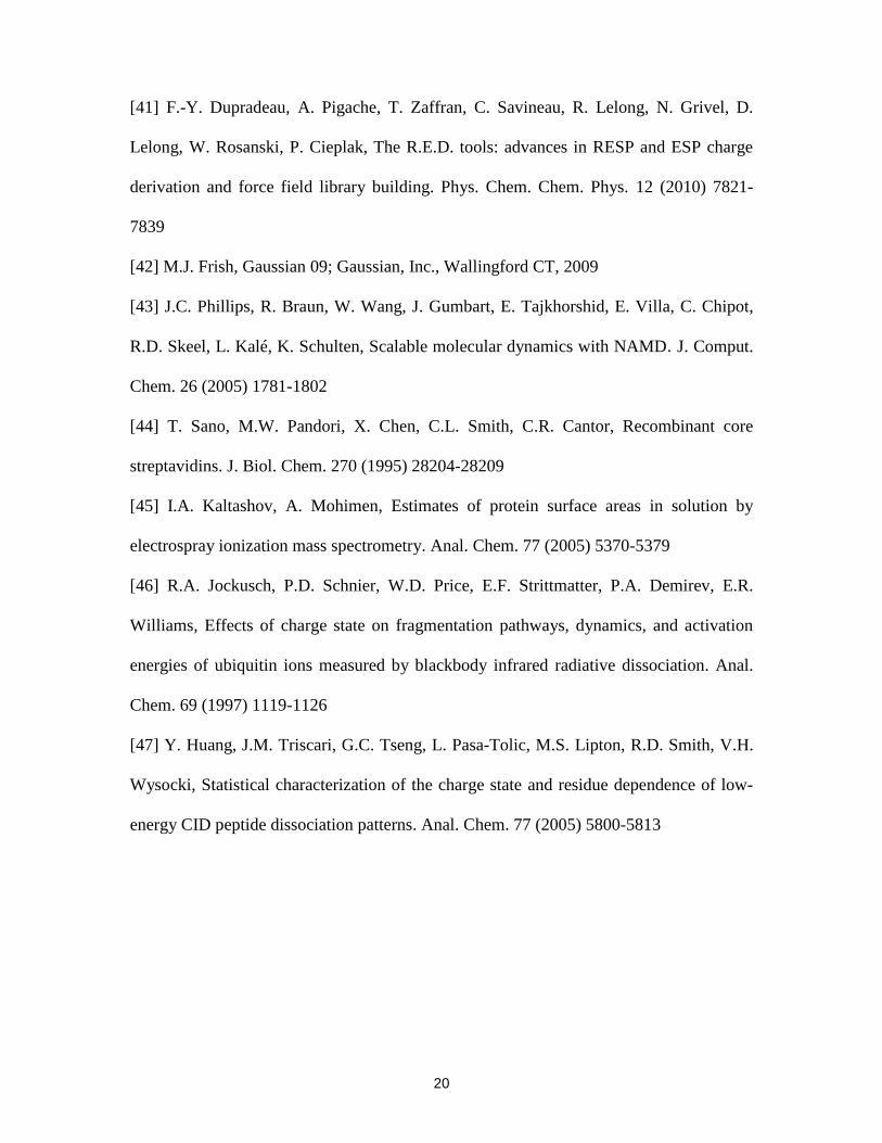
Shown in Figure 3 are representative BIRD mass spectra acquired for the S4+16
ion at 151, 141 and 131 oC. It can be seen that the BIRD results obtained at 151
oC are
qualitatively similar to those at 161 oC, with evidence for pathways involving subunit
loss, covalent fragmentation and water loss. At 141 and 131 oC, the (S3y106)
+14 ion is the
major product ion observed; ions corresponding to the water loss are also detected.
However, the y1066+
and y1067+
ions are absent from the mass spectra. This result indicates
that the (S3y106)+14
ion is kinetically more stable than the S4+16
ion, and resists dissociation
(except for the loss of water) at these temperatures. This finding can be explained by the
lower Coulombic repulsion between S3 and the truncated subunit. These results also
suggest that the activation energy (Ea) for the backbone fragmentation pathway, leading
to the (S3y106)+14
ion, is lower than that for intact subunit ejection.
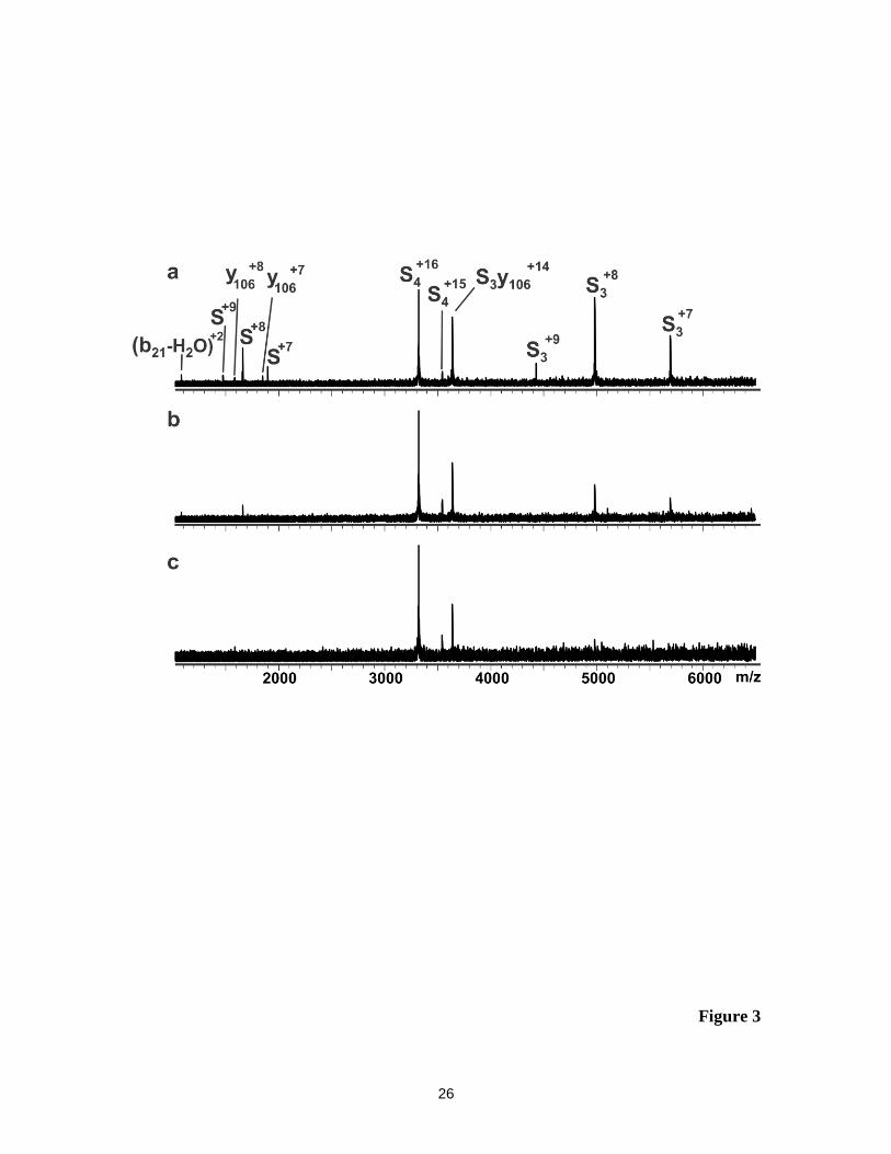
Representative BIRD mass spectra acquired for the S4+15
and S4+17
ions are shown
in Figures 4 and 5. Qualitatively, the BIRD results for these ions are similar to those

10
described for the S4+16
ion, with evidence for subunit ejection, backbone fragmentation
and water loss. As was found for S4+16
ion, the relative contribution of the backbone
cleavage pathway to the BIRD mass spectra for the S4+15
and S4+17
ions was more
significant at lower reaction temperatures. The (S3y106)+15
ion produced the expected
y106n+
ions (at n = 6 - 8) at 161 and 151 oC but not at lower temperatures, similar to what
was found for the (S3y106)+14
ion. In contrast, the (S3y106)+13
ion was found to be
kinetically stable at the temperatures investigated and no y106n+
ions were detected.
The BIRD mass spectra reveal that there are multiple dissociation channels
accessible to the S4n+
ions. An obvious question to ask is whether these represent parallel
pathways originating from a single reactant. Shown in Figure 6a are plots of the natural
logarithm of the normalized abundance of the S4+16
ion versus time measured at the four
reaction temperatures considered. It can be seen, particularly at the lower temperatures
investigated, that the plots are non-linear. Kinetic plots obtained for charge sates +15 and
+17 are also non-linear (data not shown). Plotted in Figure 6b is the ratio (RC/S) of the
abundances of the unique products produced by backbone fragmentation ((S3y106)+14
,
y1066+
and y1067+
ions) and subunit loss (S+7
, S+8
and S+9
) versus the fraction of S4+16
that
has reacted, measured at 161, 151 and 141 oC. It can be seen that RC/S increases with the
extent of dissociation of the reactant ion, indicating that the backbone fragmentation
pathway becomes more prominent as the reaction proceeds. Qualitatively similar results
were obtained for the S4+15
and S4+17
(data not shown). These findings rule out the
possibility of parallel pathways involving a single reactant and, instead, suggest the
presence of multiple non-interconverting structures or the occurrence of structural
changes to the reactant ion, on the timescale of the reaction, which influence the rate of
dissociation. However, an analysis of the average charge states of the Sn+
, S3n+
and y106n+

11
ions produced from the three S4n+
ions fails to reveal any time and temperature
dependence. Assuming that the charge states of the product ions are sensitive to their
structure and, in particular, surface area [17,45], this result would seem to argue against
structural changes taking place over the course of the reaction.
Taken together the BIRD results indicate that lower temperatures and longer
reaction times promote backbone fragmentation. As discussed above, the influence of
temperature can be explained in terms of a lower Ea for backbone fragmentation
compared to that for subunit loss. This is consistent with the Ea values reported
previously for the fragmentation of small proteins and peptides and subunit loss from
mulitprotein complexes in the gas phase [11,13,46]. The influence of reaction time on the
different pathways is explained by the presence of multiple, non-interconverting S4n+
structures, which contribute differentially to the dissociation pathways. Overall, the
kinetic stability of the S4n+
ions decreases with increasing charge state, a finding that can
be explained in terms of the destabilizing effect of Coulombic repulsion within the S4n+
ions. However, the backbone cleavage pathway was not found to be strongly dependent
on the charge state of the S4n+
ion.
That backbone fragmentation of the S4n+
ions occurs at only one site, the Ala21-
Gly22 amide linkage, is intriguing and suggests that the Ea for cleavage at this particular
linkage is small compared to the other amide bonds. It is not obvious from the primary
structure why this particular site is unusually labile, although it has been shown that
backbone cleavage on the N-terminal side of Gly residues is commonly observed in CID
of protonated peptides [18,47]. An analysis of the crystal structure of S4 indicates that the
residues that make up the b21 fragment are involved in two -strands, with the Ala21
residue located at the end of the second -strand. Based on the arguments advanced by

12
McLuckey and co-workers [26], the selectivity of the cleavage site may be related to the
unfolding of the N-terminal portion of the subunit, which promotes protonation of Ala21.
In an effort to gain more insight into the selectivity of the fragmentation reaction,
MD simulations were performed on S4+16
ions with five different charge configurations
(Table 1). For four of the configurations considered, the charges were distributed
asymmetrically between subunits: seven charges were placed on one subunit (subunit A)
and nine charges were evenly distributed between remaining three subunits. In each case,
the N-terminus, as well as Arg47, Arg72, Lys109 and Lys120 were protonated; the
position of the remaining charges was varied: Asn11 and Arg41 (configuration 16_1),
Ala21 and Arg41 (16_2), Tyr31 and Arg41 (16_3), and Arg41 and Arg 91 (16_4). A
symmetric charge configuration was also considered (16_5), in which the charges were
located on the N-terminus, Arg41, Arg47 and Lys120 of each of the subunits.
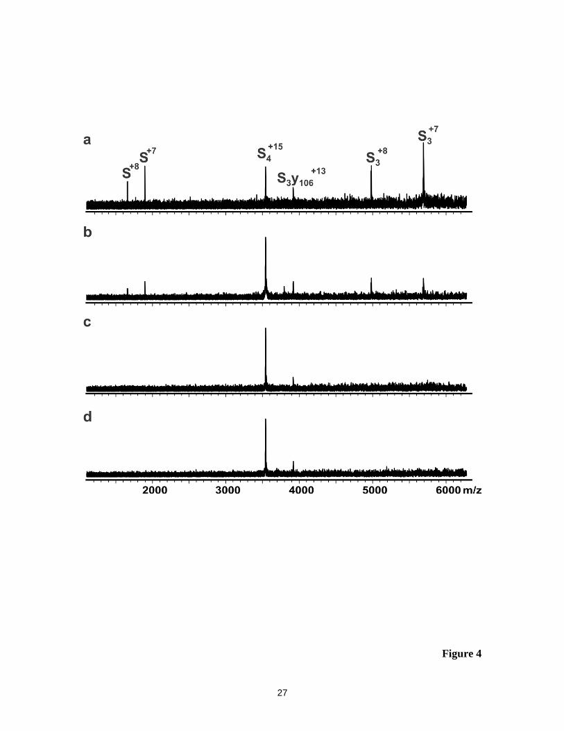
Shown in Figure 7 are representative MD snapshots of the S4+16
ions with the
different charge configurations, as well as the crystal structure of S4. The MD results
indicate that the location of the charges influences the structure of the subunits. Overall,
the structures of the S4+16
ions associated with the symmetric charge configuration (16_5)
and two of the asymmetric charge configurations (16_1 and 16_4) were found to
resemble the crystal structure. However, there are significant structural differences in
subunit A for the charge configurations 16_2 (RMSD ~7 Å, calculated for subunit A) and
16_3 (RMSD ~15 Å, calculated for subunit A). In the case of 16_3, the four -strands
comprising residues 1 – 48 of subunit A still exist but have shifted greatly from their
original positions (as seen in the crystal structure). However, in the case of configuration
16_2, protonation of Ala21 – the site of backbone cleavage – promotes complete
unfolding of the first two -strands of the N-terminal region of subunit A (RMSD ˃9 Å,

13
calculated for residues 1 – 21). While the results of the MD simulations do not provide a
conclusive explanation for the cleavage site specificity, they do raise the possibility that
thermal unfolding of the N-terminal region of one of the subunits promotes protonation
of the Ala21 residue, which may in turn promote amide bond cleavage.
Conclusions
In summary, the results of time-resolved BIRD experiments performed on gaseous
protonated S4+15
, S4+16
and S4+17
ions reveal three different primary dissociation
pathways: the loss of a subunit, covalent cleavage of the backbone and concomitant
ejection of the resultant b212+
ion, and the direct loss of water molecules. The contribution
of the different dissociation channels was found to be dependent on temperature, with the
loss of subunit dominating at the higher temperatures and backbone fragmentation
dominating at lower temperatures, and reaction time, with longer times favouring
covalent fragmentation. Analysis of the relative abundance of product ions measured as a
function of the extent of reaction reveals that backbone fragmentation and subunit loss
are not produced via parallel pathways from a single reactant. Instead, the results are
consistent with the presence of multiple non-interconverting structures, which contribute
differentially to the dissociation pathways. The results of MD simulations performed on
S4+16
ions with different charge configurations suggest that unfolding of the N-terminal
region of the subunit may be associated with the formation in the b212+
ion.
Acknowledgements
The authors acknowledge the Natural Sciences and Engineering Research Council of
Canada and the Alberta Glycomics Centre for funding, A. Broom (University of
Waterloo) for technical assistance and Professor P. Stayton (University of Washington)

14
for generously providing the plasmid for streptavidin. The MD simulations were made
possible by the facilities of the Shared Hierarchical Academic Research Computing
Network (SHARCNET: www.sharcnet.ca) and Compute/Calcul Canada.
15
References
[1] J.A. Loo, Electrospray ionization mass spectrometry: a technology for studying
noncovalent macromolecular complexes. Int. J. Mass Spectrom. 200 (2000) 175-186
[2] J.L.P. Benesch, C.V. Robinson, Mass spectrometry of macromolecular assemblies:
preservation and dissociation. Curr. Opin. Struct. Biol. 16 (2006) 245-251
[3] N.P. Barrera, C.V. Robinson, Advances in the mass spectrometry of membrane
proteins: from individual proteins to intact complexes. Annu. Rev. Biochem. 80 (2011)
247-271.
[4] C. Uetrecht, A.J. Heck, Modern biomolecular mass spectrometry and its role in
studying virus structure, dynamics, and assembly. Angew. Chemie Int. Ed. 50 (2011)
8248–8262
[5] R.H.H. van den Heuvel, A.J.R. Heck, Native protein mass spectrometry: from intact
oligomers to functional machineries. Curr. Opin. Chem. Biol. 8 (2004) 519–526
[6] B.L. Schwartz, J.E. Bruce, G.A. Anderson, S.A. Hofstadler, A.L. Rockwood, A.
Chilkoti, P.S. Stayton, R.D. Smith, Dissociation of tetrameric ions of noncovalent
streptavidin complexes formed by electrospray ionization. J. Am. Soc. Mass Spectrom. 6
(1995) 459–465
[7] J.L.P. Benesch, B.T. Ruotolo, D.A. Simmons, C.V. Robinson, Protein complexes in
the gas phase: technology for structural genomics and proteomics. Chem. Rev. 107
(2007) 3544-3567
[8] J.C. Jurchen, E.R. Williams, Origin of asymmetric charge partitioning in the
dissociation of gas-phase protein homodimers. J. Am. Chem. Soc. 125 (2003) 2817-2826

16
[9] C.M. Jones, R.L. Beardsley, A.S. Galhena, S. Dagan, G.L Cheng, V.H. Wysocki,
Gas-phase dissociation energetics of noncovalent protein complexes probed by surface-
induced dissociation mass spectrometry. J. Am. Chem. Soc. 128 (2006) 15044-15045
[10] R.C. Dunbar, T.B. McMahon, Activation of unimolecular reactions by ambient
blackbody radiation. Science 279 (1998) 194-197
[11] N. Felitsyn, E.N. Kitova, J.S. Klassen, Thermal decomposition of a gaseous
multiprotein complex studied by blackbody infrared radiative dissociation. Investigating
the origin of the asymmetric dissociation behavior. Anal. Chem. 73 (2001) 4647–4661
[12] N. Felitsyn, E.N. Kitova, J.S. Klassen, Thermal dissociation of the protein
homodimer ecotin in the gas phase. J. Am. Soc. Mass Spectrom. 13 (2002) 1432–1442
[13] I. Sinelnikov, E.N. Kitova, J.S. Klassen, Influence of Coulombic repulsion on the
dissociation pathways and energetics of multiprotein complexes in the gas phase. J. Am.
Soc. Mass Spectrom. 18 (2007) 617–631
[14] I. Sinelnikov, E.N. Kitova, J.S. Klassen, Effects of single amino acid substitution on
the dissociation of multiply charged multiprotein complexes in the gas phase. J. Am. Soc.
Mass Spectrom. 18 (2007) 688–692
[15] R.B.J. Geels, S.M. van der Vies, A.J.R. Heck, R.M.A. Heeren, Electron capture
dissociation as structural probe for noncovalent gas-phase protein assemblies. Anal.
Chem. 78 (2006) 7191-7196
[16] K.J. Light-Wahl, B.L. Schwartz, R.D. Smith, Observation of the noncovalent
quaternary associations of proteins by electrospray ionization mass spectrometry. J. Am.
Chem. Soc. 116 (1994) 5271-5278
17
[17] K. Pagel, S.-J. Hyung, B.T. Ruotolo, C.V. Robinson, Alternate dissociation
pathways identified in charge-reduced protein complex ions, Anal. Chem. 82 (2010)
5363-5372
[18] V.H. Wysocki, C.M. Jones, A.S. Galhena, A.E. Blackwell, Surface-induced
dissociation shows potential to be more informative than collision-induced dissociation
for structural studies of large systems, J. Am. Soc. Mass Spectrom. 19 (2008) 903–913
[19] B.T. Ruotolo, S.-J. Hyung, P.M. Robinson, K. Giless, R.H. Bateman, C.V.
Robinson, Ion mobility–mass spectrometry reveals long-lived, unfolded intermediates in
the dissociation of protein complexes. Angew. Chem. Int. Ed. 46, 2007, 8001-8004
[20] J.L.P. Benesch, Collisional activation of protein complexes: picking up the pieces. J.
Am. Soc. Mass Spectrom. 20 (2009) 341–348
[21] S.N. Wanasundara, M. Thachuk, Theoretical investigations of the dissociation of
charged protein complexes in the gas phase. J. Am. Soc. Mass Spectrom. 18 (2007)
2242–2253
[22] J.L.P. Benesch, J.A. Aquilina, B.T. Ruotolo, F. Sobott, C.V. Robinson, Tandem
mass spectrometry reveals the quaternary organization of macromolecular assemblies.
Chem. Biol. 13, 2006, 597-605
[23] A.J. Aquilina, The major toxin from the Australian Common Brown Snake is a
hexamer with unusual gas-phase dissociation properties. Proteins 75 (2009) 478-485
[24] R.H. van den Heuvel, E. van Duijn, H. Mazon, S.A. Synowsky, K. Lorenzen, C.
Versluis, S.J. Brouns, D. Langridge, J. van der Oost, J. Hoyes, A.J. Heck, Improving the
performance of a quadrupole time-of-flight instrument for macromolecular mass
spectrometry. Anal. Chem. 78 (2006) 7473–7483

18
[25] E.B. Erba, B.T. Ruotolo, D. Barsky, C.V. Robinson, Ion mobility-mass spectrometry
reveals the influence of subunit packing and charge on the dissociation of multiprotein
complexes. Anal. Chem. 82 (2010) 9702–9710
[26] S.J. Pitteri, P.A. Chrisman, E.R. Badmanb, S.A. McLuckey, Charge-state dependent
dissociation of a trypsin/inhibitor complex via ion trap collisional activation, Int. J. Mass
Spectrom. 253 (2006) 147–155
[27] S.N. Wanasundara, M. Thachuk, Free energy barrier estimation for the dissociation
of charged protein complexes in the gas phase. J. Phys. Chem. A 113 (2009) 3814–3821
[28] S.N. Wanasundara, M. Thachuk, Toward an improved understanding of the
dissociation mechanism of gas phase protein complexes. J. Phys. Chem. B 114 (2010)
11646–11653
[29] Z. Hall, A. Politis, M.F. Bush, L.J. Smith, C.V. Robinson, Charge-state dependent
compaction and dissociation of protein complexes: insights from ion mobility and
molecular dynamics. J. Am. Chem. Soc. 134 (2012) 3429-3438
[30] A. Chilkoti, P.H. Tan, P.S. Stayton, Site-directed mutagenesis studies of the high-
affinity streptavidin-biotin complex - contributions of tryptophan residue-79, residue-108,
and residue-120. Proc. Natl. Acad. Sci. U.S.A. 92 (1995) 1754-1758
[31] D. Bagal, E.N. Kitova, L. Liu, A. El-Hawiet, P.D. Schnier, J.S. Klassen, Gas phase
stabilization of noncovalent protein complexes formed by electrospray ionization. Anal.
Chem. 81 (2009) 7801–7806
[32] E.N. Kitova, M. Seo, P.-N. Roy, J.S. Klassen, Elucidating the intermolecular
interactions within a desolvated protein-ligand complex. An experimental and
computational study. J. Am. Chem. Soc. 130 (2008) 1214-1226
19
[33] L. Liu, D. Bagal, E.N. Kitova, P.D. Schnier, J.S. Klassen, Hydrophobic protein-
ligand interactions preserved in the gas phase. J. Am. Chem. Soc. 131 (2009) 15980-
15981
[34] J.C. Phillips, R. Braun, W. Wang, J. Gumbart, E. Tajkhorshid, E. Villa, C. Chipot,
R.D. Skeel, L. Kale, K. Schulten, Scalable molecular dynamics with NAMD. J. Comput.
Chem. 26(2005) 1781- 1802
[35] Y. Duan, C. Wu, S. Chowdhury, M.C. Lee, G. Xiong, W. Zhang, R. Yang, P.
Cieplak, R. Luo, T. Lee, J. Caldwell, J. Wang, P. Kollman, A point-charge force field for
molecular mechanics simulations of proteins based on condensed-phase quantum
mechanical calculations. J. Comput. Chem. 24 (2003) 1999-2012
[36] R.E. Stenkamp, I.L. Trong, L. Klumb, P.S. Stayton, S. Freitag, Structural studies of
the streptavidin binding loop. Protein Sci. 6 (1997) 1157-1166
[37] N. Ramasubbu, C. Ragunath, P.J. Mishra, L.M. Thomas, G. Gyémánt, L. Kandra,
Human salivary α-amylase Trp58 situated at subsite −2 is critical for enzyme activity.
Eur. J. Biochem. 271 (2004) 2517-2529
[38] L. Holm, P. Rosenström, Dali server: conservation mapping in 3D. Nucleic Acids
Res. 38 (2010) W545-W549
[39] J. Wang, W. Wang, P.A. Kollman, D.A. Case, Automatic atom type and bond type
perception in molecular mechanical calculations. J. Mol. Graphics Modell. 25 (2006)
247-260
[40] E. Vanquelef, S. Simon, G. Marquant, E. Garcia, G. Klimerak, J.C. Delepine, P.
Cieplak, F.-Y. Dupradeau, R.E.D. Server: a web service for deriving RESP and ESP
charges and building force field libraries for new molecules and molecular fragments.
Nucleic Acids Res. 39 (2011) W511-W517
20
[41] F.-Y. Dupradeau, A. Pigache, T. Zaffran, C. Savineau, R. Lelong, N. Grivel, D.
Lelong, W. Rosanski, P. Cieplak, The R.E.D. tools: advances in RESP and ESP charge
derivation and force field library building. Phys. Chem. Chem. Phys. 12 (2010) 7821-
7839
[42] M.J. Frish, Gaussian 09; Gaussian, Inc., Wallingford CT, 2009
[43] J.C. Phillips, R. Braun, W. Wang, J. Gumbart, E. Tajkhorshid, E. Villa, C. Chipot,
R.D. Skeel, L. Kalé, K. Schulten, Scalable molecular dynamics with NAMD. J. Comput.
Chem. 26 (2005) 1781-1802
[44] T. Sano, M.W. Pandori, X. Chen, C.L. Smith, C.R. Cantor, Recombinant core
streptavidins. J. Biol. Chem. 270 (1995) 28204-28209
[45] I.A. Kaltashov, A. Mohimen, Estimates of protein surface areas in solution by
electrospray ionization mass spectrometry. Anal. Chem. 77 (2005) 5370-5379
[46] R.A. Jockusch, P.D. Schnier, W.D. Price, E.F. Strittmatter, P.A. Demirev, E.R.
Williams, Effects of charge state on fragmentation pathways, dynamics, and activation
energies of ubiquitin ions measured by blackbody infrared radiative dissociation. Anal.
Chem. 69 (1997) 1119-1126
[47] Y. Huang, J.M. Triscari, G.C. Tseng, L. Pasa-Tolic, M.S. Lipton, R.D. Smith, V.H.
Wysocki, Statistical characterization of the charge state and residue dependence of low-
energy CID peptide dissociation patterns. Anal. Chem. 77 (2005) 5800-5813

21
Table 1. Analysis of results of MD simulations performed on S416+
ions with five
different charge configurations.
Configu-
ration
Subunit Protonated residuesa RMSD
b
(Å)
(Subunit)
RMSDc
(Å)
(Residues
1 – 21)
16_1 A N-Ala, Asn11, Arg41, Arg47, Arg72, Lys109, Lys120 4.9 ± 0.2 3.7 ± 0.3
B Arg47, Lys109, Lys120 4.9 ± 0.2
C Arg47, Lys109, Lys120 3.8 ± 0.1
D Arg47, Lys109, Lys120 3.7 ± 0.2
16_2 A N-Ala, Ala21, Arg41, Arg47, Arg72, Lys109, Lys120 7.4 ± 0.3 9.3 ± 0.5
B Arg47, Lys109, Lys120 3.2 ± 0.1
C Arg47, Lys109, Lys120 3.4 ± 0.2
D Arg47, Lys109, Lys120 4.1 ± 0.3
16_3 A N-Ala, Tyr31, Arg41, Arg47, Arg72, Lys109, Lys120 15.3 ±
0.7
3.9 ± 0.3
B Arg47, Lys109, Lys120 4.6 ± 0.2
C Arg47, Lys109, Lys120 4.0 ± 0.2
D Arg47, Lys109, Lys120 4.0 ± 0.3
16_4 A N-Ala, Arg41, Arg47, Arg72, Arg91, Lys109, Lys120 3.3 ± 0.1 2.7 ± 0.2
B Arg47, Lys109, Lys120 4.3 ± 0.1
C Arg47, Lys109, Lys120 3.8 ± 0.2
D Arg47, Lys109, Lys120 3.7 ± 0.2
16_5 A N-Ala, Arg41, Arg47, Lys120 4.0 ± 0.2 2.8 ± 0.1
B N-Ala, Arg41, Arg47, Lys120 5.0 ± 0.2
C N-Ala, Arg41, Arg47, Lys120 4.2 ± 0.3
D N-Ala, Arg41, Arg47, Lys120 4.2 ± 0.2
a. The residues indicated correspond to the protonation sites in each subunit. All
remaining residues are in their neutral form. Residue numbering is based on the truncated
form of the core streptavidin used in this study. b. The Cα RMSD was calculated for each
subunit with respect to the crystal structure of S4 with extended N- and C-termini. c. The
Cα RMSD was calculated for residues 1 – 21 of subunit A with respect to the crystal
structure of S4 with extended N- and C-termini.

22
Figure captions
Figure 1. Positive ion ESI mass spectra for a neutral aqueous ammonium acetate (10
mM) solution of S4 (10 M) acquired (a) with and (b) without the use of the
air flow-assisted nanoESI device.
Figure 2. BIRD mass spectra measured for the S4+16
at the reaction temperature 161 oC
and reaction times (a) 0 s, (b) 2 s and (c) 5 s.
Figure 3. BIRD mass spectra obtained for the protonated S4+16
ion at (a) reaction
temperature 151 oC and reaction time 11 s; (b) 141
oC and 20 s and (c) 131
oC
and 40 s.
Figure 4. BIRD mass spectra obtained for the protonated S4+15
ion at (a) reaction
temperature 161 oC and reaction time 20 s; (b) 151
oC and 15 s; (c) 141
oC
and 28 s and (d) 131 oC and 60 s.
Figure 5. BIRD mass spectra obtained for the protonated S4+17
ions at (a) reaction
temperature 161 oC and reaction time 2.5 s; (b) 151
oC and 10 s; (c) 141
oC
and 16 s and (d) 131 oC and 30 s.
Figure 6. (a) Plots of the natural logarithm of the normalized intensity (I/Io) of the S4+16
ion versus reaction time measured 161oC (○), 151
oC (□),141
oC (∆) and 131
oC ( ). (b) Plot of Rc/s versus the extent of reactant ion dissociation measured
for the S416+
ion at 161oC, 151
oC and 141
oC. The magnitude of Rc/s was
calculated using the following equation:
14 6 7 7 8 93 106 106 106
/ /c s S S SS y y yR Ab Ab Ab Ab Ab Ab .

23
Figure 7. (a) Crystal structure (PDB ID: 1SWB) of S4 with extended N- and C-termini.
Representative snapshots obtained from MD simulations for charge
configurations (b) 16_1, (c) 16_2, (d) 16_3, (e)16_4 and (f) 16_5. Subunits A,
B, C and D are colored as green, purple, blue and yellow, respectively. The
N-terminal region (residues 1 – 21) of subunit A is shown in black.

24
Figure 1

25
Figure 2
26
Figure 3
27
Figure 4

28
Figure 5

29
Figure 6a

30
Figure 6b
31
Figure 7