Embed Size (px)
Citation preview
N . K . K A S H U R I N A , K . S . V O L K O V
EMBRIOLOGY
E a r l y h u m a n d e v e l o p m e n t . F e t a lmembranes.
Comparing embryology
The course of illustrated lectures
TernopilTDMU
Ukrmedkniga2005
2
BBK 28.8ÿ73 KUDK 611.013(075.8)
K a s h u r i n a N . K . , V o l k o v K . S .K Embryology. Early Human Development. Fetal membranes.
Comparing Embryology. – Ternopil: TSMU, 2006. – 120 p.ISBN
ÁÁÊ 28.8ÿ73UDÊ611.013(075.8)
ISBN © N.K.Kashurina, K.S.Volkov, 2006
C o m p i z e d b y : N. K. Kashurina, Doctor of Medical Science,professor, the Department of Histology, Cytology and Embriology,Crimean State Medical University named after S.I. Georgievskyy.
K.S.Volkov, Doctor of biological Science, professor, Head ofthe Department of Histology, Cytology and Embriology TernopilState Medical University named after I.Y.Gorbachevskyy.
R e c e n s e n t s : professor Maslovskyy S.I ., Head of theDepartment Histology, Cytology and Embriology Kharkiv StateMedical University.
Professor Tverdokhlib I.V. Head of the Department Histology,Cytology and Embriology Dnipropetrovsk State Medical University.
This lecture course represents compact text and illustration of earlyhuman development and comparing embriology.
The manual presents the relevant information necessary for the medicalstudents to understand origination of cells, tissues and organs.
The chapters are organized to present a systematic and logical approachthat explains how embryo develops. The first chapter covers embrionicdevelopment beginning with the formation of gamets (progenesis, includingoocyte types of chordate animals). The second chapter describes main steepof fertilization. The next chapters introduce embryonic development:implantation of a conseptus, differentiation of derm layers, and formationof the axial organs including notochord, neural tube, and primary gut).
The last part contains clinically orientated material about the fetalmembranes (amnion, yolk sac, allantois, chorion, umbilical cord, and placenta).
We are believed that these features will help to emphasize the importanttenet of modern day Embriology.
The manual is for medical students and practitioners.
3
ContentEmbryology ....................................................................................................................................... 5H i s t o r i c a l g l e a n i n g s ................................................................................................................. 5
Human developmental periods .................................................................................. 6Progenesis ................................................................................................................................. 7Prenatal period .................................................................................................................. 7Postnatal period ................................................................................................................. 8
Gametogenesis ............................................................................................................................... 9C o n s i d e r a t i o n o f m a l e e v e n t s ......................................................................................... 10
Spermatogenesis .................................................................................................................... 10S t r u c t u r e o f t h e m a t u r e h u m a n s p e r m ................................................................. 15C o n s i d e r a t i o n o f f e m a l e e v e n t s .................................................................................... 20
Common characteristic ............................................................................................... 20of the oocytes .................................................................................................................... 20Oocyte types ........................................................................................................................ 20of Chordata animals: .................................................................................................. 20Oocyte membranes ....................................................................................................... 21
Oogenesis ..................................................................................................................................... 21Prenatal maturation of human oocytes ................................................. 21Postnatal maturation of human oocytes ................................................... 23
Human fertilization ................................................................................................................ 25Capacitation of sperms ................................................................................................ 27Distant interactions and approach of sex cells. ................................. 28Reotaxis .................................................................................................................................... 28Prostaglandins .................................................................................................................... 28Fertilizing proteins (androgamones and hynogamones) ............ 29Progesterone ........................................................................................................................ 29pH ................................................................................................................................................. 29
Contact interactions of sex cells. ....................................................................... 29Binding sperms to oocyte ..................................................................................... 30Acrosome reaction ......................................................................................................... 30Penetration of oocyte membrane ................................................................. 30
Postfusion reactions .......................................................................................................... 32Fast block to polyspermy.......................................................................................... 32Cortical reaction .............................................................................................................. 32Zona reaction .................................................................................................................... 32
Formation of male and female pronuclei ................................................ 33Formation of zygote ...................................................................................................... 33
Cleavage ........................................................................................................................................... 35
4
Cleavage in miolecithal eggs .................................................................................. 36Cleavage in medialecithal eggs ............................................................................. 37Cleavage in megalecithal eggs ............................................................................... 39Cleavage in mammalian oocytes ............................................................................ 40Human cleavage .................................................................................................................... 41Formation of human blastocyst ........................................................................ 43
H u m a n i m p l a n t a o n .................................................................................................................. 46D Adhesive mechnisms ............................................................................................. 46D Invasive mechnisms ............................................................................................... 48
Control of implantation ............................................................................................... 50Extrauterine implantation .......................................................................................... 51Results of ectopic pregnancy ................................................................................ 51
Gastrulation .................................................................................................................................. 52Invagination ............................................................................................................................. 52Epiboly .......................................................................................................................................... 54Delamination and migration ...................................................................................... 55Human gastrulation ........................................................................................................... 57Development of the extraembryonic mesoderm .......................... 58Fomation of the primitive streak ............................................................... 59Development of the intraembryonic mesoderm (embryonicmesoderm) ............................................................................................................................. 60External form of the embryo at presomite period ...... 61Fomation of the notochord ................................................................................ 62Notochord is important for the following: ........................... 63
Fomation of the neural tube (neurulation) ............................................. 64Neural crest cells give rise to following: ................................... 66
D i f f e r e n t i a t i o n o f t h e g e r m l a y e r s ..................................................................... 67Defferentiation of the mesodermal germ layer .................................. 67Development of somites ....................................................................................... 67Intermediate mesoderm ........................................................................................... 70Lateral plate ....................................................................................................................... 70
Defferentiation of the entoderm......................................................................... 72Derivations of the ectoderm .................................................................................. 73Derivations of the mesoderm ............................................................................... 74Derivations of the entoderm ................................................................................. 75
F e t a l m e m b r a n e s ...................................................................................................................... 75Amnion ......................................................................................................................................... 77Formation ............................................................................................................................... 77Amniotic fluid .................................................................................................................... 79(or liquor amnii) ............................................................................................................... 79
5
Composition of amniotic fluid ............................................... 80Production of amniotic fluid ...................................................... 80Resorption of amniotic fluid ................................................... 80Functions ................................................................................................................................. 81
Yolk sac ........................................................................................................................................ 82Formation ............................................................................................................................... 82Functions ................................................................................................................................. 84
Allantois ....................................................................................................................................... 85Formation and functions ...................................................................................... 85
Umbilical cord ........................................................................................................................ 87Placenta types ........................................................................................................................ 88Types of implantation ............................................................................................. 90
H u m a n p l a c e n t a ......................................................................................................................... 91Chorion. Chorionic villi. ................................................................................................... 91Decidua ......................................................................................................................................... 94
Further development of the chorionic villi and decidua 96 Cytotrophoblast and syncytiotrophoblast changes ...................... 100Fibrinoid ................................................................................................................................ 100
Full term placenta ............................................................................................................ 101Fetal portion of the placenta ............................................................................ 101Fetal portion changes at the end of pregnancy ............ 103Maternal portion of the placenta .................................................................. 104Changes at the end of pregnancy ..................................... 105Placental barrier ............................................................................................................ 105(placental membrane) ............................................................................................. 105
Blood placental circulation ........................................................................................ 108Maternal placental circulation .......................................................................... 109Fetal placental circulation ................................................................................ 109
Functions of the placents .......................................................................................... 110Exchange of nutrients and electrolytes ................................................... 111Exchange of gases ........................................................................................................ 111Transmission of the maternal antibodies ............................................... 112Transmission of the waste products ............................................................ 112Endocrine function ...................................................................................................... 112Regulation of myometrium contraction .................................................. 113Immunological interrelations: Maternal - fetal organisms ....... 114Factors produced by placenta; .................................................. 114Factors produced by maternal organism. ............................... 115Factors produced by embryo and fetus: ................................. 116
Literature ........................................................................................................................................ 91
6
Embryology
The term “embryology” originates from two Greek words:“embryon” – embryo and “logos” – study or science.
The value of the study of embryology to the medical studentsis fivefold:
1. Embryology gives an understanding of how the differentorgans and tissue develop from a single cell (zygote) into acomplex multicellular organism.
2. Embryology gives a rational explanation of the relationshipsand position of many normal adult structures.
3. Embryology includes not only the development of theembryo but also the development of the fetus membranes,which connect the fetus to the mother.
A knowledge of the development, relations and propertiesof these membranes is essential in order to understand obstetricsand as a basis for advances in this subject. Such knowledge isalso obviously necessary for the understanding of the physio-logical relationship between the fetus and the mother.
4. Many pathological conditions can only be understood inthe light of normal and abnormal development. Knowledge ofthe processes of development is essential for the understandingof such defects and for the study of their causes and possibleelimination.
5. As the student continues his studies through the basicmedical sciences and into the clinical subjects embryology willbe appreciated more and more as a great correlator of othermorphological disciplines such as anatomy, pathology, physicaldiagnosis and surgery, and even of many physiological aspectsof medicine.
H i s t o r i c a l g l e a n i n g s
Historically 2 contrasting theory describe the humandevelopment.
7
* The theory of preformation considered that a pre-existingdiversity is already present in the fertilized ovum (or in thesperm) and that future development consists merely in theinfolding and rendering visible of this innate diversity.
* The preformation controversy ended in 1775 when LazanoSpallanzani showed that both the ovum and sperm werenecessary for initiating the development of a new individual.From his experiments, including artificial insemination in dogs,he concluded that the sperm was the fertilizing agent that initia-ted the developmental processes.
The theory of epigenesis considered that during developmentnew cells, tissues and structures appear. In 1759 Caspar F r ied r ic hWolff examined unencumbered eggs and proposed the layerconcept. His ideas formed the basis of the theory of epigenesis,which states that development results from growth and diffe-rentiation of specialized cells.
H e i n r i c h C h r i s t i a n P a n d e r discovered the three germ layersof the embryo, which he named the blastoderm.
The embryological investigations of the past hundred yearshave demonstrated that the actual processes of development areof an epigenetic nature, the fertilized egg, possessing a simpleform and exhibiting an apparently undifferentiated structure,undergoes a series of developmental changes, which result in thespatial differentiation of the mature organism with its specializedtypes of cells, tissues and organs.
Modern genetics has shown that the genes located in thechromosomes of the nucleus of the zygote carry the infor-mation enabling normal development to occur.
H u m a n d e v e l o p m e n t a l p e r i o d s
Human development is continuous process that includes threem a i n p e r i o d s:
· progenesis· prenatal period· postnatal period
8
ProgenesisProgenesis is a period of maturation of specialized generative
cells – gametes. This maturation process is called spermatogenesisin males and oogenesis (ovogenesis) in female.
Gametogenesis is the process of formation and developmentof gametes. The number of chromosomes is reduced duringmeiosis, a special type of cell division that occurs during gameto-genesis. During gametogenesis, the chromosome number isreduced by half and the shape of the cells is altered.
The history of male and female gamete formation is different.· Spermatogenesis begins at puberty (13 to 16 years) and
continues into old age.· Oogenesis begins before birth and is completed after puberty
(12 to 15 years) and continuous to menopause.
P r e n a t a l p e r i o dPrenatal (antenatal) period begins when an oocyte (ovum)
from female is fertilized by a sperm (spermatozoon) from malewith the formation of zygote.
Zygote results from the union of the oocyte and a spermduring fertilization.
Main stages of prenatal period:· Fertilization is fusion of a female and male gamete.· Cleavage is the series of rapid cell devesions of the zygote
with the formation of blastula.· Gastrulation is the formative process by which the three
germ embryonic layers are established in embryos (ectoderm,endoderm, and mesoderm).· Formation of axial organs: notochord, neural tube, and
primordial gut.· Histogenesis.· Organogenesis with the appearance of recognizable organs
or organ primordia.
9
Human development includesseveral processes:
î growth
î morphogenesis is the development of shape, size or other
features of different organs that includes interactions andmovement of cells
î differentiation is the formation of cells, tissues and organs
that are callable of performing specialized functionsMost developmental changes occur during the embryonic and
fetal period.
î The embryonic period extends to the end of the 8th week
(56 days). The conceptus includes all structures that developfrom zygote (embryonic part and fetal membranes developingfrom zygote).
î The f e t a l p e r i o d extends from 9th week to birth. Fetus is the
developing human after the embryonic period.
P o s t n a t a l p e r i o dPostnatal period occurs after the birth and includes infancy,
childhood, puberty, adolescence, adulthood.· Infancy includes the first year after birth. Newborn is infant
aged 1 month or less.· Childhood is period from 13 month until puberty. During
this period primary and secondary teeth are appeared.· Puberty is period between the ages of 12 to 15 years,
during which secondary sex characteristics develop. Pubertyends in females with the first menstrual cycle.
· Adolescence is the period from about 11 to 19 years of age;during which rapid physical and sexual maturation occur. Thereare active ossification (formation of bone), growth of the bodyand organs. Important changes occur after birth. The brain triplesin weight between birth and 16 years.
· Adulthood is the period of full growth and maturity (18 –21 years).
The study of human development has been greatly influencedby the knowledge obtained as the result of investigations on
10
other vertebrate types.Ontogeny repeats phylogeny. Ontogeny repeats fundamental
steps in the ontogenesis of ancestral forms, especially when thesesteps are of structural or functional importance to the individual(de Beer, 1954).
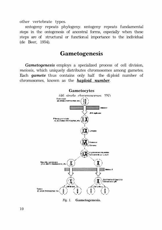
Gametogenesis
Gametogenesis employs a specialized process of cell division,meiosis, which uniquely distributes chromosomes among gametes.Each gamete thus contains only half the diploid number ofchromosomes, known as the haploid number.
Gametocytes (46 single chromosomes, 2N)
Fig. 1. Gametogenesis.
11
* Meiosis involves two sequential cell divisions, of whichonly first is preceded by duplication of chromosomes.
* The f i r s t m e i o t i c d i v i s i o n results in reduction of thechromosome complement to the haploid state and the secondresults in the production of four haploid gametes.
* Unlike the cells produced by mitosis that are geneticallyidentical with the parent cell, the cells produced by meiosis(gametes) are genetically unique.
D i f f e r e n c e b e t w e e n m e i o s i si n m a l e a n d i n f e m a l e
A fundamental difference between meiosis in male and theequivalent process in female is that the supply of primary oocytebecomes progressively depleted over a woman’s lifetime, whereasthe supply of primary spermatocytes is sustained, even in old men.
This is because the spermatogenic cell population is essentiallya continuously renewing system that depends on spermatocytereplacement from stem cells.
The sperm and oocyte, the male and female gametes arehighly specialized sex cells. Their structure is adapted to, ordetermined by the functions they perform.
C o n s i d e r a t i o n o f m a l e e v e n t s
Spermatogenesis is the process of formation and developmentof specialized male generative cells – spermatozoa or mature sperms.
Spermatogenesis
* At week 3-4 after fertilization primordial germ cells developwithin the wall of yolk sac. Primordial germ cells migrate towardthe developing testis and are incorporated into the wall offurther seminiferous tubules.
* Within the developing gonads primordial germ cellsdifferentiate into spermatogonia, which undergo mitosis in lateadolescence.
12
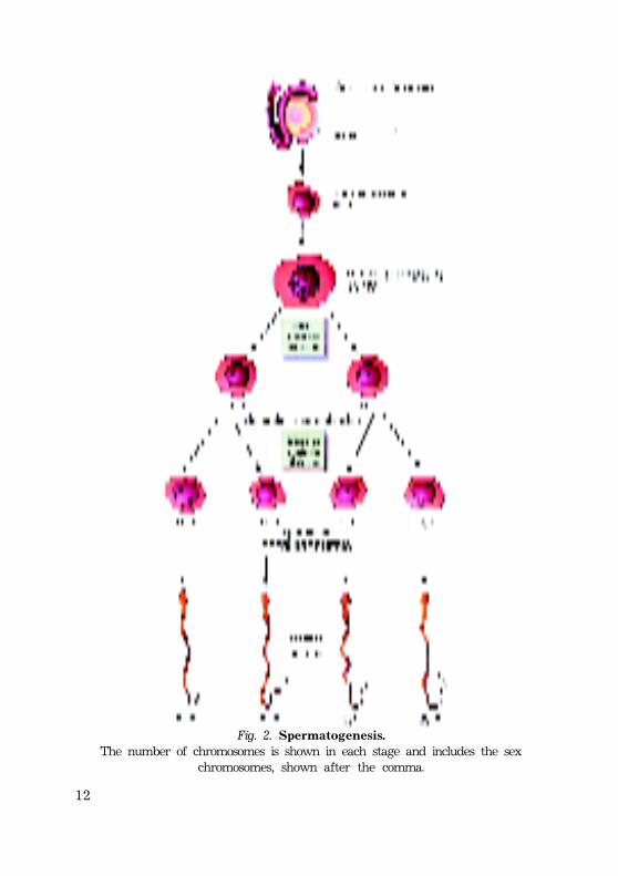
Fig. 2. Spermatogenesis.The number of chromosomes is shown in each stage and includes the sex
chromosomes, shown after the comma.
13
* Spermatogonia, which have been dormant in theseminiferous tubules of the testis since the fetal period, beginto increase in number at puberty. After several mitotic divisions,the spermatogonia grow and give rise to primary spermato-cytes. The cells of this series are diploid.
* Primary spermatocytes enter the first reduction meioticdivision by undergoing DNA replication after the puberty.Primary spermatocytes complete first meiotic division to formtwo secondary spermatocytes, which are haploid.
* Without first passing through an S-phase secondaryspermatocytes undergo the second meiotic division to form fourhaploid spermatids.
* Maturation o r spermiogenesis i s t h e s e r i e s o f c h a n g e s i ns p e r m a t i d s t h a t r e s u l t s i n t h e formation of spermatozoa (sperm).
* The total time of sperm formation (from spermatogonia tospermatozoa) is about 72 days.
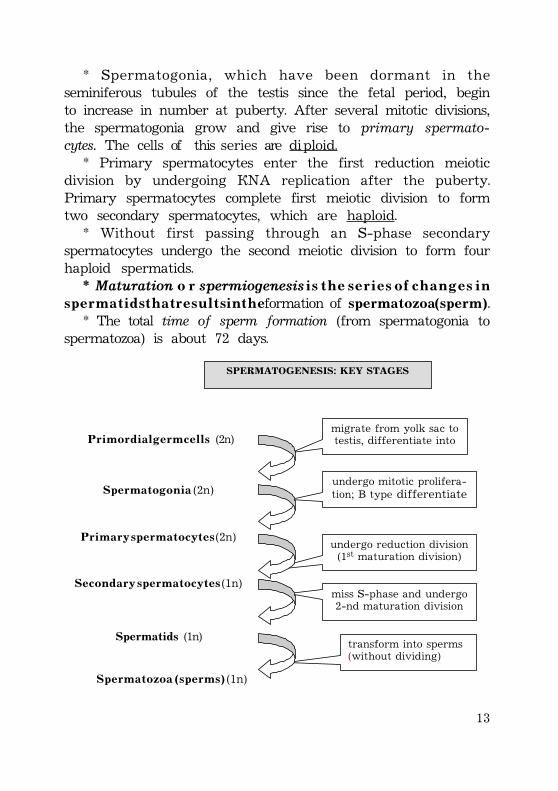
migrate from yolk sac to testis, differentiate into
undergo mitotic prolifera-tion; B type differentiate
i
undergo reduction division (1st maturation division)
miss S-phase and undergo 2-nd maturation division
transform into sperms (without dividing)
SPERMATOGENES IS : K EY STAGES
P r i m o r d i a l g e r m c e l l s (2n)
S p e r m a t o g o n i a (2n)
P r i m a r y s p e r m a t o c y t e s (2n)
S e c o n d a r y s p e r m a t o c y t e s (1n)
Spermatids (1n)
S p e r m a t o z o a ( s p e r m s ) (1n)
14
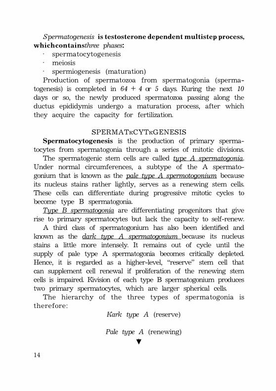
Spermatogenesis is testosterone dependent multistep process,w h i c h c o n t a i n s three phases:
· spermatocytogenesis· meiosis· spermiogenesis (maturation)Production of spermatozoa from spermatogonia (sperma-
togenesis) is completed in 64 + 4 or 5 days. During the next 10days or so, the newly produced spermatozoa passing along theductus epididymis undergo a maturation process, after whichthey acquire the capacity for fertilization.
SPERMATOCYTOGENESISSpermatocytogenesis is the production of primary sperma-
tocytes from spermatogonia through a series of mitotic divisions.The spermatogenic stem cells are called type A spermatogonia.
Under normal circumferences, a subtype of the A spermato-gonium that is known as the pale type A spermotogonium becauseits nucleus stains rather lightly, serves as a renewing stem cells.These cells can differentiate during progressive mitotic cycles tobecome type B spermatogonia.
Type B spermatogonia are differentiating progenitors that giverise to primary spermatocytes but lack the capacity to self-renew.
A third class of spermatogonium has also been identified andknown as the dark type A spermatogonium because its nucleusstains a little more intensely. It remains out of cycle until thesupply of pale type A spermatogonia becomes critically depleted.Hence, it is regarded as a higher-level, “reserve” stem cell thatcan supplement cell renewal if proliferation of the renewing stemcells is impaired. Division of each type B spermatogonium producestwo primary spermatocytes, which are larger spherical cells.
The hierarchy of the three types of spermatogonia istherefore:
Dark type A (reserve)
Pale type A (renewing)
15
Type B (differentiatingor progenitor cells)
MEIOSISMeiosis consists of two successive divisions that reduce the
chromosome number from 46 (44=XY in the male) to 23 (22+X or22+Y in spermatozoa).
Prophase–I of the first meiotic division lasts for 20-22 daysand involves four stages:
LeptosomesZygotenePachyteneDiakinesis
The exchange of segments (crossing over) of homologouschromosomes occurs during diakinesis.
Prophase ends with the dissolution of the nuclear envelope andmigration of the chromosomes to the equatorial plate.
In metaphase, the centrioles at opposite poles of the cell areconnected to the chromosomes by microtubules of the spindle.
In a n a p h a s e, the pairs of chromosomes, each consisting oftwo chromatids, separate and migrate to the poles.
In telophase, the cytoplasm constricts, separating two hap-loid secondary spermatocytes.
Secondary spermatocytes are relatively small cells. These cells,which contain 2n DNA, do not replicate their chromosomes. Theirchromosome number is haploid (23n) and their DNA is diploid (2n).Secondary spermatocytes quickly enter the second meiotic division,forming two haploid (1n DNA) spermatids.
SPERMIOGENEISSSpermiogenesis is the complex of cytodifferentiation by which
spermatids become spermatozoa.Spermiogenesis includes the following phases:
Golgi phaseCap phaseMaturation phase
16
1. During the G o l g i p h a s e hydrolytic enzymes are formed onthe rough endoplasmic reticulum (rER), modified in the Golgicomplex and packaged as small membrane-limited proacrosomalgranules. These small granules fuse with one another, formingan a c r o s o m e l v e s i c l e. The acrosomel vesicle adherents to thenucleus and assumes a hemispherical shape.
The centrioles migrate to a position near the cell surface andopposite the forming acrosome.
2. During t h e c a p p h a s e the acrosomal vesicle and granule spreadto cover the anterior half of the condensing nucleus. This reshapedstructure is then known as the acrosome or acrosomal cap.
The nucleus becomes more elongated and condensed.One of the centrioles (distal centriole) aligns at right angles
to the plasma membrane. The distal centriole initiates the synthesisof nine peripheral microtubule doublets and two centralmicrotubules that constitute the axonemal complex.
The centrioles, which had initiated the development of theflagellum, move back to the posterior surface of the nucleuswhere the proximal centriole becomes attached to a shallow groovein the nucleus.
Mitochondria aggregate around the proximal part of theflagellum, forming a thickened region known as the middle piece.
The cytoplasm is displaced posteriory. The cytoplasmicmicrotubules become organized into a cylindrical sheath, whichextends from the posterior rim of the acrosome toward theposterior pole of the spermatid.
3. M a t u r a t i o n p h a s e.During the last phase, the maturation phase, of sperma-
togenesis, the excess cytoplasm or residual cytoplasm shed andphagocytized by the Sertoli cells. Mature spermatozoa arereleased into the lumen of the seminiferous tubules.
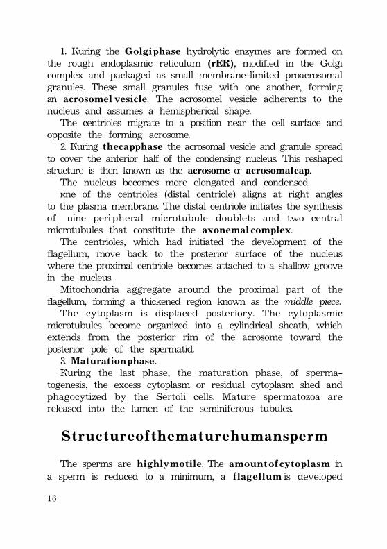
S t r u c t u r e o f t h e m a t u r e h u m a n s p e r m
The sperms are h i g h l y m o t i l e. The a m o u n t o f c y t o p l a s m ina sperm is reduced to a minimum, a f l a g e l l u m is developed
17
which renders it highly motileand the sperm head possessesa special mechanism for theperforation of the oocyte andits membranes.
The sperm cells morphologicaltraits are chiefly adaptations tothe particular problems whichthey must solve in reaching andpenetrating the oocyte.
The mature human sperm isabout 60 ìm long.
The sperms are motile cells,which contain 2 main parts -head and tail.
· The h e a d is flattened andcontains haploid nucleus.
* Nucleus is elongate,contains condensing chromatin.
Nucleus includes 22 autoso-mes and 1 sex chromosome.
50% of the sperms have Ysex chromosome and 50% - Xsex chromosome.
Nuclear envelope contains nonuclear pores.
* Bilamin ar acrosomalcap covers the anterior two-thirds of the nucleus.
The acrosomal cap oracrosome is specialized type oflysosome.
The acrosomal cap contains10-12 hydrolitic enzymes, suchas hyaluronidase, trypsin-like
A crosom al cap
H ead (5 µm long)
N eck
M itochondrial sheath
M iddle p iece (7µm long)
P rincip al p iece
(40µm long)
E nd piece (5-10µm long)
À ÂFig. 3. A l o n g i t u d i n a l s e c t i o n o f
a human sperm.Designation: A – electron micrograph
B – diagram.
Fig. 4. S p e r m a t o z o a .( Scanning electron micrograph – SEM).
Extent of acrosome and postalsegment of sperm head is evident.
Designation:1 – acrosomal cap; 2 – postacrosomal
segment; 3 – flagellum.
12
3
18
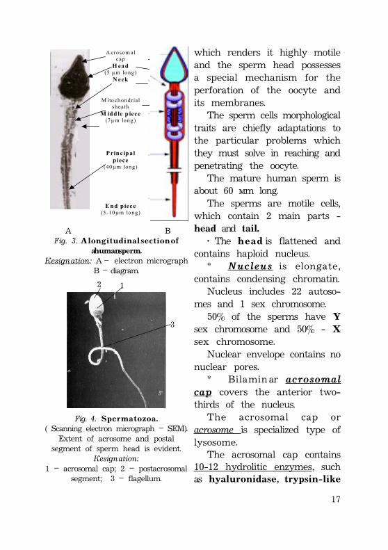
p r o t e a s e called a c r o s i n, acid phosphatase, neuraminidase,glucosidase and other.Function. These acrosomal enzymes facilitate sperm
penetration of the corona radiata and zona pellucida duringfertilization
Fig. 5. T h e l o n g i t u d i b a l s e c t i o n ( A ) a n d t r a n s v e r s e s e c t i o n ( B ) o f a p o r t i o no f h u m a n s p e r m t a i l .
(TEM – transmission electron micrograph).
Outer dense fibers
Centralfibrils
À
M i d d l e p i e c e
P r i n c i p a lp i e c e
Axonemal
complex
Axonemal
complex
Axonemal complex
B
Mitochondrialsheath
· The tail is subdivided into the neck, the middle piece, theprincipal piece and the end piece.
* The short neck is the junction between the head and tail.The neck contains the centrioles.Proximal centriole lies near the nucleus within the invagi-
nation of nuclear envelope.Distal centiole is located in the neck and gives rise to axo-
n e m a ( a x o n e m a l c o m p l e x ) that is composed of 9 pairs of peri-pheral microtubules and 1 pair centrally located microtubules.
19
Axonema continues into the all parts of the sperm tail.* The middle piece is long cylinder about 1 ìm in diameter
and 7 ìm long.It contains the mitochondria, helically wrapped around the
axonemal complex.The axonemal complex is composed of a central pair of fibril
within a symmetrical set of nine doublet fibrils, an in a typicalcilium.
Function. The mitochondria provide the energy for movementof the tail and thus are responsible for the motility of the sperm.
E n e r g y f r o m A T P produced by the mitochondrial sheathinduces sliding between each peripheral doublet and the adjacentdoublet. An outer ring of large coarse fibers (o u t e r d e n s e f i b e r s)and a d o r s a l c o l u m n and ventral column that are interconnectedby f i b r o u s r i b s, harness the forces that result from this slidingmovement and apply them to propulsive lasting of the flagellum.
* The principal piece is the motile part of the sperm and isapproximately 40 ìm long.
This part of tail contains axonemal complex that issurrounded by f i b r o u s s h e a t h.
The flagellum in principal piece is surrounded by o u t e r f i b r o u ssheath with dorsal and v e n t r a l l o n g i t u d i n a l c o l u m n s connectedby circumferential ribs. The flagellum itself has seven denseouter fibres collected in two compartments and a 9+2 coremicrotubule pattern.
The principal piece contains a supplementary cytoskeletalarrangement called the fibrous sheath that converts sliding mo-vement between microtubules into lashing movement of the fla-gellum the axonemal complex.
* The end piece is approximately 5 ìm long of the flagellum.The end piece of the flagellum is similar in construction. It
consists of the remainder of the axonema and covering cellmembrane.
20
ABNORMAL SPERMSNormally up to 10% of sperms in an ejaculate are grossly
abnormal (with two heads or two tails), but these abnormalsperms do not fertilize an oocytes owing to their l a c k o f m o t i l i t y.Most morphologically abnormal sperms are unable to passthrough the mucus in the cervical canal. Measurement offorward progression is a subjective assessment of the qualityof sperm movement.
Male fertility depends on the n u m b e r and m o t i l i t y o f s p e r m.The average volume of semen in normal, fertile male ejaculateis 3.5 ml, with a concentration of about 1 0 0 m i l l i o n s p e r m p e rmilliliter of semen; sterile males produce less than 20 millionsperm per milliliter of semen.
Immotile cilia syndrom (K a r t a g e n e r s y n d r o m) and otherimmotile cilia (ciliary dyskinesia) syndromes are characterizingby immotile spermatozoa and consequent infertility. Men withthese syndromes are usually sterile because a structural defect,lack, or transposition of any of the axonemal microtubules, ordefective action of their dynein arms (an ATP-ase), is manife-sted as a lack of propulsion.
These disorders usually coincide with chronic respiratoryinfections, since a similar deficiency exists in the ciliary axonemesof respiratory epithelial cells.
X - r a y s, severe a l l e r g i c r e a c t i o n s, and certain a n t i s p e r -matogenic agents have been reported to increase the percentageof abnormally shaped sperms. Such sperms are not believed toaffect fertility unless their number exceeds 20%.
The g e n e m u t a t i o n (change in DNA) increase with age. Theo l d e r p a r e n t s are to have accumulated mutations, that theembryo might inherit. For fathers of children with freshmutations, such as the one causing achondroplasia, this agerelationship has continually been demonstrated.
21
C o n s i d e r a t i o n o f f e m a l e e v e n t s
C o m m o n c h a r a c t e r i s t i co f t h e o o c y t e s
* The oocytes, on other hand, are always distinctly largercells than the normal somatic cells of the organism from whichthey are derived.
* Their increased cytoplasm is frequently enlarged by theaccumulation of yolk in the cytoplasmic yolk droplets.
* They possess protective envelopes, or egg membranes.* The oocytes are nonmotile cells; they can only be moved
passively along uterine tube by means the contractile activityof muscle cells within the muscle tunica of the tubes.
The o o c y t e s o f C h o r d a t a a n i m a l s (Vertebrate) can be clas-sified on the basis of the relative amounts and distribution ofyolk, and number of the sheath.
O o c y t e t y p e so f C h o r d a t a a n i m a l s :
1. M i o l e c i t h a l o r y o l k - p o o r o o c y t e s (synonyms: isolecithal,oligolecithal):
· hav e primitive aquatic forms, placental mammals, includingman;
· amount of yolk is little;· nucleus is located near center.2. M e d i a l e c i t h a l o r m e d i u m y o l k e d o o c y t e s :· have amphibia, polypteridae, holocephali (frogs, toads);· amount of yolk is medium, less near one polar region;· active cytoplasm is located near nucleus or animal pole;· nucleus is located near animal pole.3. M e g a l e c i t h a l o r l a r g e - y o l k o o c y t e s :· have avers, reptilia (birds and reptiles);· amount of yolk is much;· nucleus is located at one pole (animal pole);
22
· amount of active cytoplasm is very little and entirely atnuclear or animal pole.
Some authors also classified oocytes into the four types anddescribed forth type of eggs as the alecithal oocytes that havenot yolk.
But this type of the egg is unknown in nature.
O o c y t e m e m b r a n e sOocyte membranes of different chordate groups are varia-
ble in their structure and number.On the basis of their origination oocyte membranes can be
classified as:1. Primary oocyte membrane is plasma membrane (or vitelline
membrane) which can be demonstrated in all oocytes.2. Secondary oocyte membranes are produced by cells of
the ovarian follicle (zona pellucida, corona radiata).3. Tertiary oocyte membranes are secreted by the lining of
the oviduct or uterus.
O o g e n e s i s
Oogenesis is the process of formation and development ofspecialized female generative cells – oocytes (or eggs).
Oogenesis begins before birth and is completed after pubertyand continues to menopause.
P r e n a t a l m a t u r a t i o n o f h u m a n o o c y t e s* At week 3-4 after fertilization p r i m o r d i a l g e r m c e l l s develop
within the wall of yolk sac. These cells migrate along the wall ofyolk sac and primitive gut toward the developing ovaries.
* Within the developing gonads primordial germ cells diffe-rentiate into o o g o n i a, which actively proliferate by mitoticdivision.
* By the fourth and fifth months of human fetal develop-ment, oogonia enlarge to form p r i m a r y o o c y t e s before the birth.The primary oocyte enclosed by the follicular cells.
23
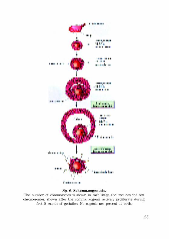
Fig. 6. S c h e m a . O o g e n e s i s .The number of chromosomes is shown in each stage and includes the sexchromosomes, shown after the comma. Oogonia actively proliferate during
first 5 month of gestation. No oogonia are present at birth.
24
* P r i m a r y o o c y t e s begin the first meiotic division before birth.Primary oocytes are arrested in the first meiotic prophase fromthe fifth month of fetal period at least until puberty.
* The follicular cells surrounding the primary oocyte arebelieved to secrete a substance, oocyte maturation inhibitor(OMI), which keeps the meiotic process of the oocyte arrested.
* No oogonia form postnatally.* * *
P o s t n a t a l m a t u r a t i o n o f h u m a n o o c y t e sEarly stages of o o g e n e s i s occur during fetal life when mitotic
divisions massively increase the number of oogonia developingfrom primordial germ cells.
1. Oogonia give rise to primary oocytes, which are surroundedby a single layer of f o l l i c u l a r c e l l s. The outer surface of thefollicular cells is bounded by a b a s a l l a m i n a. All primary oocytesare formed by the fifth month of fetal life; no oogonia arepresent at birth. Near 7 m i l l i o n primary acolytes are present at5 month of fetal life.
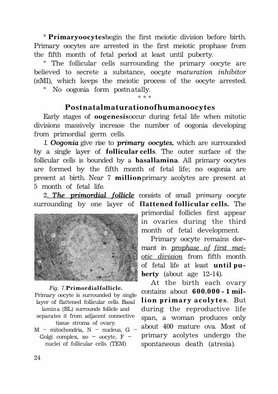
2. The primordial follicle consists of small primary oocytesurrounding by one layer of f l a t t e n e d f o l l i c u l a r c e l l s . The
primordial follicles first appearin ovaries during the thirdmonth of fetal development.
Primary oocyte remains dor-mant in prophase of first mei-otic division from fifth monthof fetal life at least u n t i l p u -berty (about age 12-14).
At the birth each ovarycontains about 6 0 0 . 0 0 0 - 1 mil-l i o n p r i m a r y a c o l y t e s. Butduring the reproductive lifespan, a woman produces onlyabout 400 mature ova. Most ofprimary acolytes undergo thespontaneous death (atresia).
Fig. 7. P r i m o r d i a l f o l l i c l e .Primary oocyte is surrounded by singlelayer of flattened follicular cells. Basallamina (BL) surrounds follicle and
separates it from adjacent connectivetissue stroma of ovary.
M – mitochondria, N – nucleus, G –Golgi complex, Oo – oocyte, F –nuclei of follicular cells. (TEM)
25
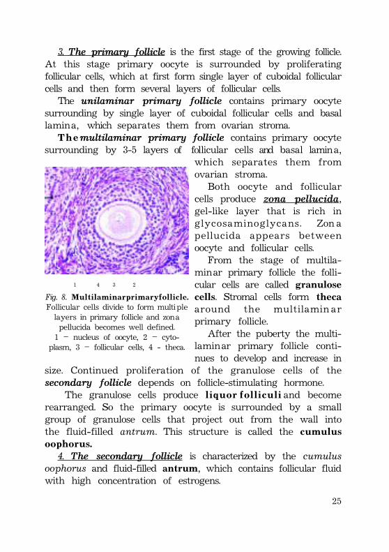
3. The primary follicle is the first stage of the growing follicle.At this stage primary oocyte is surrounded by proliferatingfollicular cells, which at first form single layer of cuboidal follicularcells and then form several layers of follicular cells.
The unilaminar primary follicle contains primary oocytesurrounding by single layer of cuboidal follicular cells and basallamina, which separates them from ovarian stroma.
T h e multilaminar primary follicle contains primary oocytesurrounding by 3-5 layers of follicular cells and basal lamina,
which separates them fromovarian stroma.
Both oocyte and follicularcells produce zona pellucida,gel-like layer that is rich inglycosaminoglycans. Zon apellucida appears betweenoocyte and follicular cells.
From the stage of multila-minar primary follicle the folli-cular cells are called granulosecells. Stromal cells form thecaaround the multilamin arprimary follicle.
After the puberty the multi-laminar primary follicle conti-nues to develop and increase in
size. Continued proliferation of the granulose cells of thesecondary follicle depends on follicle-stimulating hormone.
The granulose cells produce l i q u o r f o l l i c u l i and becomerearranged. So the primary oocyte is surrounded by a smallgroup of granulose cells that project out from the wall intothe fluid-filled antrum. This structure is called the cumuluso o p h o r u s .
4. The secondary follicle is characterized by the cumulusoophorus and fluid-filled antrum, which contains follicular fluidwith high concentration of estrogens.
1 4 3 2
Fig. 8. M u l t i l a m i n a r p r i m a r y f o l l i c l e .Follicular cells divide to form multiplelayers in primary follicle and zonapellucida becomes well defined.1 – nucleus of oocyte, 2 – cyto-
plasm, 3 – follicular cells, 4 - theca.
26
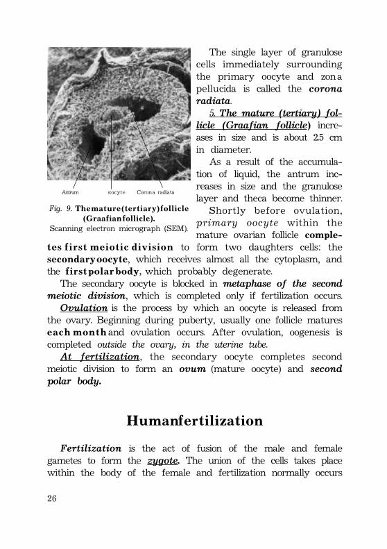
The single layer of granulosecells immediately surroundingthe primary oocyte and zonapellucida is called the coronaradiata.
5. The mature (tertiary) fol-licle (Graafian follicle) incre-ases in size and is about 2.5 cmin diameter.
As a result of the accumula-tion of liquid, the antrum inc-reases in size and the granuloselayer and theca become thinner.
Shortly before ovulation,primary oocyte within themature ovarian follicle comple-
t e s f i r s t m e i o t i c d i v i s i o n to form two daughters cells: thesecondary oocy te, which receives almost all the cytoplasm, andthe f i r s t p o l a r b o d y, which probably degenerate.
The secondary oocyte is blocked in metaphase of the secondmeiotic division, which is completed only if fertilization occurs.Ovulation is the process by which an oocyte is released from
the ovary. Beginning during puberty, usually one follicle maturese a c h m o n t h and ovulation occurs. After ovulation, oogenesis iscompleted outside the ovary, in the uterine tube.At fertilization, the secondary oocyte completes second
meiotic division to form an ovum (mature oocyte) and secondpolar body.
H u m a n f e r t i l i z a t i o n
Fertilization is the act of fusion of the male and femalegametes to form the zygote. The union of the cells takes placewithin the body of the female and fertilization normally occurs
Fig. 9. T h e m a t u r e ( t e r t i a r y ) f o l l i c l e( G r a a f i a n f o l l i c l e ) .
Scanning electron micrograph (SEM).
Antrum Oocyte Corona radiata
27
in the ampulla or at the ampulla-isthmus junction of the uterinetube. Shortly after the ovulation, the human oocyte, surroundedby the corona radiata passes into the uterine tube.
Immediately after ovulation the oocyte completely fills thezonal cavity forming by the surrounding zona pellucida. Later aspace, p e r i v i t e l l i n e s p a c e, is present between the oocyte andthe zona pellucida.
Fertilization includes:· approach and attachment of oocyte and sperms,· penetration of oocyte membranes by sperm and· fusion of male and female pronuclei with the formation
of the zygote.Fertilization includes several mechanisms whereby a sperm
approaches, become attached to, and then penetrates the surfaceof an ovum, and early series of changes, which follow.
Fertilization in the normal mature female results frominsemination depends on:
* the time interval between insemination and ovulation;* the length of time that the ovum remains fertilization;* the number of spermatozoa reaching the uterine tube
(oviduct);* the time taken by the spermatozoa to reach the ovum in
the tube;* the length of time during which the spermatozoa retain
their fertilizing power;* other factors in the semen, which influence fertilization. One
of these appears to be the presence of a sufficient quantity ofthe enzyme, hyaluronidase.
The seminal fluid, or semen, is a complex mixture derivedmainly from the testis, the seminal vesicles and prostate gland. Thefluid in man in normal ejaculation, amounts to about 3,5 ml (with arange of 2 to 6ml), and contains 200-300 million spermatozoa. Thesecretions derived from the accessory glands help to activate thesperms and at the same time provide a carrying medium for them.
A sperms pass very rapidly into the uterus and from it intothe uterine tubes. The rapid movement of the seminal fluid is
28
explained by contractions of the musculature of the uterus anduterine tube. As a result the seminal fluid is rapidly mixed withthe secretions of female genital tract. Part of this mixture isaspirated into the ampulla of the uterine (Fallopian) tube. In theabsence of contractions of the uterus sperm ascent does notappear to occur.
Fertilization includes 5 phases:* Capacitation of sperms.* Distant interaction and approach of sex cells.* Contact interactions between the sex cells and activation
of the oocyte.* Entering of the sperm into the ovum and fusion of male
and female pronuclei.* Postfusion reactions.
C a p a c i t a t i o n o f s p e r m s
Sperms are not capable of fertilizing ovum immediately uponreaching the uterine tube. This suggests that they must undergosome form of physiological changes, called capacitation.
Capacitation involves the release of the e p i d i d y m a l f l u i dglucoconjugate from the surface of the head of the sperma-rozoon. These surface glycosides are decapacitation factors addedduring sperm maturation in the epididymis and accessory malereproductive organs.
Capacitation is a process that p r e p a r e s t h e s p e r m f o r f e r t i -lization. The surface glycosides inhibit binding to the zonapellucida receptors.
During capacitation a glycoprotein coat and seminal proteinsare removed from the surface of sperm’s (acrosome) head.
So the capacitation is a process by which enzymatic secretionsof the uterus and uterine tube of the female genital tractstrips glycoproteins from the sperm cell membrane.
Cholesterol of sperm plasma membrane is removed from theplasma membrane during capacitation, which results in thei n c r e a s e d f l u i d i t y o f t h e membrane that is required for the
29
fusion of acrosomal membrane with the sperm plasmamembrane.
Capacitation lasts about 7 hours.
D i s t a n t i n t e r a c t i o n s a n d a p p r o a c h o f s e x c e l l s .
Distant interactions between the male and female gametesinvolve several factors:
1 – reotaxis;2 – influence of some chemical and hormonal factors including:a) prostaglandins;b) fertilizing proteins (androgamones, hynogamones);c) progesterone;d) pH.
ReotaxisReotaxis is the sperm ability for advance against the current
of the fluid secreting by the epithelium, which lines the cervix,uterus and uterine tube.
ProstaglandinsSeminal fluid or semen contains prostaglandins, which have
pharmacodynamic actions on the smooth muscle of the uterusand uterine tube.
Prostaglandins stimulate uterine contractions, r h y t h m i ccontractions of the smooth muscle of the uterine tube, andsmooth muscle in general. The oocyte is transported along theuterine tube by peristaltic contractions.
The oocyte and sperms are transported from opposite endsof female genital tract. The movement of sperms is much toorapid, however, to be accounted for by their intrinsic motility.
Prostaglandins assist in the movement of sperms throughthe uterus and tubes to the site of fertilization in the ampullaof the uterine tube.
30
F e r t i l i z i n g p r o t e i n s ( a n d r o g a m o n e s a n d h y n o g a m o n e s )Hemotaxis provides the approach of male and female gametes.
Important role has gamones, the chemical substances producedby the ovum and sperms that activate and agglutinate sperms.
The oocyte secrets hynogamones type I and II. HynogamoneI stimulates the movement of the sperms. Hynogamone IIagglutinates sperms.
The sperms secrete androgamones type I and II. AndrogamoneI blocks the movement of the sperms. Andogamones II lysesthe oocyte membrane.
ProgesteroneThe hormone progesterone is secreted by the corpus luteum
of the ovary after the ovulation.It stimulates the secretion of nutrient-rich fluid produced
by mucosal epithelium of female reproductive tract (glandularepithelium of uterine tube, uterus and cervix).
pHThe sperms move 2 to 3 mm per minute but the speed varies
with the pH pf the environment.Sperms move slowly in the acid environment of the vagina, but
move rapidly in the alkaline environment of the uterus.It was found that a few motile sperms were appeared in the
ampulla of the uterine tube 5 minutes after their depositionnear the external uterine os. Some sperms, however, took upto 45 minutes to complete the journey.
Only about 200 sperms reach the fertilization site. Most spermsdegenerate and are resorbed by the female genital tract.
C o n t a c t i n t e r a c t i o n s o f s e x c e l l s .
Contact interactions includes:1 – binding sperms to oocyte;2 – acrosomal reaction;3 – penetration of oocyte membrane.
31
B i n d i n g s p e r m s t o o o c y t eThe zona pellucida is important in the recognition of homolo-
gous sperms, in blocking polyspermy.Glucosyltransferase receptors of the sperm plasma memb-
rane bind to zona pellucida receptors, ZP-3 molecules.The ZP-3 molecules of the z o n a p e l l u c i d a h a v e t w o r e g i o n s:1) the sperm receptors that recognize integral proteins of
the sperm plasma membrane;2) the other region of the ZP-3 molecule binds to receptor pro-
teins located in the head of the sperm, triggering the acrosome reaction.
A c r o s o m e r e a c t i o nBinding the sperm receptor to ZP-3 molecules triggers the
acrosome reaction. The acrosome membrane fuses with plasmamembrane of sperm in anterior part of the head and acrosomalenzymes are released from acrosome.
The release of enzymes from the sperm acrosome results indigestion of the zona pellucida surrounding the oocyte, allowingpenetration by sperm.
Most important enzymes are hyaluronidase and trypsin-likeprotease, acrosin, which lyses the zona pellucida. The liberatedenzymes digest the zona pellucida, permitting the flagellamovement of the sperm to propel the sperm toward the oocyte.
Penetration is accomplished by limited proteolysis of the zonapellucida in front of the advancing sperm.Additional spermsmay be found attached to the zona pellucida. Several spermsmay penetrate the zona pellucida, but only one sperm comp-letes the fertilization process.
The sperm penetrates the zona pellucida and enters thep e r i v i t e l l i n e s p a c e, located between the zona pellucida andoocyte plasma membrane.
P e n e t r a t i o n o f o o c y t e m e m b r a n eThe sperm binds to receptors of oocyte plasma membrane.
The plasma membranes of oocyte and sperm fuse and breakdown at the area of fusion.
32
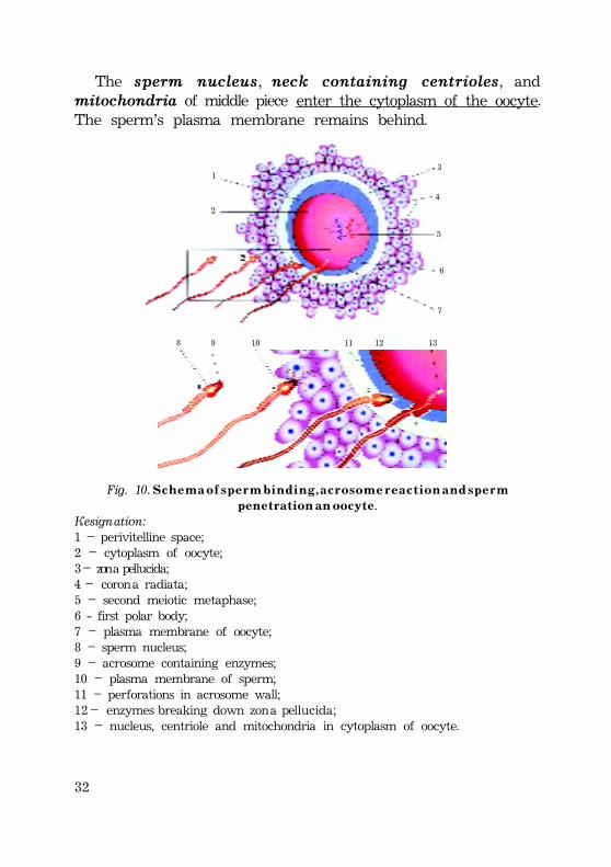
The sperm nucleus, neck containing centrioles, andmitochondria of middle piece enter the cytoplasm of the oocyte.The sperm’s plasma membrane remains behind.
1
2
3
4
5
6
7
8 9 10 11 12 13
Fig. 10. S c h e m a o f s p e r m b i n d i n g , a c r o s o m e r e a c t i o n a n d s p e r mp e n e t r a t i o n a n o o c y t e.
Designation:1 – perivitelline space;2 – cytoplasm of oocyte;3 – zona pellucida;4 – corona radiata;5 – second meiotic metaphase;6 - first polar body;7 – plasma membrane of oocyte;8 – sperm nucleus;9 – acrosome containing enzymes;10 – plasma membrane of sperm;11 – perforations in acrosome wall;12 – enzymes breaking down zona pellucida;13 – nucleus, centriole and mitochondria in cytoplasm of oocyte.
33
P o s t f u s i o n r e a c t i o n s
Postfusion reactions occur to prevent other sperms fromentering the secondary oocyte (polyspermy).
The fusion of sperm with oocyte is responsible for severalpostfusion reactions:
1) fast block to polyspermy;2) cortical reaction;3) zona reaction.
F a s t b l o c k t o p o l y s p e r m yThe fast block involves changes in the resting membrane
potential of the oocyte plasma membrane that prevent contactbetween oocyte and another sperms.
Fast block involves a large and long-lasting (few minutes)depolarization of the oolemma.
C o r t i c a l r e a c t i o nCortical reaction is slow component. Changes in the polarity
of the oolemma then trigger release of Ca++ from the ooplasmicstores. The Ca++ propagates a cortical reaction wave, in whichcortical granules move to the surface and fuse with oolemma.
The contents of c o r t i c a l g r a n u l e s are released into theperivitelline space.
Z o n a r e a c t i o nE n z y m e s within the cortical granules (proteases) act to
hydrolyze ZP-3 molecules, the sperm receptors, in zonapellucida, thus preventing additional sperms from reaching theoocyte.
These enzymes d e g r a d e t h e g l y c o p r o t e i n o o c y t e r e c e p t o r sfor sperm binding and make it impermeable to other sperms.
The contents of cortical granules, which are released intothe perivitelline space, also cause changes in the plasmamembrane that take make it impermeable to sperms.
34
These enzymes form the perivitelline barrier by cross-linkingproteins on the surface of the zona pellucida. This event createsthe final and permanent block to polyspermy.
F o r m a t i o n o f m a l e a n d f e m a l e p r o n u c l e i
Entry of the sperm nucleus triggers the secondary oocyte toresume and complete its second meiotic division.
This results in forming twohaploid cells, the ovum and thes e c o n d p o l a r b o d y.
Second polar body isextruded and the chromoso-mes, which remain in ovumreconstitute into the f e m a l epronucleus.
Within the cytoplasm ofthe oocyte, the nucleus of thesperm enlarges to form them a l e p r o n u c l e u s. Morpholo-gically the male and femalepronuclei are indistingui-shable.
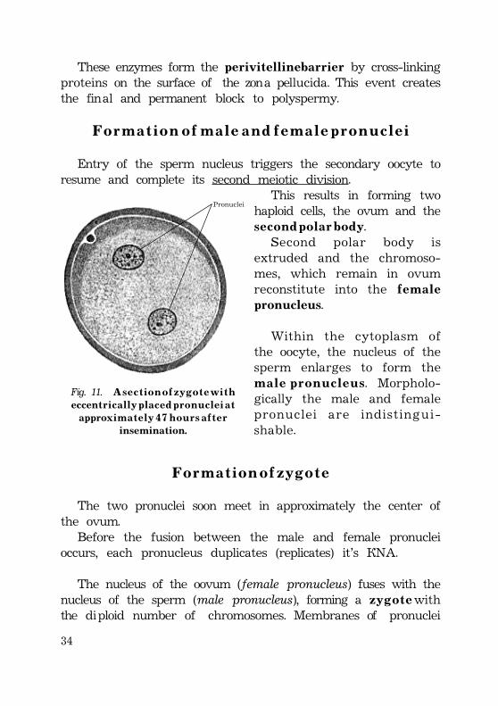
F o r m a t i o n o f z y g o t e
The two pronuclei soon meet in approximately the center ofthe ovum.
Before the fusion between the male and female pronucleioccurs, each pronucleus duplicates (replicates) it’s DNA.
The nucleus of the oovum (female pronucleus) fuses with thenucleus of the sperm (male pronucleus), forming a z y g o t e withthe diploid number of chromosomes. Membranes of pronuclei
Pronuclei
Fig. 11. A s e c t i o n o f z y g o t e w i t heccent r i c a l l y p l a c e d p r o n u c l e i a t
approx i m a t e l y 4 7 h o u r s a f t e rinsemination.
35
break down; the chromosomes condense and become arrangedfor a mitotic cell division
The fertilization oocyte or zygote is a u n i c e l l u l a r e m b r y o.The combination of 23 chromosomes in each pronucleus resultsin a zygote with 4 6 c h r o m o s o m e s.
The zygote is genetically unique because half of itschromosomes come from mother and half from farther. Thezygote contains a new combination of chromosomes that isdifferent from that in the cells of either of the parents.
The embryo’s c h r o m o s o m a l s e x is determinated at ferti-lization by the kind of sperm (X or Y) that fertilizes theoocyte.
Fertilization by an X-bearing sperm produces a 4 6 , X X z y g o t e,which normally develops into a female, whereas fertilization bya Y-sperm produces a 4 6 , X Y z y g o t e, which normally developsinto a male.
ABNORMAL SPERMSIn vitro fertilization (IVF) of oocyte and transfer of the
cleaving zygote into the uterus has provided an opportunity formany women who are sterile (e.g. owing to tubal occlusion) tobear children.Main steps:· Ovarian follicles are stimulated to grow and mature by
administration of gonadotropins.· Mature oocyte is aspirated from mature ovarian follicles
during laparoscopy.· The oocytes are placed in a Petri dish containing a culture
medium and capacitated sperms.· Fertilization of the oocyte and cleavage of the zygote are
monitored microscopically.· Cleaving zygote during the 4 to 8 cell stage is transferred
into the uterus.Intracellular sperm injectionA sperm can be injected directly into the cytoplasm of a
mature oocyte. This technique has been successfully used for
36
the treatment of couples where IVF failed or in cases wherethere are too few sperms available for in vitro inseminationSurrogate mothersSome women produce mature oocytes but are unable to
become pregnant (after hysterectomy). In these cases INF maybe performed and the embryos transferred to another woman’suterus. The surrogate mother bears the embryo and fetus anddelivers it to the natural mother at birth.
CLINICAL CONSIDERATIONSDuring meiosis, homologous chromosomes sometimes fail to
separate and go to opposite poles of the germ cell.As a result of this error of meiotic cell division (nondis-
junction) abnormal gametes with 24 single chromosomes and22 single chromosomes are produced.
- Fertilization between a normal gamete and an abnormalgamete with 24 single chromosomes will produce an individualwith 4 7 s i n g l e c h r o m o s o m e s (23+24+47), known as trisomy.
- If a gamete with 22 single chromosomes units with thenormal one, a zygote with 45 single chromosomes forms(22+23+45). This condition is known as monosomy.
Cleavage
Cleavage is a series of mitotic divisions of the zygote. Cleavageresults in a rapid increase in the number of cells without cellgrowth. These embryonic cells are called blastomeres.
Cleavage is the process w h e r e b y the zygote is divided intoblastomeres with the diminution in cell size.
Cleavage is largely dependent on the amount and distributionof the stored yolk and varies according to whether the eggs aremiolecithal, medialecithal or megalecithal.
37
Cleavage of chordates or vertebrate animals can be classifiedinto 4 types:
· Complete.· Equal.· Incomplete.· Unequal.
C l e a v a g e i n m i o l e c i t h a l e g g s
Cleavage in miolecithal oocytes is complete (holoblastic) andnearly equal.
In these oocytes the first cleavage spindle forms near thecenter of the egg so that two equal-sized blastomeres areformed.
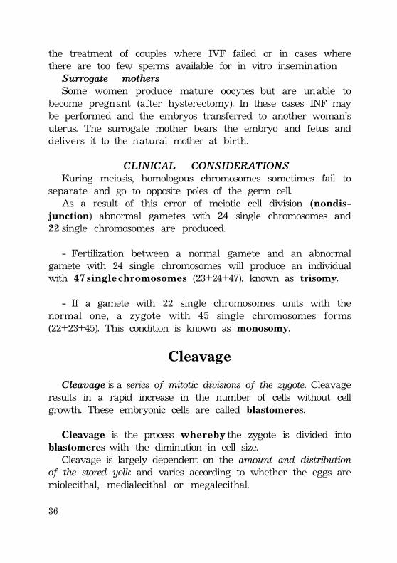
These blastomeres in turn divide equally and successiveequivalent divisions of the daughter cells result in theformation of a morula made up of many cells of nearly
equal size and containingnearly equal amounts of yolkand cytoplasm.
In practically there is a slightdifference in blastomere sizeand quality after the thirdcleavage. In Amphioxus(primitive aquatic form) afterthis third (equatorial) cleavagethe four cells at the so-called“animal” pole are slightlysmaller than the four at the“vegetal” pole.
In miolecithal eggs theblastula is called coeloblastula.
The coeloblastula of primitive aquatic form contains blastodermcomposed of unilaminar blastomeres and centrally locatedblastula cavity. The unilaminar hollow sphere of blastomeresresults from the process of cleavage.
Animal pole
Vegetal pole
Fig. 12. S c h e m a o f e a r l y c o e l o -b l a s t u l a o f p r i m i t i v e a q u a t i c f o r m s .
38
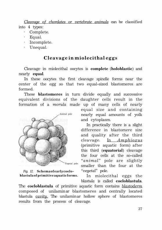
Blastula cavity Animal hemisphere
Unilaminar blastoderm Vegetal hemisphere of blastula
A B
Fig. 13. S c h e m a t i c s e c t i o n s o f c o e l o b l a s t u l a i n e q u a t o r i a l l e v e l ( A ) a n dt h r o u g h c r a n i a l - c a u d a l a x i s ( B ) .
The cavity of the blastula {blastocoele) is enclosed by a singlelayer of cells which are slightly smaller in the animal than in thevegetal hemisphere.
C l e a v a g e i n m e d i a l e c i t h a l e g g s
Cleavage in medialecithal oocytes is complete and unequal.The most familiar examples of medialecithal oocyte with
complete but unequal cleavage are a m p h i b i a n e g g s, especiallythose of frogs and toads.
In these eggs with a moderate amount of yolk the smallcells contain little yolk, while the vegetal cells are loadedwith it.
This inert nutritive material slows down the metabolic rateof the v e g e t a l p o l e cells compared with that of the animalpole cells; the latter, therefore, divide much more rapidly andso assume the major role in the formation of the embryo.
In medialecithal eggs the blastocoele (cavity of blastula) isrelatively small and, since the animal pole cells are definitelysmaller than those in the vegetal portion of the sphere, theblastocoele cavity is much nearer the animal pole.
The animal portion is usually about one-third of blastodermand a much larger is the vegetal portion.
39
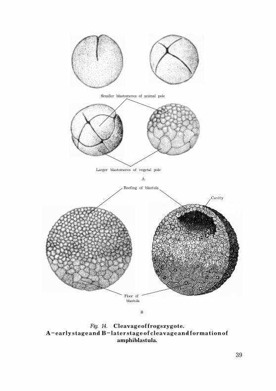
Smaller blastomeres of animal pole
Larger blastomeres of vegetal pole
Roofing of blastula
Cavity
Floor of blastula
Fig. 14. C l e a v a g e o f f r o g s z y g o t e .A – e a r l y s t a g e a n d B – l a t e r s t a g e o f c l e a v a g e a n d f o r m a t i o n o f
amphiblastula.
A
B
40
The animal pole cells are smaller in these eggs and they forma single layer r o o f i n g the blastocoele as in miolecithal eggs,while the much larger cells of the vegetal pole forming thefloor of the blastocoele exist as a multilayered mass.
C l e a v a g e i n m e g a l e c i t h a l e g g s
Cleavage in megalecithal oocytes is incomplete (discoidal ormeroblastic) and unequal.
The megalecithal eggs of reptiles, birds and the egg-laying mammals (Monotremata) represent the greatestdevelopment of yolk storage. Sharks and rays (Euselachii)have almost as much yolk, while the bony fishes (Teleostei)have noticeably less, but still enough to limit cleavage tothe incomplete type.
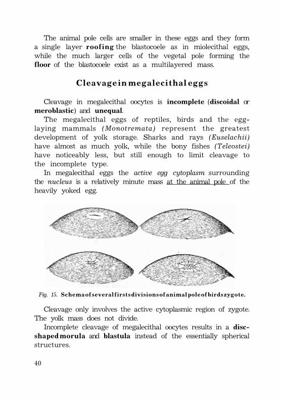
In megalecithal eggs the active egg cytoplasm surroundingthe nucleus is a relatively minute mass at the animal pole of theheavily yoked egg.
Fig. 15. S c h e m a o f s e v e r a l f i r s t s d i v i s i o n s o f a n i m a l p o l e o f b i r d s z y g o t e .
Cleavage only involves the active cytoplasmic region of zygote.The yolk mass does not divide.
Incomplete cleavage of megalecithal oocytes results in a disc-s h a p e d m o r u l a and blastula instead of the essentially sphericalstructures.
41
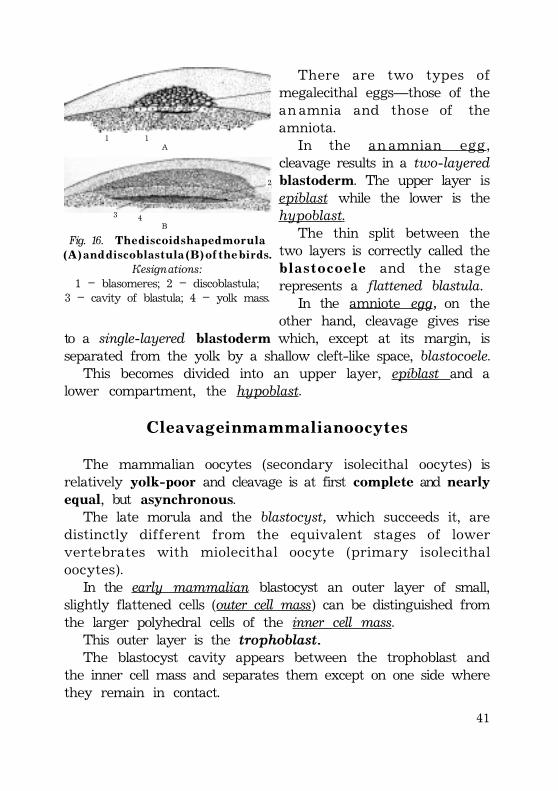
There are two types ofmegalecithal eggs—those of thean amnia and those of theamniota.
In the an amnian egg,cleavage results in a two-layeredblastoderm. The upper layer isepiblast while the lower is thehypoblast.
The thin split between thetwo layers is correctly called theb l a s t o c o e l e and the stagerepresents a flattened blastula.
In the amniote egg, on theother hand, cleavage gives rise
to a single-layered blastoderm which, except at its margin, isseparated from the yolk by a shallow cleft-like space, blastocoele.
This becomes divided into an upper layer, epiblast and alower compartment, the hypoblast.
C l e a v a g e i n m a m m a l i a n o o c y t e s
The mammalian oocytes (secondary isolecithal oocytes) isrelatively yolk-poor and cleavage is at first complete and nearlyequal, but asynchronous.
The late morula and the blastocyst, which succeeds it, aredistinctly different from the equivalent stages of lowervertebrates with miolecithal oocyte (primary isolecithaloocytes).
In the early mammalian blastocyst an outer layer of small,slightly flattened cells (outer cell mass) can be distinguished fromthe larger polyhedral cells of the inner cell mass.
This outer layer is the trophoblast.The blastocyst cavity appears between the trophoblast and
the inner cell mass and separates them except on one side wherethey remain in contact.
11A
43
2
B
Fig. 16. T h e d i s c o i d s h a p e d m o r u l a( A ) a n d d i s c o b l a s t u l a ( B ) o f t h e b i r d s .
Designations:1 – blasomeres; 2 – discoblastula;
3 – cavity of blastula; 4 – yolk mass.
42
The blastocyst is peculiar to the eutherian mammals, and isnot precisely homologous to the blastulae of lower aquatic forms.
The oocytes of marsupials are slightly larger and more yolk-laden than those of placental mammals and they differentiatemore rapidly. After the second cleavage, the blastomeres arrangethemselves in a single layer around the inner surface of thezona pellucida, forming a hollow sphere with eliminated yolk-fragments in the central cavity. Certain cells at the animal polelater migrate inwards to form the endoderm. As there is noinner cell mass in marsupials at this stage the sphere resemblesthe blastula of lower vertebrates with miolecithal eggs.
In the insectivores, a single-layered sphere is formed withoutan intervening morula stage and at first without an inner cell mass.At one pole, however, a thickening, resembling an inner cell mass,soon appears. This is perhaps caused by delamination of an innercell mass in the true sense. It is conceivable, therefore, that themarsupials and certain insectivores show some of the transitionalsteps leading up to the typical mammalian blastocyst.
There is considerable variation in the relation of thetrophoblast to the inner cell mass in the stages immediatelyfollowing the formation of the blastocyst in the differenteutherian groups.
In primates, including Man, the trophoblast at first completelycovers the inner cell mass. In others, like the p i g, thetrophoblastic cells covering the inner cell mass soon disappearexposing on the surface the e m b r y o n i c ectodermal portion ofthe inner cell mass. This remains exposed until the amnioticfolds form at a later stage.
H u m a n c l e a v a g e
Human cleavage normally occurs as the zygote passes alongthe uterine tube toward the uterus.
Human cleavage is called:– complete cleavage and– asynchronous cleavage.
43
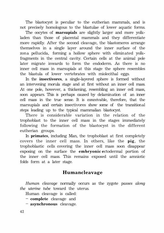
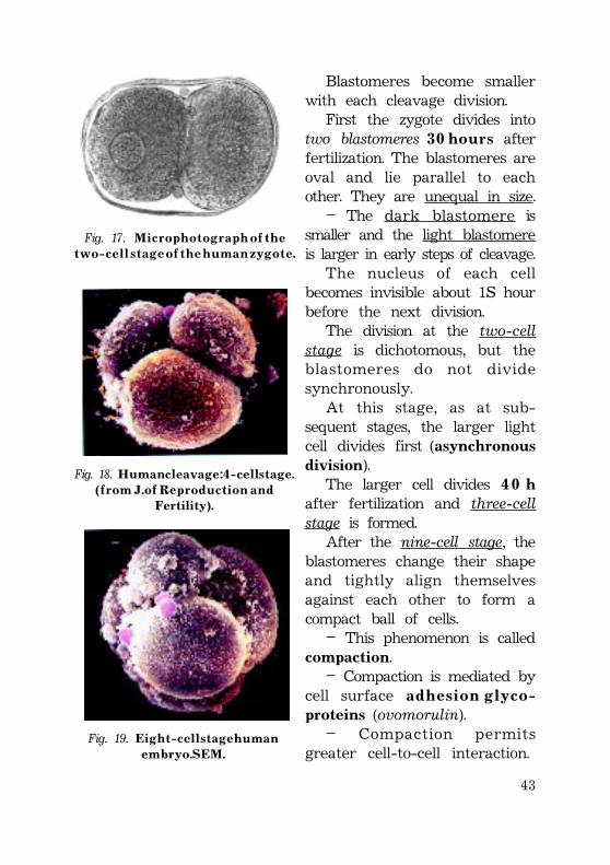
Blastomeres become smallerwith each cleavage division.
First the zygote divides intotwo blastomeres 3 0 h o u r s afterfertilization. The blastomeres areoval and lie parallel to eachother. They are unequal in size.
– The dark blastomere issmaller and the light blastomereis larger in early steps of cleavage.
The nucleus of each cellbecomes invisible about 1½ hourbefore the next division.
The division at the two-cellstage is dichotomous, but theblastomeres do not dividesynchronously.
At this stage, as at sub-sequent stages, the larger lightcell divides first (asynchronousdivision).
The larger cell divides 4 0 hafter fertilization and three-cellstage is formed.
After the nine-cell stage, theblastomeres change their shapeand tightly align themselvesagainst each other to form acompact ball of cells.
– This phenomenon is calledcompaction.
– Compaction is mediated bycell surface a d h e s i o n g l y c o -proteins (ovomorulin).
– Compaction permitsgreater cell-to-cell interaction.
Fig. 17. M i c r o p h o t o g r a p h o f t h et w o - c e l l s t a g e o f t h e h u m a n z y g o t e .
Fig. 18. H u m a n c l e a v a g e : 4 - c e l l s t a g e .( f r o m J . o f R e p r o d u c t i o n a n d
Fertility).
Fig. 19. E i g h t - c e l l s t a g e h u m a ne m b r y o . S E M .
44
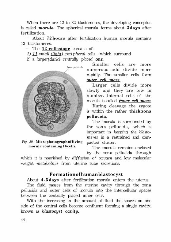
When there are 12 to 32 blastomeres, the developing conceptusis called morula. The spherical morula forms about 3 d a y s afterfertilization.
· About 7 2 h o u r s after fertilization human morula contains12 blastomeres.
· The 1 2 - c e l l s s t a g e consists of:1) 11 small (light) peripheral cells, which surround2) a larger(dark) centrally placed one.
Smaller cells are morenumerous add divide morerapidly. The smaller cells formouter cell mass.
Larger cells divide moreslowly and they are few innumber. Internal cells of themorula is called inner cell mass.
During cleavage the zygoteis within the rather t h i c k z o n apellucida.
The morula is surrounded bythe zona pellucida, which isimportant in keeping the blasto-meres in a restrained and com-pacted cluster.
The morula remains enclosedby the zona pellucida through
which it is nourished by diffusion of oxygen and low molecularweight metabolites from uterine tube secretions.
F o r m a t i o n o f h u m a n b l a s t o c y s tAbout 4 - 5 d a y s after fertilization morula enters the uterus.The fluid passes from the uterine cavity through the zona
pellucida and outer cells of morula into the intercellular spacesbetween the centrally placed inner cells.
With the increasing in the amount of fluid the spaces on oneside of the central cells become confluent forming a single cavity,known as blastocyst cavity.
Zona pellucida
Fig. 20. M i c r o p h o t o g r a p h o f l i v i n gm o r u l a , c o n t a i n i n g 1 6 c e l l s .
45
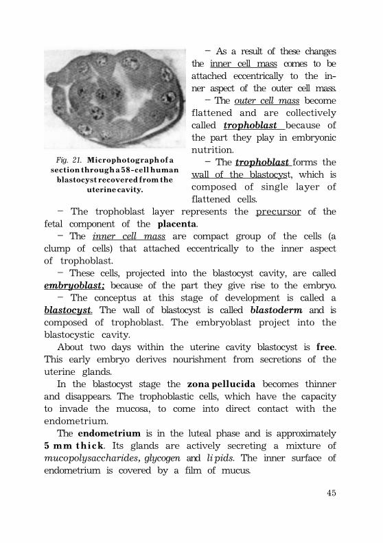
– As a result of these changesthe inner cell mass comes to beattached eccentrically to the in-ner aspect of the outer cell mass.
– The outer cell mass becomeflattened and are collectivelycalled trophoblast because ofthe part they play in embryonicnutrition.
– The trophoblast forms thewall of the blastocyst, which iscomposed of single layer offlattened cells.
– The trophoblast layer represents the precursor of thefetal component of the placenta.
– The inner cell mass are compact group of the cells (aclump of cells) that attached eccentrically to the inner aspectof trophoblast.
– These cells, projected into the blastocyst cavity, are calledembryoblast; because of the part they give rise to the embryo.
– The conceptus at this stage of development is called ablastocyst. The wall of blastocyst is called blastoderm and iscomposed of trophoblast. The embryoblast project into theblastocystic cavity.
About two days within the uterine cavity blastocyst is free.This early embryo derives nourishment from secretions of theuterine glands.
In the blastocyst stage the z o n a p e l l u c i d a becomes thinnerand disappears. The trophoblastic cells, which have the capacityto invade the mucosa, to come into direct contact with theendometrium.
The endometrium is in the luteal phase and is approximately5 m m t h i c k. Its glands are actively secreting a mixture ofmucopolysaccharides, glycogen and lipids. The inner surface ofendometrium is covered by a film of mucus.
Fig. 21. M i c r o p h o t o g r a p h o f as e c t i o n t h r o u g h a 5 8 - c e l l h u m a n
b l a s t o c y s t r e c o v e r e d f r o m t h eu t e r i n e c a v i t y .
46
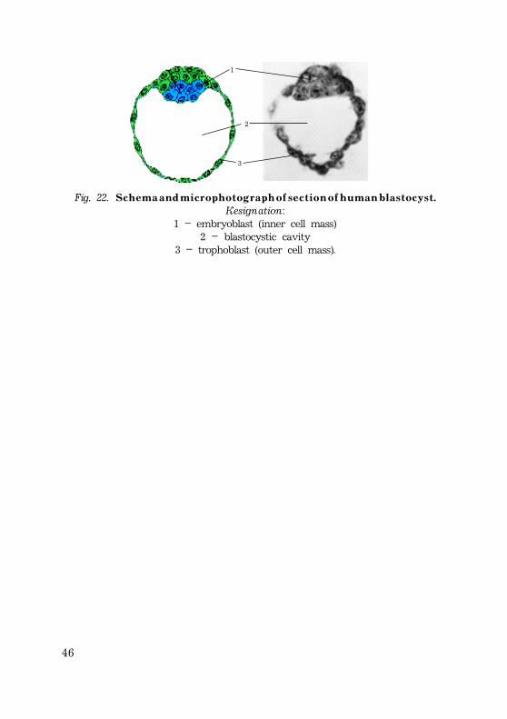
1
2
3
Fig. 22. S c h e m a a n d m i c r o p h o t o g r a p h o f s e c t i o n o f h u m a n b l a s t o c y s t .Designation:
1 – embryoblast (inner cell mass)2 – blastocystic cavity
3 – trophoblast (outer cell mass).
47
H u m a n i m p l a n t a o n
Implantation is the process of attachment and embeddingof the blastocyst into the mucosa layer or endometrium ofuterus.
Implantation continuous about 40 hours. Implantation ofhuman blastocyst is completed during the second week afterfertilization.
Implantation, or nidation, involves penetration through theuterine epithelium, with little signs of necrosis of the connectivetissue stroma and blood vessels of the endometrium.
This type of human implantation is called interstitialimplantation, in which the blastocyst comes to lie entirely withinthe endometrium.
Implantation includes two stages:· adhesion;· invasion.
∆ ∆ ∆ ∆ ∆ A d h e s i v e m e c h n i s m sThe adhesive mechanisms are those which attach the blastocyst
to a localized part of the endometrium.At a b o u t 7 d a y after fertilization blastocyst attaches to the
endometrial epithelium, lining the inner surface of endometrium.The uterine mucosal surface contains irregular depressions,
most of which represent the openings of endometrial glands.These glands are very numerous (about 15.000). The blastocystusually becomes attached between the openings of theendometrial glands.
The blastocyst attaches to the endometrial epithelium usuallyadjacent to the e m b r y o n i c p o l e, containing embryoblast.
In this period blastocyst expands to a diameter of about 0.25mm due to the absorption of fluid into the cavity from uterinesecretion.
As contact is made with the uterine wall, the trophoblastrapidly proliferates and begins to invade the endometrium.
48
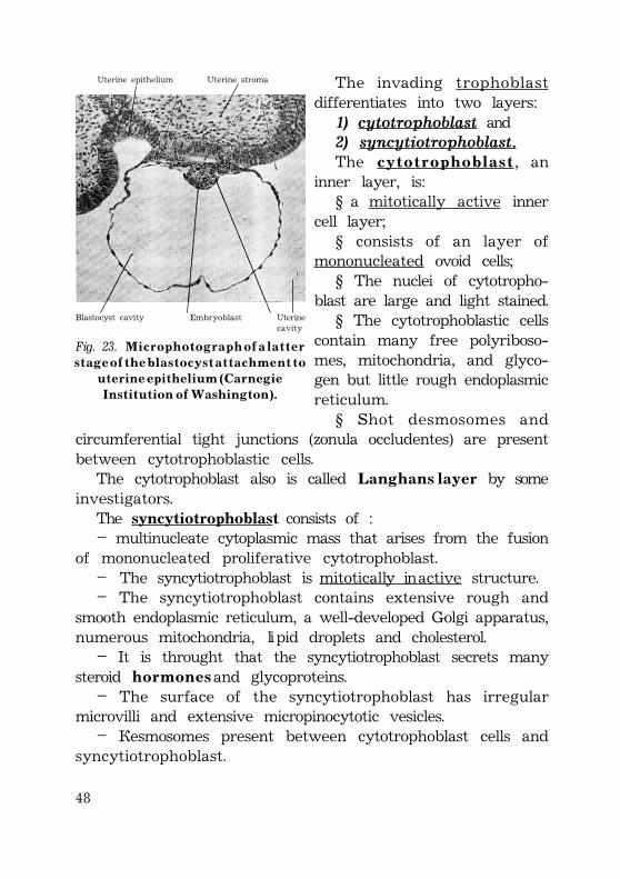
Uterine epithelium Uterine stroma
Blastocyst cavity Embryoblast Uterinecavity
Fig. 23. M i c r o p h o t o g r a p h o f a l a t t e rs t a g e o f t h e b l a s t o c y s t a t t a c h m e n t t o
u t e r i n e e p i t h e l i u m ( C a r n e g i eI n s t i t u t i o n o f W a s h i n g t o n ) .
The invading trophoblastdifferentiates into two layers:1) cytotrophoblast and2) syncytiotrophoblast.The c y t o t r o p h o b l a s t, an
inner layer, is:§ a mitotically active inner
cell layer;§ consists of an layer of
mononucleated ovoid cells;§ The nuclei of cytotropho-
blast are large and light stained.§ The cytotrophoblastic cells
contain many free polyriboso-mes, mitochondria, and glyco-gen but little rough endoplasmicreticulum.
§ Shot desmosomes andcircumferential tight junctions (zonula occludentes) are presentbetween cytotrophoblastic cells.
The cytotrophoblast also is called L a n g h a n s l a y e r by someinvestigators.
The syncytiotrophoblast consists of :– multinucleate cytoplasmic mass that arises from the fusion
of mononucleated proliferative cytotrophoblast.– The syncytiotrophoblast is mitotically inactive structure.– The syncytiotrophoblast contains extensive rough and
smooth endoplasmic reticulum, a well-developed Golgi apparatus,numerous mitochondria, lipid droplets and cholesterol.
– It is throught that the syncytiotrophoblast secrets manysteroid h o r m o n e s and glycoproteins.
– The surface of the syncytiotrophoblast has irregularmicrovilli and extensive micropinocytotic vesicles.
– Desmosomes present between cytotrophoblast cells andsyncytiotrophoblast.
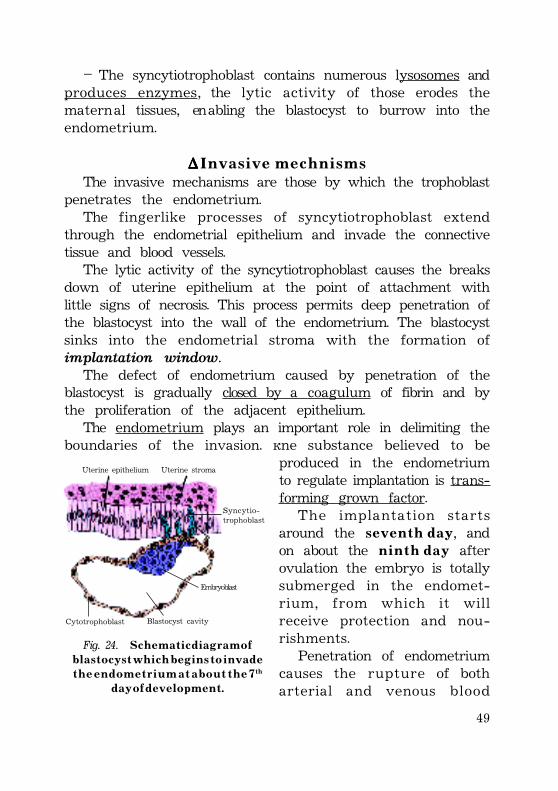
49
– The syncytiotrophoblast contains numerous lysosomes andproduces enzymes, the lytic activity of those erodes thematernal tissues, enabling the blastocyst to burrow into theendometrium.
∆∆∆∆∆ I n v a s i v e m e c h n i s m sThe invasive mechanisms are those by which the trophoblast
penetrates the endometrium.The fingerlike processes of syncytiotrophoblast extend
through the endometrial epithelium and invade the connectivetissue and blood vessels.
The lytic activity of the syncytiotrophoblast causes the breaksdown of uterine epithelium at the point of attachment withlittle signs of necrosis. This process permits deep penetration ofthe blastocyst into the wall of the endometrium. The blastocystsinks into the endometrial stroma with the formation ofimplantation window.
The defect of endometrium caused by penetration of theblastocyst is gradually closed by a coagulum of fibrin and bythe proliferation of the adjacent epithelium.
The endometrium plays an important role in delimiting theboundaries of the invasion. One substance believed to be
produced in the endometriumto regulate implantation is trans-forming grown factor.
The implantation startsaround the s e v e n t h d a y, andon about the n i n t h d a y afterovulation the embryo is totallysubmerged in the endomet-rium, from which it willreceive protection and nou-rishments.
Penetration of endometriumcauses the rupture of botharterial and venous blood
Uterine epithelium Uterine stroma
Syncytio-trophoblast
Embryoblast
Blastocyst cavityCytotrophoblast
Fig. 24. S c h e m a t i c d i a g r a m o fb l a s t o c y s t w h i c h b e g i n s t o i n v a d e
t h e e n d o m e t r i u m a t a b o u t t h e 7th
d a y o f d e v e l o p m e n t .
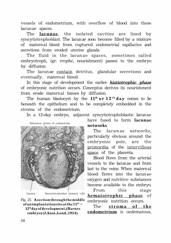
50
vessels of endometrium, with overflow of blood into theselacunae spaces.
The l a c u n a e, the isolated cavities are lined bysyncytiotrophoblast. The lacunae soon become filled by a mixtureof maternal blood from ruptured endometrial capillaries andsecretions from eroded uterine glands.
The fluid in the lacun ae spaces, sometimes calledembryotroph, (gr. trophe, nourishment) passes to the embryoby diffusion.
The lacunae contain detritus, glandular secretions andeventually, maternal blood.
In this stage of development the earlier histiotrophic phaseof embryonic nutrition occurs. Conceptus derives its nourishmentfrom erode maternal tissues by diffusion.
The human blastocyst by the 11th o r 1 2 t h d a y comes to liebeneath the epithelium and to be completely embedded in thestroma of the endometrium.
In a 12-day embryo, adjacent syncytiotrophoblastic lacunaehave fused to form l a c u n a enetworks.
The lacun ae networks,particularly obvious around theembryonic pole, are theprimordia of the intervillousspace of the placenta.
Blood flows from the arterialvessels to the lacunae and fromlast to the veins. When maternalblood flows into the lacunaeoxygen and nutritive substancesbecome available to the embryo.
From this stagehematotrophic phase ofembryonic nutrition occurs.
The s t r o m a o f t h ee n d o m e t r i u m is oedematous,
Edematous stroma of endometrium
Lacunae Syncytiotrophoblast (primary villi)
Fig. 25. A s e c t i o n t h r o u g h t h e m i d d l eo f a n i m p l a n t a t i o n s i t e a t t h e 1 1th –
12th d a y o f d e v e l o p m e n t . ( B a r n e se m b r y o ) ( J . A n a t . , L o n d . , 1 9 5 4 ) .
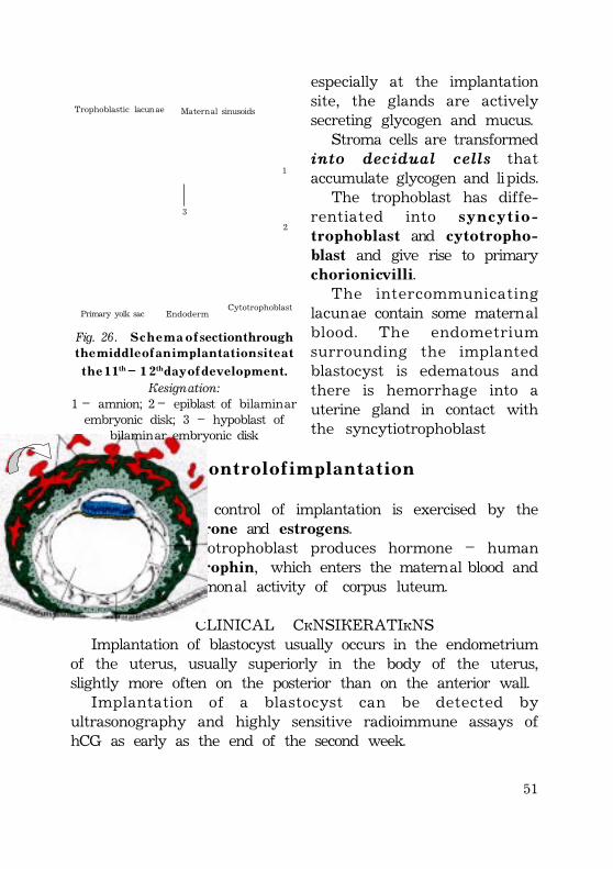
51
especially at the implantationsite, the glands are activelysecreting glycogen and mucus.
Stroma cells are transformedinto decidual cells thataccumulate glycogen and lipids.
The trophoblast has diffe-rentiated into s y n c y t i o -t r o p h o b l a s t and c y t o t r o p h o -blast and give rise to primaryc h o r i o n i c v i l l i.
The intercommunicatinglacunae contain some maternalblood. The endometriumsurrounding the implantedblastocyst is edematous andthere is hemorrhage into auterine gland in contact withthe syncytiotrophoblast
C o n t r o l o f i m p l a n t a t i o n
1. Fundamental control of implantation is exercised by thehormone progesterone and estrogens.
2. The syncytiotrophoblast produces hormone – humanchorionic gonadotrophin, which enters the maternal blood andmaintains the hormonal activity of corpus luteum.
CLINICAL CONSIDERATIONSImplantation of blastocyst usually occurs in the endometrium
of the uterus, usually superiorly in the body of the uterus,slightly more often on the posterior than on the anterior wall.
Implantation of a blastocyst can be detected byultrasonography and highly sensitive radioimmune assays ofhCG as early as the end of the second week.
1
2
3
Trophoblastic lacunae Maternal sinusoids
Primary yolk sac EndodermCytotrophoblast
Fig. 26. S c h e m a o f section throught h e m i d d l e o f a n i m p l a n t a t i o n s i t e a t
t h e 1 1th – 1 2th d a y o f d e v e l o p m e n t .Designation:
1 – amnion; 2 – epiblast of bilaminarembryonic disk; 3 – hypoblast of
bilaminar embryonic disk
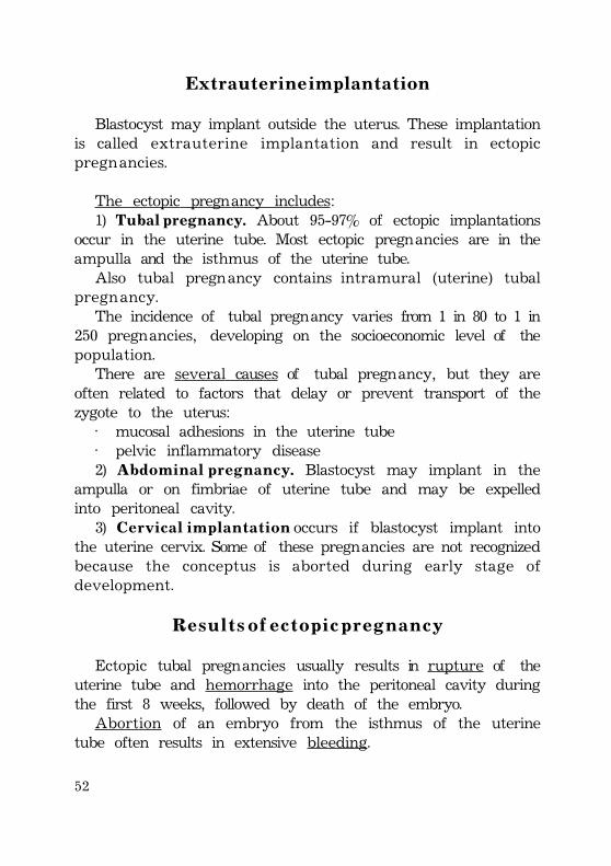
52
E x t r a u t e r i n e i m p l a n t a t i o n
Blastocyst may implant outside the uterus. These implantationis called extrauterine implantation and result in ectopicpregnancies.
The ectopic pregnancy includes:1) T u b a l p r e g n a n c y . About 95-97% of ectopic implantations
occur in the uterine tube. Most ectopic pregnancies are in theampulla and the isthmus of the uterine tube.
Also tubal pregnancy contains intramural (uterine) tubalpregnancy.
The incidence of tubal pregnancy varies from 1 in 80 to 1 in250 pregnancies, developing on the socioeconomic level of thepopulation.
There are several causes of tubal pregnancy, but they areoften related to factors that delay or prevent transport of thezygote to the uterus:
· mucosal adhesions in the uterine tube· pelvic inflammatory disease2) A b d o m i n a l p r e g n a n c y . Blastocyst may implant in the
ampulla or on fimbriae of uterine tube and may be expelledinto peritoneal cavity.
3) C e r v i c a l i m p l a n t a t i o n occurs if blastocyst implant intothe uterine cervix. Some of these pregnancies are not recognizedbecause the conceptus is aborted during early stage ofdevelopment.
R e s u l t s o f e c t o p i c p r e g n a n c y
Ectopic tubal pregnancies usually results in rupture of theuterine tube and hemorrhage into the peritoneal cavity duringthe first 8 weeks, followed by death of the embryo.
Abortion of an embryo from the isthmus of the uterinetube often results in extensive bleeding.
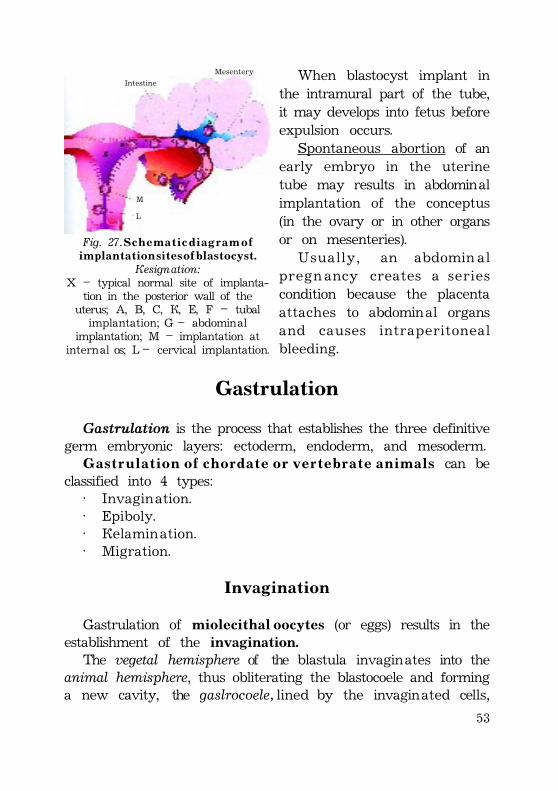
53
When blastocyst implant inthe intramural part of the tube,it may develops into fetus beforeexpulsion occurs.
Spontaneous abortion of anearly embryo in the uterinetube may results in abdominalimplantation of the conceptus(in the ovary or in other organsor on mesenteries).
Usually, an abdomin alpregn ancy creates a seriescondition because the placentaattaches to abdominal organsand causes intraperitonealbleeding.
Gastrulation
Gastrulation is the process that establishes the three definitivegerm embryonic layers: ectoderm, endoderm, and mesoderm.
G a s t r u l a t i o n o f c h o r d a t e o r v e r t e b r a t e a n i m a l s can beclassified into 4 types:
· Invagination.· Epiboly.· Delamination.· Migration.
Invagination
Gastrulation of miolecithal oocytes (or eggs) results in theestablishment of the invagination.
The vegetal hemisphere of the blastula invaginates into theanimal hemisphere, thus obliterating the blastocoele and forminga new cavity, the gaslrocoele, lined by the invaginated cells,
Fig. 27. S c h e m a t i c d i a g r a m o fi m p l a n t a t i o n s i t e s o f b l a s t o c y s t .
Designation:X – typical normal site of implanta-
tion in the posterior wall of theuterus; A, B, C, D, E, F – tubalimplantation; G – abdominal
implantation; M – implantation atinternal os; L – cervical implantation.
L
M
Mesentery
Intestine
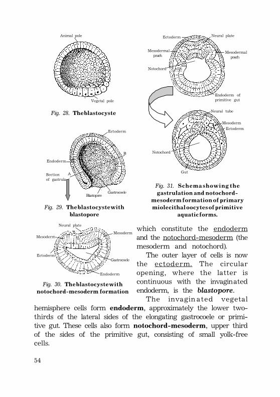
54
Neural tube
Mesoderm
Ectoderm
Ectoderm Neural plate
Mesodermal pouch
Mesodermal pouch
Notochord
Endoderm ofprimitive gut
Notochord
Gut
Fig. 31. S c h e m a s h o w i n g t h eg a s t r u l a t i o n a n d n o t o c h o r d -
m e s o d e r m f o r m a t i o n o f p r i m a r ym i o l e c i t h a l o o c y t e s o f p r i m i t i v e
a q u a t i c f o r m s .
Fig. 28. Th e b l a s t o c y s t e
Sectionof gastrula
Blastopore
Endoderm
Gastrocoele
B
A
Ectoderm
Fig. 29. Th e b l a s t o c y s t e w i t hblastopore
Animal pole
Vegetal pole
which constitute the endodermand the notochord-mesoderm (themesoderm and notochord).
The outer layer of cells is nowthe ectoderm. The circularopening, where the latter iscontinuous with the invaginatedendoderm, is the blastopore.
The invagin ated vegetalhemisphere cells form endoderm, approximately the lower two-thirds of the lateral sides of the elongating gastrocoele or primi-tive gut. These cells also form notochord-mesoderm, upper thirdof the sides of the primitive gut, consisting of small yolk-freecells.
Endoderm
Ectoderm
MesodermMesoderm
Gastrocoele
Neural plate
Fig. 30. Th e b l a s t o c y s t e w i t hn o t o c h o r d - m e s o d e r m f o r m a t i o n
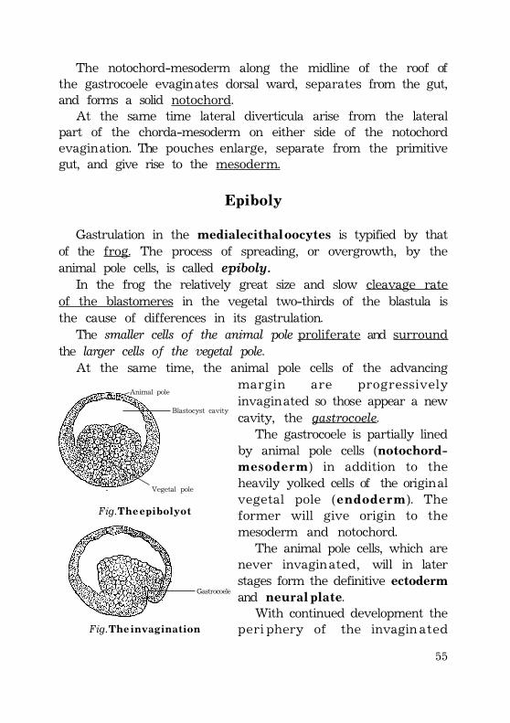
55
The notochord-mesoderm along the midline of the roof ofthe gastrocoele evaginates dorsal ward, separates from the gut,and forms a solid notochord.
At the same time lateral diverticula arise from the lateralpart of the chorda-mesoderm on either side of the notochordevagination. The pouches enlarge, separate from the primitivegut, and give rise to the mesoderm.
Epiboly
Gastrulation in the medialecithal oocytes is typified by thatof the frog. The process of spreading, or overgrowth, by theanimal pole cells, is called epiboly.
In the frog the relatively great size and slow cleavage rateof the blastomeres in the vegetal two-thirds of the blastula isthe cause of differences in its gastrulation.
The smaller cells of the animal pole proliferate and surroundthe larger cells of the vegetal pole.
At the same time, the animal pole cells of the advancingmargin are progressivelyinvaginated so those appear a newcavity, the gastrocoele.
The gastrocoele is partially linedby animal pole cells (notochord-m e s o d e r m) in addition to theheavily yolked cells of the originalvegetal pole (e n d o d e r m). Theformer will give origin to themesoderm and notochord.
The animal pole cells, which arenever invaginated, will in laterstages form the definitive ectodermand n e u r a l p l a t e.
With continued development theperi phery of the invaginated
Animal pole
Vegetal pole
Blastocyst cavity
Fig. T h e e p i b o l y o t
Gastrocoele
Fig. T h e i n v a g i n a t i o n
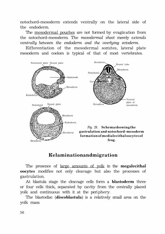
56
notochord-mesoderm extends ventrally on the lateral side ofthe endoderm.
The mesodermal pouches are not formed by evagination fromthe notochord-mesoderm. The mesodermal sheet merely extendsventrally between the endoderm and the overlying ectoderm.
Differentiation of the mesodermal somites, lateral platemesoderm and coelom is typical of that of most vertebrates.
D e l a m i n a t i o n a n d m i g r a t i o n
The presence of large amounts of yolk in the megalecithaloocytes modifies not only cleavage but also the processes ofgastrulation.
At blastula stage the cleavage cells form a blastoderm threeor four cells thick, separated by cavity from the centrally placedyolk and continuous with it at the periphery.
The blastodisc (discoblastula) is a relatively small area on theyolk mass.
Fig. 28. S c h e m a s h o w i n g t h eg a s t r u l a t i o n a n d n o t o c h o r d - m e s o d e r m
f o r m a t i o n o f m e d i a l e c i t h a l o o c y t e s o ffrog.
Endoderm
Neural plate Notochord plate
Gastrocoele
Mesoderm
Neural tube
Mesoderm
Ectoderm
Notochord
Gut
Ectoderm
Ventralplate ofmesoderm
Endoderm
Mesoderm
Neural plate Notochord
Mesoderm
Ectoderm
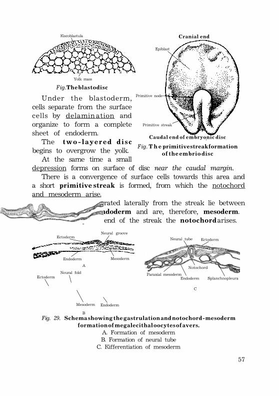
57
Discoblastula
Yolk mass
Fig. T h e b l a s t o d i s c
Under the blastoderm,cells separate from the surfacecells by delamin ation andorganize to form a completesheet of endoderm.
The t w o - l a y e r e d d i s cbegins to overgrow the yolk.
At the same time a smalldepression forms on surface of disc near the caudal margin.
There is a convergence of surface cells towards this area anda short primitive streak is formed, from which the notochordand mesoderm arise.
All the cells proliferated laterally from the streak lie betweenthe ectoderm and endoderm and are, therefore, mesoderm.
From the cephalic end of the streak the n o t o c h o r d arises.
C r a n i a l e n d
Epiblast
Primitive node
Primitive streak
C a u d a l e n d o f e m b r y o n i c d i s c
Fig. T h e p r i m i t i v e s t r e a k f o r m a t i o no f t h e e m b r i o d i s c
EctodermNeural groove
Endoderm Mesoderm
A
B
C
Neural tube Ectoderm
Endoderm
Notochord
SplanchnopleuraParaxial mesoderm
Fig. 29. S c h e m a s h o w i n g t h e g a s t r u l a t i o n a n d n o t o c h o r d - m e s o d e r mf o r m a t i o n o f m e g a l e c i t h a l o o c y t e s o f a v e r s .
A. Formation of mesodermB. Formation of neural tube
C. Differentiation of mesoderm
EctodermNeural fold
Mesoderm Endoderm
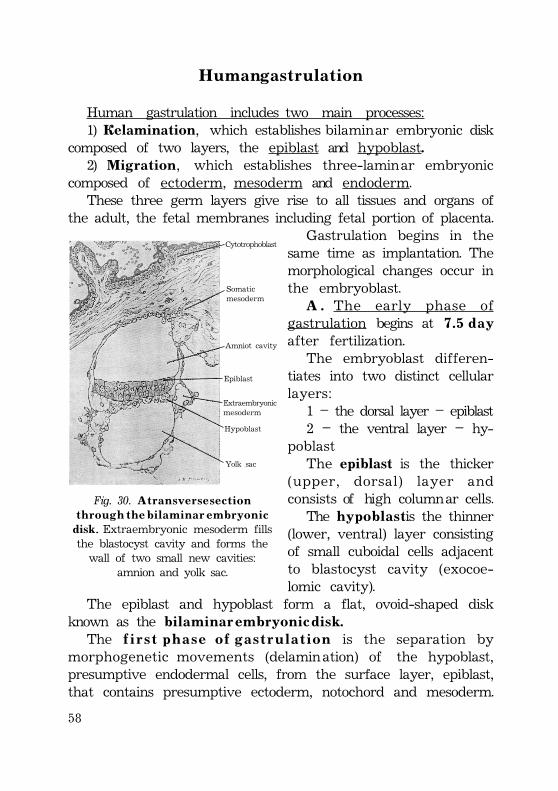
58
H u m a n g a s t r u l a t i o n
Human gastrulation includes two main processes:1) Delamination, which establishes bilaminar embryonic disk
composed of two layers, the epiblast and hypoblast.2) Migration, which establishes three-laminar embryonic
composed of ectoderm, mesoderm and endoderm.These three germ layers give rise to all tissues and organs of
the adult, the fetal membranes including fetal portion of placenta.Gastrulation begins in the
same time as implantation. Themorphological changes occur inthe embryoblast.
A . The early phase ofgastrulation begins at 7 . 5 d a yafter fertilization.
The embryoblast differen-tiates into two distinct cellularlayers:
1 – the dorsal layer – epiblast2 – the ventral layer – hy-
poblastThe epiblast is the thicker
(upper, dorsal) layer andconsists of high columnar cells.
The h y p o b l a s t is the thinner(lower, ventral) layer consistingof small cuboidal cells adjacentto blastocyst cavity (exocoe-lomic cavity).
The epiblast and hypoblast form a flat, ovoid-shaped diskknown as the b i l a m i n a r e m b r y o n i c d i s k .
The f i r s t p h a s e o f g a s t r u l a t i o n is the separation bymorphogenetic movements (delamination) of the hypoblast,presumptive endodermal cells, from the surface layer, epiblast,that contains presumptive ectoderm, notochord and mesoderm.
Fig. 30. A t r a n s v e r s e s e c t i o nt h r o u g h t h e b i l a m i n a r e m b r y o n i c
disk. Extraembryonic mesoderm fillsthe blastocyst cavity and forms thewall of two small new cavities:
amnion and yolk sac.
Yolk sac
Hypoblast
Extraembryonicmesoderm
Epiblast
Amniot cavity
Somaticmesoderm
Cytotrophoblast
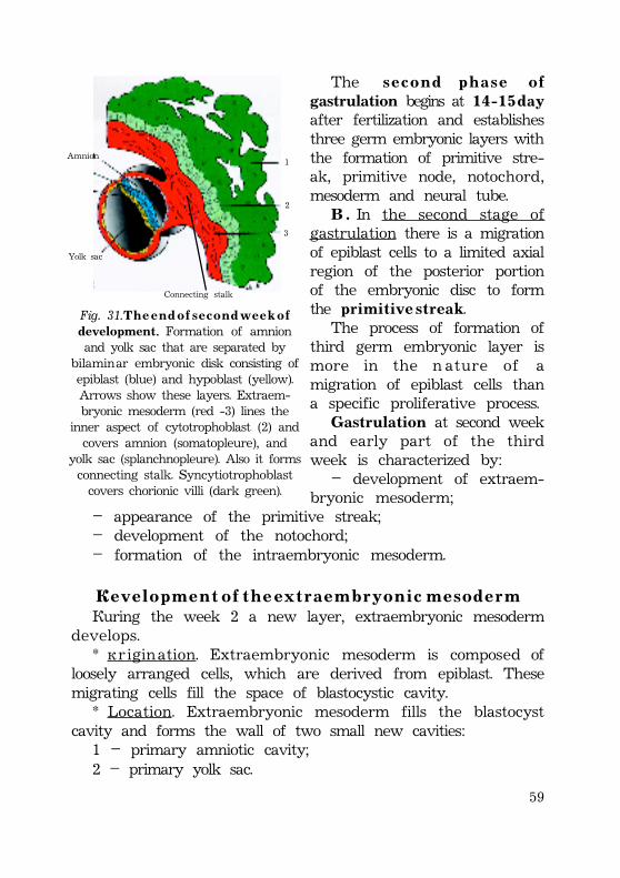
59
The s e c o n d p h a s e o fgastrulation begins at 1 4 - 1 5 d a yafter fertilization and establishesthree germ embryonic layers withthe formation of primitive stre-ak, primitive node, notochord,mesoderm and neural tube.
B . In the second stage ofgastrulation there is a migrationof epiblast cells to a limited axialregion of the posterior portionof the embryonic disc to formthe primitive streak.
The process of formation ofthird germ embryonic layer ismore in the n ature of amigration of epiblast cells thana specific proliferative process.
Gastrulation at second weekand early part of the thirdweek is characterized by:
– development of extraem-bryonic mesoderm;
– appearance of the primitive streak;– development of the notochord;– formation of the intraembryonic mesoderm.
D e v e l o p m e n t o f t h e e x t r a e m b r y o n i c m e s o d e r mDuring the week 2 a new layer, extraembryonic mesoderm
develops.* Origination. Extraembryonic mesoderm is composed of
loosely arranged cells, which are derived from epiblast. Thesemigrating cells fill the space of blastocystic cavity.
* Location. Extraembryonic mesoderm fills the blastocystcavity and forms the wall of two small new cavities:
1 – primary amniotic cavity;2 – primary yolk sac.
Amnion
Yolk sac
Connecting stalk
1
2
3
Fig. 31. T h e e n d o f s e c o n d w e e k o fdevelopment. Formation of amnionand yolk sac that are separated by
bilaminar embryonic disk consisting ofepiblast (blue) and hypoblast (yellow).Arrows show these layers. Extraem-bryonic mesoderm (red -3) lines the
inner aspect of cytotrophoblast (2) andcovers amnion (somatopleure), and
yolk sac (splanchnopleure). Also it formsconnecting stalk. Syncytiotrophoblastcovers chorionic villi (dark green).
60
P r i m a r y a m n i o t i c c a v i t y is located between the epiblast andinner aspect of cytotrophoblast.
P r i m a r y y o l k s a c is located under the hypoblast.The bilaminar embryonic disk lies between two cavities - the
amniotic cavity and cavity of primary yolk sac.The extraembryonic mesodermsubdivides into two parts:1. extraembryonic somatic mesoderm;2. extraembryonic visceral mesoderm.E x t r a e m b r y o n i c s o m a t i c m e s o d e r m (somatopleure) lines the
trophoblast (inner surface of cytotrophoblast) and covers theamnion, and forms the c o n n e c t i n g s t a l k.
Extraembryonic visceral mesoderm (splanchnopleure) coversthe yolk sac.
The fluid-filled cavity surrounds the amnion and yolk sac,except where they are attached to the trophoblast by connectingstalk, is called extraembryonic coelom.
Extraembryonic somatic mesoderm, lining the inner aspectof cytotrophoblast, and two layer of trophoblast(cytotrophoblast + syncytiotrophoblast) constitute the chorion.
The extraembryonic coelom is now called the chorionic cavity.
F o m a t i o n o f t h e p r i m i t i v e s t r e a kIn a human embryo of 15th d a y the primitive streak appears
at the caudal end of the embryonic disc. The primitive streakresults from the proliferation and migration of the cells of theepiblast to the median plane of the embryonic disc.
These cells form a thickened linear band of epiblast. In thisarea epiblast cells proliferate, dividing more frequently thenelsewhere in epiblast. A narrow primitive groove develops inthe primitive streak.
Cranial end of primitive streak forms a p r i m i t i v e n o d e,known as Hensen’s node.
Some of the cells of primitive node form an invagination, asmall depression, which is called the p r i m i t i v e p i t, surroundedby a slightly elevated area.
61
1
2
3
45
6
7
C r a n i a l e n d
C a u d a l e n d
Fig. 32. S c h e m a i l l u s t r a t e s t h e m i g r a t i o no f e p i b l a s t c e l l s d u r i n g t h e f o r m a t i o n o f p r i m i t i v e s t r e a k .
Designation:1 – epiblast; 2 – material of neural plate; 3 – material of notochord; 4 –primitive node; 5 – primitive pit; 6 – primitive streak; 7 – primitive groove
Cells of primitive pit migrate in the axial region betweenepiblast and hypoblast and give rise to the notochord.
D e v e l o p m e n t o f t h e i n t r a e m b r y o n i c m e s o d e r m( e m b r y o n i c m e s o d e r m )
The term intraembryonic mesoderm describes the germ layerthat forms during w e e k 3 (gastrulation) in contrast to theextraembryonic mesoderm, which forms during week 2.
Shortly after the primitive streak appears, cells leave itsdeep surface and migrate in lateral direction between the epiblastand hypoblast.
Migrating cells actively proliferate and move laterally andcranially, alone the midline. This new forming cellular layer iscalled intraembryonic (embryonic) mesoderm.
The intraembryonic mesoderm progressively extends laterallyuntil it reaches the margins of embryonic disc in each side eventually.There, these mesodermal cells continue with the extraembryonicmesoderm, which covers the amnion and yolk sac.
62
Primitive groove and node
Epiblast
Primitive streak
Migrating mesodermal cells Hypoblast
Fig. 33. D o r s a l s u r f a c e o f e m b r y o a t16th d a y o f d e v e l o p m e n t . Cells ofprimitive streak migrate betweenepiblast and hypoblast laterally and
give rise to intraembryonicmesodermal cells.
Some cells of intraembryonicmesoderm migrate cranially.Here they meet cranilly to formthe cardiogenic mesoderm inthe c a r – d i o g e n i c a r e a, wherethe primordium of the heartbegins to develop at the endof the third week.
As a result of the formationof intraembryonic mesodermbilamin ar embryonic discbecomes t r i l a m i n a r d i s c.
External form of the embryo at presomiteperiod
This period extends from the time of appearance of theprimitive streak until the differentiation of the f i r s t s o m i t e.
The embryonic body at the 14th o r 1 5th day after fertilizationconsists of a bilaminar disc of epiblast and hypoblast lyingbetween the amniotic and the yolk sac cavities.
In the caudal half of the disk the primitive streak can beseen in the midline as a linear thickening which terminatescranially in the primitive (Hensen’s) node.
The presence of the primitive streak together with then o t o c h o r d confers an obvious bilateral symmetry on the disk.
Caudally the primitive streak does not quite reach the hinderedge of the disk, being separated from it by the cloacal membrane.
With further growth the embryonic disk increases in size,especially in the cranio-caudal axis, and the primitive streakand node appear to be carried in a caudal direction since thestreak and the area lateral to it grow relatively slowly, whilethe area cephalic to the primitive node grows rapidly.
During this process of caudal migration of the primitivestreak the embryonic disk elongates and changes its shape,becoming first oval, then pear-shaped.
At the same time the embryonic disc develops head and tailfolds and bulges slightly upwards into the amniotic cavity.
63
Towards the end of the presomatic period of developmenta broad, a n e u r a l g r o o v e appears in the n e u r a l p l a t e region ofthe embryonic disc cranial to the primitive node.
F o m a t i o n o f t h e n o t o c h o r dDuring the formation of the intraembryonic mesoderm noto-
chord develops. The small depression, called primitive pit forms inthe center of the primitive node.
The notochordal process elongates by invagination of cellsfrom the primitive pit.
The notochordal process is now a cellular tube that extendscranially from the primitive node to the prechordal plate betweenepiblast and hypoblast.
Beginning at the cranial end of the embryo, the notochordalcells proliferate and the notochordal plate infolds to form therod-shaped notochord.
Notochord is a s o l i d c y l i n d e r c o r d located in the midline ofthe embryonic disc between the ectoderm and endoderm.
At the end of the 4th week notochord is in its final positionbetween endoderm and ventral aspect of the neural groove orneural tube.
Neural plate Primitive pit
Primitivestreak
Cloacalmembrane
Allantois
1
Embryonicendoderm
Yolk sac
Notochordal canal
Fig. 35. F o r m a t i o n o f n o t o c h o r d a lc a n a l . The opening of notochordalcanal rapidly disappears and remainsof notochordal process form a solid
cord. (1 – connecting stalk).
Primitive node
Primitivestreak
Cloacal membrane
Primitive pit
Notochord-underectoderm
Embryonic disk
Prechordal plate
C r a n i a l e n d
C a u d a l e n d
Fig. 34. D e v e l o p m e n t o f n o t o c h o r -d a l p r o c e s s . The notochordal processelongates by invagination of cellsfrom primitive pit and extendscranially from primitive node.
64
The notochord degenerates and disappears as the body forms,but it persists as the n u c l e u s p u l p o s u s of each intervertebraldisc.
Notochord is important for the following:1. Notochord induces the overlying embryonic ectoderm to
differentiate into the neural plate, the primordium of thenervous system.
2. Notochord serves as the basis for development of theaxial skeleton (bones of head and vertebral column).
3. Notochord induces the formation of vertebral bodies.4. Notochord provides changes involving endoderm.
Endoderm is involved in the formation of the future digestivesystem, respiratory system, central parts of the urogenitalsystem and pharyngeal pouches.
CLINICAL CONSIDERATIONS1 . S a c r o c o c c y g e a l t e r a t o m a.· is tumor that arises from remnants of the primitive streak,
which normally degenerates and disappears;· is derived from pluripotent cells of the primitive streak
and often contains various types of tissue (e.g. bone, nerve,hair);
· occurs more commonly in female infants;· usually becomes malignant during infancy and must be
removed by age 6 months.2. Chordoma.· is a tumor that arises from remnants of the notochord;· may be found either intracranially or in the sacral region;· may be either benign or malignant.After the formation of primitive streak, intraembryonic
mesoderm and notochord, remainder of epiblast is calledectodermal germ layer (ectoderm).
After the formation of the extraembryonic endodermal layer,lining the inner surface of secondary yolk sac, remainder ofhypoblast is called e n d o d e r m a l g e r m l a y e r (endoderm).
65
* P r e c h o r d a l p l a t e (prochordal plate) is a small circular areaof columnar endodermal cells located in cranial part ofembryonic disk.
Prechordal plate is firmly attached to the overlying ectoderm.These fused germ layers form the oropharyngeal membrane,
located at the future site of the oral cavity (mouth).* Caudal to the primitive streak there is a circular area known
as the c l o a c a l m e m b r a n e, which indicates the future site of theanus.
*The embryonic disk remains bilaminar at the oropharyngealand cloacal membranes because the embryonic ectoderm andendoderm are fused at the sites, thereby preventing migrationof mesodermal cells between them.
F o m a t i o n o f t h e n e u r a l t u b e ( n e u r u l a t i o n )
• At the beginning of the t h i r d w e e k of development theectodermal germ layer has a shape of a flat disc.
Under the inductive influence of the notochord, theectodermal disc changes in form and give rise to the nervoussystem.
S i g n a l i n g m o l e c u l e s appear to involve members of thetransforming growth factor-â (TGF- â) family, which includesactivin and fibroblast growth factors (FGFs).
• At the end of the third week of development, as thenotochord develops, the embryonic ectoderm over it thickensto form an elongated slipper-shaped plate that is called neuralplate. Neural plate formation induces the signaling molecules(activin, FGFs), produced by the developing notochord.
• N e u r a l p l a t e appears cranial to the primitive node anddorsal to the notochord and mesoderm adjacent to it. Asnotochord elongates cranially, the neural plate extends craniallyas far as the oropharyngeal membrane.
• About the 18th day of development, the neural plateinvaginates along its central axis to form a longitudinal mediann e u r a l g r o o v e, which has n e u r a l f o l d s on each side.
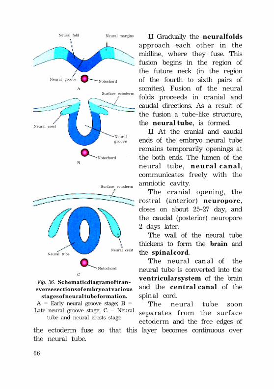
66
Notochord
Surface ectoderm
Neural tubeNeural crest
C
Fig. 36. S c h e m a t i c d i a g r a m o f t r a n -s v e r s e s e c t i o n s o f e m b r y o a t v a r i o u s
s t a g e s o f n e u r a l t u b e f o r m a t i o n .A – Early neural groove stage; B –Late neural groove stage; C – Neural
tube and neural crests stage
Neural fold Neural margins
Neural groove Notochord
ASurface ectoderm
Neuralgroove
Neural crest
NotochordB
• Gradually the n e u r a l f o l d sapproach each other in themidline, where they fuse. Thisfusion begins in the region ofthe future neck (in the regionof the fourth to sixth pairs ofsomites). Fusion of the neuralfolds proceeds in cranial andcaudal directions. As a result ofthe fusion a tube-like structure,the n e u r a l t u b e, is formed.
• At the cranial and caudalends of the embryo neural tuberemains temporarily openings atthe both ends. The lumen of theneural tube, n e u r a l c a n a l,communicates freely with theamniotic cavity.
The cranial opening, therostral (anterior) n e u r o p o r e,closes on about 25-27 day, andthe caudal (posterior) neuropore2 days later.
The wall of the neural tubethickens to form the brain andthe s p i n a l c o r d.
The neural can al of theneural tube is converted into theventricular system of the brainand the c e n t r a l c a n a l of thespinal cord.
The neural tube soonseparates from the surfaceectoderm and the free edges of
the ectoderm fuse so that this layer becomes continuous overthe neural tube.
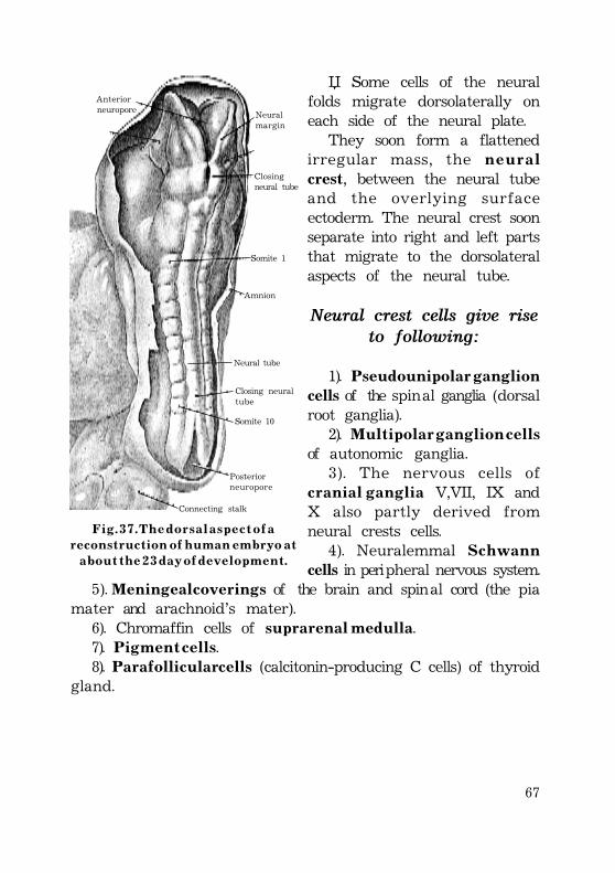
67
• Some cells of the neuralfolds migrate dorsolaterally oneach side of the neural plate.
They soon form a flattenedirregular mass, the n e u r a lcrest, between the neural tubeand the overlying surfaceectoderm. The neural crest soonseparate into right and left partsthat migrate to the dorsolateralaspects of the neural tube.
Neural crest cells give riseto following:
1). Pseudounipolar ganglioncells of the spinal ganglia (dorsalroot ganglia).
2). M u l t i p o l a r g a n g l i o n c e l l sof autonomic ganglia.
3). The nervous cells ofc r a n i a l g a n g l i a V,VII, IX andX also partly derived fromneural crests cells.
4). Neuralemmal S c h w a n ncells in peripheral nervous system.
5). Meningeal coverings of the brain and spinal cord (the piamater and arachnoid’s mater).
6). Chromaffin cells of suprarenal medulla.7). P i g m e n t c e l l s.8). Parafollicular cells (calcitonin-producing C cells) of thyroid
gland.
Connecting stalk
Posteriorneuropore
Somite 10
Closing neuraltube
Neural tube
Amnion
Somite 1
Closingneural tube
Neuralmargin
Anterior neuropore
F i g . 3 7 . T h e d o r s a l a s p e c t o f ar e c o n s t r u c t i o n o f h u m a n e m b r y o a t
a b o u t t h e 2 3 d a y o f d e v e l o p m e n t .
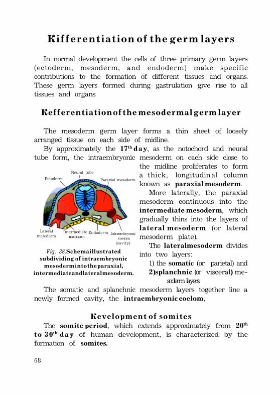
68
D i f f e r e n t i a t i o n o f t h e g e r m l a y e r s
In normal development the cells of three primary germ layers(ectoderm, mesoderm, and endoderm) make specificcontributions to the formation of different tissues and organs.These germ layers formed during gastrulation give rise to alltissues and organs.
D e f f e r e n t i a t i o n o f t h e m e s o d e r m a l g e r m l a y e r
The mesoderm germ layer forms a thin sheet of looselyarranged tissue on each side of midline.
By approximately the 17th d a y, as the notochord and neuraltube form, the intraembryonic mesoderm on each side close to
the midline proliferates to forma thick, longitudinal columnknown as paraxial mesoderm.
More laterally, the paraxialmesoderm continuous into theintermediate mesoderm, whichgradually thins into the layers ofl a t e r a l m e s o d e r m (or lateralmesoderm plate).
The l a t e r a l m e s o d e r m dividesinto two layers:
1) the somatic (or parietal) and2) splanchnic (or visceral) me-
soderm layers.The somatic and splanchnic mesoderm layers together line a
newly formed cavity, the intraembryonic coelom,
D e v e l o p m e n t o f s o m i t e sThe s o m i t e p e r i o d, which extends approximately from 20th
t o 3 0th d a y of human development, is characterized by theformation of somites.
Ectoderm
Neural tube
Paraxial mesoderm
Lateralmesoderm
Intermediate mesoderm
Endoderm Intraembryonic coelom(cavity)
Fig. 38. S c h e m a i l l u s t r a t e ds u b d i v i d i n g o f i n t r a e m b r y o n i c
m e s o d e r m i n t o t h e p a r a x i a l ,i n t e r m e d i a t e a n d l a t e r a l m e s o d e r m .
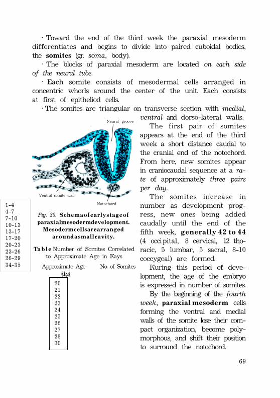
69
· Toward the end of the third week the paraxial mesodermdifferentiates and begins to divide into paired cuboidal bodies,the somites (gr. soma, body).
· The blocks of paraxial mesoderm are located on each sideof the neural tube.
· Each somite consists of mesodermal cells arranged inconcentric whorls around the center of the unit. Each consistsat first of epitheliod cells.
· The somites are triangular on transverse section with medial,ventral and dorso-lateral walls.
The first pair of somitesappears at the end of the thirdweek a short distance caudal tothe cranial end of the notochord.From here, new somites appearin craniocaudal sequence at a ra-te of approximately three pairsper day.
The somites increase innumber as development prog-ress, new ones being addedcaudally until the end of thefifth week, g e n e r a l l y 4 2 t o 4 4(4 occipital, 8 cervical, 12 tho-racic, 5 lumbar, 5 sacral, 8-10coccygeal) are formed.
During this period of deve-lopment, the age of the embryois expressed in number of somites.
By the beginning of the fourthweek, p a r a x i a l m e s o d e r m cellsforming the ventral and medialwalls of the somite lose their com-pact organization, become poly-morphous, and shift their positionto surround the notochord.
Neural groove
Ventral somite wall
Notochord
Fig. 39. S c h e m a o f e a r l y s t a g e o fp a r a x i a l m e s o d e r m d e v e l o p m e n t .
M e s o d e r m c e l l s a r e a r r a n g e da r o u n d a s m a l l c a v i t y .
Ta b l e Number of Somites Correlatedto Approximate Age in Days
Approximate Age No. of Somites (days)
20 21 22 23 24 25 26 27 28 30
1-4 4-7 7-10 10-13 13-17 17-20 20-23 23-26 26-29 34-35
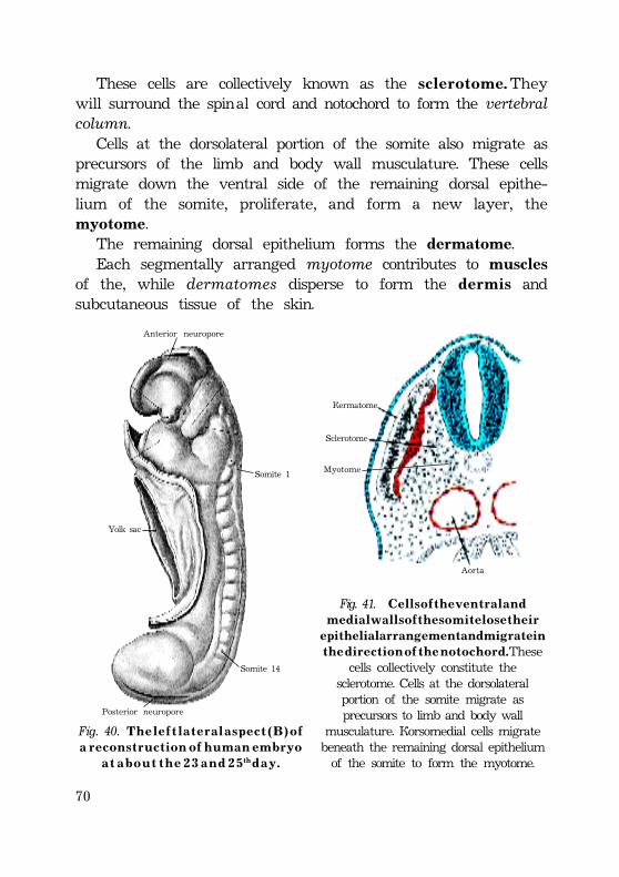
70
Posterior neuropore
Somite 14
Somite 1
Yolk sac
Anterior neuropore
Fig. 40. T h e l e f t l a t e r a l a s p e c t ( B ) o fa r e c o n s t r u c t i o n o f h u m a n e m b r y o
a t a b o u t t h e 2 3 a n d 2 5th d a y .
Fig. 41. C e l l s o f t h e v e n t r a l a n dm e d i a l w a l l s o f t h e s o m i t e l o s e t h e i r
epithelial arrangement and migrate int h e d i r e c t i o n o f t h e n o t o c h o r d . These
cells collectively constitute thesclerotome. Cells at the dorsolateralportion of the somite migrate asprecursors to limb and body wall
musculature. Dorsomedial cells migratebeneath the remaining dorsal epitheliumof the somite to form the myotome.
Aorta
Dermatome
Sclerotome
Myotome
These cells are collectively known as the s c l e r o t o m e . Theywill surround the spinal cord and notochord to form the vertebralcolumn.
Cells at the dorsolateral portion of the somite also migrate asprecursors of the limb and body wall musculature. These cellsmigrate down the ventral side of the remaining dorsal epithe-lium of the somite, proliferate, and form a new layer, themyotome.
The remaining dorsal epithelium forms the dermatome.Each segmentally arranged myotome contributes to muscles
of the, while dermatomes disperse to form the d e r m i s andsubcutaneous tissue of the skin.
71
Each myotome and dermatome retains its innervation fromits segment of origin, no matter where the cells migrate.
Each somite forms:1) its own s c l e r o t o m e (the cartilage and bone component),2) its own m y o t o m e (providing the segmental muscle
component), and3) its own d e r m a t o m e , the segmental skin component.Each myotome and dermatome also has its own segmental
nerve component.Paraxial mesoderm differentiates into the:· sclerotome· myotome· dermatomeS o m e t o m e r e s 1 - 7 do not form somites but contribute
mesoderm to the pharyngeal arches.
I n t e r m e d i a t e m e s o d e r mIntermediate mesoderm connects the paraxial mesoderm and
lateral mesoderm.It forms a longitudinal elevation along the dorsal body wall
known as the urogenital ridge.In cervical and upper thoracic regions, it forms segmental
cell clusters (future n e p h r o t o m e s ) , whereas more caudally, itforms an unsegmented mass of tissue, the n e p h r o g e n i c c o r d,which gives rise to future kidneys and gonads.
L a t e r a l p l a t eThe l a t e r a l m e s o d e r m divides into two layers:1) the somatic (or parietal) and2 ) splanchnic (or visceral) mesoderm layers.1). The somatic or parietal mesoderm layer, which continuous
with the extraembryonic mesoderm covering the amnion.The parietal layer together with overlying ectoderm will form
the lateral and ventral body wall.The parietal mesoderm surrounding the intraembryonic cavity
will form the thin mesothelium, which will line the peritoneal,
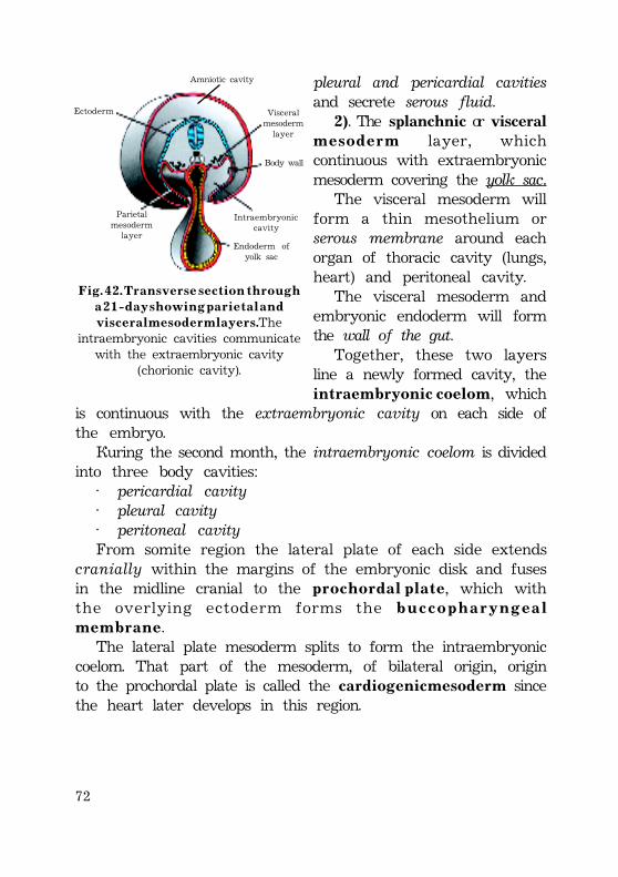
72
pleural and pericardial cavitiesand secrete serous fluid.
2). The splanchnic or visceralm e s o d e r m layer, whichcontinuous with extraembryonicmesoderm covering the yolk sac.
The visceral mesoderm willform a thin mesothelium orserous membrane around eachorgan of thoracic cavity (lungs,heart) and peritoneal cavity.
The visceral mesoderm andembryonic endoderm will formthe wall of the gut.
Together, these two layersline a newly formed cavity, theintraembryonic coelom, which
is continuous with the extraembryonic cavity on each side ofthe embryo.
During the second month, the intraembryonic coelom is dividedinto three body cavities:
· pericardial cavity· pleural cavity· peritoneal cavityFrom somite region the lateral plate of each side extends
cranially within the margins of the embryonic disk and fusesin the midline cranial to the p r o c h o r d a l p l a t e, which withthe overlying ectoderm forms the b u c c o p h a r y n g e a lm e m b r a n e.
The lateral plate mesoderm splits to form the intraembryoniccoelom. That part of the mesoderm, of bilateral origin, originto the prochordal plate is called the cardiogenic mesoderm sincethe heart later develops in this region.
Amniotic cavity
Visceralmesodermlayer
Body wall
Intraembryoniccavity
Ectoderm
Parietalmesoderm
layerEndoderm of
yolk sac
F i g . 4 2 . T r a n s v e r s e s e c t i o n t h r o u g ha 2 1 - d a y s h o w i n g p a r i e t a l a n d
v i s c e r a l m e s o d e r m l a y e r s . Theintraembryonic cavities communicate
with the extraembryonic cavity(chorionic cavity).
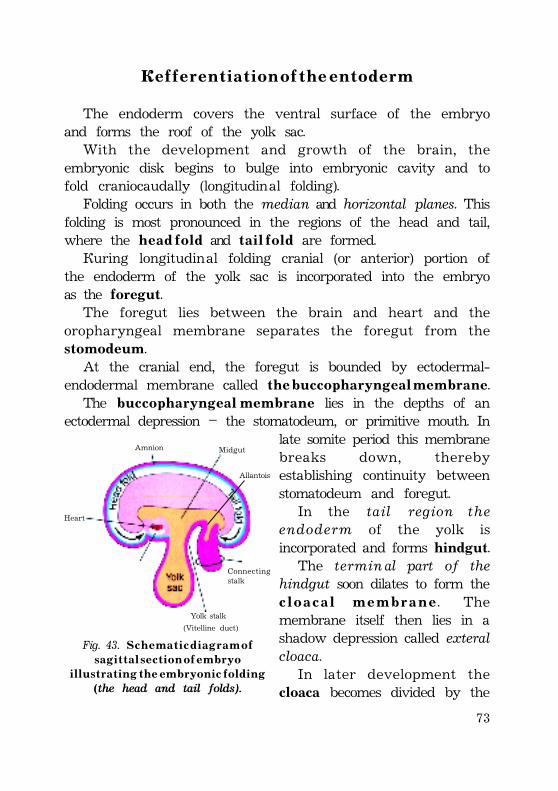
73
D e f f e r e n t i a t i o n o f t h e e n t o d e r m
The endoderm covers the ventral surface of the embryoand forms the roof of the yolk sac.
With the development and growth of the brain, theembryonic disk begins to bulge into embryonic cavity and tofold craniocaudally (longitudinal folding).
Folding occurs in both the median and horizontal planes. Thisfolding is most pronounced in the regions of the head and tail,where the h e a d f o l d and t a i l f o l d are formed.
During longitudinal folding cranial (or anterior) portion ofthe endoderm of the yolk sac is incorporated into the embryoas the foregut.
The foregut lies between the brain and heart and theoropharyngeal membrane separates the foregut from thestomodeum.
At the cranial end, the foregut is bounded by ectodermal-endodermal membrane called t h e b u c c o p h a r y n g e a l m e m b r a n e.
The b u c c o p h a r y n g e a l m e m b r a n e lies in the depths of anectodermal depression – the stomatodeum, or primitive mouth. In
late somite period this membranebreaks down, therebyestablishing continuity betweenstomatodeum and foregut.
In the tail region theendoderm of the yolk isincorporated and forms hindgut.
The terminal part of thehindgut soon dilates to form thec l o a c a l m e m b r a n e . Themembrane itself then lies in ashadow depression called exteralcloaca.
In later development thecloaca becomes divided by the
Fig. 43. S c h e m a t i c d i a g r a m o fs a g i t t a l s e c t i o n o f e m b r y o
i l l u s t r a t i n g t h e e m b r y o n i c f o l d i n g(the head and tail folds).
Amnion Midgut
Allantois
Heart
Connectingstalk
Yolk stalk(Vitelline duct)
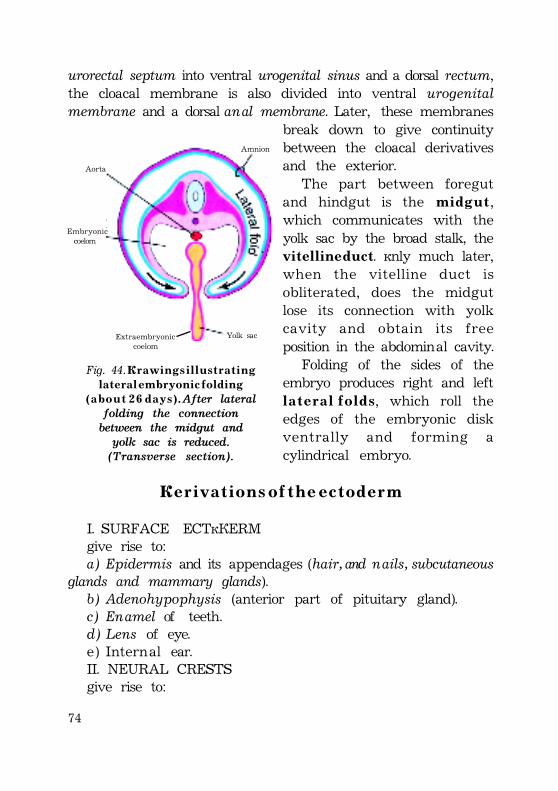
74
urorectal septum into ventral urogenital sinus and a dorsal rectum,the cloacal membrane is also divided into ventral urogenitalmembrane and a dorsal anal membrane. Later, these membranes
break down to give continuitybetween the cloacal derivativesand the exterior.
The part between foregutand hindgut is the m i d g u t,which communicates with theyolk sac by the broad stalk, thev i t e l l i n e d u c t. Only much later,when the vitelline duct isobliterated, does the midgutlose its connection with yolkcavity and obtain its freeposition in the abdominal cavity.
Folding of the sides of theembryo produces right and leftl a t e r a l f o l d s, which roll theedges of the embryonic diskventrally and forming acylindrical embryo.
D e r i v a t i o n s o f t h e e c t o d e r m
I. SURFACE ECTODERMgive rise to:a) Epidermis and its appendages (hair, and nails, subcutaneous
glands and mammary glands).b) Adenohypophysis (anterior part of pituitary gland).c) Enamel of teeth.d) Lens of eye.e) Internal ear.II. NEURAL CRESTSgive rise to:
Amnion
Aorta
Embryonic coelom
Yolk sacExtraembryoniccoelom
Fig. 44. D r a w i n g s i l l u s t r a t i n gl a t e r a l e m b r y o n i c f o l d i n g
( a b o u t 2 6 d a y s ) . After lateralfolding the connectionbetween the midgut andyolk sac is reduced.(Transverse section).
75
a) Pseudounipolar ganglion cells of the spinal ganglia (dorsalroot ganglia).
b) Multipolar ganglion cells of autonomic ganglia.c) The nervous cells of cranial ganglia V,VII, IX and X also
partly derived from neural crests cells.d) Neuralemmal Schwann cells in peripheral nervous system.e) Meningeal coverings of the brain and spinal cord (the pia
mater and arachnoid mater).f) Chromaffin cells of suprarenal medulla.g) Pigment cells.h) Parafollicular cells (calcitonin-producing C cells) of thyroid
glands.III. NEURAL TUBEgive rise to:a) The wall of the neural tube thickens to form the brain
and the spinal cord.b) The neural canal of the neural tube is converted into the
ventricular system of the brain and the central canal of thespinal cord.
D e r i v a t i o n s o f t h e mesoderm
I. PARAXIAL MESODERMgive rise to:a ) Sclerotome gives rise to skeleton (vertebral column) except
cranium.b) .Myotome gives rise to striated skeletal muscle (trunk, limbs),
muscles of head.c ) Dermatome give rise to dermis and subcutaneous tissue of
the skin.II. INTERMEDIARE MESODERMgive rise to:a) Urinary system (pronephros, mesonephros, metanephros,)
including ducts.b) Gonads and accessory glands.
76
III. LATERAL PLATE of MESODERMgive rise to:a) Serous membranes of pleura, pericardium, and peritoneum.b) Suprarenal gland cortex.c) Germinal epithelium of gonads.d) Myocardium, endocardium of heart.e) Connective tissue and muscle of viscera.IV. HEAD MESODERMgive rise to:a) Cranium.b) Dentin.c) Connective tissue of head.
D e r i v a t i o n s o f t h e e n t o d e r m
I. ENDODERM of the GUTgive rise to:a) Epithelium lining gastrointestinal tract (stomach, small intestine,
most part of large intestine except caudal portion of rectum).b) The parenchyma of liver and pancreas.c) Epilelium lining respiratory tract (pharynx, trachea,bronchi,
lungs).d) The parenchyma of the thyroid gland, parathyroid gland.e) The reticular stroma of thymus.f) The epithelium lining of the urinary bladder and urethra.g) The epithelium lining of the tympanic cavity and auditory
tube.
F e t a l m e m b r a n e s
Fetal membranes are p r o v i s o r y o r g a n s o f e m b r y o a n d f e t u s ,which provide the normal human development during prenatalperiod.
Fetal membranes are developed from the fertilized egg, yetnot forming part of the embryonic body.
77
M a i n f u n c t i o n s :–Fetal membranes are of functional importance during
embryonic life, being concerned with the supply or storage ofnutriment, respiratory exchange, excretion and mechanicalprotection of the embryo.
– Fetal membranes are also known to transmit antibodiesfrom mother to young, thus providing for passive immunity ofthe newborn.
– They produce hormones, which resemble those of theovary and adenohypophysis, and are an essential part of theendocrine system during pregnancy.
– They are largely shed or absorbed at hatching or birth.
T y p e s o f F e t a l m e m b r a n e s :1. yolk sac;2. amnion;3. allantois;4. chorion;5. placenta;6. umbilical cord.
The first phase of gastrulation (delamination) begins at 7th –7,5 day of development and proceed at the same time with theimplantation.
At once fetal membranes form (yolk sac, amnion and chorion),which provide entrance of nutrients and further developmentof embryo.
The formations of these fetal membranes take place aboutduring s e c o n d w e e k.
First of all amnion and yolk sac develop.
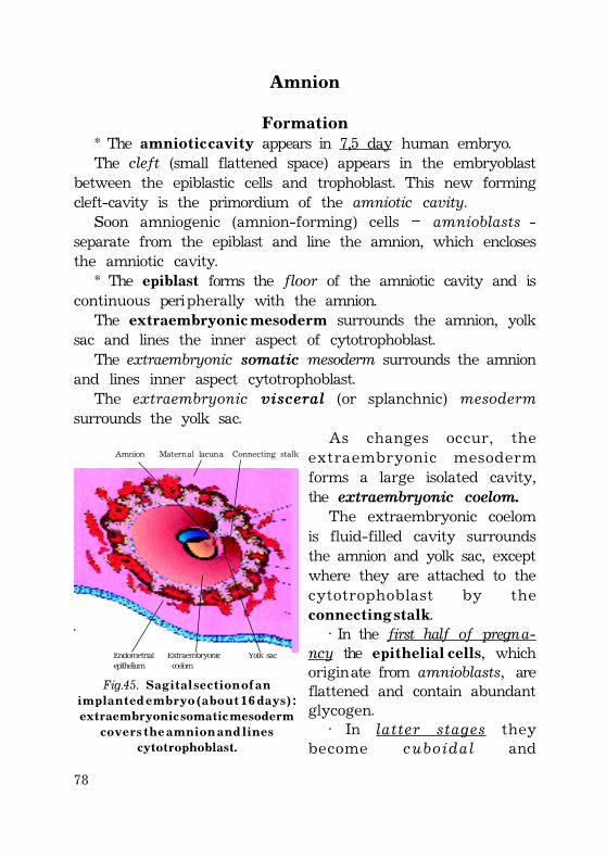
78
Amnion
Formation* The a m n i o t i c c a v i t y appears in 7,5 day human embryo.The cleft (small flattened space) appears in the embryoblast
between the epiblastic cells and trophoblast. This new formingcleft-cavity is the primordium of the amniotic cavity.
Soon amniogenic (amnion-forming) cells – amnioblasts -separate from the epiblast and line the amnion, which enclosesthe amniotic cavity.
* The epiblast forms the floor of the amniotic cavity and iscontinuous peripherally with the amnion.
The extraembryonic mesoderm surrounds the amnion, yolksac and lines the inner aspect of cytotrophoblast.
The extraembryonic somatic mesoderm surrounds the amnionand lines inner aspect cytotrophoblast.
The extraembryonic visceral (or splanchnic) mesodermsurrounds the yolk sac.
As changes occur, theextraembryonic mesodermforms a large isolated cavity,the extraembryonic coelom.
The extraembryonic coelomis fluid-filled cavity surroundsthe amnion and yolk sac, exceptwhere they are attached to thecytotrophoblast by thec o n n e c t i n g s t a l k.
· In the first half of pregna-ncy the e p i t h e l i a l c e l l s, whichoriginate from amnioblasts, areflattened and contain abundantglycogen.
· In latter stages theybecome cuboidal and
Amnion Maternal lacuna Connecting stalk
Endometrial Extraembryonic Yolk sacepithelium coelom
Fig.45. S a g i t a l s e c t i o n o f a ni m p l a n t e d e m b r y o ( a b o u t 1 6 d a y s ) :
e x t r a e m b r y o n i c s o m a t i c m e s o d e r mc o v e r s t h e a m n i o n a n d l i n e s
cytotrophoblast.
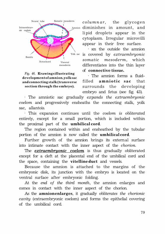
79
column ar , the glycogendiminishes in amount, andli pid droplets appear in thecytoplasm. Irregular microvilliappear in their free surface.
· On the outside the amnionis covered by extraembryonicsomatic mesoderm, whichdifferentiates into the thin layerof connective tissue.
· The amnion forms a fluid-filled a m n i o t i c s a c thatsurrounds the developingembryo and fetus (see fig. 43).
· The amniotic sac gradually expands the extraembryoniccoelom and progressively ensheaths the connecting stalk, yolksac, allantois.
· This expansion continues until the coelom is obliteratedentirely, except for a small portion, which is included withinthe proximal part of the u m b i l i c a l c o r d.
The region contained within and ensheathed by the tubularportion of the amnion is now called the u m b i l i c a l c o r d.
Further growth of the amnion brings its external surfaceinto intimate contact with the inner aspect of the chorion.
The extraembryonic coelom is thus gradually obliteratedexcept for a cleft at the placental end of the umbilical cord andthe space, containing the v i t e l l i n e d u c t and vessels.
Because the amnion is attached to the margins of theembryonic disk, its junction with the embryo is located on theventral surface after embryonic folding.
At the end of the third month, the amnion enlarges andcomes in contact with the inner aspect of the chorion.
As the a m n i o n e n l a r g e s, it gradually obliterates the chorioniccavity (extraembryonic coelom) and forms the epithelial coveringof the umbilical cord.
Neural tube Somatic mesoderm
Intraembryo-nic coelom
Amnion
Yolk sac
Visceralmesoderm
Notochord
Somite
Fig. 46. D r a w i n g s i l l u s t r a t i n gd e v e l o p m e n t o f a m n i o n , y o l k s a ca n d c o n n e c t i n g s t a l k ( t r a n s v e r s e
s e c t i o n t h r o u g h t h e e m b r y o ) .
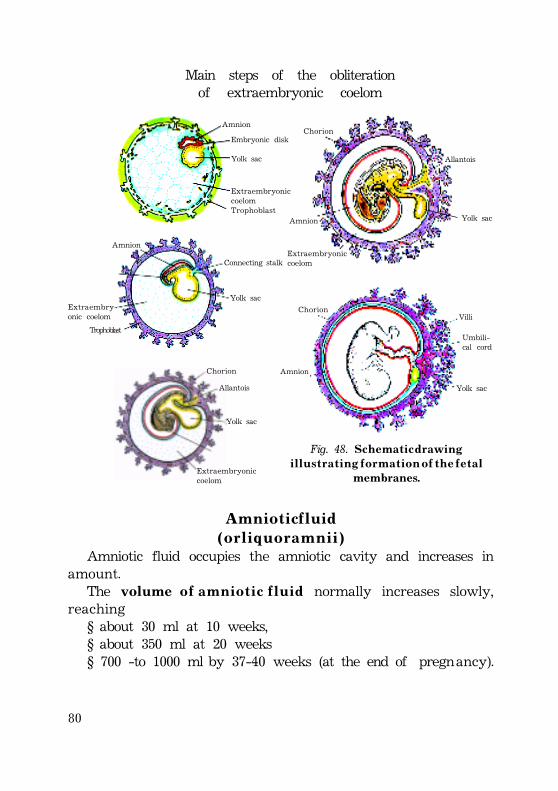
80
Main steps of the obliterationof extraembryonic coelom
Amnion
Extraembry-onic coelom
Trophoblast
Embryonic disk
Yolk sac
ExtraembryoniccoelomTrophoblast
Amnion
Connecting stalk
Yolk sac
Yolk sac
Extraembryoniccoelom
Chorion
Allantois
Yolk sac
Extraembryoniccoelom
Chorion
Allantois
Amnion
Yolk sac
Chorion
Amnion
Villi
Umbili-cal cord
Fig. 48. S c h e m a t i c d r a w i n gi l l u s t r a t i n g f o r m a t i o n o f t h e f e t a l
membranes.
A m n i o t i c f l u i d( o r l i q u o r a m n i i )
Amniotic fluid occupies the amniotic cavity and increases inamount.
The volume o f a m n i o t i c f l u i d normally increases slowly,reaching
§ about 30 ml at 10 weeks,§ about 350 ml at 20 weeks§ 700 -to 1000 ml by 37-40 weeks (at the end of pregnancy).
81
Composition of amniotic fluidAmniotic fluid is basically water (about 99%) that contains:• organic substances• inorganic substances• desquamated fetal epithelial cellsHalf organic substances are proteins. The other half consists
of carbohydrates, lipids, enzymes, hormones, pigments, and fetalurine.
As pregnancy advances, the composition of the amnioticfluid changes as fetal excreta (meconium and urine) are added.
Production of amniotic fluidMost amniotic fluid is derived f r o m m a t e r n a l t i s s u e:1) by dialysis (diffusion) maternal blood in the decidua
parietalis through the amniochorionic membrane ;2) by diffusion through the chorionic plate from maternal
blood in the intervillous spaces of the placenta;Amniotic fluid is derived f r o m t h e fetus:1) by dialysis of fetal blood through blood vessels in the
umbilical cord;2) by diffusion through the skin of the fetus before
keratinization of the skin;3) Fluid is also secreted by the fetal respiratory tract and
enters the amniotic cavity. The daily rate of contribution of fluidto the amniotic cavity from the respiratory tract is 300 to 4 0 0 m l .
4) Beginning in the eleventh week, the fetus contributes tothe amniotic fluid by excreting urine into the amniotic cavity.By late pregnancy about a 5 0 0 m l of urine is added daily.
Resorption of amniotic fluidAmniotic fluid is constantly resorbed during pregnancy by
several possible routes.1 . F e t u s s w a l l o w s amniotic fluid and absorbs by the fetus
digestive and respiratory tracts.§ During the final stages of pregnancy, the fetus swallows
u p t o 4 0 0 m l of amniotic fluid p e r d a y .
82
§ Amniotic fluid is absorbed into the fetal bloodstream throughthe wall of gastrointestinal tract.
§ The fluid passes into the fetal blood and enter the maternalblood in the intervillous spaces of the placenta.
§ Excess water in the fetal blood is excreted by the fetalkidney and returned to the amniotic sac through the fetalurinary tract.
2. There is rapid exchange between the amniotic fluid andmaternal and fetal circulations probably by way of the placentaand fetal kidney.
The water content of amniotic fluid changes every 3 h o u r s.
FunctionsAmniotic fluid plays a major role in fetal growth and
development.1) Amniotic fluid provides a buoyant medium which supports
the delicate tissues of the embryo.2) Amniotic cavity containing fluid allows the free movement
of the fetus.3) Amniotic fluid (A.f.) absorbs mechanical pressures, thereby
protecting the embryo from most external trauma.4) Absorption the amniotic fluid with swallowing permits
normal fetal lung development.5) A.f. prevents adherence of the amnion to the embryo and fetus.6) A.f. acts as a barrier to infection and contains antibodies.7) A.f. assists in regulation of fetal body temperature.8) A.f. provides homeostasis of fluid and electrolytes.
CLINICAL CONSIDERATIONSOligohydramnios is low volume of amniotic fluid. It results in
most cases of diminished placental blood flow when there isrenal agenesis (failure of kidney formation). The absence offetal urine contribution to the amniotic fluid is main cause ofoligohydramnios.
Obstructive uropathy (urinary tract obstruction) results insimilar decrease of fluid.
83
Compression of the umbilical cord is also a potentialcomplication.
Polihydramnios is high volume of amniotic fluid in excess of2000 ml.
It results when the fetus does not swallow the usual amountof amniotic fluid (esophageal atresia).
It may be associated with severe anomalies of CNS(anencephaly).
Amniocentesis –is the aspiration of fluid from amniotic sac.Amniocentesis is performed between weeks 12 and 14 after
fertilization. It has a 0,5% risk of miscarriage.Amniocentesis is used to perform the following studies:1 . A l f a - f e t o p r o t e i n a s s a y .High level of á-fetoprotein (AFP) in the amniotic fluid usually
indicates the presence of a neural tube defect (spina bifida,anencephaly, meroanencephaly).
Low levels of AFP indicate chromosomal aberrations such astrisomy 21.
2 . S p e c t r o p h o t o m e t r y is used to diagnose hemolytic disease.3 . S e x c h r o m o s o m a t i n s t u d i e s are used to diagnose sex-
linked diseases (X-linked muscular dystrophy, hemophilia).4 . D N A a n a l y s i s are used to identify defective chromosomes
early in pregnancy (cystic fibrosis, hematologic diseases, neurologicdisease, phenylketonuria).
5 . E n z y m e a n a l y s i s are used to identify steroid sulfatasedeficiency, disorders of connective tissue (hypophosphatasia),lysosomal storage diseases.
Y o l k s a c
FormationThe end of the second week of development is characterized
by the appearance of extraembryonic mesoderm, which is afine, loose connective tissue.
Extraembryonic splanchnic (splanchnopleuric) mesoderm formsthe wall of the p r i m a r y y o l k s a c.
84
The extraembryonic coelom appears between extraembryonicsplanchnic mesoderm, which forms the wall of the primary yolksac, and the extraembryonic somatic (somatopleuric) mesoderm,which lines the inner aspect of cytotrophoblast. In the sametime the primary yolk sac decreases in size.
Some cells of hypoblast migrate along the inner surface ofprimary yolk sac and a smaller s e c o n d a r y y o l k s a c forms.
The s e c o n d a r y y o l k s a c iscovered on its outer aspect bythe extraembryonic splanchnicmesoderm and on its inneraspect by the extraembryonicendoderm.
During the fourth week thef o ldin g end od er m of the dorsalwall of the yolk sac is incor-porated into the embryo andgives rise to the primordial gut(foregut, midgut, and hindgut).
The yolk sac remainsattached to the midgut by anarrow y o l k s t a l k.
The communication betweenthe gut and yolk sac (7th week)is reduced to a relatively slenderduct called the yolk stalk.
As the amniotic cavityexpands and obliterates most of
the extraembryonic coelom, the amnion forms the epithelialcovering of umbilical cord.
The yolk stalk compressed into the umbilical cord.The remainder of the yolk sac extends out of the embryo
ventrally and is pushed against the connecting stalk by theamnion.
The yolk sac then is gradually obliterated (look schema “Mainsteps of the obliteration of extraembryonic coelom”).
Amnion
Embryonic disk
Connecting stalk
Yolk sac
Extraembryonic Extraembryonic splanchnicmesodermendoderm
Fig. 49. S c h e m a t i c d r a w i n gi l l u s t r a t i n g f o r m a t i o n o f a m n i o n
a n d y o l k s a c . T h e w a l l o f t h e y o l ks a c i s c o m p o s e d o f t w o l a y e r s :
1 – internal endodermal layer origi-nating from hypoblast; 2 – externalmesodermal layer originating from
epiblast.
85
By 1 0 w e e k the yolk sac shrunk to pear-shaped remnantabout 5 m m in diameter.
By 2 0 w e e k the yolk sac is very small and is usually novisible.
Functions1. The human yolk sac contains n o y o l k. It may have a role in
the selective transfer of nutrients to the embryo at early stageof human development.
The yolk sac has a role in the t r a n s f e r o f n u t r i e n t s to theembryo during the second and third weeks, when theuteroplacental circulation is being established.
The trophoblast absorbs nutritive fluid from the lacunaenetworks, which is transferred to the embryo.
2 . The yolk sac mesoderm gives rise to the angioblastic tissue.It is first formed in the deepest layer of mesoderm early inthird week.
· Tissue spaces form the angioblastic tissue and the cells, whichline these spaces, take on the characters of typical flattenedendothelial cells of capillaries.
· The capillaries of yolk sac fuse with each other and giveraise the v i t e l l i n e v e i n s that grow toward the embryo.
· The p o r t a l v e i n develops from an anastomotic networkformed by the vitelline veins around the duodenum.
· The localized groups of mesodermal cells project into theinterior of developing capillary plexuses and become cut off toform b l o o d i s l e t s.
· These cells soon differentiating into the first generation ofstem blood cells that give rise to primary erythrocytes and then– to secondary erythrocytes.
The yolk sac is the first s o u r c e o f b l o o d cells that enter theembryonic circulation via vitelline veins.
The yolk sac is important as a s o u r c e o f b l o o d until the fetalliver replaces this function in about the 6th week of thedevelopment.
86
3 . The p r i m o r d i a l g e r m c e l l s( g o n a d o b l a s t s ) appear in theendoderm layer of the yolk sacwall in the third week of thedevelopment.
The newly forming primordialgerm cells subsequently migrateto the developing sex gonadsalong the wall of the yolk sactoward the wall of the hindgut.
The gonadoblasts embeddingthe developing sex gonad diffe-rentiate into the spermatogoniain males and oogonia in females.
· Vessels of the yolk sac forma vascular plexus that isconnected to the heart tubes byvitelline veins.
· The umbilical vein carries oxygenated blood and nutrientsfrom the chorion.
AllantoisF o r m a t i o n a n d f u n c t i o n s
In the 3d week of the development (on about day 16) diver-ticulum from the caudal wall of the yolk sac appears and extendsinto the connecting stalk. This diverticulum is called allantois.
The diverticulum arises early as a solid outgrowth. It soonbecomes canalized.
The allantoises wall is composed of two germ layers:1 – internal endodermal layer;2 – external layer of extraembryonic splanchnic mesoderm.The allantois grows for a variable distance into the connecting
stalk mesoderm but does not reach to the chorion (see fig. 44).The blood vessels develops within the extraembryonic
mesoderm of allantois. The vascular plexus in the wall of allantoisfuse and give rise to umbilical arteries and veins.
heart
Vitelline arteryVascular systemof the y o l k s a c
(1 )
Chorion
(3)Umbilicalcord withumbilicalvessels
Cardiovascular system of an embryos body (2)
Fig. 50. D i a g r a m o f t h e p r i m o r d i a lc a r d i o v a s c u l a r s y s t e m i n a n e m b r y o
o f a b o u t 2 1 d a y s .Vascular system contains:
(1) – vascular system of the yolk sac,(2) - cardiovascular system of an emb-ryos body,(3) – vascular system of the chorion.
87
B l o o d f o r m a t i o n occurs in its wall during the third to fifthweeks.
The umbilical vessels connect the circulatory system of theembryo and chorion.
The paired umbilical arteries pass through the connectingstalk (later the umbilical cord) and become continuous withvessels in the chorion.
The umbilical arteries carry poorly oxygenated blood fromembryo to the placenta.
P r o x i m a l p a r t of the umbilical arteries becomes the internaliliac arteries and superior vesical arteries after the birth.
The u m b i l i c a l v e i n s carry well-oxygenated blood from theplacenta to the embryo.
The right umbilical vein disappears during the seventh weekof the development.
The d i s t a l p o r t i o n o f t h e u m b i l i c a l v e s s e l s connect with thechorionic vessels, which differentiate at the same time in thechorionic mesoderm and form arterio-capillary-venous systemof the chorionic villi.
D i s t a l p a r t s of umbilical vessels obliterate after birth andbecome the median umbilical ligaments.
· Folding of the caudal ending of the embryo results partiallyincorporation of allantois into the embryo. After the folding theallantois is partially incorporated into the body of the embryo,where it forms the cloaca.
· The intraembryonic part of the allantois runs from theumbilical cord to the urinary bladder, with which it is continuous.
· The allantois soon constricts and becomes a thick fibrouscord, the urachus. It extends from the apex of the urinarybladder to the umbilical cord.
· During the second month, the extraembryonic part of theallantois degenerates.
· After the birth the urachus becomes a fibrous cord, them e d i a n u m b i l i c a l l i g a m e n t .
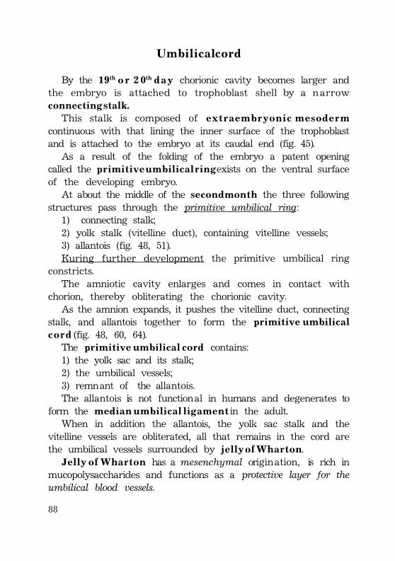
88
U m b i l i c a l c o r d
By the 19th o r 2 0th d a y chorionic cavity becomes larger andthe embryo is attached to trophoblast shell by a narrowconnecting stalk.
This stalk is composed of e x t r a e m b r y o n i c m e s o d e r mcontinuous with that lining the inner surface of the trophoblastand is attached to the embryo at its caudal end (fig. 45).
As a result of the folding of the embryo a patent openingcalled the p r i m i t i v e u m b i l i c a l r i n g exists on the ventral surfaceof the developing embryo.
At about the middle of the s e c o n d m o n t h the three followingstructures pass through the primitive umbilical ring:
1) connecting stalk;2) yolk stalk (vitelline duct), containing vitelline vessels;3) allantois (fig. 48, 51).During further development the primitive umbilical ring
constricts.The amniotic cavity enlarges and comes in contact with
chorion, thereby obliterating the chorionic cavity.As the amnion expands, it pushes the vitelline duct, connecting
stalk, and allantois together to form the p r i m i t i v e u m b i l i c a lc o r d (fig. 48, 60, 64).
The p r i m i t i v e u m b i l i c a l c o r d contains:1) the yolk sac and its stalk;2) the umbilical vessels;3) remnant of the allantois.The allantois is not functional in humans and degenerates to
form the m e d i a n u m b i l i c a l l i g a m e n t in the adult.When in addition the allantois, the yolk sac stalk and the
vitelline vessels are obliterated, all that remains in the cord arethe umbilical vessels surrounded by j e l l y o f W h a r t o n.
J e l l y o f W h a r t o n has a mesenchymal origination, is rich inmucopolysaccharides and functions as a protective layer for theumbilical blood vessels.
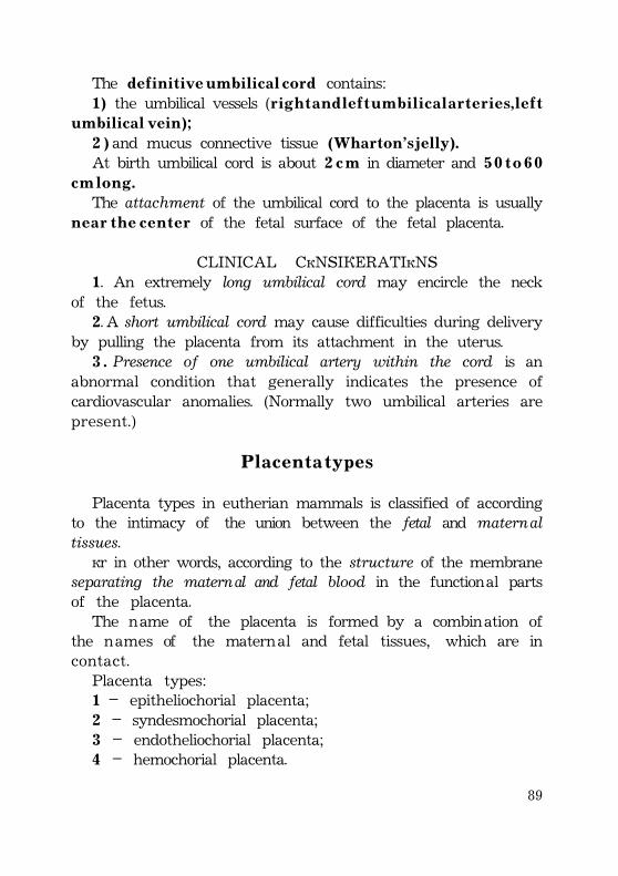
89
The d e f i n i t i v e u m b i l i c a l c o r d contains:1) the umbilical vessels (r i g h t a n d l e f t u m b i l i c a l a r t e r i e s , l e f t
u m b i l i c a l v e i n ) ;2 ) and mucus connective tissue (Wharton’s jelly).At birth umbilical cord is about 2 c m in diameter and 5 0 t o 6 0
c m l o n g .The attachment of the umbilical cord to the placenta is usually
n e a r t h e c e n t e r of the fetal surface of the fetal placenta.
CLINICAL CONSIDERATIONS1. An extremely long umbilical cord may encircle the neck
of the fetus.2. A short umbilical cord may cause difficulties during delivery
by pulling the placenta from its attachment in the uterus.3 . Presence of one umbilical artery within the cord is an
abnormal condition that generally indicates the presence ofcardiovascular anomalies. (Normally two umbilical arteries arepresent.)
P l a c e n t a t y p e s
Placenta types in eutherian mammals is classified of accordingto the intimacy of the union between the fetal and maternaltissues.
Or in other words, according to the structure of the membraneseparating the maternal and fetal blood in the functional partsof the placenta.
The name of the placenta is formed by a combination ofthe names of the maternal and fetal tissues, which are incontact.
Placenta types:1 – epitheliochorial placenta;2 – syndesmochorial placenta;3 – endotheliochorial placenta;4 – hemochorial placenta.
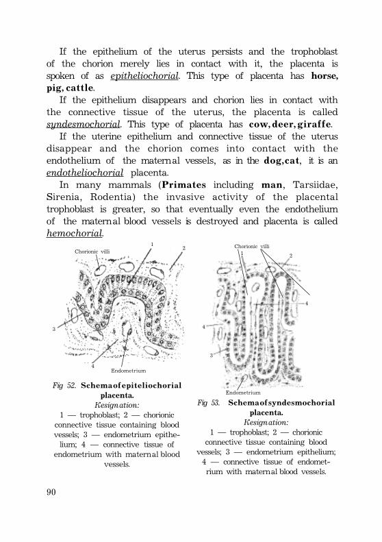
90
If the epithelium of the uterus persists and the trophoblastof the chorion merely lies in contact with it, the placenta isspoken of as epitheliochorial. This type of placenta has horse,p i g , c a t t l e.
If the epithelium disappears and chorion lies in contact withthe connective tissue of the uterus, the placenta is calledsyndesmochorial. This type of placenta has c o w , d e e r , g i r a f f e.
If the uterine epithelium and connective tissue of the uterusdisappear and the chorion comes into contact with theendothelium of the maternal vessels, as in the d o g , c a t, it is anendotheliochorial placenta.
In many mammals (P r i m a t e s including m a n, Tarsiidae,Sirenia, Rodentia) the invasive activity of the placentaltrophoblast is greater, so that eventually even the endotheliumof the maternal blood vessels is destroyed and placenta is calledhemochorial.
Chorionic villi
Endometrium
3
12
4
Fig 52. S c h e m a o f e p i t e l i o c h o r i a lplacenta.
Designation:1 — trophoblast; 2 — chorionic
connective tissue containing bloodvessels; 3 — endometrium epithe-lium; 4 — connective tissue of
endometrium with maternal bloodvessels.
1 2
4
4
3
Endometrium
Chorionic villi
Fig 53. S c h e m a o f s y n d e s m o c h o r i a lplacenta.
Designation:1 — trophoblast; 2 — chorionic
connective tissue containing bloodvessels; 3 — endometrium epithelium;4 — connective tissue of endomet-rium with maternal blood vessels.
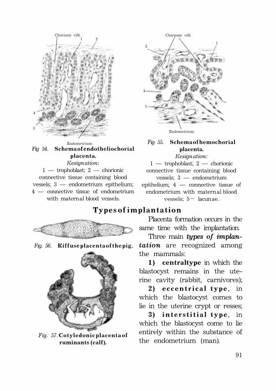
91
1 2
4
3
Chorionic villi
Fig 54. S c h e m a o f e n d o t h e l i o c h o r i a l p l a c e n t a .Designation:
1 — trophoblast; 2 — chorionicconnective tissue containing blood
vessels; 3 — endometrium epithelium;4 — connective tissue of endometrium
with maternal blood vessels.
Fig 55. S c h e m a o f h e m o c h o r i a lplacenta.
Designation:1 — trophoblast; 2 — chorionic
connective tissue containing bloodvessels; 3 — endometrium
epithelium; 4 — connective tissue ofendometrium with maternal blood
vessels; 5 – lacunae.
Endometrium
Endometrium
Chorionic villi
12
4
5
T y p e s o f i m p l a n t a t i o nPlacenta formation occurs in the
same time with the implantation.Three main types of implan-
tation are recognized amongthe mammals:
1 ) central type in which theblastocyst remains in the ute-rine cavity (rabbit, carnivores);
2 ) e c c e n t r i c a l t y p e, inwhich the blastocyst comes tolie in the uterine crypt or resses;
3 ) i n t e r s t i t i a l t y p e, inwhich the blastocyst come to lieentirely within the substance ofthe endometrium (man).
Fig. 56. D i f f u s e p l a c e n t a o f t h e p i g .
Fig. 57. C o t y l e d o n i c p l a c e n t a o fr u m i n a n t s ( c a l f ) .
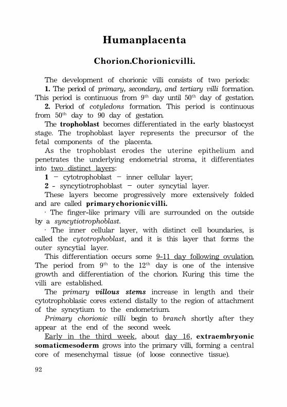
92
H u m a n p l a c e n t a
C h o r i o n . C h o r i o n i c v i l l i .
The development of chorionic villi consists of two periods:1. The period of primary, secondary, and tertiary villi formation.
This period is continuous from 9th day until 50th day of gestation.2. Period of cotyledons formation. This period is continuous
from 50th day to 90 day of gestation.The trophoblast becomes differentiated in the early blastocyst
stage. The trophoblast layer represents the precursor of thefetal components of the placenta.
As the trophoblast erodes the uterine epithelium andpenetrates the underlying endometrial stroma, it differentiatesinto two distinct layers:
1 – cytotrophoblast – inner cellular layer;2 - syncytiotrophoblast – outer syncytial layer.These layers become progressively more extensively folded
and are called p r i m a r y c h o r i o n i c v i l l i .· The finger-like primary villi are surrounded on the outside
by a syncytiotrophoblast.· The inner cellular layer, with distinct cell boundaries, is
called the cytotrophoblast, and it is this layer that forms theouter syncytial layer.
This differentiation occurs some 9-11 day following ovulation.The period from 9th to the 12th day is one of the intensivegrowth and differentiation of the chorion. During this time thevilli are established.
The primary villous stems increase in length and theircytotrophoblasic cores extend distally to the region of attachmentof the syncytium to the endometrium.
Primary chorionic villi begin to branch shortly after theyappear at the end of the second week.
Early in the third week, about day 16, e x t r a e m b r y o n i cs o m a t i c m e s o d e r m grows into the primary villi, forming a centralcore of mesenchymal tissue (of loose connective tissue).
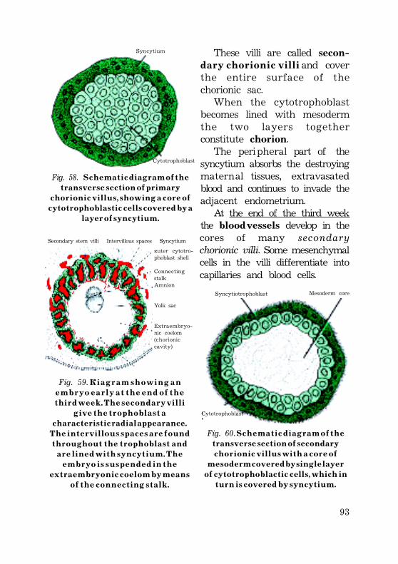
93
Syncytium
Cytotrophoblast
Fig. 58. S c h e m a t i c d i a g r a m o f t h et r a n s v e r s e s e c t i o n o f p r i m a r y
c h o r i o n i c v i l l u s , s h o w i n g a c o r e o fc y t o t r o p h o b l a s t i c c e l l s c o v e r e d b y a
l a y e r o f s y n c y t i u m .
These villi are called secon-d a r y c h o r i o n i c v i l l i and coverthe entire surface of thechorionic sac.
When the cytotrophoblastbecomes lined with mesodermthe two layers togetherconstitute chorion.
The peripheral part of thesyncytium absorbs the destroyingmaternal tissues, extravasatedblood and continues to invade theadjacent endometrium.
At the end of the third weekthe blood vessels develop in thecores of many secondarychorionic villi. Some mesenchymalcells in the villi differentiate intocapillaries and blood cells.
Secondary stem villi Intervillous spaces SyncytiumOuter cytotro-phoblast shell
ConnectingstalkAmnion
Yolk sac
Extraembryo-nic coelom(chorioniccavity)
Fig. 59. D i a g r a m s h o w i n g a ne m b r y o e a r l y a t t h e e n d o f t h e
t h i r d w e e k . T h e s e c o n d a r y v i l l ig i v e t h e t r o p h o b l a s t a
c h a r a c t e r i s t i c r a d i a l a p p e a r a n c e .T h e i n t e r v i l l o u s s p a c e s a r e f o u n d
t h r o u g h o u t t h e t r o p h o b l a s t a n da r e l i n e d w i t h s y n c y t i u m . T h e
e m b r y o i s s u s p e n d e d i n t h ee x t r a e m b r y o n i c c o e l o m b y m e a n s
o f t h e c o n n e c t i n g s t a l k .
Mesoderm coreSyncytiotrophoblast
Cytotrophoblast
Fig. 60. S c h e m a t i c d i a g r a m o f t h et r a n s v e r s e s e c t i o n o f s e c o n d a r y
c h o r i o n i c v i l l u s w i t h a c o r e o fm e s o d e r m c o v e r e d b y s i n g l e l a y e r
o f c y t o t r o p h o b l a c t i c c e l l s , w h i c h i nt u r n i s c o v e r e d b y s y n c y t i u m .
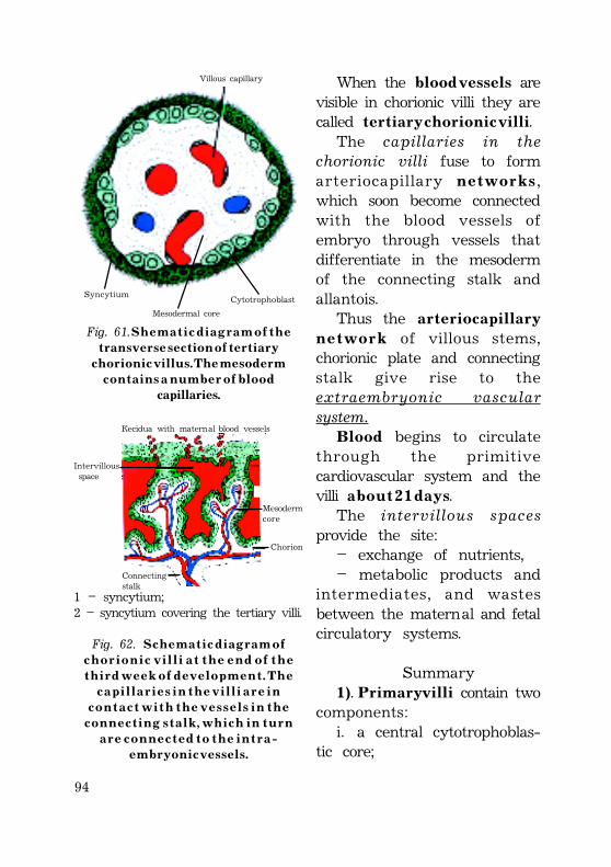
94
When the b l o o d v e s s e l s arevisible in chorionic villi they arecalled t e r t i a r y c h o r i o n i c v i l l i.
The capillaries in thechorionic villi fuse to formarteriocapillary n e t w o r k s,which soon become connectedwith the blood vessels ofembryo through vessels thatdifferentiate in the mesodermof the connecting stalk andallantois.
Thus the a r t e r i o c a p i l l a r yn e t w o r k of villous stems,chorionic plate and connectingstalk give rise to theextraembryonic vascularsystem.
B l o o d begins to circulatethrough the primitivecardiovascular system and thevilli a b o u t 2 1 d a y s.
The intervillous spacesprovide the site:
– exchange of nutrients,– metabolic products and
intermediates, and wastesbetween the maternal and fetalcirculatory systems.
Summary1). P r i m a r y v i l l i contain two
components:i. a central cytotrophoblas-
tic core;
Cytotrophoblast
Villous capillary
Syncytium
Mesodermal core
Fig. 61. S h e m a t i c d i a g r a m o f t h et r a n s v e r s e s e c t i o n o f t e r t i a r y
c h o r i o n i c v i l l u s . T h e m e s o d e r mc o n t a i n s a n u m b e r o f b l o o d
capillaries.
Decidua with maternal blood vessels
Intervillous space
Mesodermcore
Chorion
Connectingstalk
1 – syncytium;2 – syncytium covering the tertiary villi.
Fig. 62. S c h e m a t i c d i a g r a m o fc h o r i o n i c v i l l i a t t h e e n d o f t h e
t h i r d w e e k o f d e v e l o p m e n t . T h ec a p i l l a r i e s i n t h e v i l l i a r e i n
c o n t a c t w i t h t h e v e s s e l s i n t h ec o n n e c t i n g s t a l k , w h i c h i n t u r n
a r e c o n n e c t e d t o t h e i n t r a -e m b r y o n i c v e s s e l s .
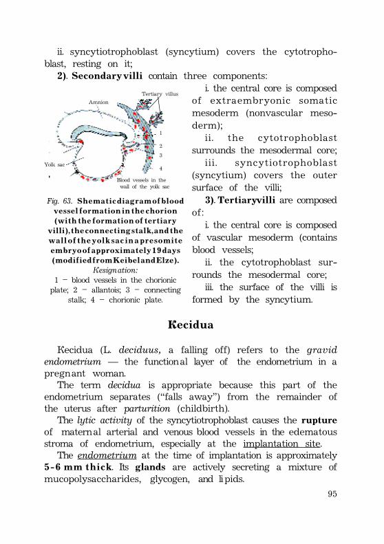
95
ii. syncytiotrophoblast (syncytium) covers the cytotropho-blast, resting on it;
2). S e c o n d a r y v i l l i contain three components:i. the central core is composed
of extraembryonic somaticmesoderm (nonvascular meso-derm);
ii. the cytotrophoblastsurrounds the mesodermal core;
iii. syncytiotrophoblast(syncytium) covers the outersurface of the villi;
3). Tertiary villi are composedof:
i. the central core is composedof vascular mesoderm (containsblood vessels;
ii. the cytotrophoblast sur-rounds the mesodermal core;
iii. the surface of the villi isformed by the syncytium.
Decidua
Decidua (L. deciduus, a falling off) refers to the gravidendometrium — the functional layer of the endometrium in apregnant woman.
The term decidua is appropriate because this part of theendometrium separates (“falls away”) from the remainder ofthe uterus after parturition (childbirth).
The lytic activity of the syncytiotrophoblast causes the ruptureof maternal arterial and venous blood vessels in the edematousstroma of endometrium, especially at the implantation site.
The endometrium at the time of implantation is approximately5 - 6 m m t h i c k. Its glands are actively secreting a mixture ofmucopolysaccharides, glycogen, and lipids.
Tertiary villus
Amnion
1
2
3
4
Blood vessels in the wall of the yolk sac
Yolk sac
Fig. 63. S h e m a t i c d i a g r a m o f b l o o dv e s s e l f o r m a t i o n i n t h e c h o r i o n
( w i t h t h e f o r m a t i o n o f t e r t i a r yv i l l i ) , t h e c o n n e c t i n g s t a l k , a n d t h e
w a l l o f t h e y o l k s a c i n a p r e s o m i t ee m b r y o o f a p p r o x i m a t e l y 1 9 d a y s
( m o d i f i e d f r o m K e i b e l a n d E l z e ) .Designation:
1 – blood vessels in the chorionicplate; 2 – allantois; 3 – connecting
stalk; 4 – chorionic plate.
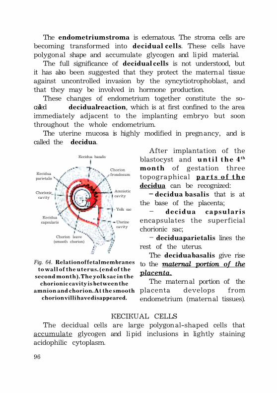
96
The endometrium stroma is edematous. The stroma cells arebecoming transformed into d e c i d u a l c e l l s. These cells havepolygonal shape and accumulate glycogen and lipid material.
The full significance of d e c i d u a l c e l l s is not understood, butit has also been suggested that they protect the maternal tissueagainst uncontrolled invasion by the syncytiotrophoblast, andthat they may be involved in hormone production.
These changes of endometrium together constitute the so-called decidual reaction, which is at first confined to the areaimmediately adjacent to the implanting embryo but soonthroughout the whole endometrium.
The uterine mucosa is highly modified in pregnancy, and iscalled the decidua.
After implantation of theblastocyst and u n t i l t h e 4 t h
m o n t h of gestation threetopographical p a r t s o f t h edecidua can be recognized:
– d e c i d u a b a s a l i s that is atthe base of the placenta;
– d e c i d u a c a p s u l a r i sencapsulates the superficialchorionic sac;
– decidua parietalis lines therest of the uterus.
The d e c i d u a b a s a l i s give riseto the maternal portion of theplacenta.
The maternal portion of theplacenta develops fromendometrium (maternal tissues).
DECIDUAL CELLSThe decidual cells are large polygonal-shaped cells that
accumulate glycogen and lipid inclusions in lightly stainingacidophilic cytoplasm.
Decidua basalis
Chorionfrondosum
Amnioticcavity
Yolk sac
Uterinecavity
Chorion leave(smooth chorion)
Deciduacapsularis
Deciduaparietalis
Chorioniccavity
Fig. 64. R e l a t i o n o f f e t a l m e m b r a n e st o w a l l o f t h e u t e r u s . ( e n d o f t h e
s e c o n d m o n t h ) . T h e y o l k s a c i n t h ec h o r i o n i c c a v i t y i s b e t w e e n t h e
a m n i o n a n d c h o r i o n . A t t h e s m o o t hc h o r i o n v i l l i h a v e d i s a p p e a r e d .
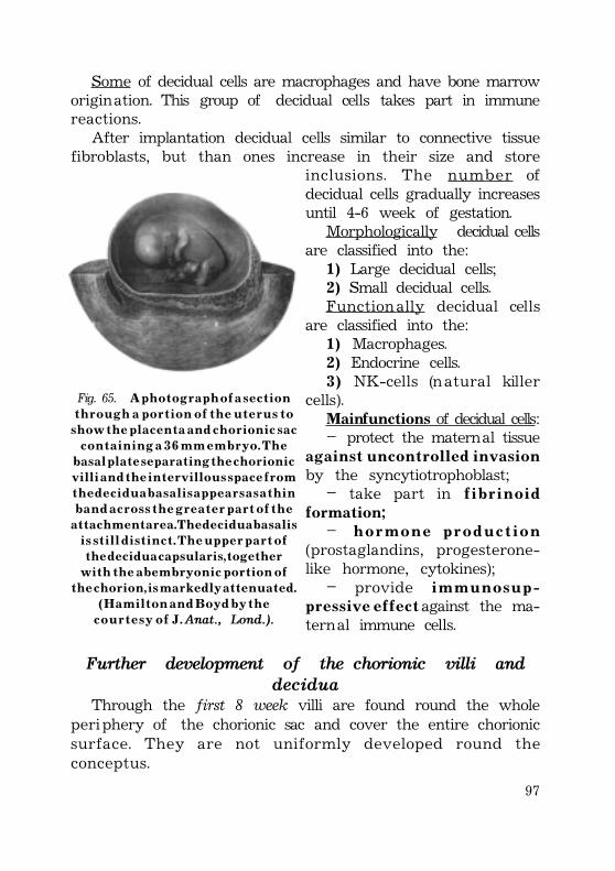
97
Some of decidual cells are macrophages and have bone marroworigination. This group of decidual cells takes part in immunereactions.
After implantation decidual cells similar to connective tissuefibroblasts, but than ones increase in their size and store
inclusions. The number ofdecidual cells gradually increasesuntil 4-6 week of gestation.
Morphologically decidual cellsare classified into the:
1) Large decidual cells;2) Small decidual cells.Functionally decidual cells
are classified into the:1) Macrophages.2) Endocrine cells.3 ) NK-cells (natural killer
cells).Main functions of decidual cells:– protect the maternal tissue
a g a i n s t u n c o n t r o l l e d i n v a s i o nby the syncytiotrophoblast;
– take part in f i b r i n o i dformation;
– h o r m o n e p r o d u c t i o n(prostaglandins, progesterone-like hormone, cytokines);
– provide i m m u n o s u p -p r e s s i v e e f f e c t against the ma-ternal immune cells.
Further development of the chorionic villi anddecidua
Through the first 8 week villi are found round the wholeperiphery of the chorionic sac and cover the entire chorionicsurface. They are not uniformly developed round theconceptus.
Fig. 65. A p h o t o g r a p h o f a s e c t i o nt h r o u g h a p o r t i o n o f t h e u t e r u s t o
s h o w t h e p l a c e n t a a n d c h o r i o n i c s a cc o n t a i n i n g a 3 6 m m e m b r y o . T h e
b a s a l p l a t e s e p a r a t i n g t h e c h o r i o n i cv i l l i a n d t h e i n t e r v i l l o u s s p a c e f r o m
t h e d e c i d u a b a s a l i s a p p e a r s a s a t h i nb a n d a c r o s s t h e g r e a t e r p a r t o f t h e
a t t a c h m e n t a r e a . T h e d e c i d u a b a s a l i si s s t i l l d i s t i n c t . T h e u p p e r p a r t o f
t h e d e c i d u a c a p s u l a r i s , t o g e t h e rw i t h t h e a b e m b r y o n i c p o r t i o n o f
t h e c h o r i o n , i s m a r k e d l y a t t e n u a t e d .( H a m i l t o n a n d B o y d b y t h e
c o u r t e s y o f J . Anat., Lond.).
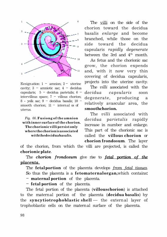
98
The villi on the side of thechorion toward the deciduabasalis enlarge and becomebranched, while those on theside toward the deciduacapsularis rapidly degeneratebetween the 3rd and 4th month.
As fetus and the chorionic sacgrow, the chorion expendsand, with it now very thincovering of decidua capsularis,projects into the uterine cavity.
The villi associated with thedecidua capsularis soondegenerate, producing arelatively avascular area, thes m o o t h c h o r i o n .
The villi associated withdecidua parietalis rapidlyincrease in number and enlarge.This part of the chorionic sac iscalled the v i l l o u s c h o r i o n orc h o r i o n f r o n d o s u m. The layer
of the chorion, from which the villi are projected, is called thec h o r i o n i c p l a t e.
The chorion frondosum give rise to fetal portion of theplacenta.
The f e t a l p o r t i o n of the placenta develops from fetal tissues.So thus the placenta is a f e t o m a t e r n a l o r g a n , which contains:– m a t e r n a l p o r t i o n of the placenta.– f e t a l p o r t i o n of the placenta.The fetal portion of the placenta (v i l l o u s c h o r i o n) is attached
to the maternal portion of the placenta (d e c i d u a b a s a l i s) bythe s y n c y t i o t r o p h o b l a s t i c s h e l l — the external layer oftrophoblastic cells on the maternal surface of the placenta.
1
2
3
4
5
6
7
8
9
10
11
Designation: 1 – amnion; 2 – uterinecavity; 3 – amniotic sac; 4 – deciduacapsularis; 5 – decidua parietalis; 6 –intervillous space; 7 – villous chorion;8 - yolk sac; 9 – decidua basalis; 10 –smooth chorion; 11 – internal os ofuterus.
Fig. 66. F u s i o n g o f t h e a m n i o nw i t h i n n e r s u r f a c e o f t h e c h o r i o n .
T h e c h o r i o n i c v i l l i p e r s i s t o n l yw h e r e t h e c h o r i o n i s a s s o c i a t e d
w i t h t h e d e c i d u a b a s a l i s .
99
The chorionic villi attach firmly to the decidua basalis throughthe syncytotropho-blastic shell and anchor the c h o r i o n i c s a c tothe decidua basalis.
Villi that attach to the maternal tissues through thecytotrophoblastic shell are s t e m v i l l i (anchoring villi).
The villi that grow from the sides of the stem villi are branchvilli (terminal villi).
At is through the walls of the b r a n c h v i l l i that the mainexchange of material between the blood of the mother andthe embryo takes place.
The b r a n c h v i l l i are bathed in continually changing maternalblood in the intervillous space.
The d e c i d u a c a p s u l a r i s disappears by degeneration. Thesmooth chorion intimately related to the decidua parietalis.
· At 4 , 5 m o n t h s the s m o o t h c h o r i o n consists of several layersof cytotrophoblastic cells only.
The surface epithelium of the decidua parietalis soondisappears and its stroma is intimately fused with cytotrophoblastof the smooth chorion.
· By the f i f t h m o n t h, the two layers fuse and the uterinelumen is obliterated.
· At this time the amnion is also fuses with the inside of thechorion.
The increased t h i c k n e s s o f t h e p l a c e n t a is due to growth insize and length of the villi of the chorion frondosum, and is notcaused by further penetration into maternal tissues.
Until the end of the fourth month the normal placenta growsboth in thickness and circumference.
After this period there is no increase in thickness but theplacenta continues to grow circumferentially until near the endof pregnancy.
As the placenta continues to mature during the 4-5 monthsthe number of s t e m v i l l i increases. The villous trees grow intothe intervillous blood lakes.
The intervillous space, containing maternal blood, graduallybecomes subdivided by the formation of septa, which project
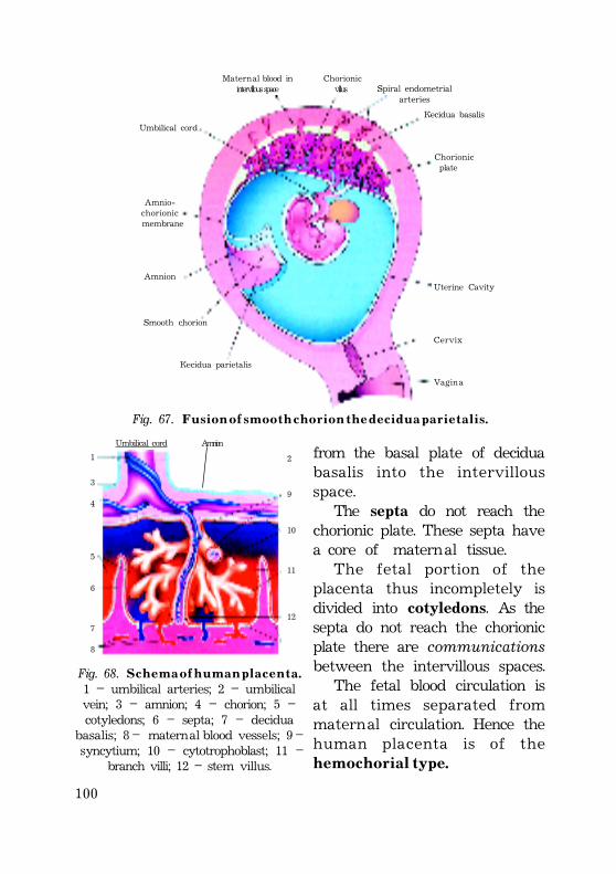
100
Maternal blood in Chorionicintervillous space villus
Decidua basalis
Spiral endometrialarteries
Chorionic plate
Uterine Cavity
Cervix
Vagina
Decidua parietalis
Smooth chorion
Amnio-chorionic membrane
Amnion
Umbilical cord
Fig. 67. F u s i o n o f s m o o t h c h o r i o n t h e d e c i d u a p a r i e t a l i s .
Umbilical cord Amnion
1
3
4
5
6
7
8
2
9
10
11
12
Fig. 68. S c h e m a o f h u m a n p l a c e n t a .1 – umbilical arteries; 2 – umbilicalvein; 3 – amnion; 4 – chorion; 5 –cotyledons; 6 – septa; 7 – decidua
basalis; 8 – maternal blood vessels; 9 – syncytium; 10 – cytotrophoblast; 11 –
branch villi; 12 – stem villus.
from the basal plate of deciduabasalis into the intervillousspace.
The septa do not reach thechorionic plate. These septa havea core of maternal tissue.
The fetal portion of theplacenta thus incompletely isdivided into cotyledons. As thesepta do not reach the chorionicplate there are communicationsbetween the intervillous spaces.
The fetal blood circulation isat all times separated frommaternal circulation. Hence thehuman placenta is of thehemochorial type.
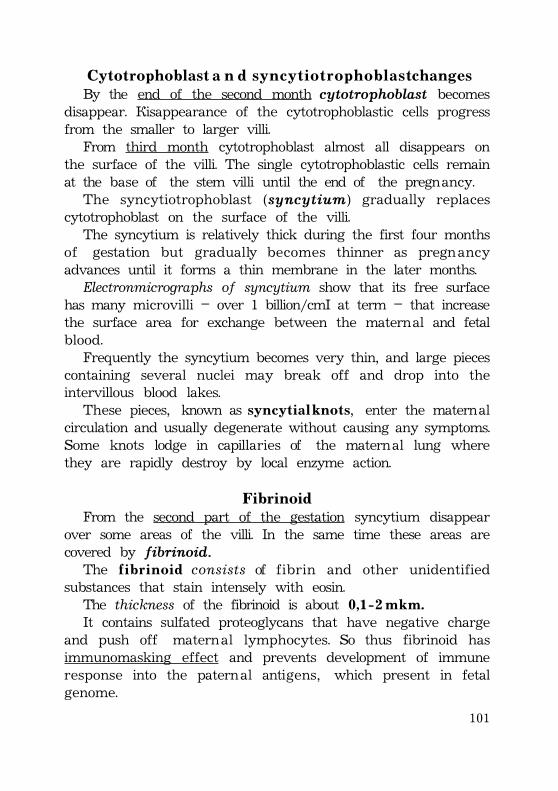
101
Cytotrophoblast a n d s y n c y t i o t r o p h o b l a s t changesBy the end of the second month cytotrophoblast becomes
disappear. Disappearance of the cytotrophoblastic cells progressfrom the smaller to larger villi.
From third month cytotrophoblast almost all disappears onthe surface of the villi. The single cytotrophoblastic cells remainat the base of the stem villi until the end of the pregnancy.
The syncytiotrophoblast (syncytium) gradually replacescytotrophoblast on the surface of the villi.
The syncytium is relatively thick during the first four monthsof gestation but gradually becomes thinner as pregnancyadvances until it forms a thin membrane in the later months.
Electronmicrographs of syncytium show that its free surfacehas many microvilli – over 1 billion/cm² at term – that increasethe surface area for exchange between the maternal and fetalblood.
Frequently the syncytium becomes very thin, and large piecescontaining several nuclei may break off and drop into theintervillous blood lakes.
These pieces, known as s y n c y t i a l k n o t s, enter the maternalcirculation and usually degenerate without causing any symptoms.Some knots lodge in capillaries of the maternal lung wherethey are rapidly destroy by local enzyme action.
FibrinoidFrom the second part of the gestation syncytium disappear
over some areas of the villi. In the same time these areas arecovered by fibrinoid.
The f i b r i n o i d consists of fibrin and other unidentifiedsubstances that stain intensely with eosin.
The thickness of the fibrinoid is about 0 , 1 - 2 m k m .It contains sulfated proteoglycans that have negative charge
and push off maternal lymphocytes. So thus fibrinoid hasimmunomasking effect and prevents development of immuneresponse into the paternal antigens, which present in fetalgenome.
102
Some authors suppose that fibrinoid is destroying materialof syncytium and decidual cells. It main function is to preventthe invasion of syncytium into the maternal organism.
F u l l t e r m p l a c e n t a
The human placenta is classified as:· h e m o c h o r i a l because of the nature of the placental
membrane;· deciduate because maternal decidua is shed at birth along
with the fetal placenta;· villous because of its villi· discoidal because of its circular shape.The fully developed placenta covers 1 5 t o 3 0 % of the decidua
and weighs about one-sixth that of the fetus.At the full term the human placenta has the shape of a
f l a t t e n e d c a k e (plakuos=placenta=cake).It has a d i a m e t e r o f 1 5 - 1 6 c m (6-7 inches) and a thickness is
about 3 cm.Its weight is about 5 0 0 - 6 0 0 g r a m s. During the second half of
pregnancy placenta increases in weight much less rapidly thanthe fetus.
The placenta is a f e t o m a t e r n a l o r g a n , which contains:a . maternal portionb . f e t a l p o r t i o n
F e t a l p o r t i o n o f t h e p l a c e n t aThe fetal portion of the human placenta at full term contains:– about 15-20 large cotyledons,– about 40-50 small cotyledons,– until 150 rudimental cotyledons.Each cotyledon (placental lobes) consists of two or more main
s t e m v i l l i and many b r a n c h v i l l i that are incompletelly separatedby matenal septa.
The s t e r m v i l l i (synonym – a n c h o r i n g v i l l i ) give rise tonumerous highly branched t e r m i n a l v i l li (syn.: b r a n c h or freevilli). The chorionic villi originate from the chorionic plate.
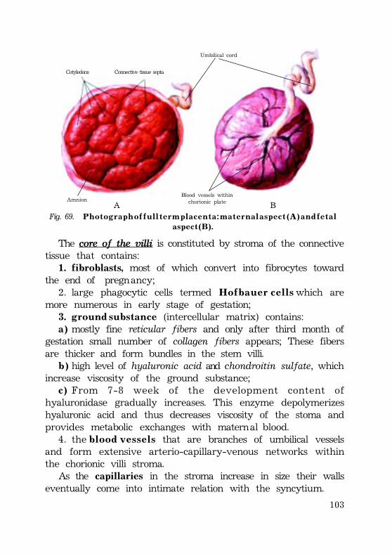
103
Cotyledons Connective tissue septa
Umbilical cord
AmnionBlood vessels within
chorionic plateA BFig. 69. P h o t o g r a p h o f f u l l t e r m p l a c e n t a : m a t e r n a l a s p e c t ( A ) a n d f e t a l
a s p e c t ( B ) .
The core of the villi is constituted by stroma of the connectivetissue that contains:
1 . fibroblasts, most of which convert into fibrocytes towardthe end of pregnancy;
2. large phagocytic cells termed H o f b a u e r c e l l s which aremore numerous in early stage of gestation;
3 . g r o u n d s u b s t a n c e (intercellular matrix) contains:a ) mostly fine reticular fibers and only after third month of
gestation small number of collagen fibers appears; These fibersare thicker and form bundles in the stem villi.
b ) high level of hyaluronic acid and chondroitin sulfate, whichincrease viscosity of the ground substance;
c ) From 7-8 week of the development content ofhyaluronidase gradually increases. This enzyme depolymerizeshyaluronic acid and thus decreases viscosity of the stoma andprovides metabolic exchanges with maternal blood.
4. the b l o o d v e s s e l s that are branches of umbilical vesselsand form extensive arterio-capillary-venous networks withinthe chorionic villi stroma.
As the capillaries in the stroma increase in size their wallseventually come into intimate relation with the syncytium.

104
The capillaries approachthe surface of the villi butare always separated fromthe basement membrane ofthe trophoblast by somedelicate connective tissue.
· B a s a m e n t m e m b r a n eseparates connective tissuecore and persisting cytotro-phoblast cells that remainnear the base of the stemvilli
· Thin layer of syncytiumcovers the villi.
· F i b r i n o i d forms theoutermost layer of the villi.
T h e c h o r i o n i c p l a t e iscovered:
1) on its fetal aspect bythe a m n i o t i c e p i t h e l i u m,followed by a connectivetissue layer carring the mainbranches of the umbilicalvessels. The connective tissuelayer is derived from fusionbetween the mesoderma-covered surface of theamnion and chorion and ismore fibrous.
2) on its maternal aspecta diminishing layer of cytotrophoblast with thin layer ofsyncytium.
Fetal portion changes at the end of pregnancyAt the end of pregnancy changes of fetal placenta include:a. an increase in fibrous tissue in the core of the villi;
Endothelium of fetal capillary
Connectivetissue coreof villus
Placentalmembrane
Hofbauercells
Cytotrophoblast
Syncytium
A
B
Syncytial knot
Oxygen-rich fetalblood
Fibrinoid
Oxygen-poorblood in fetalcapillary
Placentalbarrier
Persistingcytotrophoblastcells
Fig. 70. S h e m a t i c d i a g r a m o f s e c t i o n st h r o u g h a b r a n c h v i l l u s a t 1 0 w e e k s
( A ) a n d f u l l t e r m ( B ) . T h e p l a c e n t a lb a r r i e r s e p a r a t e s t h e m a t e r n a l b l o o d i n
t h e m a t e r n a l l a c u n a e f r o m t h e f e t a lb l o o d i n t h e c a p i l l a r i e s i n t h e v i l l i . A t
f u l l t e r m p l a c e n t a l b a r r i e r b e c o m e sv e r y t h i n . H o f b a u e r c e l l s a r e
p h a g o c y t i c c e l l s .
105
b. thickening of basement membranes in fetal capillaries;c. obliterative changes in small capillaries of the villi;d. deposition of fibrinoid on the surface of the villi in the
junctional zone and in the chorionic plate.
M a t e r n a l p o r t i o n o f t h e p l a c e n t aThe maternal portion of the placenta is derived from the
endometrium.At full term maternal portion of the human placenta contains:– basal plate– septa– lacunae (or intervillous spaces) with maternal blood– junctional zoneBy the end of the 4th m o n t h the decidua basalis is replaced by
the fetal part of the placenta. The chorionic villi invade and erodethe decidua basalis, thus intervillous spaces develop and enlarge.
The intervillous spaces or lacunae are filled with maternalblood.
The deepest part of the decidua basalis, which forms thefloor of the lacunae, is called the b a s a l p l a t e (or d e c i d u a l p l a t e).It contains opening of the destroying maternal blood vessels.
The R o h r ’ s s t r i a o f fibrinoid is amorphous extracellularmaterial. It covers the surface of basal plate, and preventsdevelopment of immune response.
Between the intervillous spaces are located projections ofdecidua, which are called p l a c e n t a l s e p t a .
T h e p l a c e n t a l s e p t a project toward the c h o r i o n i c p l a t e , buts e p t a do not reach the chorionic plate. The septa incompletelydivide the fetal portion of the placenta into cotyledons. Thereforethe intervillous spaces with the matern al blood arecommunicated.
The j u n c t i o n a l z o n e is located at the periphery of the placenta,near the boundary between the villous chorion and smoothchorion. The junctional zone is the zone of strong attachment offetal and maternal portions of the placenta. The junctional zoneprevents blood outpouring from the maternal lacunae.
106
The fetal portion of the placenta (c h o r i o n i c v i l l i ) attachesfirmly to the maternal portion of the placenta (basalis plate)through the cytotrophoblastic shell and anchor the chorionics a c to the decidua basalis.
Changes at the end of pregnancyThroughout the second half of pregnancy the basal plate is
thinned and becomes progressively modified. These changes include:i. a relative diminution of the decidual elements; The glands
disappear after 6th month in the deeper portion of the deciduabasalis.
ii. an increasing deposition of fibrinoid;iii. admixture of fetal and maternal derivatives.
P l a c e n t a l b a r r i e r(placental membrane)
The p l a c e n t a l b a r r i e r is called also the placental membrane.The placental barrier separates maternal and fetal blood. It
contains the extrafetal tissues separating the maternal and fetalblood.
The placental barrier is composed of four layers:1. the endothelium lining the fetal vessels;2. the connective tissue in the villus core;3. the diminished cytotrophoblastic layer, which lies on the
trophoblast basement membrane;4. the syncytium;5. the fibrinoid.Eventually cytotrophoblast cells disappear over large areas
of the villi leaving only thin patches of syncytium. As a result,the placental barrier consists of four layers (1 , 2 , 4 , 5).
As pregnancy advances, the placental barrier b e c o m e sprogressively thinner and fetal capillaries are located near thebasement membrane of syncytium. So that blood in many fetalcapillaries is extremely close to the maternal blood in thematernal lacunae.
The placental barrier is not a true barrier, since manysubstances pass through it freely.
107
Substances with a molecular weight of 600 to 1000 tend toeasily cross the placental barrier.
Some metabolites, hormones, drugs, and toxins, through presentin the maternal blood, do not pass through the placental barrier.
* Hormones.1) Most maternal hormones do not cross the placenta. The
hormones that do cross, such as thyroxine, do so only at a slow rate.2 ) Testosterone and certain synthetic progestins rapidly cross
the placenta and may cause masculinization of female fetusesin some cases.
3) Even more dangerous was the use of the synthetic estrogendiethylstilbestrol, which easily crosses the placenta. Thiscompound produced carcinoma of the vagina and abnormalitiesof the testes in individuals who were exposed to it during theirintrauterine life
*Infectious Agents.a) Viruses. Cytomegalovirus, rubella and coxsackie viruses,
and viruses associated with variola, varicella, measles, andpoliomyelitis may pass through the placental membrane andcause fetal infection. In some cases, such as the r u b e l l a v i r u s ,congenital anomalies, such as cataracts, may be produced.b) Microorganisms. Microorganisms such as Treponema
pallidum that causes syphilis and Tbxoplasma gondii thatproduces destructive changes in the brain and eyes cross theplacental membrane. Toxoplasma gondii infect the placenta bycreating lesions and then cross the placental membrane throughthe defects that are created.
These organisms enter the fetal blood, often causing congenitalanomalies and/or death of the embryo or fetus.
* Drugs and Drug Metabolites.Most drugs and drug metabolites cross the placenta by simple
diffusion.1) Some drugs cause major congenital anomalies. F e t a l d r u g
addiction may occur after maternal use of drugs such as heroinand 50 to 75% of these newborns experience withdrawal symptoms.
Because psychic dependence on these drugs is notdeveloped during the fetal period, no liability to subsequent
108
n a r c o t i c a d d i c t i o n exists in the infant after withdrawal iscomplete.
2) All s e d a t i v e s and a n a l g e s i c s affect the fetus to some degree.Drugs taken by the mother can affect the embryo/fetus directlyor indirectly by interfering with maternal or placental metabolism.
The amount of drug or metabolite reaching the placenta iscontrolled by the maternal blood level and blood flow throughthe placenta.
S u b s t a n c e s t h a t c r o s s t h e p l a c e n t a l b a r r i e r
Beneficia substances
Harmful substances
Designation: *Cross at a very low rate; most fetal serum pby the fetus.
+In general, most drugs and their metabolites cross the placental membrane.
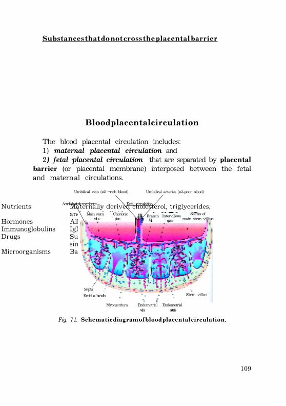
109
S u b s t a n c e s t h a t d o n o t c r o s s t h e p l a c e n t a l b a r r i e r
Nutrients Maternally derived cholesterol, triglycerides, and phospholipids
Hormones All protein hormones (including insulin) Immunoglobulins IgM, IgS Drugs Succinylcholine, curare, heparin, drugs
similar to amino acids (methyldopa) Microorganisms Bacteria
B l o o d p l a c e n t a l c i r c u l a t i o n
The blood placental circulation includes:1) maternal placental circulation and2) fetal placental circulation that are separated by placental
barrier (or placental membrane) interposed between the fetaland maternal circulations.
Main stem Chorionicvillus plate
Branch Intervillous Stamn ofmain stem villiusVilli space
Umbilical vein (O2 –rich blood) Umbilical arteries (O2-poor blood)
Amniochorionic membrane Fetal circulation
Septa Decidua basalis
Myometrium Endometrial Endometrial veins arteries
Stem villus
Fig. 71. S c h e m a t i c d i a g r a m o f b l o o d p l a c e n t a l c i r c u l a t i o n .
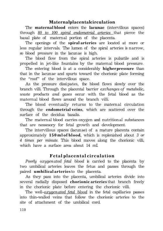
110
M a t e r n a l p l a c e n t a l c i r c u l a t i o nThe m a t e r n a l b l o o d enters the lacunae (intervillous spaces)
through 80 to 100 spiral endometrial arteries that pierce thebasal plate of maternal portion of the placenta.
The openings of the s p i r a l a r t e r i e s are located at more orless regular intervals. The lumen of the spiral arteries is narrow,so blood pressure in the lacunae is high.
The blood flow from the spiral arteries is pulsatile and ispropelled in jet-like fountains by the maternal blood pressure.
The entering blood is at a considerably h i g h e r p r e s s u r e thanthat in the lacunae and spurts toward the chorionic plate formingthe “roof” of the intervillous space.
As the pressure dissipates, the blood flows slowly over thebranch villi. Through the placental barrier exchanges of metabolic,waste products and gases occur with the fetal blood as thematernal blood flows around the branch villi.
The blood eventually returns to the maternal circulationthrough the endometrial veins, which are scattered over thesurface of the decidua basalis.
The maternal blood carries oxygen and nutritional substancesthat are nessesory for fetal growth and development.
The intervillous spaces (lacunae) of a mature placenta containapproximately 1 5 0 m l o f b l o o d, which is replenished about 3 or4 times per minute. This blood moves along the chorionic villi,which have a surface area about 14 m².
F e t a l p l a c e n t a l c i r c u l a t i o nPoorly oxygenated fetal blood is carried to the placenta by
two umbilical arteries leaves the fetus and passes through thepaired u m b i l i c a l a r t e r i e s to the placenta.
As they pass into the placenta, umbilical arteries divide intoseveral radially disposed c h o r i o n i c a r t e r i e s that branch freelyin the chorionic plate before entering the chorionic villi.
The well-oxygenated fetal blood in the fetal capillaries passesinto thin-walled veins that follow the chorionic arteries to thesite of attachment of the umbilical cord.
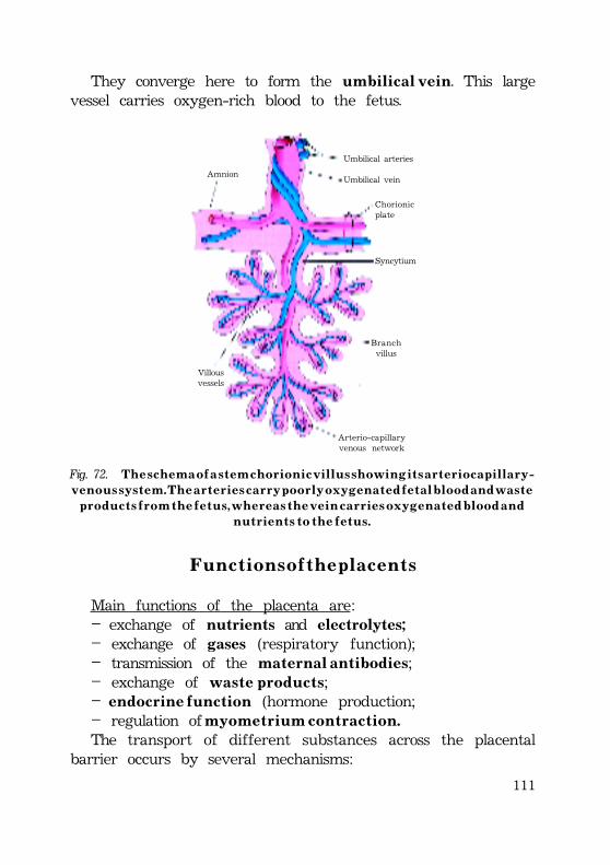
111
They converge here to form the u m b i l i c a l v e i n. This largevessel carries oxygen-rich blood to the fetus.
Fig. 72. T h e s c h e m a o f a s t e m c h o r i o n i c v i l l u s s h o w i n g i t s a r t e r i o c a p i l l a r y -v e n o u s s y s t e m . T h e a r t e r i e s c a r r y p o o r l y o x y g e n a t e d f e t a l b l o o d a n d w a s t e
p r o d u c t s f r o m t h e f e t u s , w h e r e a s t h e v e i n c a r r i e s o x y g e n a t e d b l o o d a n dn u t r i e n t s t o t h e f e t u s .
Umbilical arteries
Umbilical vein
Chorionicplate
Syncytium
Branchvillus
Arterio-capillaryvenous network
Villousvessels
Amnion
F u n c t i o n s o f t h e p l a c e n t s
Main functions of the placenta are:– exchange of nutrients and electrolytes;– exchange of gases (respiratory function);– transmission of the maternal antibodies;– exchange of w a s t e p r o d u c t s;– endocrine function (hormone production;– regulation of m y o m e t r i u m c o n t r a c t i o n .The transport of different substances across the placental
barrier occurs by several mechanisms:
112
simple diffusionfacilitated diffusionactive transportpinocytosisNormally, carbon dioxide, metabolic waste products, water,
hormones are transferred from the fetal blood to maternal blood.Oxygen, nutrients, vitamins, some maternal antibodies,
hormones, metabolites, water pass in the opposite direction.
E x c h a n g e o f n u t r i e n t s a n d e l e c t r o l y t e sExchange of nutrients and electrolytes is rapid and increases
as pregnancy advances.The placenta, especially during early pregnancy, synthesizes
glycogen, cholesterol, and f a t t y a c i d s, which serve as sourcesof nutrients and energy for the embryo/fetus. The fetal portionof the placenta is the site of synthesis and storage
A m i n o a c i d s are actively transported and are essential forfetal growth. The concentration of most amino acids is higher infetal plasma than ones in maternal blood.
G l u c o s e is quickly transferred to the embryo/fetus bydiffusion from maternal blood.
Vitamins cross the placental membrane and are essential fornormal development. Water soluble vitamins cross the placentalmembrane more quickly than fat-soluble ones.
The transport of maternal lipid substances (cholesterol, trigly-cerides, phospholipids, and fatty acids) is relatively small or no.
Electrolytes are freely exchanged. Water is rapidly exchangedby simple diffusion.
E x c h a n g e o f g a s e sThe respiratory function of the placenta approaches to one
of adult respiratory system.O x y g e n, c a r b o n d i o x i d e, and c a r b o n m o n o x i d e cross the
placental membrane by simple diffusion.The oxygen that is joined to the hemoglobin in maternal
blood crosses the placental barrier by diffusion. Than in fetal
113
blood oxygen binds to fetal hemoglobin and is carried to theembryo/fetus with the fetal blood (umbilical vein).
The umbilical arteries contain poorly oxygenated fetal bloodwith higher concentration of c a r b o n d i o x i d e than highlyoxygenated maternal blood in maternal lacunae.
The c a r b o n d i o x i d e crosses the placental barrier by diffusionfrom fetal blood to maternal blood (lacunae) and carries tomaternal organism.
The exchange of oxygen and carbon dioxide is limited moreby blood flow than by the efficiency of diffusion.
At full term placenta the fetus extracts 2 0 t o 3 0 m l o f o x y g e np e r m i n u t e from maternal blood and even a short-terminterruption of the oxygen supply is fatal to the fetus.
T r a n s m i s s i o n o f t h e m a t e r n a l a n t i b o d i e sFetus immunological competence begins to develop late in
the first trimester of pregnancy.The fetus produces only small amounts of antibodies because
of its immature immune system.The passage of maternal antibodies across the placental
barrier confers some degree of p a s s i v e i m m u n i t y o n t h e f e t u s.Maternal antibodies, immunoglobu-lins G (IgG) begin to be
transported from the mother to fetus at approximately 14 weekby pinocytosis.
Maternal antibodies provide passive immunity against a varietyof infectious agents such as diphtheria, smallpox, and measles.
Newborns begin to produce their own IgG, but adult levelsare not attained until the age of 3 years.
T r a n s m i s s i o n o f t h e w a s t e p r o d u c t sFetal urea and uric acid pass through the placental barrier
by simple diffusion.
E n d o c r i n e f u n c t i o nThe syncytium is site of the placenta synthesizing of protein
and steroid hormones.
114
The p r o t e i n hormones are:¦ human chorionic gonadotropin (hCG)¦ human chorionic somatomammotropin (hCS), or human
placental lactogen (hPL)¦ human chorionic thyrotropin (hCT)¦ human chorionic corticotropin (hCACTH)The s t e r o i d hormones are:¦ estrogens (predominantly estriol);¦ progesterone.From the 4th month placenta is the major site of synthesis of
several hormones, which regulate fetal growth and development.Human c h o r i o n i c g o n a d o t r o p i n is first secreted by the
syncytium during the second week. During the first two monthsof pregnancy, the syncytium produces human c h o r i o n i c g o -nadotropin (hCG), which maintains the corpus luteum. Thishormone is excreted by the mother in the urine, and in theearly stages of gestation, its presence is used as an i n d i c a t o r o fpregnancy.
By the end of the 4th month the placenta produces progesteronein sufficient amounts to maintain pregnancy if the ovarian corpusluteum is removed or fails to function properly.
Progesterone can be obtained from the placenta at all stagesof gestation, indicating that it is essential for the maintenanceof pregnancy. The placenta forms progesterone from maternalcholesterol or pregnenolone.
The placenta produces increasing amounts of estrogens untiljust before the end of pregnancy, when a maximum level isreached. These high levels of estrogens stimulate uterine growthand development of the mammary glands.
Placental lactogen is a growth hormone-like substance thatgives the fetus priority on maternal blood glucose.
R e g u l a t i o n o f m y o m e t r i u m c o n t r a c t i o nThe fetal hypothalamus initiates the birth process.Several hormones trigger the initiation of uterine contractions
during the childbirth.
115
There are: – oxytocin– prostaglandins– estrogens– cortisol
The fetal hypothalamus secretes corticotropin-releasinghormone (CRH), which stimulates the anterior hypophysis toproduce adrenocorticotropin (ACTH). ACTH causes the secretionof cortisol from the suprarenal cortex. Cortisol is involved inthe synthesis of estrogens. These steroids stimulate uterinecontraction.
Peristaltic contractions of uterine smooth muscle are elicitedby o x y t o c i n , which is released by the posterior hypophysis. Thishormone is administered clinically when it is necessary to inducelabor. Oxytocin also stimulates release of p r o s t a g l a n d i n s fromthe decidua that stimulates myometrial contractility.
E s t r o g e n s also increase myometrial contractile activity andstimulate the release of oxytocin and prostaglandins.
Im m u n o l o g i c a l in t e r r e l a t i o n s : M a t e r n a l - f e t a lorganisms
Fetal organism contains 50% of the foreign antigens for thematernal organism.
Fetal portion of the placenta contains maternal and paternala n t i g e n s, but normally rejection of fetus by the mother’simmune system doesn’t occur.
P r o t e c t i v e m e c h a n i s m s i n c l u d e :A. Factors produced by placenta;B. Factors produced by maternal organism;C. Factors synthesized by embryo and fetus.
Factors produced by placenta;Syncytium produces several factors that block immune
reactions between fetal tissues of placenta and maternal tissues.Ø 1 . F i b r i n o i d has immunomasking effect and prevents
development of immune response into the paternal antigens,which present in fetal genome.
116
Fibrinoid contains sulfated proteoglycans and sialomucinogensthat have negative charge and push off maternal lymphocytes.
2 . Syncytium synthesizes p r o t e i n s which block maternalimmune system (for example, transferrin).
3. Syncytium looses ability to synthesis immunogenic antigens.Syncytium does not contain H L A - G antigens. Expression ofH L A - G is restricted to a few tissues including placental syncytiumcells.
Its strategic location in the placenta is believed to provide adual immunoprotective role:
1) evasion of T cell recognition owing to its nonpolymorphicnature, and
2) recognition by the “killer-inhibitory receptors” on NK cells,thus turning off their killer function.
4 . Syncytium produces and maintains high concentration ofh o r m o n e s that have immunosuppressive effect (chorionicgonadotropin, progesterone, estrogen, and cortisol-bindingglobulin).
5 . Syncytium synthesis lysine that destroys T-lymphocytesand NK-cells (natural killer cells) of maternal organism.
6. M a t e r n a l p o r t i o n of the placenta contains decidual cells,some of which synthesis proteins with immunosuppressive effect.
Factors produced by maternal organism.1. The endocrine system of maternal organism (suprarenal glands)
produces glucocorticoids, which have immunosuppressive effect.2. Through 6-72 h after fertilization maternal blood contains
f a c t o r o f e a r l y p r e g n a n c y . This earliest factor hasimmunosuppressive effect.
3 . M a t e r n a l l y m p h n o d e s actively produce T-suppressorlymphocytes, which modulate immunosuppressive effect.
4 . Immunoprotection is provided locally by certainimmunosuppressor molecules, e.g., p r o s t a g l a n d i n E
2,
t r a n s f o r m i n g g r o w t h f a c t o r and i n t e r l e u k i n (IL-10). Decidua-derived prostaglandins block activation of maternal T cells, aswell as NK cells in situ.
117
Factors produced by embryo and fetus:1. T—suppressor lymphocytes;2. Lymphokines;3 . Alpha-fetoproteins;4. Amniotic fluid accumulates immunosuppressive factors.5. Zona pellucida has the same antigenic content as the
maternal organism, and so thus zona pellucida preventspenetration of maternal lymphocytes toward the conceptus.
However it was shown that zona pelucida contains someantigens, which maternal organism designates as foreignantigens.
The blood of sterility women often contains antibodies to ZP.
CLINICAL CONSIDERATIONS1. A b n o r m a l p l a c e n t a l s h a p e s1). Velamentous placenta-is a placenta in which the umbilical vessels abnormally travel
through the amniochorionic membrane before reaching theplacenta proper.
If the umbilical vessels cross the internal os, a serious conditioncalled v a s a p r e v i a exists. In vasa previa, if one of the umbilicalvessels ruptures during pregnancy, labor, or delivery, the fetuswill bleed to death.
2). B i p a r t i t e o r t r i p a r t i t e p l a c e n t a-is a placenta made up of two or three connected lobes.3). D u p l e x o r t r i p l e x p l a c e n t a-is a placenta made up of two or three separate lobes.4). Succenturiate placenta—is a placenta consisting of small accessory lobes completely
separate from the main placenta. -Care must be taken to assurethat the accessory lobes are eliminated in the afterbirth.
5). M e m b r a n o u s p l a c e n t a-is a t h i n p l a c e n t a that forms over the greater part of the
uterine cavity. Care must be taken to assure that the entireplacenta is eliminated during the afterbirth; may requirecurettage.
118
2. P l a c e n t a p r e v i a-occurs when the placenta attaches in the lower part of the
uterus, covering the internal os. (The placenta normally implantsin the posterior superior wall of the uterus.)
Uterine vessels rupture during the later part of pregnancyas the uterus begins to gradually dilate.
The mother may bleed to death, and the fetus will also beplaced in jeopardy because of the compromised blood supply.
Because the placenta blocks the cervical opening, delivery isusually accomplished by cesarean section.
Placenta previa is clinically associated with repeated episodesof b r i g h t r e d v a g i n a l b l e e d i n g .
3 . P l a c e n t a l a b r u p t i o n-occurs when a normally implanted placenta prematurely
separates from the uterus before delivery of the fetus.Placental abruption is associated with maternal hypertension.4 . E r y t h r o b l a s t o s i s f e t a l i s-occurs when R h - p o s i t i v e f e t a l r e d b l o o d c e l l s (RBCs) cross
the placental membrane into the maternal circulation of anRh-negative mother; the mother forms anti-Rh antibodies thatcross the placental membrane and destroy fetal RBCs.
Destruction of fetal RBCs releases large amounts of bilirubin,leading to cerebral damage and sometimes death of the fetus.
5. Choriocarcinoma-is a malignant tumor of the trophoblast.Choriocarcinoma presents clinically as repeated u t e r i n e
bleeding.Choriocarcinoma may occur following a normal pregnancy,
a b o r t i o n , o r h y d a t i d i f o r m m o l e .
119
Literature.
1. Allen C.A., Green D.P.L. The mammalian acrosomereaction:gateway to sperm fusion with the oocyte.//BioEssays. -1997.-v.19.-p.241.
2. Butler H., Juurlink B.H..J An Atlas for Stading Mammalian andChick Embryos. 1987.
3. Bianchi D.W., Wilkins-Hang L.E., Enders A.C., Hay E.D. Origin ofextraembryonic mesoderm in experimental animals: Relevance tochorionic mosaicism in humans. //Am.J.Med.Genet.- 1993.- v.46, p.542.
4. Âîëêîâà Î.Â., Åëåöêèé Þ.Ê. Ãèñòîëîãèÿ, öèòîëîãèÿ èýìáðèîëîãèÿ. Àòëàñ.-1996.-542 ñ.
5. Ãèñòîëîãèÿ (ââåäåíèå â ïàëîòîãèþ)þ ïîä ðåä Óëóìáåêîâà Ý.Ã.,×åëûøåâà Þ.À.- 1997.- 947 ñ.
6. Ãèñòîëîãèÿ. Ïîä ðåä Àôàíàñüåâà Þ.È., Þðèíîé Í.À.-1999.-743 ñ.7. Êàðëñîí Á. Îñíîâû ýìáðèîëîãèè ïî Ïýòòåíó.- 1983.- 357 ñ.8. Cormack D.H. Clinically integrated Histology.-1997.- 317 p.9. Cross J.C. Formation of the placenta and extraembryonic
membranes. //Ann. NY Acad. Sci.- 1998.-v.857, p.23.10. Dunstan G.R. The Human Embryo. Aristotle and the Arabic and
European Traditions. 1990.11. Hamilton W.J., Boyd J.D., Mossaman H.W. Human Embryology.-
Cambridge, 1964.- 464 p.12. Êíîððå À.Ã. Êðàòêèé î÷åðê ýìáðèîëîãèè ÷åëîâåêà.-1959.-221 ñ.13. Kraemer K., Noerr B. Placental transfer of drugs. //J.Obstet.
Gynecol. Neonatal Network.-1997.-v.16, p.65.14. Ëóöèê Î.Ä., ²âàíîâà À.É., Êàáàê Ê.Ñ. óñòîëîã³ÿ ëþäèíè.- 1993.-
398 ñ.15. Moore K.L., Persaud T.V.N. The Developing Human. Clinically
oriented embryology.-2003.-560 p.16. Ìÿäåëåö Î.Ä. Îñíîâû öèòîëîãèè, ýìáðèîëîãèè è îáùåé
ãèñòîëîãèè.-2002.-361 ñ.17. Needham J. History of Embryology. Cambridge, 1959.18. Ross M.H., Kaye G.I., Pawlina W. Histology. A text and Atlas.-
2002.-875 p.19. Gartner L.P., James L.H. Color Textbook of Histology.-2001.- 577 p.20. Guraya S.S. Cellular and molecular biology of capacitation and
acrosome reaction in spermatozoa. //Int.Rev.Cytol.-2000.- v.199, p.1.21. Fawcett D.W. Concise Histology.-2001.-312 p.
120
22. Fix J.D., Dudek R.W. Embryology.- 1994.-266 p.23. Sjoberg N.O., Hamberger L. Blastocyst Development and Early
implantation. //Human Reprod.-2000.- v.15.24. Schneider H. Placental transport function. //Reprod. Fert. Develop.-
1991.- v.3, p.345.25. Sandler T.W. langman’s Medical Embryology.-2003.-534 p.26. Òåïïåðìåí Ä., Òåïïåðìåí Õ. Ôèçèîëîãèÿ îáìåíà âåùåñòâ è
ýíäîêðèííîé ñèñòåìû.-1989.-653 ñ.27. Jungueira L.C., Carneiro J. Basic Histology. Text and Atlas.-2003.-
515 p.28.Øìèäò Ã.À. Ýìáðèîëîãèÿ æèâîòíûõ.- 1953.-403 ñ.29. Õâàòîâ Á.Ï. Ê ìåòîäèêå èçó÷åíèÿ ïðîöåññà îïëîäîòâîðåíèÿ è
äâèæåíèÿ ÿèö â ÿéöåâîäàõ ìëåêîïèòàþùèõ.//Òð.Êðûìñê.ìåä.èí., 1950.-ò.12.
30. Õâàòîâ Á.Ï. Ðàñïîëîæåíèå ÿèö â ÿéöåâîäàõ ìëåêîïèòàþùèõ âðàííèå ñðîêè äðîáëåíèÿ.// Òð.Êðûìñê. ìåä.èí.- 1952.-ò.12.
31. Õâàòîâ Á. îïëîäîòâîðåíèå è ðàííèå ñòàäèè ðàçâèòèÿ çàðîäûøåéñåëüñêîõîçÿéñòâåííûõ æèâîòíûõ. Ê., 1954.- 129 ñ.
121
Ê à ø ó ð ³ í à Í . Ê . î ë ê î â Ê . Ñ .
ÅÌÁвÎËÎòß
Í à â ÷ à ë ü í å â è ä à í í ÿ
ϳäïèñàíî äî äðóêó _____. Ôîðìàò 60õ84/16.Ïàï³ð îôñåòíèé ¹1. Ãàðí³òóðà Journal.
Äðóê îôñåòíèé. Óì.äð.àðê. _____. Îáë.-âèä. àðê. ______.Íàêëàä _____. Çàì. ¹ ____.
Îðèã³íàë-ìàêåò ï³äãîòîâëåíî ó â³ää³ë³ êîìï’þòåðíî¿ âåðñòêèÒåðíîï³ëüñüêîãî äåðæàâíîãî ìåäè÷íîãî óí³âåðñèòåòó ³ì. ².ß.
Ãîðáà÷åâñüêîãîÌàéäàí Âîë³, 1, ì. Òåðíîï³ëü, 46001, Óêðà¿íà.
Íàäðóêîâàíî ó äðóêàðí³ Òåðíîï³ëüñüêîãî äåðæàâíîãîìåäè÷íîãî óí³âåðñèòåòó ³ì. ².ß. Ãîðáà÷åâñüêîãîÌàéäàí Âîë³, 1, ì. Òåðíîï³ëü, 46001, Óêðà¿íà.
Ñâ³äîöòâî ïðî âíåñåííÿ äî äåðæàâíîãî ðåºñòðóñóá’ºêò³â âèäàâíè÷î¿ ñïðàâè ÄÊ ¹ 2215 â³ä 16.06.2005 ð.