Embed Size (px)
Citation preview
Embryonic Expression of the Myelin Basic Protein Gene:Identification of a Promoter Region That Targets TransgeneExpression to Pioneer Neurons
Charles F. Landry,1 Thomas M. Pribyl,1 Julie A. Ellison,1 M. Irene Givogri,1 Kathy Kampf,1Celia W. Campagnoni,1 and Anthony T. Campagnoni1,2
1Developmental Biology Group, Neuropsychiatric Institute, and 2Brain Research Institute, University of California at LosAngeles, School of Medicine, Los Angeles, California 90024
The myelin basic protein (MBP) gene produces two families ofstructurally related proteins from three different promoters—thegolli products, generated from the most upstream promoter,and the MBPs, produced from the two downstream promoters.In this report we describe the expression of golli proteins withinsome of the earliest neuronal populations of the brain, includingCajal–Retzius cells and preplate neurons of the forebrain, rep-resenting a new marker for these cells. To identify elementsresponsible for neuronal expression of the golli products, wegenerated transgenic animals from constructs containing dif-ferent portions of the upstream promoter. A construct contain-ing 1.1 kb immediately upstream of the golli transcription startsite targeted expression of b-galactosidase to preplate neuronsand a subset of Cajal–Retzius cells in transgenic mice—the firstreported genetic element to target expression to these pioneer
cortical populations. Although expression in Cajal–Retzius cellsdeclined with embryonic development, preplate cells continuedto express the transgene after arriving at their final destinationin the subplate. Interestingly, expression persisted in subplateneurons found within a distinct layer between the white matterand cortical layer VI well into postnatal life. Birth dating studieswith bromodeoxyuridine indicated that these neurons wereborn between E10.5 and E12.5. Thus, the transgene markedsubplate neurons from their birth, providing a fate marker forthese cells. This work suggests a role for the MBP gene in theearly developing brain long before myelination and especially inthe pioneer cortical neurons important in the formation of thecortical layers.
Key words: subplate neurons; Cajal–Retzius neurons; pioneerneurons; golli-mbp gene; promoter elements
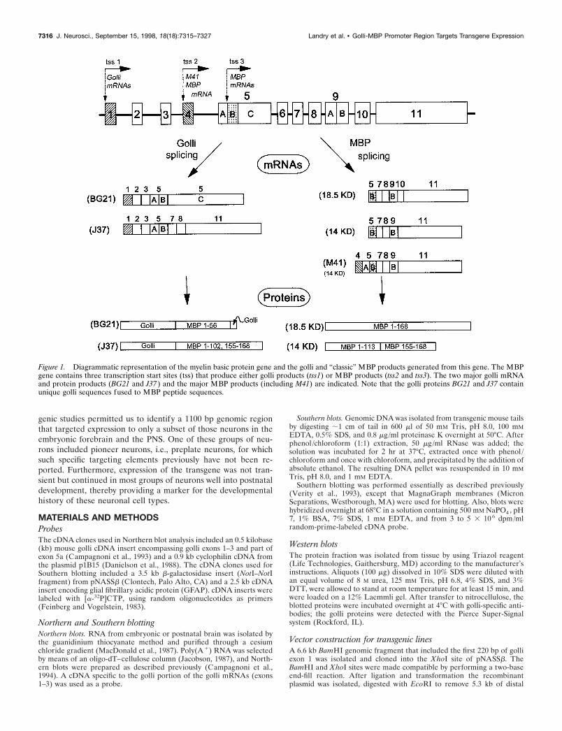
Myelin proteins are among the most abundant in the nervoussystem, and, generally, they have been considered to be expressedonly in myelin-forming cells. One of the two major classes ofmyelin proteins is the myelin basic proteins (MBPs), a family ofproteins derived by alternative splicing of the MBP gene. Re-cently, the MBP gene structure has been found to be larger andmore complicated than originally conceived (Campagnoni et al.,1993; Pribyl et al., 1993). This transcription unit, which we calledthe golli-mbp gene, is ;105 kb in mice; its structure and the twofamilies of products it encodes are shown in Figure 1. The gollimRNAs are produced from the most upstream promoter at thefirst transcription start site (tss1). The MBP mRNAs are pro-duced from two downstream promoters: tss2, which gives rise tothe M41-MBP mRNA, and tss3, which produces the majority ofthe MBP mRNAs.
The golli-mbp gene structure is unusual in many respects. It
numbers among the relatively small group of genes .100 kb andis an example of a very rare arrangement of overlapping genes.For example, the transcription unit defining BG21, the mostabundant golli product, extends from exon 1 to exon 5C, overlap-ping both MBP transcription units. On the other hand, the tran-scription unit defining golli J37 extends from exon 1 to exon 11,completely encompassing the entire MBP transcription unit. TheMBP and BG21 portions of the golli-mbp gene are not simplyincluded within an intron of the larger gene, but they sharealternatively spliced exons in common. This type of alternativesplicing in overlapping transcription units of this size is veryunusual.
Another unusual feature of the golli-mbp gene is its regulation.Tss3 is under tight developmental and cellular control and isactive only in myelin-forming cells. There is evidence that thisregulation resides solely within the promoter for tss3 (Goujet-Zalc et al., 1993). In contrast, tss1, which controls the expressionof golli products, appears to be under less stringent cellularcontrol because it is expressed in selected populations of neurons,in oligodendrocytes in the postnatal brain, and in cells and tissuesof the immune system (Pribyl et al., 1993; Fritz and Kalvakolanu,1995; Landry et al., 1996; Pribyl et al., 1996a,b). Nothing is knownabout golli expression in the mouse embryonic nervous system.
The goals of this study were to define the cellular expression ofgolli mRNAs and proteins in the embryonic nervous system andto identify promoter elements that specified the cell and devel-opmental expression of the golli promoter in the nervous system.We found extensive expression of golli in neurons within theembryonic CNS and peripheral nervous system (PNS). Trans-
Received March 6, 1998; revised June 23, 1998; accepted June 29, 1998.This work was supported by National Institutes of Health Grants NS23022 and
NS33091 and National Multiple Sclerosis Society Grants RG2233 and RG2693. Wethank Vance Handley for assistance in managing the transgenic lines and optimizingthe lac Z staining, and Edwina Skinner and Lauren Cherman for assistance withtissue preparation. We also thank Dr. Andre M. Goffinet for providing us with reelinmonoclonal antibody.
C.F.L. and T.M.P. contributed equally to this work.Correspondence should be addressed to Dr. A. T. Campagnoni, MRRC/NPI,
Room 47–448, University of California at Los Angeles, School of Medicine, 760Westwood Plaza, Los Angeles, CA 90024.
Dr. Pribyl’s present address: Digital Gene Technologies, La Jolla, CA 92037.Dr. Ellison’s present address: SmithKline Beecham Pharmaceuticals, King of
Prussia, PA 19406.Copyright © 1998 Society for Neuroscience 0270-6474/98/187315-13$05.00/0
The Journal of Neuroscience, September 15, 1998, 18(18):7315–7327
genic studies permitted us to identify a 1100 bp genomic regionthat targeted expression to only a subset of those neurons in theembryonic forebrain and the PNS. One of these groups of neu-rons included pioneer neurons, i.e., preplate neurons, for whichsuch specific targeting elements previously have not been re-ported. Furthermore, expression of the transgene was not tran-sient but continued in most groups of neurons well into postnataldevelopment, thereby providing a marker for the developmentalhistory of these neuronal cell types.
MATERIALS AND METHODSProbesThe cDNA clones used in Northern blot analysis included an 0.5 kilobase(kb) mouse golli cDNA insert encompassing golli exons 1–3 and part ofexon 5a (Campagnoni et al., 1993) and a 0.9 kb cyclophilin cDNA fromthe plasmid p1B15 (Danielson et al., 1988). The cDNA clones used forSouthern blotting included a 3.5 kb b-galactosidase insert (NotI–NotIfragment) from pNASSb (Clontech, Palo Alto, CA) and a 2.5 kb cDNAinsert encoding glial fibrillary acidic protein (GFAP). cDNA inserts werelabeled with [a-32P]CTP, using random oligonucleotides as primers(Feinberg and Vogelstein, 1983).
Northern and Southern blottingNorthern blots. RNA from embryonic or postnatal brain was isolated bythe guanidinium thiocyanate method and purified through a cesiumchloride gradient (MacDonald et al., 1987). Poly(A 1) RNA was selectedby means of an oligo-dT–cellulose column (Jacobson, 1987), and North-ern blots were prepared as described previously (Campagnoni et al.,1994). A cDNA specific to the golli portion of the golli mRNAs (exons1–3) was used as a probe.
Southern blots. Genomic DNA was isolated from transgenic mouse tailsby digesting ;1 cm of tail in 600 ml of 50 mM Tris, pH 8.0, 100 mMEDTA, 0.5% SDS, and 0.8 mg/ml proteinase K overnight at 50°C. Afterphenol /chloroform (1:1) extraction, 50 mg/ml RNase was added; thesolution was incubated for 2 hr at 37°C, extracted once with phenol /chloroform and once with chloroform, and precipitated by the addition ofabsolute ethanol. The resulting DNA pellet was resuspended in 10 mMTris, pH 8.0, and 1 mM EDTA.
Southern blotting was performed essentially as described previously(Verity et al., 1993), except that MagnaGraph membranes (MicronSeparations, Westborough, MA) were used for blotting. Also, blots werehybridized overnight at 68°C in a solution containing 500 mM NaPO4 , pH7, 1% BSA, 7% SDS, 1 mM EDTA, and from 3 to 5 3 10 6 dpm/mlrandom-prime-labeled cDNA probe.
Western blotsThe protein fraction was isolated from tissue by using Triazol reagent(Life Technologies, Gaithersburg, MD) according to the manufacturer’sinstructions. Aliquots (100 mg) dissolved in 10% SDS were diluted withan equal volume of 8 M urea, 125 mM Tris, pH 6.8, 4% SDS, and 3%DTT, were allowed to stand at room temperature for at least 15 min, andwere loaded on a 12% Laemmli gel. After transfer to nitrocellulose, theblotted proteins were incubated overnight at 4°C with golli-specific anti-bodies; the golli proteins were detected with the Pierce Super-Signalsystem (Rockford, IL).
Vector construction for transgenic linesA 6.6 kb BamHI genomic fragment that included the first 220 bp of golliexon 1 was isolated and cloned into the XhoI site of pNASSb. TheBamHI and XhoI sites were made compatible by performing a two-baseend-fill reaction. After ligation and transformation the recombinantplasmid was isolated, digested with EcoRI to remove 5.3 kb of distal
Figure 1. Diagrammatic representation of the myelin basic protein gene and the golli and “classic” MBP products generated from this gene. The MBPgene contains three transcription start sites (tss) that produce either golli products (tss1) or MBP products (tss2 and tss3). The two major golli mRNAand protein products (BG21 and J37 ) and the major MBP products (including M41) are indicated. Note that the golli proteins BG21 and J37 containunique golli sequences fused to MBP peptide sequences.
7316 J. Neurosci., September 15, 1998, 18(18):7315–7327 Landry et al. • Golli-MBP Promoter Region Targets Transgene Expression
mouse genomic sequence, and then religated. This new recombinantplasmid now contained 1.3 kb of mouse genomic DNA (1.1 kb of golligene promoter plus 220 bp of golli exon 1), followed by an SV40 splicedonor–acceptor site, the b-galactosidase gene, and a polyadenylationsignal. The recombinant plasmid was digested with ScaI–SphI to removeexcess plasmid DNA, and the resulting insert (6.1 kb) was purified byagarose gel electrophoresis. The production of the transgenic founders wasperformed by the UCLA Transgenic Core Facility (Los Angeles, CA).
Analysis of transgenic animalsFounders. Transgenic mouse lines were identified by Southern blot anal-ysis of isolated tail DNA. Founder lines stably transmitted the transgenewith Mendelian inheritance, as assessed by Southern analysis. Estimatesof the copy number of the transgene in transgenic lines were made bycomparing the strengths of the hybridization signals of theb-galactosidase probe with those of a known single-copy gene probe(GFAP). Progeny screened from every generation that was examinedexhibited no detectable change in transgene copy number.
b-Galactosidase activity in tissue extracts. Animals were anesthetizedwith barbiturates (halothane) and killed by cervical dislocation. Tissueswere removed and immediately homogenized for 20 sec in a PolytronPT3000 (Brinkmann, Westbury, NY) at 15,000 rpm in 1–2 ml of 100 mMKHPO4 , pH 7.8, 0.2% Triton X-100, 1 mM dithiothreitol, 0.2 mM phe-nylmethylsulfonylfluoride, and 5 mg/ml leupeptin. The homogenateswere centrifuged in a microfuge (14,000 rpm) for 5 min. The supernatantwas assayed for b-galactosidase activity by the Galacto-Light Chemilu-minescent Reporter assay (Tropix, Bedford, MA). Reactions wereperformed with 5–15 mg of protein for 30 min at room temperature, andthe light emissions were read in a luminometer. Protein determinationswere made with a detergent-compatible protein assay (Bio-Rad,Hercules, CA).
b-Galactosidase staining of embryonic mouse tissues. Timed-pregnantfemales were anesthetized and killed by cervical dislocation. Embryoswere removed, rinsed briefly in ice-cold PBS, and incubated for 1 hr at4°C in fixative solution (2% formaldehyde, 0.2% glutaraldehyde, and 0.1M NaHPO4 , pH 7.3) with gentle agitation. Then the embryos werewashed three times with rinse solution (0.01% sodium deoxycholate,0.02% NP-40, 2 mM MgCl2 , and 0.1 M NaHPO4 , pH 7.3) and then placedin X-gal stain solution [containing (in mM) 2 MgCl2 , 5 K3Fe(CN)6 , and5 K4Fe(CN)6 , plus 0.1 M NaHPO4 , pH 7.3, 0.01% sodium deoxycholate,0.02% NP-40, and 8 mg/ml X-gal (5-bromo-4-chloro-3-indolyl-b-D-galactoside)] for 12–16 hr at 37°C. Stained embryos were rinsed andequilibrated in sucrose solution (20% sucrose, 0.05% NaN3 , and 0.1 MNaHPO4 , pH 7.3) at 4°C.
b-galactosidase staining in postnatal mice. Postnatal transgenic micewere killed and perfused as described (Landry et al., 1996). Then thebrains were removed and rinsed in ice-cold 0.1 M NaHPO4 , pH 7.3. Theentire brain was cut into 1-mm-thick slices, processed with fixative,rinsed, and stained identically to the embryos. After equilibration insucrose, the brain slices were embedded in OCT embedding compound,frozen at 220°C, cut into 20 mm cryostat sections, and mounted on slides.
Bromodeoxyuridine birth dating and detection. Timed-pregnant femalemice were injected intraperitoneally with 100 mg/gm body weight of5-bromo-29-deoxyuridine (BrdU) in sterile PBS. At specific ages afterbirth, the animals were perfused and processed as described below.Cryostat sections (20 mm) from forebrain were incubated in X-gal stain-ing solution for 2 hr at 37°C, preincubated in 2N HCl for 30 min at 65°C,and then incubated overnight in anti-BrdU monoclonal antibody (BectonDickinson, San Jose, CA). Detection of the antibody was performed withthe Vectastain Elite ABC reagents and peroxidase substrate according tothe manufacturer’s instructions (Vector Laboratories, Burlingame, CA).
ImmunocytochemistryFrozen cryostat sections (20 mm) of transgenic or normal mouse braintissue were prepared and processed for immunohistochemistry as de-scribed (Landry et al., 1996). Postnatal animals were killed and perfusedwith 4% paraformaldehyde (PBS-buffered); the brain tissue was equili-brated in sucrose solution, frozen in OCT, and sectioned. Whole em-bryos were emersion-fixed in 2% paraformaldehyde (PBS-buffered) for12 hr, frozen in OCT, and sectioned. The mounted sections were stainedwith polyclonal antibodies against GFAP (1:1000; Chemicon, Temecula,CA), tau protein (1:2000; Chemicon), calretinin (1:1000; Chemicon),golli protein (1:5000; Landry et al., 1996), or a monoclonal antibodyagainst reelin (1:1500; a gift kindly provided by Dr. Andre M. Goffinet,University of Namur, Belgium). Immunocytochemistry was visualized
with the Vectastain Elite ABC reagents and peroxidase substrate accord-ing to the manufacturer’s instructions (Vector Laboratories). To com-bine b-galactosidase staining with immunocytochemistry, we first treatedsections with X-gal staining solution for 2 hr to overnight (37°C) and thenprocessed them for immunocytochemistry as described above. Doubleimmunofluorescence was performed by incubating the tissue overnight(4°C) in the presence of golli polyclonal (1:1500) and reelin monoclonal(1:1500) antibodies. Detection was with anti-rabbit fluorescein or anti-mouse rhodamine (Boehringer Mannheim, Indianapolis, IN) to detectanti-golli or anti-reelin antibody, specifically. Images from fluorescentstaining were obtained on a Zeiss LSM 310 laser-scanning confocalmicroscope (Oberkochen, Germany).
In situ hybridizationThe tissue preparation, 33P-labeled cRNA probe synthesis, and in situhybridizations were performed as described by Ellison et al. (1996).Embryos were removed from pregnant females after anesthesia andcervical dislocation and immersion-fixed in 2% paraformaldehyde inPBS for 5 d at 4°C. Then the embryos were rinsed and equilibrated insucrose solution, embedded in OCT embedding compound, and frozen at280°C. The embryos were sectioned sagittally at 10–14 mm, mounted onSuperfrost slides (Fisher Scientific, Pittsburgh, PA), and stored at 280°Cuntil used. Golli-specific sense and antisense 33P–UTP-labeled ribo-probes (corresponding to exons 2, 3, and 5a) were synthesized fromlinearized plasmids. The specific activity of the probes ranged from 1 to2 3 10 9 cpm/mg. The tissue sections were hydrated and then treated asfollows: 0.2 M HCl for 10 min; 3 mg/ml proteinase K in 10 mM Tris and1 mM EDTA, pH 8.0, for 10 min; and 0.1 M triethanolamine, pH 8.0, and0.25% (v/v) acetic anhydride for 10 min. Finally, the sections weredehydrated through a graded ethanol series. Then the tissue sectionswere prehybridized for 2 hr at 60°C in 13 hybridization buffer [43 SET(600 mM NaCl, 4 mM EDTA, and 80 mM Tris, pH 7.8)/13 Denhardt’ssolution, 0.2% SDS, 100 mM DTT, 250 mg/ml tRNA, 25 mg/ml each ofpoly(A 1) and poly-C/1 U/ml RNasin (Promega, Madison, WI), and 50%formamide]. The slides were incubated in 13 hybridization buffer and10% dextran-sulfate containing 0.2 ng/ml cRNA probe overnight at 60°Cin a humid chamber. Posthybridization washes were 43 SSC at roomtemperature (RT); 23 SSC at RT, 20 mg/ml RNase A at RT, 23 SSC atRT, 0.13 SSC at 55°C, and 0.53 SSC at RT. Slides then were dehydratedin an ascending ethanol series containing 300 mM NH4OAc, with a finaldehydration in 100% ethanol. Last, the slides were exposed to Hyperfilmb-Max (Amersham, Arlington Heights, IL).
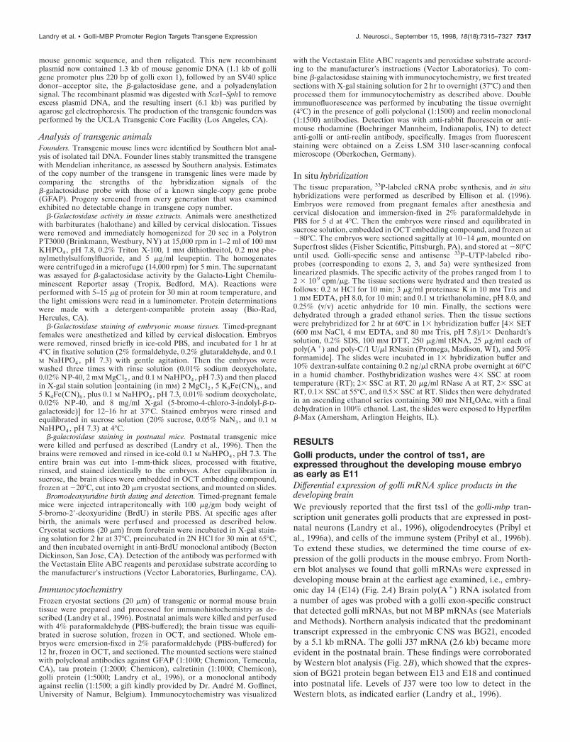
RESULTSGolli products, under the control of tss1, areexpressed throughout the developing mouse embryoas early as E11Differential expression of golli mRNA splice products in thedeveloping brainWe previously reported that the first tss1 of the golli-mbp tran-scription unit generates golli products that are expressed in post-natal neurons (Landry et al., 1996), oligodendrocytes (Pribyl etal., 1996a), and cells of the immune system (Pribyl et al., 1996b).To extend these studies, we determined the time course of ex-pression of the golli products in the mouse embryo. From North-ern blot analyses we found that golli mRNAs were expressed indeveloping mouse brain at the earliest age examined, i.e., embry-onic day 14 (E14) (Fig. 2A) Brain poly(A1) RNA isolated froma number of ages was probed with a golli exon-specific constructthat detected golli mRNAs, but not MBP mRNAs (see Materialsand Methods). Northern analysis indicated that the predominanttranscript expressed in the embryonic CNS was BG21, encodedby a 5.1 kb mRNA. The golli J37 mRNA (2.6 kb) became moreevident in the postnatal brain. These findings were corroboratedby Western blot analysis (Fig. 2B), which showed that the expres-sion of BG21 protein began between E13 and E18 and continuedinto postnatal life. Levels of J37 were too low to detect in theWestern blots, as indicated earlier (Landry et al., 1996).
Landry et al. • Golli-MBP Promoter Region Targets Transgene Expression J. Neurosci., September 15, 1998, 18(18):7315–7327 7317
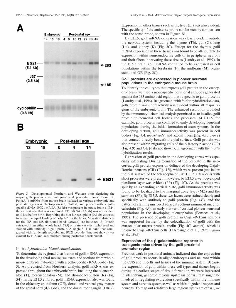
In situ hybridization histochemical studiesTo determine the regional distribution of golli mRNA expressionin the developing fetal mouse, we examined sections from whole-mouse embryos hybridized with a golli-specific cRNA probe (Fig.3). As predicted from Northern analysis, golli mRNA was ex-pressed throughout the embryonic brain, including the telenceph-alon (T), mesencephalon (M), and rhombencephalon (R) (Fig.3A). In the E11.5 embryo, golli mRNA expression also was foundin the olfactory epithelium (OE), dorsal and ventral gray matterof the spinal cord (d/v GM), and the dorsal root ganglia (DRG).
Expression in other tissues such as the liver (Li) was also evident.The specificity of the antisense probe can be seen by comparisonwith the sense probe, shown in Figure 3B.
By E15.5, golli mRNA expression was clearly evident outsidethe nervous system, including the thymus (Th), gut (G), lung(Lu), and kidney (K) (Fig. 3C). Except for the thymus, gollimRNA expression in these tissues was found to be attributable toexpression within neuroendocrine cells or in peripheral neuronsand their fibers innervating these tissues (Landry et al., 1997). Inthe E15.5 brain, golli mRNA continued to be expressed in cellpopulations within the forebrain (F), the midbrain (M), brain-stem, and OE (Fig. 3C).
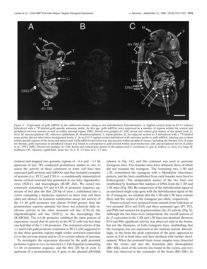
Golli proteins are expressed in pioneer neuronalpopulations in the embryonic mouse brainTo identify the cell types that express golli protein in the embry-onic brain, we used a monospecific polyclonal antibody generatedagainst the 133 amino acid region that is specific to golli proteins(Landry et al., 1996). In agreement with in situ hybridization data,golli protein immunoreactivity was evident within all major re-gions of the embryonic brain. The enhanced resolution providedby the immunocytochemical analysis permitted us to localize golliprotein to neuronal cell bodies and processes. At E11.5, forexample, golli protein was confined to early developing neuronalpopulations during the initial formation of axon systems. In thedeveloping tectum, golli immunoreactivity was present in cellbodies (Fig. 4A, arrowheads) and axonal fibers (Fig. 4A, arrows)that coursed directly beneath the pial surface. Golli protein wasalso present within migrating cells of the olfactory placode (OP)(Fig. 4B) and OE (data not shown), in agreement with the in situhybridization results.
Expression of golli protein in the developing cortex was espe-cially interesting. During formation of the preplate in the neo-cortex, golli protein expression delineated the developing Cajal–Retzius neurons (CR) (Fig. 4B), which were present just belowthe pial surface of the telencephalon. At E11.5 a few cells withshort processes were present; however, by E13.5 a well developedplexiform layer was evident (PP) (Fig. 4C). As the preplate wassplit by an expanding cortical plate, golli immunoreactivity wasfound to be localized to the marginal zone layer (MZ) and thesubplate (SP). By E15.5, these two layers were stained clearly andspecifically with antibody to golli protein (Fig. 4E), and thepattern of staining mirrored adjacent sections immunostained forcalretinin (Fig. 4F), an early marker of cortical pioneer neuronalpopulations in the developing telencephalon (Fonseca et al.,1995). The presence of golli protein in Cajal–Retzius neuronswas supported further by the colocalization of golli with theextracellular matrix protein, reelin (Fig. 4G, arrows), which isunique to Cajal–Retzius cells (D’Arcangelo et al., 1995; Ogawaet al., 1995).
Expression of the b-galactosidase reporter intransgenic mice driven by the golli proximalpromoter regionPrevious work in postnatal animals indicated that the expressionof golli products occurs in oligodendrocytes and neurons withinthe CNS and in cells and tissues of the immune system. Becausethe expression of golli within these cell types and tissues beginsduring the earliest stages of tissue formation, we were interestedin identifying genomic regions upstream of tss1 that might beinvolved in regulating expression specifically within the immunesystem and nervous system as well as within oligodendrocytes andneurons. To map out relatively large regions upstream of tss1, we
Figure 2. Developmental Northern and Western blots depicting themajor golli products in embryonic and postnatal mouse brain. A,Poly(A 1) mRNA from mouse brain isolated at various embryonic andpostnatal ages was electrophoresed, blotted, and probed with a golli-specific cDNA. BG21 mRNA (5.1 kb) was present in mouse brain at E14,the earliest age that was examined. J37 mRNA (2.6 kb) was not evidentuntil just before birth. Reprobing the blot for cyclophilin (0.8 kb) was usedto assess the equal loading of poly(A 1) in the lanes. Migration distancesfor the 28S and 18S ribosomal bands (arrows) are indicated. B, Proteinextracted from either whole head (E13) or brain was electrophoresed andstained with antibody to golli protein. A single 31 kDa band that comi-grated with full-length recombinant BG21 peptide (lane not shown) wasevident by E18 and accumulated during postnatal development.
7318 J. Neurosci., September 15, 1998, 18(18):7315–7327 Landry et al. • Golli-MBP Promoter Region Targets Transgene Expression
isolated and mapped two genomic regions of ;6.4 and ;1.1 kbupstream of tss1. We conducted preliminary studies in vitro toassess the activity of these constructs in some cell lines thatexpressed golli proteins and mRNAs and that included examplesof neurons (i.e., PC12 and CN1.4—a conditionally immortalizedmouse cortical neuronal line generated in our lab), oligodendro-cytes (N20.1), and macrophages (RAW 264). We tested twoconstructs containing 0.9 and 6.4 kb of promoter sequence up-stream of tss1 plus the first 220 bp of exon 1 (subcloned into avector containing a luciferase reporter) in these four cell lines(data not shown). In transient transfection assays the activity ofthe 1.1 kb golli promoter was almost 50-fold greater than thepromoterless reporter plasmid, but there was relatively little lu-ciferase activity in the other neuronal cell line (CN1.4), theoligodendroglial cell line (N20.1), or the macrophage line(RAW264). The 6.6 kb promoter exhibited the same pattern ofexpression, except that its activity in the PC12 cells was less thanone-half that of the 1.1 kb promoter. The restricted activity of the1.1 and 6.6 kb golli promoter constructs to PC12 cells suggested tous that these genomic regions might confer restricted expression(1) to the nervous system and/or (2) to certain subsets of neurons.
To investigate the expression directed by the golli proximalpromoter region in vivo, we inserted a 1.3 kb fragment (containing1.1 kb of promoter sequence and the first 220 bp of exon 1)upstream of a promoterless lac Z gene in the plasmid pNASSb
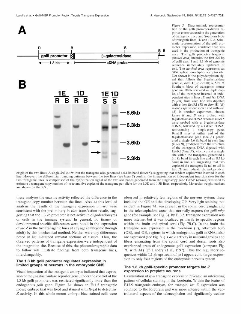
(shown in Fig. 5A), and this construct was used to generatetransgenic mice. Five founder mice were obtained, three of whichdid not transmit the transgene. The remaining two, 1.3D and1.3E, transmitted the transgene with a Mendelian inheritancepattern, and the lines established from each founder were bred tohomozygosity. The independent nature of the two lines wasestablished by Southern blot analysis of DNA from the 1.3D and1.3E mice (Fig. 5B). By comparison of the hybridization signal ofan unrelated single-copy gene with the hybridization signal of thelac Z transgene, we estimate that the 1.3D and 1.3E mice containthree and five copies of the transgene per allele, respectively.
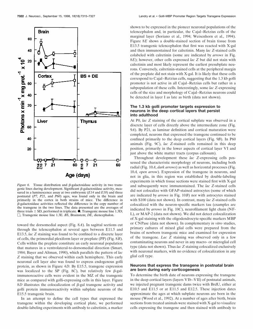
Tissues extracts were prepared from animals from both lines attwo prenatal (E14 and E18) and three postnatal ages (P7, P21,and P60) and assayed for b-galactosidase (lac Z) activity (Fig. 6).Although the two lines were independent, the overall pattern oflac Z expression in the 1.3D and 1.3E lines was identical. BetweenE14 and P60, significant activity was observed only in the brains,but not the thymuses, of both transgenic lines, establishing thatthe transgene was not expressed in the immune system. Interest-ingly, in the brain the peak expression of the gene appeared tooccur at E18 in both lines and then declined with further devel-opment. When the postnatal brain tissue samples were dissectedinto the cortex and into the brainstem plus diencephalon(BS1DE), most of the activity was found in the cortex, and verylittle was observed in the remainder of the brain (BS1DE). In
Figure 3. Expression of golli mRNA in the embryonic mouse, using in situ hybridization histochemistry. A, Sagittal section from an E11.5 embryohybridized with a 33P-labeled golli-specific antisense probe. At this age, golli mRNAs were expressed in a number of regions within the central andperipheral nervous systems as well as within internal organs. DRG, Dorsal root ganglia; d/v GM, dorsal and ventral gray matter of the spinal cord; Li,liver; M, mesencephalon; OE, olfactory epithelium; R, rhombencephalon; T, telencephalon. B, An adjacent section to A hybridized with a 33P-labeledsense probe did not label above background levels. C, In an E15.5 sagittal section hybridized with antisense probe to golli mRNA, labeling was evidentwithin specific regions of the brain and spinal cord. Golli mRNA expression was also present within peripheral tissues, including the thymus (Th). Exceptfor thymus, golli expression in peripheral tissues was found to correspond to golli protein within neuroendocrine cells and peripheral nerves (Landryet al., 1997). DRG, Dorsal root ganglia; d/v GM, dorsal and ventral gray matter of the spinal cord; F, forebrain; G, gut; K, kidney; Li, liver; Lu, lung; M,midbrain; OE, olfactory epithelium. Scale bar: in A, B, 1.9 mm; in C, 1.5 mm.
Landry et al. • Golli-MBP Promoter Region Targets Transgene Expression J. Neurosci., September 15, 1998, 18(18):7315–7327 7319
Figure 4. Golli protein is present in early developing neuronal systems within the embryonic mouse brain. A, Sagittal sections from E11.5 embryos wereimmunostained for golli protein by incubating the tissue with immunopurified golli-specific polyclonal antibody. Neuronal cell bodies (arrowheads) andaxonal fibers (arrows), seen coursing directly beneath the pial surface in the developing tectum, stained intensely. Dorsal ( D) and rostral ( R) orientationmarkers are indicated. B, Golli immunoreactivity was evident within neurons of the olfactory placode (OP) as well as within Cajal–Retzius (CR) neuronsat the pial surface of the E11.5 telencephalon. Dorsal (D) and rostral (R) orientation markers are indicated. C, Sagittal sections of E13.5 embryos at thelevel of the telencephalon showed prominent immunostaining of golli protein within the primordial plexiform layer (PP, preplate). D, An E11.5 sectionincubated with antibody preabsorbed with golli peptide illustrates background. E, By E15.5, stain corresponding to golli protein was present withinneurons of the subplate (SP) and marginal zone (MZ) of coronal sections. The cortical plate (CP) was stained only lightly. F, An adjacent section to Ethat was immunostained with antibody to the early neuronal marker calretinin. Note that the pattern of staining in the MZ and SP is very similar to sectionsthat were stained with antibody to golli protein (see E). G, Coronal sections of E15.5 mouse brain were double-labeled with polyclonal antibody to golliprotein and monoclonal antibody to the Cajal–Retzius marker reelin. Green and red corresponds to golli and reelin immunoreactivity, respectively. Acolocalization of the proteins within a single neuron is indicated by yellow (arrows). Scale bar: in A–C, E, F, 30 mm; in D, 25 mm; in G, 10 mm.
7320 J. Neurosci., September 15, 1998, 18(18):7315–7327 Landry et al. • Golli-MBP Promoter Region Targets Transgene Expression
these analyses the enzyme activity reflected the difference in thetransgene copy number between the lines. Also, at this level ofanalysis the results of the transgene expression in vivo wereconsistent with the preliminary in vitro transfection results, sug-gesting that the 1.3 kb promoter is not active in oligodendrocytesor cells in the immune system. In general, no tissue- ordevelopmental-specific differences were noted in the expressionof lac Z in the two transgenic lines at any age (embryonic throughadult) by this biochemical method. Neither were any differencesnoted in lac Z-stained cryostat sections of tissues. Thus, theobserved patterns of transgene expression were independent ofthe integration site. Because of this, the photomicrographic datato follow will illustrate findings from both transgenic lines,interchangeably.
The 1.3 kb golli promoter regulates expression inlimited groups of neurons in the embryonic CNS
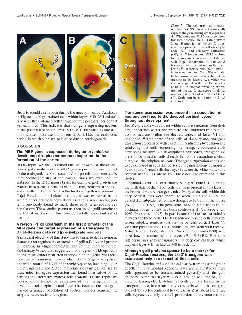
Visual inspection of the transgenic embryos indicated that expres-sion of the b-galactosidase reporter gene, under the control of the1.3 kb golli promoter, was restricted significantly more than theendogenous golli gene. Figure 7A shows an E11.5 transgenicmouse embryo that was fixed and stained with X-gal to detect lacZ activity. In this whole-mount embryo blue-stained cells were
observed in relatively few regions of the nervous system; theseincluded the OE and the developing OP. Very light staining, notevident in Figure 7A, was present in the spinal cord ganglia andin the telencephalon, areas that normally express the golli-mbpgene (for example, see Fig. 3). By E13.5, transgene expression wasmore intense, but it was localized primarily to specific regionswithin the brain and spinal cord (Fig. 7B,C). In the brain thetransgene was expressed in the forebrain (F), olfactory bulb(OB), and OE, regions in which endogenous golli mRNAs alsoare expressed (see Fig. 3C). Lac Z activity in neuronal groups andfibers emanating from the spinal cord and dorsal roots alsooverlapped areas of endogenous golli expression (compare Fig.7C with 3A) (cf. Landry et al., 1997). Thus the regulatory se-quences within 1.1 kb upstream of tss1 appeared to target expres-sion to only four regions of the embryonic nervous system.
The 1.3 kb golli-specific promoter targets lac Zexpression to preplate neuronsExamination of golli transgene expression revealed an interestingpattern of cellular staining in the forebrain. Within the brains ofE13.5 transgenic embryos, for example, lac Z expression wasconfined to the forebrain and was more intense within the ven-trolateral aspects of the telencephalon and significantly weaker
Figure 5. Diagrammatic representa-tion of the golli promoter-driven re-porter construct used in the generationof transgenic mice and Southern blotsof transgenic lines 1D and 1E. A, Sche-matic representation of the golli pro-moter expression construct that wasused in the production of transgenicmice. The golli promoter fragment(shaded area) includes the first 220 bpof golli exon 1 and 1.1 kb of genomicsequence immediately upstream oftss1. The hatched area represents anSV40 splice donor/splice acceptor site.Not shown is the polyadenylation sig-nal that follows the b-galactosidasegene; B, BamHI; R, EcoRI; S, SstI. B,Southern blots of transgenic mousegenomic DNA revealed multiple cop-ies of the transgene inserted at inde-pendent sites in lines 1E and 1D. DNA(5 mm) from each line was digestedwith either EcoRI ( R) or BamHI (B)in one experiment shown and with SstI(S) in another experiment shown.Lanes R and B were probed withb-galactosidase cDNA whereas lanes Swere probed with a b-galactosidasecDNA, followed by a GFAP cDNA,representing a single-copy gene.BamHI sites at either end of theb-galactosidase gene (see A) gener-ated a single 3.6 kb band in each line(lanes B), predicted from the structureof the transgene. DNA digested withEcoRI (lanes R), which cuts at a singlesite within the transgene, generated a6.1 kb band in each line and an 8.5 kbband in line 1E, suggesting that twocopies of the transgene lie tail to tail inline 1E and indicate the independent
origin of the two lines. A single SstI cut within the transgene also generated a 6.1 kb band (lanes S), suggesting that tandem copies were inserted in eachline. However, the different SstI banding patterns between the two lines (see lanes S) confirm the interpretation of independent insertion sites for thetwo transgenic lines. A comparison of the hybridization signal of the two SstI bands generated from the single-copy gene GFAP (arrows) was used toestimate a transgene copy number of three and five copies of the transgene per allele for the 1.3D and 1.3E lines, respectively. Molecular weight markersare shown on the lef t.
Landry et al. • Golli-MBP Promoter Region Targets Transgene Expression J. Neurosci., September 15, 1998, 18(18):7315–7327 7321
toward the dorsomedial aspect (Fig. 8A). In sagittal sections cutthrough the telencephalon at several ages between E11.5 andE13.5, lac Z staining was found to be confined to a discrete layerof cells, the primordial plexiform layer or preplate (PP) (Fig. 8B).Cells within the preplate constitute an early neuronal populationthat matures in a ventrolateral-to-dorsomedial direction (Smart,1984; Bayer and Altman, 1990), which parallels the pattern of lacZ staining that we observed within each hemisphere. This earlyneuronal cell layer also was found to express endogenous golliprotein, as shown in Figure 4D. By E15.5, transgene expressionwas localized to the SP (Fig. 8C), but relatively few b-gal-immunoreactive cells were evident in the MZ of the transgenicmice as compared with golli-expressing cells in this layer. Figure8D illustrates the colocalization of b-gal transgene activity andgolli protein immunoreactivity within subplate neurons of theE15.5 transgenic brain.
In an attempt to define the cell types that expressed thetransgene within the developing cortical plate, we performeddouble-labeling experiments with antibody to calretinin, a marker
shown to be expressed in the pioneer neuronal populations of thetelencephalon and, in particular, the Cajal–Retzius cells of themarginal layer (Soriano et al., 1994; Weisenhorn et al., 1994).Figure 8E shows a double-stained section of brain tissue fromE13.5 transgenic telencephalon that first was reacted with X-galand then immunostained for calretinin. Many lac Z-stained cellscolabeled with calretinin (some are indicated by arrows in Fig.8E); however, other cells expressed lac Z but did not stain withcalretinin and most likely represent the earliest presubplate neu-rons. Conversely, calretinin-stained cells at the peripheral marginof the preplate did not stain with X-gal. It is likely that these cellscorrespond to Cajal–Retzius cells, suggesting that the 1.3 kb gollipromoter is not active in all Cajal–Retzius cells but rather in asubpopulation of these cells. Interestingly, some lac Z-expressingcells of the size and morphology of Cajal–Retzius neurons couldbe detected in layer I as late as birth (data not shown).
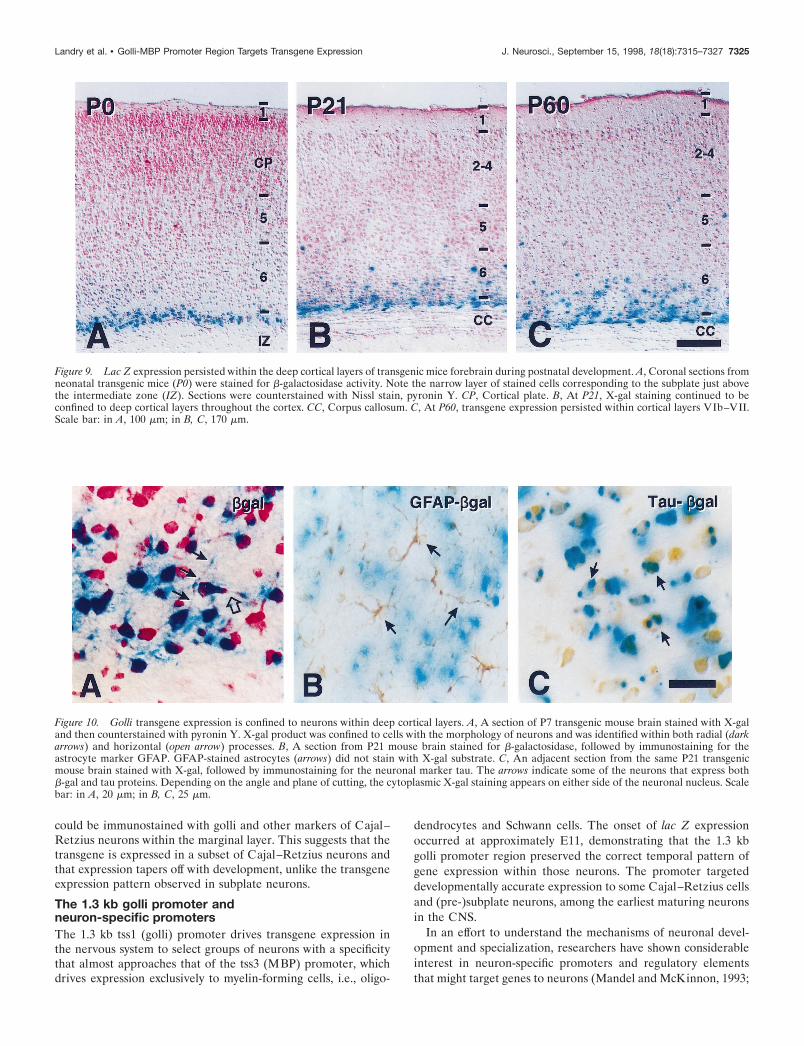
The 1.3 kb golli promoter targets expression toneurons in the deep cortical layers that persistinto adulthoodAt P0, lac Z staining of the cortical subplate was observed in adiscrete layer of cells directly above the intermediate zone (Fig.9A). By P21, as laminar definition and cortical maturation werecompleted, neurons that expressed the transgene continued to beconfined primarily to the deep cortical layers (Fig. 9B). In P60animals (Fig. 9C), lac Z-stained cells remained in this deepposition, primarily in the lower aspects of cortical layer VI andjust above the white matter tracts (corpus callosum).
Throughout development these lac Z-expressing cells pos-sessed the characteristic morphology of neurons, including bothradial (Fig. 10A, dark arrows) as well as horizontal processes (Fig.10A, open arrow). Expression of the transgene in neurons, andnot in glia, in this region was established by double-labelingexperiments in which tissue sections were stained first with X-galand subsequently were immunostained. The lac Z-stained cellsdid not colocalize with GFAP-stained astrocytes (some of whichare indicated by arrows in Fig. 10B) nor with astrocytes stainedwith S100 (data not shown). In contrast, many lac Z-stained cellscolocalized with the neuron-specific markers tau (examples areindicated by arrows in Fig. 10C), neurofilament light chain (NF-L), or MAP-2 (data not shown). We did not detect colocalizationof X-gal staining with the oligodendrocyte-specific markers MBPor CNPase (data not shown). In complementary in vitro studies,primary cultures of mixed glial cells were prepared from thebrains of newborn transgenic mice and examined for expressionof the transgene. Lac Z staining was observed only in a fewcontaminating neurons and never in any macro- or microglial celltype (data not shown). Thus lac Z staining colocalized exclusivelywith neuronal markers, with no evidence of colocalization in anyglial cell type.
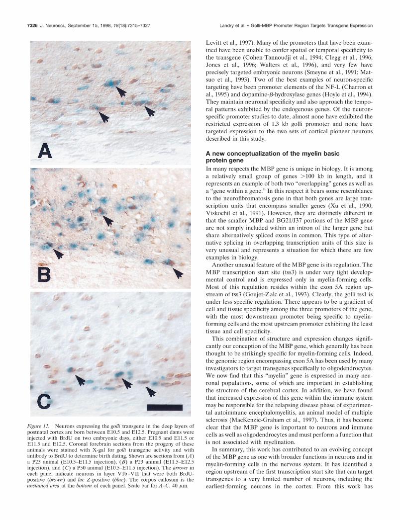
Neurons that express the transgene in postnatal brainare born during early corticogenesisTo determine the birth date of neurons expressing the transgenein the deep cortical layers (layers VIb–VII) of postnatal animals,we injected pregnant transgenic dams twice with BrdU, either atE10.5 and E11.5 or at E11.5 and E12.5. These injection datesapproximate the ages at which subplate neurons are born in themouse (Wood et al., 1992). At a number of ages after birth, brainsections from treated animals were stained with X-gal to visualizecells expressing the transgene and then stained with antibody to
Figure 6. Tissue distribution and b-galactosidase activity in two trans-genic lines during development. Significant b-galactosidase activity, mea-sured in a luminescence assay at two embryonic (E14 and E18) and threepostnatal (P7, P21, and P60) ages, was found only in the brain andprimarily in the cortex in both strains of mice. The difference inb-galactosidase activities reflected the difference in the copy number ofthe transgene in the two lines. The data presented are the averages ofthree trials 6 SD, performed in triplicate. F, Transgenic mouse line 1.3D;M, Transgenic mouse line 1.3E. BS, Brainstem; DE, diencephalon.
7322 J. Neurosci., September 15, 1998, 18(18):7315–7327 Landry et al. • Golli-MBP Promoter Region Targets Transgene Expression
BrdU to identify cells born during the injection period. As shownin Figure 11, X-gal-stained cells within layers VIb–VII colocal-ized with BrdU-stained cells throughout the postnatal period thatwas examined. This indicates that transgene-expressing neuronsin the postnatal subplate layer (VIb–VII) identified as late as 2months after birth are born from E10.5–E12.5, the embryonicperiod in which subplate cells arise during corticogenesis.
DISCUSSIONThe MBP gene is expressed during embryonic braindevelopment in pioneer neurons important in theformation of the cortexIn this report we have extended our earlier work on the expres-sion of golli products of the MBP gene in postnatal developmentto the embryonic nervous system. Golli protein was detected byimmunocytochemistry at the earliest times we examined theembryos. In the E11.5 mouse brain, for example, golli protein wasevident in superficial neurons of the tectum, neurons of the OP,and in cells of the OE. Within the forebrain, golli was present inCajal–Retzius and subplate neurons and was expressed in thesame pioneer neuronal populations as calretinin and reelin, pro-teins previously found to mark these early telencephalic cellpopulations. These studies permit us, then, to add golli proteins tothe list of markers for this developmentally important set ofneurons.
A region ;1 kb upstream of the first promoter of theMBP gene can target expression of a transgene toCajal–Retzius cells and (pre-)subplate neuronsA principal objective of this study was to begin to define genomicelements that regulate the expression of golli mRNAs and proteinin neurons, in oligodendrocytes, and in the immune system.Preliminary in vitro data suggested that a region ;1 kb upstreamof tss1 might confer restricted expression on the gene. We there-fore created transgenic mice in which the lac Z gene was placedunder the control of 1.3 kb of genomic sequence, including 1.1 kbdirectly upstream and 220 bp immediately downstream of tss1. Inthese mice, transgene expression was found in a subset of theneurons that normally express golli proteins. In this report wefocused our attention on expression of the transgene in thedeveloping telencephalon and forebrain, because the transgenemarked a unique population of cortical pioneer neurons, thesubplate neurons, in this region.
Transgene expression was present in a population ofneurons confined to the deepest cortical layersthroughout developmentLac Z expression was evident within subplate neurons from theirfirst appearance within the preplate and continued in a popula-tion of neurons within the deepest aspects of layer VI intoadulthood. Within some of the cells of the subplate, transgeneexpression colocalized with calretinin, confirming by position andcolabeling that cells expressing the transgene represent earlydeveloping neurons. As development proceeded, transgene ex-pression persisted in cells directly below the expanding corticalplate, i.e., the subplate neurons. Transgene expression continuedto be expressed in cells that possessed the morphology of subplateneurons and formed a distinct layer between the white matter andcortical layer VI as late as P60 (the oldest age examined in thisstudy).
Bromodeoxyuridine experiments were performed to determinethe birth date of the “blue” cells that were present in this layer inthe brains of mature transgenic mice. Many of the cells within thisdeep cortical layer were “born” between E10.5 and E12.5, theperiod that subplate neurons are thought to be born in the mouse(Wood et al., 1992). The persistence of subplate neurons in thepostnatal rodent cortex has been controversial (Valverde et al.,1995; Price et al., 1997), in part because of the lack of suitablemarkers for these cells. The transgene-expressing cells may rep-resent subplate neurons that survive beneath cortical layer VIwell into postnatal life. These results are consistent with those ofValverde et al. (1989, 1995) and Reep and Goodwin (1988), whohave shown that neurons born between E11–E13 (E12–E14 in therat) persist in significant numbers in a deep cortical layer, whichthey call layer VII, as late as P60 in rodents.
Although golli proteins appear to be a marker forCajal–Retzius neurons, the lac Z transgene wasexpressed only in a subset of these cellsThe Cajal–Retzius and subplate cells arise from the same groupof cells in the primordial plexiform layer, and in our studies thesecells appeared to be immunostained generally with the golliantibody. After this layer was split into the MZ and SP, golliimmunostaining clearly delineated both of these layers. In thetransgenic mice, in contrast, only some cells within the marginallayer of the cortex continued to express lac Z as late as P0. Thesecells represented only a small proportion of the neurons that
Figure 7. The golli proximal promoteris active in CNS neurons that normallyexpress the gene during embryogenesis.A, Whole-mount E11.5 embryo fromtransgenic mouse line 1.3D stained withX-gal. Expression of the lac Z trans-gene was present in the olfactory pla-code (OP) and olfactory epithelium(OE). B, Whole-mount E13.5 embryofrom transgenic mouse line 1.3D stainedwith X-gal. Expression of the lac Ztransgene was evident within the fore-brain (F), olfactory bulb (OB), and ol-factory epithelium (OE). We also de-tected variable and inconsistent X-galstaining in the kidney (K ), which wasnot investigated further. C, Dorsal viewof an E13.5 embryo revealing expres-sion of the lac Z transgene in dorsalroot ganglia (D) and ventral root fibers(V ). Scale bar: in A, 1.1 mm; in B, 1.9mm; in C, 1 mm.
Landry et al. • Golli-MBP Promoter Region Targets Transgene Expression J. Neurosci., September 15, 1998, 18(18):7315–7327 7323
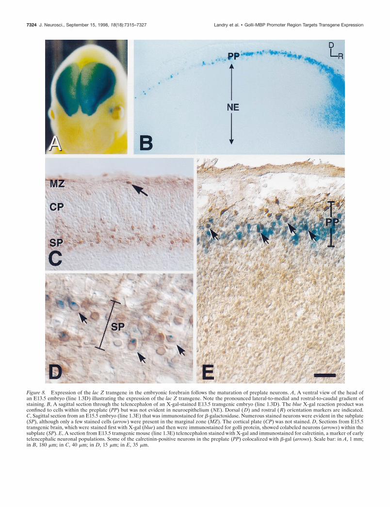
Figure 8. Expression of the lac Z transgene in the embryonic forebrain follows the maturation of preplate neurons. A, A ventral view of the head ofan E13.5 embryo (line 1.3D) illustrating the expression of the lac Z transgene. Note the pronounced lateral-to-medial and rostral-to-caudal gradient ofstaining. B, A sagittal section through the telencephalon of an X-gal-stained E13.5 transgenic embryo (line 1.3D). The blue X-gal reaction product wasconfined to cells within the preplate (PP) but was not evident in neuroepithelium (NE). Dorsal ( D) and rostral ( R) orientation markers are indicated.C, Sagittal section from an E15.5 embryo (line 1.3E) that was immunostained for b-galactosidase. Numerous stained neurons were evident in the subplate(SP), although only a few stained cells (arrow) were present in the marginal zone (MZ). The cortical plate (CP) was not stained. D, Sections from E15.5transgenic brain, which were stained first with X-gal (blue) and then were immunostained for golli protein, showed colabeled neurons (arrows) within thesubplate (SP). E, A section from E13.5 transgenic mouse (line 1.3E) telencephalon stained with X-gal and immunostained for calretinin, a marker of earlytelencephalic neuronal populations. Some of the calretinin-positive neurons in the preplate (PP) colocalized with b-gal (arrows). Scale bar: in A, 1 mm;in B, 180 mm; in C, 40 mm; in D, 15 mm; in E, 35 mm.
7324 J. Neurosci., September 15, 1998, 18(18):7315–7327 Landry et al. • Golli-MBP Promoter Region Targets Transgene Expression
could be immunostained with golli and other markers of Cajal–Retzius neurons within the marginal layer. This suggests that thetransgene is expressed in a subset of Cajal–Retzius neurons andthat expression tapers off with development, unlike the transgeneexpression pattern observed in subplate neurons.
The 1.3 kb golli promoter andneuron-specific promotersThe 1.3 kb tss1 (golli) promoter drives transgene expression inthe nervous system to select groups of neurons with a specificitythat almost approaches that of the tss3 (MBP) promoter, whichdrives expression exclusively to myelin-forming cells, i.e., oligo-
dendrocytes and Schwann cells. The onset of lac Z expressionoccurred at approximately E11, demonstrating that the 1.3 kbgolli promoter region preserved the correct temporal pattern ofgene expression within those neurons. The promoter targeteddevelopmentally accurate expression to some Cajal–Retzius cellsand (pre-)subplate neurons, among the earliest maturing neuronsin the CNS.
In an effort to understand the mechanisms of neuronal devel-opment and specialization, researchers have shown considerableinterest in neuron-specific promoters and regulatory elementsthat might target genes to neurons (Mandel and McKinnon, 1993;
Figure 9. Lac Z expression persisted within the deep cortical layers of transgenic mice forebrain during postnatal development. A, Coronal sections fromneonatal transgenic mice (P0) were stained for b-galactosidase activity. Note the narrow layer of stained cells corresponding to the subplate just abovethe intermediate zone (IZ). Sections were counterstained with Nissl stain, pyronin Y. CP, Cortical plate. B, At P21, X-gal staining continued to beconfined to deep cortical layers throughout the cortex. CC, Corpus callosum. C, At P60, transgene expression persisted within cortical layers VIb–VII.Scale bar: in A, 100 mm; in B, C, 170 mm.
Figure 10. Golli transgene expression is confined to neurons within deep cortical layers. A, A section of P7 transgenic mouse brain stained with X-galand then counterstained with pyronin Y. X-gal product was confined to cells with the morphology of neurons and was identified within both radial (darkarrows) and horizontal (open arrow) processes. B, A section from P21 mouse brain stained for b-galactosidase, followed by immunostaining for theastrocyte marker GFAP. GFAP-stained astrocytes (arrows) did not stain with X-gal substrate. C, An adjacent section from the same P21 transgenicmouse brain stained with X-gal, followed by immunostaining for the neuronal marker tau. The arrows indicate some of the neurons that express bothb-gal and tau proteins. Depending on the angle and plane of cutting, the cytoplasmic X-gal staining appears on either side of the neuronal nucleus. Scalebar: in A, 20 mm; in B, C, 25 mm.
Landry et al. • Golli-MBP Promoter Region Targets Transgene Expression J. Neurosci., September 15, 1998, 18(18):7315–7327 7325
Levitt et al., 1997). Many of the promoters that have been exam-ined have been unable to confer spatial or temporal specificity tothe transgene (Cohen-Tannoudji et al., 1994; Clegg et al., 1996;Jones et al., 1996; Walters et al., 1996), and very few haveprecisely targeted embryonic neurons (Smeyne et al., 1991; Mat-suo et al., 1993). Two of the best examples of neuron-specifictargeting have been promoter elements of the NF-L (Charron etal., 1995) and dopamine-b-hydroxylase genes (Hoyle et al., 1994).They maintain neuronal specificity and also approach the tempo-ral patterns exhibited by the endogenous genes. Of the neuron-specific promoter studies to date, almost none have exhibited therestricted expression of 1.3 kb golli promoter and none havetargeted expression to the two sets of cortical pioneer neuronsdescribed in this study.
A new conceptualization of the myelin basicprotein geneIn many respects the MBP gene is unique in biology. It is amonga relatively small group of genes .100 kb in length, and itrepresents an example of both two “overlapping” genes as well asa “gene within a gene.” In this respect it bears some resemblanceto the neurofibromatosis gene in that both genes are large tran-scription units that encompass smaller genes (Xu et al., 1990;Viskochil et al., 1991). However, they are distinctly different inthat the smaller MBP and BG21/J37 portions of the MBP geneare not simply included within an intron of the larger gene butshare alternatively spliced exons in common. This type of alter-native splicing in overlapping transcription units of this size isvery unusual and represents a situation for which there are fewexamples in biology.
Another unusual feature of the MBP gene is its regulation. TheMBP transcription start site (tss3) is under very tight develop-mental control and is expressed only in myelin-forming cells.Most of this regulation resides within the exon 5A region up-stream of tss3 (Goujet-Zalc et al., 1993). Clearly, the golli tss1 isunder less specific regulation. There appears to be a gradient ofcell and tissue specificity among the three promoters of the gene,with the most downstream promoter being specific to myelin-forming cells and the most upstream promoter exhibiting the leasttissue and cell specificity.
This combination of structure and expression changes signifi-cantly our conception of the MBP gene, which generally has beenthought to be strikingly specific for myelin-forming cells. Indeed,the genomic region encompassing exon 5A has been used by manyinvestigators to target transgenes specifically to oligodendrocytes.We now find that this “myelin” gene is expressed in many neu-ronal populations, some of which are important in establishingthe structure of the cerebral cortex. In addition, we have foundthat increased expression of this gene within the immune systemmay be responsible for the relapsing disease phase of experimen-tal autoimmune encephalomyelitis, an animal model of multiplesclerosis (MacKenzie-Graham et al., 1997). Thus, it has becomeclear that the MBP gene is important to neurons and immunecells as well as oligodendrocytes and must perform a function thatis not associated with myelination.
In summary, this work has contributed to an evolving conceptof the MBP gene as one with broader functions in neurons and inmyelin-forming cells in the nervous system. It has identified aregion upstream of the first transcription start site that can targettransgenes to a very limited number of neurons, including theearliest-forming neurons in the cortex. From this work has
Figure 11. Neurons expressing the golli transgene in the deep layers ofpostnatal cortex are born between E10.5 and E12.5. Pregnant dams wereinjected with BrdU on two embryonic days, either E10.5 and E11.5 orE11.5 and E12.5. Coronal forebrain sections from the progeny of theseanimals were stained with X-gal for golli transgene activity and withantibody to BrdU to determine birth dating. Shown are sections from (A)a P23 animal (E10.5–E11.5 injection), ( B) a P23 animal (E11.5–E12.5injection), and (C) a P50 animal (E10.5–E11.5 injection). The arrows ineach panel indicate neurons in layer VIb–VII that were both BrdU-positive (brown) and lac Z-positive (blue). The corpus callosum is theunstained area at the bottom of each panel. Scale bar for A–C, 40 mm.
7326 J. Neurosci., September 15, 1998, 18(18):7315–7327 Landry et al. • Golli-MBP Promoter Region Targets Transgene Expression
emerged a transgenic mouse that will be important for futurestudies on determining the fate and function of subplate neurons.
REFERENCESBayer SA, Altman J (1990) Development of layer I and the subplate in
the rat neocortex. Exp Neurol 107:48–62.Campagnoni AT, Pribyl TM, Campagnoni CW, Kampf K, Amur-
Umarjee S, Landry CF, Handley VW, Newman SL, Garbay B, Kita-mura K (1993) Structure and developmental regulation of golli-mbp, a105 kb gene that encompasses the myelin basic protein gene and isexpressed in cells in the oligodendrocyte lineage in the brain. J BiolChem 268:4930–4938.
Campagnoni CW, Kampf K, Mason B, Handley VW, Campagnoni AT(1994) Isolation and characterization of a cDNA encoding the zebrafinch myelin proteolipid protein. Neurochem Res 19:1061–1065.
Charron G, Guy L-G, Bazinet M, Julien J-P (1995) Multiple neuron-specific enhancers in the human neurofilament light chain. J Biol Chem270:30604–30610.
Clegg CH, Haugen HS, Boring LF (1996) Promoter sequences in the RIb-subunit gene of cAMP-dependent protein kinase required for trans-gene expression in mouse brain. J Biol Chem 271:1638–1644.
Cohen-Tannoudji M, Babinet C, Wassef M (1994) Early determinationof a mouse somatosensory cortex marker. Nature 368:460–463.
Danielson PE, Forss-Petter S, Brow MA, Calavetta L, Douglass J, MilnerRJ, Sutcliffe JG (1988) p1B15: a cDNA clone of the rat mRNA en-coding cyclophilin. DNA 7:261–267.
D’Arcangelo G, Miao GG, Chen SC, Soares HD, Morgan JI, Curran T(1995) A protein related to extracellular matrix proteins deleted in themouse mutant reeler. Nature 374:719–723.
Ellison JA, Scully SA, de Vellis J (1996) Evidence for neuronal regula-tion of oligodendrocyte development: cellular localization of platelet-derived growth factor a-receptor and A-chain mRNA during cerebralcortex development in the rat. J Neurosci Res 45:28–39.
Feinberg AP, Vogelstein B (1983) A technique for radiolabeling DNArestriction endonuclease fragments to high specific activity. Anal Bio-chem 132:6–13.
Fonseca M, del Rio JA, Martinez A, Gomez S, Soriano E (1995) Devel-opment of calretinin immunoreactivity in the neocortex of the rat.J Comp Neurol 361:177–192.
Fritz RB, Kalvakolanu I (1995) Thymic expression of the golli-myelinbasic protein gene in the SJL/J mouse. J Neuroimmunol 57:93–99.
Goujet-Zalc C, Babinet C, Monge M, Timsit S, Cabon F, Gansmuller A,Miura M, Sanchez M, Pournin S, Mikoshiba K, Zalc B (1993) Theproximal region of the MBP gene promoter is sufficient to induceoligodendroglial-specific expression in transgenic mice. Eur J Neurosci5:624–632.
Hoyle GW, Mercer EH, Palmiter RD, Brinster RL (1994) Cell-specificexpression from the human dopamine b-hydroxylase promoter in trans-genic mice is controlled via a combination of positive and negativeregulatory elements. J Neurosci 14:2455–2463.
Jacobson L (1987) Purification and fractionation of poly(A 1) RNA. In:Methods in enzymology, Vol 152, Guide to molecular cloning tech-niques (Berger SI, Kimmel AR, eds), pp 254–261. San Diego:Academic.
Jones A, Bahn S, Grant AL, Kohler M, Wisden W (1996) Characteriza-tion of a cerebellar granule cell-specific gene encoding the g-aminobutyric acid type A receptor a6 subunit. J Neurochem 67:907–916.
Landry CF, Ellison JA, Pribyl TM, Campagnoni C, Kampf K, Campag-noni AT (1996) Myelin basic protein gene expression in neurons:developmental and regional changes in protein targeting within neuro-nal nuclei, cell bodies, and processes. J Neurosci 16:2452–2462.
Landry CF, Ellison JA, Skinner E, Campagnoni AT (1997) Golli-MBPproteins mark the earliest stages of fiber extension and terminal arbo-ration in the mouse peripheral nervous system. J Neurosci Res50:265–271.
Levitt P, Barbe MF, Eagleson KL (1997) Patterning and specification ofthe cerebral cortex. Annu Rev Neurosci 20:1–24.
Macdonald RJ, Swift GH, Przybyla AE, Chirgwin JM (1987) Isolationof RNA using guanidinium salts. In: Methods in enzymology, Vol 152,Guide to molecular cloning techniques (Berger SI, Kimmel AR, eds),pp 219–227. San Diego: Academic.
MacKenzie-Graham AJ, Pribyl TM, Kim S, Porter VR, Campagnoni AT,Voskuhl RR (1997) Myelin protein expression is increased in lymphnodes of mice with relapsing experimental autoimmune encephalomy-elitis. J Immunol 159:4602–4610.
Mandel G, McKinnon D (1993) Molecular basis of neural-specific geneexpression. Annu Rev Neurosci 16:323–345.
Matsuo K, Ikeshima H, Shimoda K, Umezawa A, Hata J, Maejima K,Nojima H, Takano T (1993) Expression of the rat calmodulin gene IIin the central nervous system: a 294-base promoter and 68-base leadersegment mediates neuron-specific gene expression in transgenic mice.Mol Brain Res 20:9–20.
Ogawa M, Miyata T, Nakajima K, Yagyu K, Seike M, Ikenaka K,Yamamoto H, Mikoshiba K (1995) The reeler gene-associated antigenon Cajal–Retzius neurons is a crucial molecule for laminar organiza-tion of cortical neurons. Cell 14:899–912.
Pribyl TM, Campagnoni CW, Kampf K, Kashima T, Handley VW,McMahon J, Campagnoni AT (1993) The 179 kilobase human golli-mbp gene: structure and expression in the immune and central nervoussystems. Proc Natl Acad Sci USA 90:10695–10699.
Pribyl TM, Campagnoni CW, Kampf K, Ellison JA, Landry CF, KashimaT, McMahon J, Campagnoni AT (1996a) Expression of the myelinbasic protein gene locus in neurons and oligodendrocytes in the humanfetal central nervous system. J Comp Neurol 374:342–353.
Pribyl TM, Campagnoni CW, Kampf K, Handley VW, Campagnoni AT(1996b) Expression of major myelin proteins in the human thymus.J Neurosci Res 45:812–819.
Price DJ, Aslam S, Tasker L, Gillies K (1997) Fates of the earliestgenerated cells in the developing murine neocortex. J Comp Neurol377:414–422.
Reep RL, Goodwin GS (1988) Layer VII of rodent cerebral cortex.Neurosci Lett 90:15–20.
Smart IH (1984) Histogenesis of the mesocortical area of the mousetelencephalon. J Anat 138:537–552.
Smeyne RJ, Oberdick J, Schilling K, Berrebi AS, Mugnaini E, Morgan JI(1991) Dynamic organization of developing Purkinje cells revealed bytransgene expression. Science 254:719–721.
Soriano E, Del Rio JA, Martinez A, Super H (1994) Organization of theembryonic and early postnatal murine hippocampus. I. Immunocyto-chemical characterization of neuronal populations in the subplate andmarginal zone. J Comp Neurol 342:571–595.
Valverde F, Vacal-Valverde MV, Santacana M, Heredia M (1989) De-velopment and differentiation of early generated cells of sublayer VIbin the somatosensory cortex of the rat: a correlated Golgi and autora-diographic study. J Comp Neurol 290:118–140.
Valverde F, Lopez-Mascaraque L, Santacana M, De Carlos JA (1995)Persistence of early-generated neurons in the rodent subplate: assess-ment of cell death in neocortex during the early postnatal period.J Neurosci 15:5014–5024.
Verity AN, Bredesen D, Vonderscher C, Handley VW, Campagnoni AT(1993) Expression of myelin protein genes and other myelin compo-nents in an oligodendrocytic cell line conditionally immortalized with atemperature-sensitive retrovirus. J Neurochem 60:577–587.
Viskochil D, Cawthon R, O’Connell P, Xu GF, Stevens J, Culver M,Carey J, White R (1991) The gene encoding the oligodendrocyte-myelin glycoprotein is embedded within the neurofibromatosis type 1gene. Mol Cell Biol 11:906–912.
Walters E, Grillo M, Tarazzo G, Stein-Izsak C, Corbin J, Bocchiaro C,Margolis FL (1996) Proximal regions of the olfactory marker proteingene promoter direct olfactory neuron-specific expression in transgenicmice. J Neurosci Res 43:146–160.
Weisenhorn DM, Prieto E, Celio MR (1994) Localization of calretininin cells of layer I (Cajal–Retzius cells) of the developing cortex of therat. Brain Res Dev Brain Res 82:293–297.
Wood JG, Martin S, Price DJ (1992) Evidence that the earliest gener-ated cells of the murine cerebral cortex form a transient population inthe subplate and marginal zone. Brain Res Dev Brain Res 66:137–140.
Xu GF, O’Connell P, Viskochil D, Cawthon R, Robertson M, Culver M,Dunn D, Stevens J, Gesteland R, White R, Weiss R (1990) Theneurofibromatosis type 1 gene encodes a protein related to GAP. Cell62:599–608.
Landry et al. • Golli-MBP Promoter Region Targets Transgene Expression J. Neurosci., September 15, 1998, 18(18):7315–7327 7327