Embed Size (px)
Citation preview
11
Gene Expression in Embryonic Neural Development and Stem Cell Differentiation
C. Y. Irene Yan1, Felipe M. Vieceli1, Tatiane Y. Kanno1, José Antonio O. Turri1 and Mirian A. F. Hayashi2
1Departamento de Biologia Celular e do Desenvolvimento, Universidade de São Paulo (USP)
2Departamento de Farmacologia, Universidade Federal de São Paulo (UNIFESP)
Brazil
1. Introduction
Since all cells ultimately are derived from a single cell – the fertilized egg – a complete overview on the neuron development should peruse through initial steps of neural induction in the ectoderm of the blastula embryo, and the sequential activation of the neurogenic program in the neural tube through neurite outgrowth during final differentiation step.
The concept of neural induction, i.e. the definition of the neural plate domain in the ectoderm, was first proposed by Spemman and Mangold after the classic experiment in which transplantation of the frog embryo’s dorsal blastopore lip induced a complete neural axis from the acceptor embryo’s ectoderm. Since then, much effort has been made aimed at identify the signals that confer the neural bias to the ectoderm. The resulting picture clearly indicates that neural induction is a multi-step process that requires the interplay of various pathways. The result of neural induction is the definition of a neural plate composed by proliferating neuroepithelial cells expressing pan-neural genes.
However, acquisition of neural bias is not sufficient to propel the neuroepithelial cell towards terminal neural differentiation path. However, acquisition of neural bias is not sufficient to propel the neuroepithelial cell towards A terminal neural differentiation path. Cell fate plasticity remains high and demands continuous reinforcement to proceed towards a specific differentiation path. The transition from proliferating precursor cell to post-mitotic state is also a highly regulated step. Thus, proneural genes have an important role, regulating both cell cycle arrest and initiation of neural differentiation.
In recent years, the potential and promise held by embryonic stem cells as a source for new cell-reposition therapies have attracted the attention of the scientific and lay community. Stem cells are, by definition, self-propagating cells that are extremely plastic and can potentially differentiate into multiple types of cells. However, the same plasticity that holds the promise of generation of multiple tissues from a single cell line is also the characteristic
Embryogenesis
250
that makes stem cell differentiation difficult to control. This has led to intense research aimed at understanding the process of cell differentiation. More often than not, stem cell biologists have approached differentiation from a developmental biology perspective. After all, the newly-fertilized egg is a single cell at its most undifferentiated and uncommitted state, and is exposed to all the signalling events necessary for generating all the differentiated tissues of a complete organism.
In support of this, several of the embryonic proneural genes and signalling pathways are also present during induction of ES-cell neural differentiation. An example of a protein that is active both in normal development and ES-cell differentiation is Ndel1. Ndel1 is a microtubule associated neuronal protein, which has been shown to be essential for neuronal differentiation and cell migration during the central nervous system development. Albeit the abundant literature on its functional role, expression modulation and protein positioning during the neuronal differentiation process, marginal attention has been paid for its localization and function in early neuronal development step. More recently, we have also demonstrated that its enzymatic activity plays an essential role in neurite outgrowth in differentiating PC12 cells. The Ndel1 gene expression modulation during the neuronal differentiation has been intensively studied and its expression in pluripotent ES cells undergoing neuronal differentiation has also been explored. Taken together, all these data strongly suggests that Ndel1 is a relevant component in the embryogenesis of the nervous system and in the differentiation of cells to neuronal phenotype.
2. Embryonic neural induction
The neural lineage derives from the ectodermal germ layer, which in turn originates through gastrulation from the epiblast. The ectoderm also gives rise to the epidermal lineage, and one of the first events that define the neural lineage is the choice between these two cell fates: neural or epidermal. Both lineages must be delimited both molecularly and anatomically. The earliest time point when we can detect this segregation is at the pre-gastrula epiblast. The epiblast receives signals that will generate a neural bias. Thereafter, this bias is progressively stabilized during neural specification and finally, the neural region is patterned in the three axes. Thus, neural induction can be subdivided into the response of the epiblast to neuralizing signals by adopting a neural bias at its central region, and the progressive stabilization of this bias through additional signals. Much of what we have learned about these events was gathered from experiments in the chick and amphibian embryo.
The precise stage at which the epiblast first demonstrates that it is competent to follow neural fate has been progressively pushed back as more molecular markers have become available. For instance, the early neural marker Sox3 and late marker Sox2 have been used as standard indicators of neural bias and specification (Fernandez-Garre et al., 2002; Rex et al., 1997; Streit et al., 2000, 1997; Uchikawa, 2003; Wood & Episkopou, 1999). These two genes have slightly different temporal expression pattern with an overlap at the neural induction stage. Sox3 is detected throughout the epiblast before neural induction in pre-gastrula embryos and becomes restricted to the future neuroepithelium as development progresses. Sox2 is first detected around the time when neural induction is believed to occur and thereafter its expression is limited to the neuroepithelium (Muhr et al., 1999; Rex et al., 1997).
Gene Expression in Embryonic Neural Development and Stem Cell Differentiation
251
The induction of neural fate in the ectoderm for a long time was claimed to be the ‘default’ fate, where the absence of additional extracellular signals is sufficient to drive towards neural bias. This model was mainly sustained on data obtained from dissociated amphibian ectodermal cell cultures (Wilson & Hemmati-Brivanlou, 1995). The mainstay of this model was that activation of ectodermal BMP signalling pathway conferred an epidermal bias. Thus, neural bias could be promoted by the absence signalling; i.e. inhibition of BMP signalling either through the addition of extracellular BMP inhibitors (e.g. Chordin, Noggin) or decrease of extracellular BMP concentration through dilution (Wilson & Hemmati-Brivanlou, 1995; reviewed in Almeida et al., 2010).
Lately, the default model has been modified by experiments done in whole avian and amphibian embryos. The current model sustains that ectopic expression of Sox2 and Sox3 and other neural bias markers is achieved when there is concomitant inhibition of BMP and stimulation of FGF signalling (Linker & Stern, 2004). In this revised model, FGF is an early neural inducer that acts by counteracting BMP signalling in the epiblast (Pera et al., 2003; Streit et al., 2000; Wilson et al., 2000, 2001). Thereafter, the presence of extracellular BMP inhibitors such as Chordin is required to maintain and stabilize the neuroepithelium’s neural bias during gastrulation (Streit et al., 1998).
3. Cell cycle exit and neurogenic differentiation
The vertebrate neuroepithelium starts with a relatively small number of proliferative progenitor cells. At early developmental stages, progenitor cells proliferate rapidly through symmetric division and give rise only to additional progenitor cells, thus increasing the population of progenitor cells. Vertebrate neurons are generated in the ventricular zone, an epithelial layer that delimits the ventricles. Proliferation at the ventricular zone occurs in an unsynchronized fashion and is characterized by the process known as the interkinetic nuclear migration (Hayes & Nowakowski, 2000). This movement spans the apical-basal cell axis and positions the nucleus at the basal side during the G1 and S phase of mitosis and at the apical side during G2 and M phases (reviewed in Latasa et al. 2009). Once a certain critical mass is attained, the neuroepithelium produces neurons through asymmetric neurogenic divisions. In this scenario, one daughter cell remains proliferative and maintains the neuroblast pool, while the other arrests from the cell cycle and proceeds towards neurogenic differentiation to populate the central nervous system. The difference in fate is given by unequal distribution of proteins amongst the daughter cells, which will direct towards self-renewal or differentiation. The mechanism that controls this asymmetric distribution is still being investigated.
One of the hypotheses is that the choice between symmetric and asymmetric segregation depends on the position of the mitotic spindle. This proposal derives from results obtained in the ferret cortex. In this model system, asymmetrical cell division is determined by the position of the mitotic spindle relative to the apical surface of the neuroepithelium (Chenn & McConnell, 1995). When the cleavage plane is perpendicular, both daughter cells inherit equal portions of apical and basal membrane, thus generating proliferating progenitor cells symmetrically. Conversely, when the mitotic spindle is parallel, the unequal distribution of apical and basal membranes amongst the daughter cells leads to the birth of an apically-located proliferating progenitor and a basally-located postmitotic progenitor (Chenn & McConnell, 1995). However, in other vertebrates, the role of mitotic spindle positioning in
Embryogenesis
252
determining the balance between asymmetric and symmetric division has been controversial (Konno et al., 2008; reviewed in Shioi et al., 2009; Zigman et al., 2005). The discrepancies observed in the various reports could be attributed to technical difficulties in imaging the apical domain of the pseudostratified neuroepithelial cells of the mammalian embryo. Irrespective of the role of mitotic spindle in the control of symmetric and asymmetric cell division, it is a consensus that the distinct cell fates arise from the asymmetric distribution of cellular components. As such, the PAR polarity proteins have been recently associated with unequal segregation of the progenitor cell components (Bultje et al., 2009; Ossipova et al., 2009; Tabler et al., 2010).
Naturally, the question arises about the nature of the proteins that direct towards self-renewal or differentiation of the neural progenitor cells. The cell-surface transmembrane Notch receptor has an evolutionary conserved role in determining cell-fate specification (reviewed in Pierfelice et al., 2011). Overwhelming evidence has indicated that Notch signalling is one of the main players in regulating the choice between proliferation and differentiation in the vertebrate nervous system. Activation of the Notch pathway is regulated by cell-cell signalling. In brief, Notch receptors are activated by Delta-like or Jagged proteins expressed on the membranes of neighbouring cells. Receptor activation results in the cleavage of the intracellular domain of Notch, its translocation to the nucleus and transcription of target genes. Of these, the Hes family of basic helix-loop-helix (bHLH) transcription factors has been consistently associated with the repression of proneural transcription factors expression, and consequently of neural differentiation. Thus, cells whose Notch pathway is triggered will remain in mitosis at the ventricular zone (Akai et al., 2005; Hammerle & Tejedor, 2007; Kawaguchi et al., 2008; Latasa et al., 2009; Le Roux et al., 2003). Conversely, inhibition of Notch signalling removes progenitor cells from mitosis (Hammerle & Tejedor, 2007). In other words, the Notch signalling pathway is intimately related to the binary cell fate choice between proliferation and differentiation. Although inhibition of Notch signalling is required for cell cycle arrest (Kawaguchi et al., 2008), it is insufficient to drive differentiation. Overexpression of the truncated form of the Delta ligand or of the Notch receptor induces cell cycle arrest, but does not increase the proportion of cells expressing differentiation markers (Akai et al., 2005; Hammerle & Tejedor, 2007).
4. The neurogenic transcriptional cascade
The transition from proliferative to postmitotic neuron is a highly-regulated multi-stepped process. Initiation of neurogenic differentiation requires expression of proneural bHLH transcription factors such as Neurogenin 1 and 2, which trigger a transcriptional cascade that culminates in the expression of terminal differentiation genes (Bertrand et al., 2002). Several of the vertebrate proneural genes were identified through homology with Drosophila achaete-scute (asc) and atonal (ato) family of genes. Overexpression of the orthologues of the asc (Xash1; Talikka et al., 2002) or ato (XNgnr1; Ma et al., 1996) induces ectopic neurogenesis and expression of downstream neurogenic bHLH transcription factors (Lin et al., 2004; Ma et al., 1996).
However, instead of generating multiple neural lineages, the overexpression of a single vertebrate proneural gene affects only specific neural subsets. For instance, Mash1-/- mice display severe defects in neurogenesis in the ventral telencephalon and the olfactory sensory epithelium (Casarosa et al., 1999; Horton et al., 1999). Similarly, Neurogenin1 (Ngn1) or
Gene Expression in Embryonic Neural Development and Stem Cell Differentiation
253
Neurogenin2 (Ngn2) single-mutant mice lack cranial sensory ganglia while Ngn1/2 double mutants also lack components of the peripheral nervous system (Fode et al., 1998; Ma et al., 1998, 1999). The complexity of the phenotypes generated confirm the diversity of existing genetic programs underlying the development of each neuronal subtype and implies that the importance of a single bHLH factor depends on the neural cell lineage (Powell & Jarman, 2008). This emphasizes the importance of using a wide array of marker genes to identify progression of neural differentiation, as a single proneural gene might not be involved in the differentiation of the neural lineage under investigation. Furthermore, it has important implications for experimental approaches that aim to direct the in vitro differentiation of stem cell lines.
Vertebrate proneural genes are first expressed, while precursor cells are still at the ventricular zone. Indeed, several of the above-mentioned proneural genes expression is regulated by the Delta-Notch pathway (Kageyama et al., 2008; Ma et al., 1996). However, neural differentiation does not occur in the ventricular zone. Rather, postmitotic neural precursors undergo migration towards outer layers of the neural tube. Proneural bHLH proteins are also involved in this migratory behaviour. Overexpression of Neurogenin1, Neurogenin2, NeuroD and Mash1 increases progenitor cell migration in the mouse cortex and regulates the expression of the cytoskeleton-regulating GTPases RhoA (Ge et al., 2006).
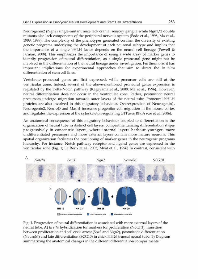
An anatomical consequence of this migratory behaviour coupled to differentiation is the organization of neural tube in distinct cell layers, compartmentalizing differentiation stages progressively in concentric layers, where internal layers harbour younger, more undifferentiated precursors and more external layers contain more mature neurons. This spatial organization facilitates the positioning of marker genes in the neurogenic programs hierarchy. For instance, Notch pathway receptor and ligand genes are expressed in the ventricular zone (Fig. 1; Le Roux et al., 2003; Myat et al., 1996) In contrast, consistent with
Fig. 1. Progression of neural differentiation is associated with more external layers of the neural tube. A) In situ hybridization for markers for proliferation (Notch1), transition between proliferation and cell cycle arrest (Sox3 and Ngn2), postmitotic differentiation (NeuroM) and late differentiation (SCG10) in chick HH26 truncal neural tube. B) Diagram summarizing the anatomical changes in the different differentiation compartments.
Embryogenesis
254
their role in initiating differentiation, Sox3 and Neurogenin1 are expressed at the ventricular layer and slightly beyond the proliferative zone as well (Fig. 1; Bylund et al., 2003). Ath3/NeuroM is mainly expressed by post-mitotic neural precursors, and the expression domain borders the external perimeter of Neurogenin1 and Sox3 (Fig. 1; Roztocil et al., 1997). This domain is also known as the intermediate layer and contains neural progenitors in the early stages of differentiation. Other markers for this stage include the RNA-binding protein Hu and the RNA splicing factor NeuN/Fox3 (Dent et al., 2010; Kim et al., 2009; Wakamatsu & Weston, 1997). Finally, the late differentiation marker SCG10 is expressed by cells at the outer border (mantle layer) of the neural tube (Fig. 1; Stein et al., 1988).
SCG10 encodes a membrane-associated protein associated with the growth cones of developing neurons (Stein et al., 1988). Its presence in the developing neural tube correlates with the onset of late differentiation events such as neuritogenesis. An additional marker that is widely used to characterize post-mitotic differentiating neurons is beta III tubulin (recognized by the monoclonal antibody Tuj1; Lewis & Cowan, 1988; Lee et al., 1990; Menezes and Luskin, 1994).
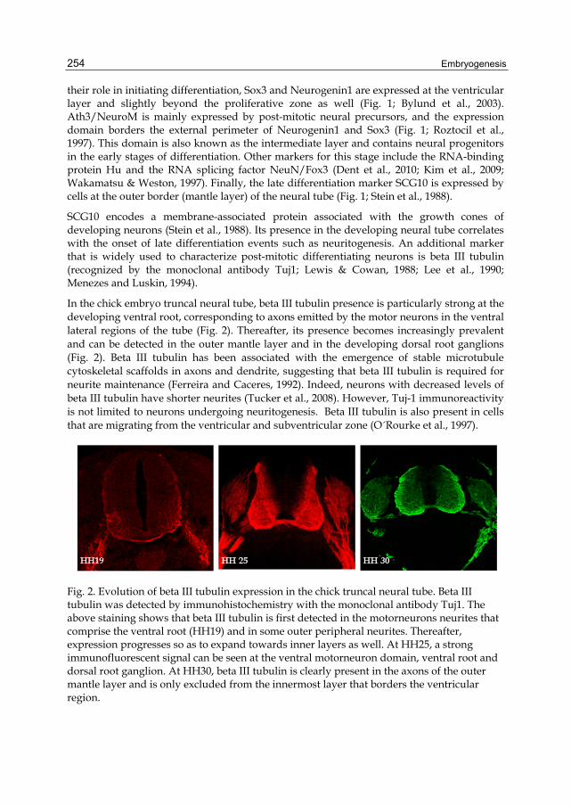
In the chick embryo truncal neural tube, beta III tubulin presence is particularly strong at the developing ventral root, corresponding to axons emitted by the motor neurons in the ventral lateral regions of the tube (Fig. 2). Thereafter, its presence becomes increasingly prevalent and can be detected in the outer mantle layer and in the developing dorsal root ganglions (Fig. 2). Beta III tubulin has been associated with the emergence of stable microtubule cytoskeletal scaffolds in axons and dendrite, suggesting that beta III tubulin is required for neurite maintenance (Ferreira and Caceres, 1992). Indeed, neurons with decreased levels of beta III tubulin have shorter neurites (Tucker et al., 2008). However, Tuj-1 immunoreactivity is not limited to neurons undergoing neuritogenesis. Beta III tubulin is also present in cells that are migrating from the ventricular and subventricular zone (O´Rourke et al., 1997).
Fig. 2. Evolution of beta III tubulin expression in the chick truncal neural tube. Beta III tubulin was detected by immunohistochemistry with the monoclonal antibody Tuj1. The above staining shows that beta III tubulin is first detected in the motorneurons neurites that comprise the ventral root (HH19) and in some outer peripheral neurites. Thereafter, expression progresses so as to expand towards inner layers as well. At HH25, a strong immunofluorescent signal can be seen at the ventral motorneuron domain, ventral root and dorsal root ganglion. At HH30, beta III tubulin is clearly present in the axons of the outer mantle layer and is only excluded from the innermost layer that borders the ventricular region.
Gene Expression in Embryonic Neural Development and Stem Cell Differentiation
255
Consistent with the importance of microtubule cytoskeleton in the latter stages of neural differentiation, several microtubule-associated proteins such as MAP2 and Tau are also up-regulated. MAP2 and MAP1B double knockout mice have fiber tract malformations and retarded neuronal migration. Additionally, primary neuronal cultures derived from these mice display reduced neurite outgrowth (Teng et al., 2001).
5. Stem cell neural differentiation recapitulates embryogenesis
In vitro differentiation of embryonic stem cells into neural lineages aims to recapitulate the multistep process – from induction to terminal differentiation - of neural embryogenesis described above. Indeed as in embryonic epiblast induction, some cell lines, neural induction is more efficiently induced by the combination of fibroblast growth factor (FGF) signalling and bone morphogenetic protein (BMP) inhibition (LaVaute et al., 2009; Tropepe et al., 2001; Ying et al., 2003). In these reports, the endogenous production of BMP inhibitors was sufficient to avoid epidermal fate. However, conservation of embryogenic signalling is not a rule for all cell lines. Some iPSCs (induced Pluripotent Stem Cells) do not improve their neural differentiation rate with FGF signalling and/or BMP inhibition (Hu et al., 2010). Thus, the extent of recall of embryogenesis in these experimental paradigms is still an open question and begs for future analysis.
There are multiple protocols for in vitro neural induction and the depth of analysis regarding similarity with embryogenesis varies. Some groups provide a detailed comparison with embryogenesis. For instance, Abranches and collaborators report expression of Sox genes during the early phases of induced differentiation, interkinetic nuclear migration and Notch-signalling and subsequent expression of the postmitotic neural (Hu) and glial markers (GFAP) (Abranches et al., 2009). However, most reports concentrate on the detection of late developmental neural markers such as MAP2, Tau, NeuN and beta III tubulin, which have been generally accepted in the community as indication for stem cell neuronal differentiation (Tropepe et al., 2001).
For instance, Kerkis and collaborators detected the presence microtubule-associated proteins (MAPs), such as Lis1 and Ndel1, as neural markers at early stages of in vitro model for neuronal differentiation from pluripotent stem cells (Kerkis et al., 2011).
6. Lis1 and Ndel1: Microtubule associated proteins involved in neural development
The microtubule associated proteins (MAPs), Lis1 and Ndel1 are involved in neuronal differentiation and cell migration during the CNS development.
Lis1, also known as platelet-activating factor acetylhydrolase (PAF-AH), regulates microtubule function and is essential for proper neuronal migration during cortical development (Arai, 2002). Mutations in Lis1 gene have been associated with neuronal migration defects and abnormal layering of the cortex (Reiner et al., 1995; Saillour et al., 2009; Youn et al., 2009). For instance, haploinsufficiency of Lis1 alone causes congenital malformation of brain folds and grooves, i.e. lissencephaly. Lis 1 microdeletion is also part of the genetic causes of Miller-Dieker syndrome (MDS; Miller, 1963; Dieker et al., 1969; Reiner et al., 1993). Besides lissencephaly, MDS patients also present hypoplasic corpus callosum. Together, these data underscore the importance of Lis1 in proper neuronal migration and axon formation.
Embryogenesis
256
In support of the importance of Lis1 in neural development, Lis1-binding protein Ndel1 (Nuclear-distribution Element like-1) also plays a relevant role in the proper establishment of the nervous system. Lis1 and Ndel1 co-localize predominantly in the centrosome in early neuroblasts, and later, redistribute to axons during neuronal development (Shu et al., 2004; Guo et al., 2006; Bradshaw et al., 2008; Hayashi et al., 2010). The direct association of Lis1 with the Ndel1 fungal homologue was first shown in 2000 (Kitagawa et al., 2000), and soon after the interaction with the mammalian homologue was also demonstrated (Sweeney et al., 2001).
Ndel1 is also known as endooligopeptidase A or EOPA and was first isolated due to its ability to inactivate bioactive peptides. Ndel1/EOPA, is a thiol-sensitive enzyme inactivates physiologically important peptides such as bradykinin and neurotensin, and also converts opioid oligopeptides into enkephalins (Camargo et al., 1973, 1983, 1987; Gomes et al., 1993; Hayashi et al., 2000, 2005). The contribution of bradykinin and neurotensin neuropeptides in neurite outgrowth was also previously described (Zhao et al., 2003; Tischler et al., 1991; Robson and Burgoyne, 1989; Tischler et al., 1984).
In normal cortical development Ndel1 is involved in microtubule organization, nuclear translocation and neuronal positioning (Shu et al., 2004; Youn et al., 2009; Bradshaw et al., 2011). Knockdown or ablation of cortical Ndel1 function also results in impaired migration of neocortical projection neurons (Sasaki et al., 2005; Youn et al., 2009). Deletion of Ndel1 by RNAi leads to deficits in neuronal positioning and uncouples the centrosome from the nucleus, resulting in aberrant neuronal migration (Shu et al., 2004). Ndel1 homozygous knockout mice have similar deficits in neuronal positioning (Sasaki et al., 2005; Youn et al., 2009).
7. Expression of Ndel1 in the developing CNS and ES cells
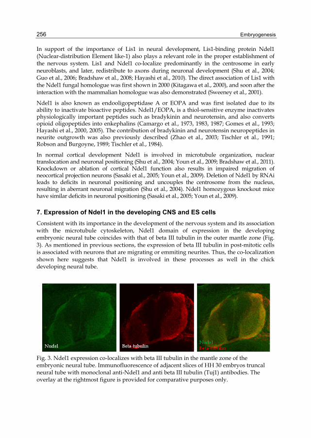
Consistent with its importance in the development of the nervous system and its association with the microtubule cytoskeleton, Ndel1 domain of expression in the developing embryonic neural tube coincides with that of beta III tubulin in the outer mantle zone (Fig. 3). As mentioned in previous sections, the expression of beta III tubulin in post-mitotic cells is associated with neurons that are migrating or emmiting neurites. Thus, the co-localization shown here suggests that Ndel1 is involved in these processes as well in the chick developing neural tube.
Fig. 3. Ndel1 expression co-localizes with beta III tubulin in the mantle zone of the embryonic neural tube. Immunofluorescence of adjacent slices of HH 30 embryos truncal neural tube with monoclonal anti-Ndel1 and anti beta III tubulin (Tuj1) antibodies. The overlay at the rightmost figure is provided for comparative purposes only.
Gene Expression in Embryonic Neural Development and Stem Cell Differentiation
257
Likewise, the dynamics of Ndel1 localization in stem cells during neural differentiation suggests that it is recruited for neurite extension. In undifferentiated ES cells, both Lis1 and Ndel1 show a perinuclear co-localization (Hayashi et al., 2011). In contrast, after the onset of neuronal differentiation, Lis1 presents a cytoplasmic and Ndel1 a perinuclear localization. Following differentiation, both Lis1 and Ndel1 co-localize in the outgrowing neurites (Kerkis et al., 2011).
The presence of Ndel1 persists in adult brains. Northern blot analysis confirmed its preferential expression in the rabbit and rodent CNS (Hayashi et al., 1996; 2000), although this could not be confirmed for humans (Guerreiro et al., 2005). Later, the presence of Ndel1 in the adult brain was confirmed by in situ hybridization studies, with higher expression in some regions, such as the hippocampus, cerebellum, and basal nucleus of Meynert (Hayashi et al., 2001). This study provided a basis for phenotypic identification of Ndel1-expressing neurons throughout the rat brain and showed a correlation between the distribution of Ndel1 neurons and systems responsible for motor, sensory, endocrine, and possibly for other functions. Together, these expression patterns argue in favour of a role for Ndel1 in neurite growth and maintenance.
8. In vitro assays for Ndel1 cellular function
As mentioned previously, clinical correlation data suggested strongly that Lis1 and Ndel1 are involved in neuronal migration during cortical layer formation. Lis1 and Ndel1 participate in nuclear and centrosomal transport in migrating neurons (Shu et al., 2004; Tsai et al., 2005). Additionally, they influence centrosome positioning in migrating non-neuronal cells (Dujardin et al., 2003; Stehman et al., 2007; Shen et al., 2008). Moreover, dominant negative overexpression of either the enzymatic active form of Ndel1 or its orthologue mNudE disrupted CNS lamination in Xenopus laevis embryos (Hayashi et al., 2004; Feng et al., 2000).
In an attempt to elucidate the exact role of Lis1 and Ndel1 in neuronal migration during cortical layer formation, we have used long-term adherent neurosphere cultures to mimic the development of cortical layers in vitro (Hayashi et al., 2011). In this experimental model, the neuropsheres grow for two weeks without splitting and the resulting aggregates present an inner core that would correspond to the inner cortical layer where migrating neurons originate from, and an outer layer that harbors neurons that finished their migration. In this experimental paradigm, a significant variation in spatial distribution of Lis1 and Ndel1 proteins was observed (Kerkis et al., 2011). Lis1, but not Ndel1, was detected in the rosette cells localized at the inner part of the cellular aggregates. In contrast, co-localization of both Lis1 and Ndel1 was observed in the cells at the peripheral layer of the cellular aggregates (Kerkis et al., 2011). Although further analysis with other MAPs would provide a better picture of the role of Lis1 and Ndel1 in neuronal migration during establishment of cortical layers, these data nonetheless indicate that these two proteins play a differential role in the establishment and maintenance of neuronal layers.
The role of Ndel1 in neurite outgrowth has been better characterized. Knockdown of Ndel1 expression in rat pheochromocytoma PC12 cell line inhibits neurite outgrowth. This inhibition can be rescued by wild-type Ndel1 (Ndel1WT), but not by a mutant (Ndel1mut273), which does not have enzymatic activity (Hayashi et al., 2010). This result indicates that
Embryogenesis
258
Ndel1 enzymatic activity plays a crucial role in neurite outgrowth. In support to this, a significant increase of Ndel1 promoter activity during the period of maximal neurite outgrowth was observed (Hayashi et al., 2010).
Clearly, the expression of Ndel1 shows strong correlation with the onset of various aspects of embryonic neural development and ES cells and PC12 cells neural differentiation. Thus, we directed our attention towards cis-regulatory elements that could regulate neuro-specific Ndel1 expression in a variety of experimental models.
9. Control of Ndel1 expression
The promoter of both rabbit and human Ndel1 gene was analyzed by the group in cultured cell lines. Interestingly, the Ndel1 promoter activity was shown to be very different in neuronal and non-neuronal cells, with a stronger activity in NH15 neuronal compared to C6 glial cells for the rabbit full-length promoter, thus confirming the preferential neuronal expression of. However, such difference was not observed for the human full-length promoter under the same conditions (Guerreiro et al., 2005).
We’ve isolated the rabbit promoter fragment -888/-744 as the region responsible for determining the neuronal-specific expression. This DNA segment contains potential binding motifs for the CP2 and SRY (sex-determining region Y) transcription factors. SRY is the founding member of the Sox (Sry-related HMG box) gene family (Sekido, 2010). Moreover, strong negative regulator elements were found within positions -755/-450 and -314/-245 in both human and rabbit promoters. Of these, at least one common negative cis-regulating region seems to be acting in the control Ndel1 expression in both species (Guerreiro et al., 2005). During neural development, these elements may restrict Ndel1 promoter activity to a neuronal subtype or a specific period of differentiation.
In the human Ndel1 promoter, the critical regulatory domain lies between -314/-245. Within this region we also found a single putative binding site for a member of the Sox transcription factor family. It is tempting to speculate on the identity of members of the Sox family, which now number more than 20, that regulate Ndel1 expression (Lefebvre et al., 2007). There are certain members of the Sox family, which we could speculatively nominate as candidates to mediate the increased expression of Ndel1. For instance, accumulated evidence has shown that Sox 11 is relevant for neurite outgrowth. As Neuro2a cells undergo retinoic acid-induced neuronal differentiation, Sox 11 levels increase significantly. Conversely, RNAi knockdown of Sox 11 inhibits axon outgrowth in Neuro2a cells, Dorsal Root Ganglion neurons and regeneration in nerve injury models (Jankowski et al., 2006; Jankowski et al., 2009).
10. Conclusion
Microtubule-associated proteins (MAPs) are essential for neuronal differentiation and cell migration during the central nervous system (CNS) development and also in the adult nervous system. In particular the distribution and role of lissencephaly (Lis1) and nuclear distribution element-like (Ndel1) allows the comparison between neural differentiation in stem cells and during embryo development. They are very powerful tools not only due to
Gene Expression in Embryonic Neural Development and Stem Cell Differentiation
259
their putative role as expression markers of the differentiation process, but also due to their confirmed role in the cell maturation and migration processes. Furthermore, the study of cis-regulatory regions that confer neural-specificity to Ndel1 expression can increase our understanding of gene expression control during neural differentiation.
11. Acknowledgment
The authors are grateful for the continuous support by Fundação de Amparo à Pesquisa do Estado de São Paulo (FAPESP) and Conselho Nacional de Desenvolvimento Científico e Tecnológico (CNPq).
12. References
Abranches, E.; Silva, M.; Pradier, L.; Schulz, H.; Hummel, O.; Henrique, D. & Bekman, E. (2009). Neural differentiation of embryonic stem cells in vitro: a road map to neurogenesis in the embryo. PLoS One, Vol. 4(7), e6286.
Akai, J.; Halley, P.A. & Storey, K.G. (2005). FGF-dependent Notch signaling maintains the spinal cord stem zone. Genes & Development, Vol. 19(23), pp. 2877-2887.
Arai H. (2002). Platelet-activating factor acetylhydrolase. Prostaglandins Other Lipid Mediat., Vol. 68-69, pp 83-94. Review.
Almeida, K.L.; Abreu, J.G. & Yan, C.Y.I. (2010). Neural Induction. In: Perspectives of Stem Cells-From Tools for Studying Mechanisms of Neuronal Differentiation towards Therapy, H. Ulrich, (Org.), Dordresch: Springer, ISBN 978-90-481-3374-1.
Bertrand, N.; Castro, D.S. & Guillemot, F. (2002). Proneural genes and the specification of neural cell types. Nature Reviews Neuroscience, Vol. 3(7), pp. 517-530.
Bradshaw, N.J.; Ogawa, F.; Antolin-Fontes, B.; Chubb, J.E.; Carlyle, B.C.; Christie, S.; Claessens, A.; Porteous, D.J. & Millar, J.K. (2008). DISC1, PDE4B, and NDE1 at the centrosome and synapse. Biochemical and Biophysical Research Communications, Vol. 377(4), pp. 1091-1096. Erratum in: Biochemical and Biophysical Research Communications, Vol. 384(3), pp. 400 (2009).
Bradshaw, N.J.; Soares, D.C.; Carlyle, B.C.; Ogawa, F.; Davidson-Smith, H.; Christie, S.; Mackie, S.; Thomson, P.A.; Porteous, D.J. & Millar, J.K. (2011). PKA phosphorylation of NDE1 is DISC1/PDE4 dependent and modulates its interaction with LIS1 and NDEL1. J Neurosci. Vol. 31(24), pp. 9043-54.
Brandon, N.J.; Handford, E.J.; Schurov, I.; Rain, J.C.; Pelling, M.; Duran-Jimeniz, B.; Camargo, L.M.; Oliver, K.R.; Beher, D.; Shearman, M.S. & Whiting, P.J. (2004). Disrupted in Schizophrenia 1 and Nudel form a neurodevelopmentally regulated protein complex: implications for schizophrenia and other major neurological disorders. Molecular and Cellular Neurosciences, Vol. 25(1), pp. 42-55.
Bultje, R.S.; Castaneda-Castellanos, D.R.; Jan, L.Y.; Jan, Y.N.; Kriegstein, A.R. & Shi, S.H. (2009). Mammalian Par3 regulates progenitor cell asymmetric division via notch signaling in the developing neocortex. Neuron, Vol. 63(2), pp. 189-202.
Burdick, K.E.; Kamiya, A.; Hodgkinson, C.A.; Lencz, T.; DeRosse, P.; Ishizuka, K.; Elashvili, S.; Arai, H.; Goldman, D.; Sawa, A. & Malhotra, A.K. (2008). Elucidating the relationship between DISC1, NDEL1 and NDE1 and the risk for schizophrenia:
Embryogenesis
260
evidence of epistasis and competitive binding. Hum. Mol. Genet., Vol. 17, pp. 2462–2473.
Bylund, M.; Andersson, E.; Novitch, B.G. & Muhr, J. (2003). Vertebrate neurogenesis is counteracted by Sox1-3 activity. Nature Neuroscience, Vol. 6(11), pp. 1162-1168.
Camargo, A.C.; Shapanka, R. & Greene, L.J. (1973). Preparation, assay, and partial characterization of a neutral endopeptidase from rabbit brain. Biochemistry, Vol. 12(9), pp. 1838-1844.
Camargo, A.C.; Caldo, H. & Emson, P.C. (1983). Degradation of neurotensin by rabbit brain endo-oligopeptidase A and endo-oligopeptidase B (proline-endopeptidase). Biochemical and Biophysical Research Communications, Vol. 116(3), pp. 1151-1159.
Camargo, A.C.; Oliveira, E.B.; Toffoletto, O.; Metters, K.M. & Rossier, J. (1987). Brain endo-oligopeptidase A, a putative enkephalin converting enzyme. Journal of Neurochemistry, Vol. 48(4), pp. 1258-1263.
Casarosa, S.; Fode, C. & Guillemot, F. (1999). Mash1 regulates neurogenesis in the ventral telencephalon. Development, Vol.126(3), pp. 525-534.
Chenn, A. & McConnell, S.K. (1995). Cleavage orientation and the asymmetric inheritance of Notch1 immunoreactivity in mammalian neurogenesis. Cell, Vol. 82(4), pp. 631-641.
Dent, M.A.; Segura-Anaya, E.; Alva-Medina, J. & Aranda-Anzaldo, A. (2010). NeuN/Fox-3 is an intrinsic component of the neuronal nuclear matrix. FEBS Letters, Vol. 584(13), pp. 2767-2771.
Dieker, H.; Edwards, R.H.; Zurhein, G.; Chou, S.M.; Hartman, H.A. & Optiz, J.M. (1969). The lissencephaly syndrome. Birth Defects, Vol. 5, pp. 53-64.
Dobyns, W.B.; Stratton, R.F. & Greenberg, F. (1984). Syndromes with lissencephaly. I: Miller-Dieker and Norman-Roberts syndromes and isolated lissencephaly. Am J Med Genet., Vol. 18(3), pp. 509-26.
Duan, X.; Chang, J.H.; Ge, S.; Faulkner, R.L.; Kim, J.Y.; Kitabatake, Y.; Liu, X.B.; Yang, C.H.; Jordan, J.D.; Ma, D.K.; Liu, C.Y.; Ganesan, S.; Cheng, H.J.; Ming, G.L.; Lu, B. & Song, H. (2007). Disrupted-In-Schizophrenia 1 regulates integration of newly generated neurons in the adult brain. Cell, Vol. 130(6), pp. 1146-58.
Dujardin, D.L.; Barnhart, L.E.; Stehman, S.A.; Gomes, E.R.; Gundersen, G.G. & Vallee, R.B. (2003). A role for cytoplasmic dynein and LIS1 in directed cell movement. J Cell Biol., Vol. 163(6), pp. 1205-11.
Faulkner, N.E.; Dujardin, D.L.; Tai, C.Y.; Vaughan, K.T.; O'Connell, C.B.; Wang, Y. & Vallee, R.B. (2000). A role for the lissencephaly gene LIS1 in mitosis and cytoplasmic dynein function. Nat Cell Biol., Vol. 2(11), pp. 784-91.
Feng, Y.; Olson, E.C.; Stukenberg, P.T.; Flanagan, L.A.; Kirschner, M.W. & Walsh, C.A. (2000). LIS1 regulates CNS lamination by interacting with mNudE, a central component of the centrosome. Neuron, Vol. 28(3), pp. 665-79.
Fernández-Garre, P.; Rodríguez-Gallardo, L.; Gallego-Díaz, V.; Alvarez, I.S. & Puelles, L. (2002). Fate map of the chicken neural plate at stage 4. Development, Vol. 129(12), pp. 2807-2822.
Fode, C.; Gradwohl, G.; Morin, X.; Dierich, A.; LeMeur, M.; Goridis, C. & Guillemot, F. (1998). The bHLH protein NEUROGENIN 2 is a determination factor for epibranchial placode-derived sensory neurons. Neuron, Vol. 20(3), pp. 483-494.
Gene Expression in Embryonic Neural Development and Stem Cell Differentiation
261
Ge, W.; He, F.; Kim, K.J.; Blanchi, B.; Coskun, V.; Nguyen, L.; Wu, X.; Zhao, J.; Heng, J.I.; Martinowich, K.; Tao, J.; Wu, H.; Castro, D.; Sobeih, M.M.; Corfas, G.; Gleeson, J.G.; Greenberg, M.E.; Guillemot, F. & Sun, Y.E. (2006). Coupling of cell migration with neurogenesis by proneural bHLH factors. PNAS, Vol. 103(5), pp. 1319-1324.
Gomes, M.D.; Juliano, L.; Ferro, E.S.; Matsueda, R.; Camargo, A.C. (1993). Dynorphin-derived peptides reveal the presence of a critical cysteine for the activity of brain endo-oligopeptidase A. Biochem. Biophys. Res. Commun. Vol. 197(2), pp. 501-507.
Guerreiro, J.R.; Winnischofer, S.M.; Bastos, M.F.; Portaro, F.C.; Sogayar, M.C.; de Camargo, A.C. & Hayashi, M.A. (2005). Cloning and characterization of the human and rabbit NUDEL-oligopeptidase promoters and their negative regulation. Biochimica et Biophysica Acta, Vol. 1730(1), pp. 77-84.
Guo, J.; Yang, Z.; Song, W.; Chen, Q.; Wang, F.; Zhang, Q. & Zhu, X. (2006). Nudel contributes to microtubule anchoring at the mother centriole and is involved in both dynein-dependent and -independent centrosomal protein assembly. Molecular Biology of the Cell, Vol. 17(2), pp. 680-689.
Hämmerle, B. & Tejedor, F.J. (2007). A novel function of DELTA-NOTCH signalling mediates the transition from proliferation to neurogenesis in neural progenitor cells. PLoS One, Vol. 2(11), e1169.
Hattori, M.; Adachi, H.; Tsujimoto, M.; Arai, H. & Inoue, K. (1994). Miller-Dieker lissencephaly gene encodes a subunit of brain platelet-activating factor acetylhydrolase [corrected]. Nature, Vol. 370(6486), pp. 216-8. Erratum in: Nature, Vol. 370(6488), pp. 391.
Hayashi, M.A.; Gomes, M.D.; Rebouças, N.A.; Fernandes, B.L.; Ferro, E.S. & de Camargo, A.C. (1996). Species specificity of thimet oligopeptidase (EC 3.4.24.15). Biological Chemistry Hoppe-Seyler, Vol. 377(5), pp. 283-291.
Hayashi, M.A.; Portaro, F.C.; Tambourgi, D.V.; Sucupira, M.; Yamane, T.; Fernandes, B.L.; Ferro, E.S.; Rebouças, N.A. & de Camargo, A.C. (2000). Molecular and immunochemical evidences demonstrate that endooligopeptidase A is the predominant cytosolic oligopeptidase of rabbit brain. Biochemical and Biophysical Research Communications, Vol. 269(1), pp. 7-13. Erratum in: Biochemical and Biophysical Research Communications, Vol. 272(1), pp. 309 (2000).
Hayashi, M.A.; Pires, R.S.; Rebouças, N.A.; Britto, L.R. & Camargo, A.C. (2001). Expression of endo-oligopeptidase A in the rat central nervous system: a non-radioactive in situ hybridization study. Brain Research. Molecular Brain Research, Vol. 89(1-2), pp. 86-93.
Hayashi, M.A.; Portaro, F.C.; Bastos, M.F.; Guerreiro, J.R.; Oliveira, V.; Gorrão, S.S.; Tambourgi, D.V.; Sant'Anna, O.A.; Whiting, P.J.; Camargo, L.M.; Konno, K.; Brandon, N.J. & Camargo, A.C. (2005). Inhibition of NUDEL (nuclear distribution element-like)-oligopeptidase activity by disrupted-in-schizophrenia 1. PNAS, Vol. 102(10), pp. 3828-3833.
Hayashi, M.A.F.; Guerreiro, J.R.; Charych, E.; Kamiya, A.; Barbos, R.L.; Machado, M.F.; Campeiro, J.D.; Oliveira, V.; Sawa, A.; Camargo, A.C. and Brandon, N.J. (2010) Assessing the role of endooligopeptidase activity of Ndel1 (nuclear-distribution
Embryogenesis
262
gene E homolog like-1) in neurite outgrowth. Mol Cell Neurosci. Vol. 44(4), pp. 353-61.
Hayashi, M.A.F.; Lizier, N.F.; Pereira, L. & Kerkis, A. (2011). Pluripotent stem cells as an in vitro model of neuronal differentiation. Embryonic Stem Cells – Differentiation and Pluripotent Alternatives, Book 3, Vol. 4, pp. 81-98.
Hayashi, M.A.F.; Portaro, F.C.V. & Camargo, A.C.M. (2005). Cytosolic oligopeptidases: features and possible physiopathological roles in the immune and nervous systems. Current Medicinal Chemistry (Hilversum), Vol. 4, pp. 269-277.
Hayes, N.L. & Nowakowski, R.S. (2000). Exploiting the dynamics of S-phase tracers in developing brain: interkinetic nuclear migration for cells entering versus leaving the S-phase. Developmental Neuroscience, Vol. 22(1-2), pp. 44-55.
Horton, S.; Meredith, A.; Richardson, J.A. & Johnson, J.E. (1999). Correct coordination of neuronal differentiation events in ventral forebrain requires the bHLH factor MASH1. Molecular and Cellular Neurosciences, Vol. 14(4-5), pp. 355-369.
Hu, B.Y.; Weick, J.P.; Yu, J.; Ma, L.X.; Zhang, X.Q.; Thomson, J.A. & Zhang, S.C. (2010). Neural differentiation of human induced pluripotent stem cells follows developmental principles but with variable potency. PNAS, Vol. 107(9), pp. 4335-4340.
Jaaro-Peled, H.; Hayashi-Takagi, A.; Seshadri, S.; Kamiya, A.; Brandon, N.J. & Sawa, A. (2009). Neurodevelopmental mechanisms of schizophrenia: understanding disturbed postnatal brain maturation through neuregulin-1-ErbB4 and DISC1. Trends Neurosci., Vol. 32(9), pp. 485-95.
Jankowski, M.P.; Cornuet, P.K.; McIlwrath, S.; Koerber, H.R. & Albers, K.M. (2006). SRY-box containing gene 11 (Sox11) transcription factor is required for neuron survival and neurite growth. Neuroscience, Vol. 143(2), pp. 501-14.
Jankowski, M.P.; McIlwrath, S.L.; Jing, X.; Cornuet, P.K.; Salerno, K.M.; Koerber, H.R. & Albers, K.M. (2009). Sox11 transcription factor modulates peripheral nerve regeneration in adult mice. Brain Res., Vol. 1256, pp. 43-54.
Kageyama, R.; Ohtsuka, T.; Shimojo, H. and Imayoshi, I. (2008). Dynamic Notch signaling in neural progenitor cells and a revised view of lateral inhibition. Nature Neuroscience, Vol. 11(11), pp. 1247-1251.
Kamiya, A.; Tomoda, T.; Chang, J.; Takaki, M.; Zhan, C.; Morita, M.; Cascio, M.B.; Elashvili, S.; Koizumi, H.; Takanezawa, Y.; Dickerson, F.; Yolken, R.; Arai, H. & Sawa, A. (2006). DISC1–NDEL1/NUDEL protein interaction, an essential component for neurite outgrowth, is modulated by genetic variations of DISC1. Hum. Mol. Genet., Vol. 15, pp. 3313–3323.
Kamiya, A.; Kubo, K.; Tomoda, T.; Takaki, M.; Youn, R.; Ozeki, Y.; Sawamura, N.; Park, U.; Kudo, C.; Okawa, M.; Ross, C.A.; Hatten, M.E.; Nakajima, K. & Sawa, A. (2005). A schizophrenia-associated mutation of DISC1 perturbs cerebral cortex development. Nat Cell Biol., Vol. 7(12), pp. 1167-78. Erratum in: Nat Cell Biol., 2006 Vol. 8(1), pp. 100.
Kawaguchi, D.; Yoshimatsu, T.; Hozumi, K. & Gotoh, Y. (2008). Selection of differentiating cells by different levels of delta-like 1 among neural precursor cells in the developing mouse telencephalon. Development, Vol. 135(23), pp. 3849-3858.
Gene Expression in Embryonic Neural Development and Stem Cell Differentiation
263
Kerkis, I.; Hayashi, M.A.F.; Lizier, N.F.; Cassola, A.C.; Pereira, L.V. & Kerkis, A. (2011). Pluripotent Stem Cells as an In Vitro Model of Neuronal Differentiation, InTech, ISBN 978-953-307-632-4, Vienna, Austria.
Kim, K.K.; Adelstein, R.S. & Kawamoto, S. (2009). Identification of neuronal nuclei (NeuN) as Fox-3, a new member of the Fox-1 gene family of splicing factors. The Journal of Biological Chemistry. Vol. 284(45), pp. 31052-31061.
Kitagawa, M.; Umezu, M.; Aoki, J.; Koizumi, H.; Arai, H. & Inoue, K. (2000). Direct association of LIS1, the lissencephaly gene product, with a mammalian homologue of a fungal nuclear distribution protein, rNUDE. FEBS Lett., Vol. 479(1-2), pp. 57-62.
Konno, D.; Shioi, G.; Shitamukai, A.; Mori, A.; Kiyonari, H.; Miyata, T. & Matsuzaki, F. (2008). Neuroepithelial progenitors undergo LGN-dependent planar divisions to maintain self-renewability during mammalian neurogenesis. Nature Cell Biology, Vol. 10(1), pp. 93-101.
Latasa, M.J.; Cisneros, E. & Frade, J.M. (2009). Cell cycle control of Notch signaling and the functional regionalization of the neuroepithelium during vertebrate neurogenesis. The International Journal of Developmental Biology, Vol.53(7), pp. 895-908.
LaVaute, T.M.; Yoo, Y.D.; Pankratz, M.T.; Weick, J.P.; Gerstner, J.R. & Zhang, S.C. (2009). Regulation of neural specification from human embryonic stem cells by BMP and FGF. Stem Cells, Vol. 27(8), pp. 1741-1749.
Lefebvre, V.; Dumitriu, B.; Penzo-Méndez, A.; Han, Y. & Pallavi, B. (2007). Control of cell fate and differentiation by Sry-related high-mobility-group box (Sox) transcription factors. Int J Biochem Cell Biol., Vol. 39(12), pp. 2195-214.
le Roux, I.; Lewis, J. and Ish-Horowicz, D. (2003). Notch activity is required to maintain floorplate identity and to control neurogenesis in the chick hindbrain and spinal cord. The International Journal of Developmental Biology, Vol. 47(4), pp. 263-272.
Lee, M.K.; Tuttle, J.B.; Rebhun, L.I., Cleveland, D.W. and Frankfuter, A. (1990) The expression and posttranslational modification of a neuron-specific beta-tubulin isotype during chick embryogenesis. Cell Mot Cytosk, Vol. 17: 118-132
Lewis, S.A. & Cowan, N.J. (1988). Complex regulation and functional versatility of mammalian alpha- and beta-tubulin isotypes during the differentiation of testis and muscle cells. The Journal of Cell Biology, Vol. 106(6), pp. 2023-2033.
Liang, Y.; Yu, W.; Li, Y.; Yu, L.; Zhang, Q.; Wang, F.; Yang, Z.; Du, J.; Huang, Q.; Yao, X. & Zhu, X. (2007). Nudel modulates kinetochore association and function of cytoplasmic dynein in M phase. Mol Biol Cell, Vol. 18(7), pp. 2656-66.
Lin, C.H.; Stoeck, J.; Ravanpay, A.C.; Guillemot, F.; Tapscott, S.J. & Olson, J.M. (2004). Regulation of neuroD2 expression in mouse brain. Developmental Biology, Vol. 265(1), pp. 234-245.
Linker, C. & Stern, C.D. (2004). Neural induction requires BMP inhibition only as a late step, and involves signals other than FGF and Wnt antagonists. Development, Vol. 131(22), pp. 5671-5681.
Ma, Q.; Kintner, C. & Anderson, D.J. (1996). Identification of neurogenin, a vertebrate neuronal determination gene. Cell, Vol. 87(1),pp. 43-52.
Ma, Q.; Sommer, L.; Cserjesi, P. & Anderson, D.J. (1997). Mash1 and neurogenin1 expression patterns define complementary domains of neuroepithelium in the developing
Embryogenesis
264
CNS and are correlated with regions expressing notch ligands. The Journal of Neuroscience, Vol. 17(10), pp. 3644-3652.
Ma, Q.; Chen, Z.; del Barco Barrantes, I.; de la Pompa, J.L. & Anderson, D.J. (1998). neurogenin1 is essential for the determination of neuronal precursors for proximal cranial sensory ganglia. Neuron, Vol. 20(3), pp. 469-482.
Ma, Q.; Fode, C.; Guillemot, F. & Anderson, DJ. (1999). Neurogenin1 and neurogenin2 control two distinct waves of neurogenesis in developing dorsal root ganglia. Genes & Development, Vol. 13(13), pp. 1717-1728.
McKenney, R.J.; Vershinin, M.; Kunwar, A.; Vallee, R.B. & Gross, S.P. (2010). LIS1 and NudE induce a persistent dynein force-producing state. Cell, Vol. 141(2), pp. 304-14.
Menezes, J.R.L. and Luskin, M.B. (1994). Expression of neuron-specific tubulin defines a novel population in the proliferative layers of the developing telencephalon. J. Neurosci. 14(9): 5399-5416.
Millar, J.K.; Wilson-Annan, J.C.; Anderson, S.; Christie. S.; Taylor, M.S.; Semple, C.A.; Devon, R.S.; St Clair, D.M.; Muir, W.J.; Blackwood, D.H. & Porteous, D.J. (2000). Disruption of two novel genes by a translocation co-segregating with schizophrenia. Hum Mol Genet., Vol. 9(9), pp. 1415-23.
Miller, J.Q. (1963). Lissencephaly in two siblings. Neurology, Vol. 13, pp. 841-850. Mori, D.; Yamada, M.; Mimori-Kiyosue, Y.; Shirai, Y.; Suzuki, A.; Ohno, S.; Saya, H.;
Wynshaw-Boris, A. & Hirotsune, S.(2009). An essential role of the aPKC-Aurora A-NDEL1 pathway in neurite elongation by modulation of microtubule dynamics. Nat Cell Biol., Vol. 11(9), pp. 1057-68.
Muhr, J.; Graziano, E.; Wilson, S.; Jessell, T.M. & Edlund, T. (1999). Convergent inductive signals specify midbrain, hindbrain, and spinal cord identity in gastrula stage chick embryos. Neuron, Vol. 23(4), pp. 689-702.
Myat, A.; Henrique, D.; Ish-Horowicz, D. & Lewis, J. (1996). A chick homologue of Serrate and its relationship with Notch and Delta homologues during central neurogenesis. Developmental Biology, Vol. 174(2), pp. 233-247.
Nguyen, M.D.; Shu, T.; Sanada, K.; Larivière, R.C.; Tseng, H.C.; Park, S.K.; Julien, J.P. & Tsai L.H. (2004). A NUDEL-dependent mechanism of neurofilament assembly regulates the integrity of CNS neurons. Nat Cell Biol., Vol. 6(7), pp. 595-608.
Nicodemus, K.K.; Callicott, J.H.; Higier, R.G.; Luna, A.; Nixon, D.C.; Lipska, B.K.; Vakkalanka, R.; Giegling, I.; Rujescu, D.; Clair, D.S.; Muglia, P.; Shugart, Y.Y. & Weinberger, D.R. (2010). Evidence of statistical epistasis between DISC1, CIT and NDEL1 impacting risk for schizophrenia: biological validation with functional neuroimaging. Hum. Genet., Vol. 127 (4), pp. 453-454.
O´Rourke, N.A.; Chenn, A. And McConnell, S.K. (1997) Postmitotic neurons migrate tangentially in the cortical ventricular zone. Development, 124: 997-1005.
Ossipova, O.; Ezan, J. & Sokol, S.Y. (2009). PAR-1 phosphorylates Mind bomb to promote vertebrate neurogenesis. Developmental Cell, Vol. 17(2), pp. 222-233.
Pera, E.M.; Ikeda, A.; Eivers, E. & De Robertis, E.M. (2003). Integration of IGF, FGF, and anti-BMP signals via Smad1 phosphorylation in neural induction. Genes & Development, Vol. 17(24), pp. 3023-3028.
Gene Expression in Embryonic Neural Development and Stem Cell Differentiation
265
Pierfelice, T.; Alberi, L. & Gaiano, N. (2011). Notch in the vertebrate nervous system: an old dog with new tricks. Neuron, Vol. 69(5), pp. 840-855.
Powell, L.M. & Jarman, A.P. (2008). Context dependence of proneural bHLH proteins. Current Opinion in Genetics & Development, Vol. 18(5), pp. 411-417.
Reiner, O.; Carrozzo, R.; Shen, Y.; Wehnert, M.; Faustinella, F.; Dobyns, W.B.; Caskey, C.T. & Ledbetter, D.H. (1993). Isolation of a Miller-Dieker lissencephaly gene containing G protein beta-subunit-like repeats. Nature, Vol. 364(6439), pp. 717-721.
Reiner, O.; Albrecht, U.; Gordon, M.; Chianese, K.A.; Wong, C.; Gal-Gerber, O.; Sapir, T.; Siracusa, L.D.; Buchberg, A.M.; Caskey, C.T. & Eichele, G. (1995) Lissencephaly gene (LIS1) expression in the CNS suggests a role in neuronal migration. J Neurosci., Vol. 15(5 Pt 2), pp. 3730-8.
Reiner, O.; Sapoznik, S. & Sapir, T. (2006). Lissencephaly 1 linking to multiple diseases: mental retardation, neurodegeneration, schizophrenia, male sterility, and more. Neuromolecular Med., Vol. 8(4), pp. 547-65
Rex, M.; Orme, A.; Uwanogho, D.; Tointon, K.; Wigmore, P.M.; Sharpe, P.T. & Scotting, P.J. (1997). Dynamic expression of chicken Sox2 and Sox3 genes in ectoderm induced to form neural tissue. Developmental Dynamics, Vol. 209(3), pp. 323-332.
Robson, S.J. & Burgoyne, R.D. (1989). L-type calcium channels in the regulation of neurite outgrowth from rat dorsal root ganglion neurons in culture. Neurosci Lett, Vol. 104(1-2), pp. 110-4.
Roztocil, T.; Matter-Sadzinski, L.; Alliod, C.; Ballivet, M. & Matter, J.M. (1997). NeuroM, a neural helix-loop-helix transcription factor, defines a new transition stage in neurogenesis. Development, vol. 124(17), pp. 3263-3672.
Saillour, Y.; Carion, N.; Quelin, C.; Leger, P.L.; Boddaert, N.; Elie, C.; Toutain, A.; Mercier, S.; Barthez, M.A.; Milh, M.; Joriot, S.; des Portes, V.; Philip, N.; Broglin, D.; Roubertie, A.; Pitelet, G.; Moutard, M.L.; Pinard, J.M.; Cances, C.; Kaminska, A.; Chelly, J.; Beldjord, C. & Bahi-Buisson, N. (2009). LIS1-related isolated lissencephaly: spectrum of mutations and relationships with malformation severity. Archives of Neurology, Vol. 66(8), pp. 1007-1015.
Sasaki, S.; Mori, D.; Toyo-oka, K.; Chen, A.; Garrett-Beal, L.; Muramatsu, M.; Miyagawa, S.; Hiraiwa, N.; Yoshiki, A.; Wynshaw-Boris, A. & Hirotsune, S. (2005). Complete loss of Ndel1 results in neuronal migration defects and early embryonic lethality. Molecular and Cellular Biology, Vol. 25(17), pp. 7812-7827.
Sekido, R. (2010). SRY: A transcriptional activator of mammalian testis determination. Int J Biochem Cell Biol., Vol. 42(3), pp. 417-20.
Shen, Y.; Li, N.; Wu, S.; Zhou, Y.; Shan, Y.; Zhang, Q.; Ding, C.; Yuan, Q.; Zhao, F.; Zeng, R. & Zhu, X. (2008). Nudel binds Cdc42GAP to modulate Cdc42 activity at the leading edge of migrating cells. Dev Cell, Vol. 14(3), pp. 342-53.
Shim, S.Y.; Samuels, B.A.; Wang, J.; Neumayer, G.; Belzil, C.; Ayala, R.; Shi, Y.; Shi, Y.; Tsai, L.H. & Nguyen, M.D. (2008). Ndel1 controls the dynein-mediated transport of vimentin during neurite outgrowth. J Biol Chem., Vol. 283(18), pp. 12232-40
Shioi, G.; Konno, D.; Shitamukai, A. & Matsuzaki, F. (2009). Structural basis for self-renewal of neural progenitors in cortical neurogenesis. Cerebral Cortex, Vol. 19, pp. i55-61.
Embryogenesis
266
Shu, T.; Ayala, R.; Nguyen, M.D.; Xie, Z.; Gleeson, J.G. & Tsai, L.H. (2004). Ndel1 operates in a common pathway with LIS1 and cytoplasmic dynein to regulate cortical neuronal positioning. Neuron, Vol. 44(2), pp. 263-277.
Siller, K.H.; Serr, M.; Steward, R.; Hays, T.S. & Doe, C.Q. (2005). Live imaging of Drosophila brain neuroblasts reveals a role for Lis1/dynactin in spindle assembly and mitotic checkpoint control. Mol Biol Cell Vol. 16(11), pp. 5127-40.
St Clair, D.; Blackwood, D.; Muir, W.; Carothers, A.; Walker, M.; Spowart, G.; Gosden, C. & Evans, H.J. (1990). Association within a family of a balanced autosomal translocation with major mental-illness. Lancet, Vol. 336, pp. 13–16.
Stein, R.; Mori, N.; Matthews, K.; Lo, L.C. & Anderson, D.J. (1988). The NGF-inducible SCG10 mRNA encodes a novel membrane-bound protein present in growth cones and abundant in developing neurons. Neuron, Vol. 1(6), pp. 463-476.
Stehman, S.A.; Chen, Y.; McKenney, R.J. & Vallee, R.B. (2007). NudE and NudEL are required for mitotic progression and are involved in dynein recruitment to kinetochores. J Cell Biol., Vol. 178(4), pp. 583-94.
Streit, A.; Sockanathan, S.; Pérez, L.; Rex, M.; Scotting, P.J.; Sharpe, P.T.; Lovell-Badge, R. & Stern C.D. (1997). Preventing the loss of competence for neural induction: HGF/SF, L5 and Sox-2. Development, Vol. 124(6), pp. 1191-1202.
Streit, A.; Lee, K.J.; Woo, I.; Roberts, C.; Jessell, T.M. & Stern, C.D. (1998). Chordin regulates primitive streak development and the stability of induced neural cells, but is not sufficient for neural induction in the chick embryo. Development, Vol. 125(3), pp. 507-519.
Streit, A.; Berliner, A.J.; Papanayotou, C.; Sirulnik, A. & Stern, C.D. (2000). Initiation of neural induction by FGF signalling before gastrulation. Nature, Vol. 406(6791), pp. 74-78.
Sweeney, K.J.; Prokscha, A. & Eichele, G. (2001). NudE-L, a novel Lis1-interacting protein, belongs to a family of vertebrate coiled-coil proteins. Mech Dev., Vol. 101(1-2), pp. 21-33.
Tabler, J.M.; Yamanaka, H. & Green, J.B. (2010). PAR-1 promotes primary neurogenesis and asymmetric cell divisions via control of spindle orientation. Development, Vol. 137(15), pp. 2501-2505.
Talikka, M.; Perez, S.E. & Zimmerman, K. (2002). Distinct patterns of downstream target activation are specified by the helix-loop-helix domain of proneural basic helix-loop-helix transcription factors. Developmental Biology, Vol. 247(1), pp. 137-148.
Tarricone, C.; Perrina, F.; Monzani, S.; Massimiliano, L.; Kim, M.H.; Derewenda, Z.S.; Knapp, S.; Tsai, L.H. & Musacchio, A. (2004). Coupling PAF signaling to dynein regulation: structure of LIS1 in complex with PAF-acetylhydrolase. Neuron, Vol. 44(5), pp. 809-21.
Taya, S.; Shinoda, T.; Tsuboi, D.; Asaki, J.; Nagai, K.; Hikita, T.; Kuroda, S.; Kuroda, K.; Shimizu, M.; Hirotsune, S.; Iwamatsu, A. & Kaibuchi, K. (2007). DISC1 regulates the transport of the NUDEL/LIS1/14-3-3epsilon complex through kinesin-1. J Neurosci., Vol. 27(1), pp. 15-26.
Gene Expression in Embryonic Neural Development and Stem Cell Differentiation
267
Teng, J.; Takei, Y.; Harada, A.; Nakata, T.; Chen. J. & Hirokawa, N. (2001). Synergistic effects of MAP2 and MAP1B knockout in neuronal migration, dendritic outgrowth, and microtubule organization. The Journal of Cell Biology, Vol. 155(1), pp. 65-76.
Tischler, A.S.; Lee, Y.C.; Perlman, R.L.; Costopoulos, D.; Slayton, V.W.; Bloom, S.R. (1984). Production of "ectopic" vasoactive intestinal peptide-like and neurotensin-like immunoreactivity in human pheochromocytoma cell cultures. J Neurosci, Vol. 4(5), pp. 1398-404.
Tischler, A.S.; Ruzicka, A.; Dobner, P.R. (1991). A protein kinase inhibitor, staurosporine, mimics nerve growth factor induction of neurotensin/neuromedin N gene expression. J Biol Chem, Vol. 266(2), pp. 1141-6.
Toth, C.; Shim, S.Y.; Wang, J.; Jiang, Y.; Neumayer, G.; Belzil, C.; Liu, W.Q.; Martinez, J.; Zochodne, D. & Nguyen, M.D. (2008). Ndel1 promotes axon regeneration via intermediate filaments. PLoS One, Vol. 3(4), pp. e2014.
Tropepe, V.; Hitoshi, S.; Sirard, C.; Mak, T.W.; Rossant, J. & van der Kooy, D. (2001). Direct neural fate specification from embryonic stem cells: a primitive mammalian neural stem cell stage acquired through a default mechanism. Neuron, Vol. 30(1), pp. 65-78.
Tsai, J.W.; Chen, Y.; Kriegstein, A.R. & Vallee, R.B. (2005). LIS1 RNA interference blocks neural stem cell division, morphogenesis, and motility at multiple stages. J Cell Biol., Vol. 170(6), pp. 935-45.
Tucker, R.P.; Tran, H; Gong, Q (2008) Neurogenesis and neurite outgrowth in the spical cord of chicken embryos and in primary cultures of spinal neurons following knockdown of Class III beta tubulin with antisense morpholinos. Protoplasma 234: 97-101.
Uchikawa, M.; Ishida, Y.; Takemoto, T.; Kamachi, Y. & Kondoh H. (2003). Functional analysis of chicken Sox2 enhancers highlights an array of diverse regulatory elements that are conserved in mammals. Developmental Cell, Vol. 4(4), pp. 509-519.
Vergnolle, M.A. & Taylor, S.S. (2007). Cenp-F links kinetochores to Ndel1/Nde1/Lis1/dynein microtubule motor complexes. Curr Biol., Vol. 17(13), pp. 1173-9.
Wakamatsu, Y. & Weston, J.A. (1997). Sequential expression and role of Hu RNA-binding proteins during neurogenesis. Development, Vol. 124(17), pp. 3449-3460.
Wilson, P.A. & Hemmati-Brivanlou, A. (1995). Induction of epidermis and inhibition of neural fate by Bmp-4. Nature, Vol. 376(6538), pp. 331-333.
Wilson, S.I.; Graziano, E.; Harland, R.; Jessell, T.M. & Edlund, T. (2000). An early requirement for FGF signalling in the acquisition of neural cell fate in the chick embryo. Current Biology, Vol. 10(8), pp. 421-429.
Wilson, S.I.; Rydström, A.; Trimborn, T.; Willert, K.; Nusse, R.; Jessell, T.M. & Edlund, T. (2001). The status of Wnt signalling regulates neural and epidermal fates in the chick embryo. Nature, Vol. 411(6835), pp. 325-330.
Wood, H.B. & Episkopou, V. (1999). Comparative expression of the mouse Sox1, Sox2 and Sox3 genes from pre-gastrulation to early somite stages. Mechanisms of Development, Vol. 86(1-2), pp. 197-201.
Embryogenesis
268
Ying, Q.L.; Stavridis, M.; Griffiths, D.; Li, M. & Smith, A. (2003). Conversion of embryonic stem cells into neuroectodermal precursors in adherent monoculture. Nature Biotechnology, Vol. 21(2), pp. 183-186.
Youn, Y.H.; Pramparo, T.; Hirotsune, S. & Wynshaw-Boris, A. (2009). Distinct dose-dependent cortical neuronal migration and neurite extension defects in Lis1 and Ndel1 mutant mice. The Journal of Neuroscience, Vol. 29(49), pp. 15520-15530.
Zhao, Y.; Biermann, T., Luther C, Unger T, Culman J, Gohlke P. (2003) Contribution of bradykinin and nitric oxide to AT2 receptor-mediated differentiation in PC12 W cells. J Neurochem, Vol. 85(3), pp. 759-67.
Zigman, M.; Cayouette, M.; Charalambous, C.; Schleiffer, A.; Hoeller, O.; Dunican, D.; McCudden, C.R.; Firnberg, N.; Barres, B.A.; Siderovski, D.P. & Knoblich, J.A. (2005). Mammalian inscuteable regulates spindle orientation and cell fate in the developing retina. Neuron, Vol. 48(4), pp. 539-545.