Embed Size (px)
Citation preview
EMRO Technical Publications Series 30
Guidelines for the earlydetection and screening of
breast cancer
Editors
Oussama M.N. Khatib (MD, PhD, FRCP)Regional Adviser
Noncommunicable DiseasesWHO Regional Office for the Eastern Mediterranean
Atord ModjtabaiProfessor of Pathology
United States of America

© World Health Organization 2006
All rights reserved.
The designations employed and the presentation of the material in this publication do not imply the expression of any opinion whatsoever on the part of the World Health Organization concerning the legal status of any country, territory, city or area or of its authorities, or concerning the delimitation of its frontiers or boundaries. Dotted lines on maps represent approximate border lines for which there may not yet be full agreement.
The mention of specific companies or of certain manufacturers’ products does not imply that they are endorsed or recommended by the World Health Organization in preference to others of a similar nature that are not mentioned. Errors and omissions excepted, the names of proprietary products are distinguished by initial capital letters.
The World Health Organization does not warrant that the information contained in this publication is complete and correct and shall not be liable for any damages incurred as a result of its use.
The named authors alone are responsible for the views expressed in this publication.
Publications of the World Health Organization can be obtained from Distribution and Sales, World Health Organization, Regional Office for the Eastern Mediterranean, PO Box 7608, Nasr City, Cairo 11371, Egypt (tel: +202 670 2535, fax: +202 670 2492; email: [email protected]). Requests for permission to reproduce WHO EMRO publications, in part or in whole, or to translate them – whether for sale or for noncommercial distribution – should be addressed to the Regional Adviser, Health and Biomedical Information, at the above address (fax: +202 276 5400; email [email protected]).
Cover design and layout by Ahmad Hassanein
Printed by Fikra Advertising Agency
WHO Library Cataloguing in Publication Data
Khatib, Oussama M.N.Guidelines for the early detection and screening of breast cancer / Edited by Oussama M.N. Khatib, Atord Modjtabai
p. (EMRO Technical Publications Series ; 30)
1. Breast neoplasms – Diagnosis 2. Breast neoplasms – Prevention and Control3. Breast neoplasms – Epidemiology I. Modjtabai Atord II. Title III. WHO Regional Office for the Eastern Mediterranean IV. Series
ISBN : 978-92-9021-406-9 (NLM Classification : WP 870) ISSN : 1020-0428
Contents
Foreword ..........................................................................................................5Preface ......................................................................................................7Acknowledgements .........................................................................................9
Epidemiology of breast cancer ....................................................................11Global overview .........................................................................................11Regional overview .....................................................................................11International collaboration in breast cancer control ..................................13
Natural history, etiology and risk factors ....................................................14Natural history ...........................................................................................14Etiology .....................................................................................................14Genetic predisposition ..............................................................................14Hormonal factors .......................................................................................15Environmental factors ...............................................................................16Sociobiological factors ..............................................................................16Physiological factors .................................................................................17
Pathology of the breast .................................................................................18Breast disease ..........................................................................................18Staging ......................................................................................................19Clinical course ...........................................................................................20
Managerial aspects of breast cancer detection .........................................21Managerial approach ................................................................................21Surveillance ..............................................................................................21Protection ..................................................................................................21Continuing education ................................................................................22Prevention .................................................................................................22Early detection ..........................................................................................22Care/disease intervention .........................................................................23
Cancer detection programmes .....................................................................24Early detection of cancer ..........................................................................24Cancer screening ......................................................................................24Screening for breast cancer ......................................................................26Screening approach ..................................................................................26
4 Guidelines for the early detection and screening of breast cancer
Breast self-examination ................................................................................27Overview ...................................................................................................27Tactile examination ....................................................................................27Visual examination ....................................................................................30Breast self-examination costs ...................................................................32Mechanisms for improving breast self-examination ..................................32
Clinical breast examination ..........................................................................33Overview ...................................................................................................33Clinical breast examination technique ......................................................33Clinical breast examination effectiveness .................................................34
Screening mammography ............................................................................36Overview ...................................................................................................36Mammograms ...........................................................................................36Mammography reading .............................................................................38
Other breast imaging techniques ................................................................40Ultrasonography ........................................................................................40Computed tomography (CT) .....................................................................40Magnetic resonance imaging (MRI) ..........................................................40Nuclear medicine breast imaging ..............................................................40Positron emission tomographic screening (PET) ......................................41Guided breast biopsy ................................................................................41Breast mass evaluation .............................................................................41
Follow-up and cost-effectiveness ................................................................42Physical health follow-up ...........................................................................42Mental health follow-up .............................................................................42Cost-effectiveness .....................................................................................43Organization of screening programmes ....................................................44
Evaluation of screening programmes .........................................................45
Framework for implementation of the guidelines .......................................46
References ....................................................................................................49
Annex 1 Participants in the consultation on early detection and screening of breast cancer ...........................................................................51
Annex 2 Consensus opinion of the regional task force for developing breast cancer prevention, screening and management guidelines .........52
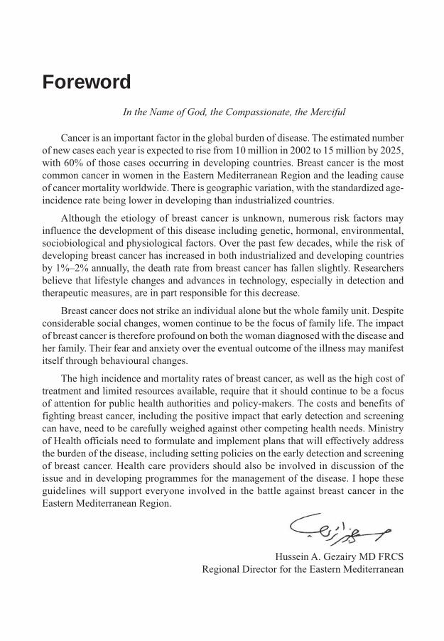
Foreword
Cancer is an important factor in the global burden of disease. The estimated number of new cases each year is expected to rise from 10 million in 2002 to 15 million by 2025, with 60% of those cases occurring in developing countries. Breast cancer is the most common cancer in women in the Eastern Mediterranean Region and the leading cause of cancer mortality worldwide. There is geographic variation, with the standardized age-incidence rate being lower in developing than industrialized countries.
Although the etiology of breast cancer is unknown, numerous risk factors may influence the development of this disease including genetic, hormonal, environmental, sociobiological and physiological factors. Over the past few decades, while the risk of developing breast cancer has increased in both industrialized and developing countries by 1%–2% annually, the death rate from breast cancer has fallen slightly. Researchers believe that lifestyle changes and advances in technology, especially in detection and therapeutic measures, are in part responsible for this decrease.
Breast cancer does not strike an individual alone but the whole family unit. Despite considerable social changes, women continue to be the focus of family life. The impact of breast cancer is therefore profound on both the woman diagnosed with the disease and her family. Their fear and anxiety over the eventual outcome of the illness may manifest itself through behavioural changes.
The high incidence and mortality rates of breast cancer, as well as the high cost of treatment and limited resources available, require that it should continue to be a focus of attention for public health authorities and policy-makers. The costs and benefits of fighting breast cancer, including the positive impact that early detection and screening can have, need to be carefully weighed against other competing health needs. Ministry of Health officials need to formulate and implement plans that will effectively address the burden of the disease, including setting policies on the early detection and screening of breast cancer. Health care providers should also be involved in discussion of the issue and in developing programmes for the management of the disease. I hope these guidelines will support everyone involved in the battle against breast cancer in the Eastern Mediterranean Region.
Hussein A. Gezairy MD FRCSRegional Director for the Eastern Mediterranean
In the Name of God, the Compassionate, the Merciful

Vacat page
Preface
Breast cancer is a major killer of women both globally and regionally. Studies have shown that most patients with breast cancer in the Region present for the first time at stages two and three, indicating the need for increased community awareness and early detection of the disease. This publication aims to assist countries to develop national breast cancer detection programmes by describing the key elements of such programmes. It discusses the epidemiology of breast cancer, its natural history and risk factors, and gives a brief description of various pathological subtypes. A regional overview of the epidemiological situation in the Eastern Mediterranean Region is also provided.
Cancer is a leading cause of death and disability in the Eastern Mediterranean Region, and Member States are becoming increasingly aware of the importance of including cancer control programmes within their national health plans. During the past few years, development of national cancer control programmes has been a principal component of national health planning and several of the resolutions of the WHO Regional Committee for the Eastern Mediterranean have addressed the importance of cancer control. The Forty-third Session of the Regional Committee recognized the importance of cancer control in 1996, adopting resolution EM/RC43/R.12 identifying cancer as a major health problem and calling on Member States to initiate national programmes for cancer control.
Experience has shown that no matter what resource restraints a country faces, a well conceived and well managed national cancer control programme is able to lower cancer incidence and improve the lives of people living with cancer. The function of these programmes is to evaluate the processes for controlling the disease and to implement those that are the most cost-effective and beneficial for the general population. Programmes should promote the development of treatment guidelines and place emphasis on the prevention and early detection of cancers, while providing as much comfort as possible to patients with advanced disease.
Breast cancer is a heterogeneous disease in both its biology and clinical manifestations. Advances in knowledge and progress in the therapy of breast cancer have been based upon a multidisciplinary approach, which is required for the development of early detection and screening guidelines as well as the proper treatment and follow-up of patients. A standardized protocol requires a systematic review of the literature to address the core questions of who is to benefit from the health intervention, i.e. those at high risk of early morbidity and mortality, and what health intervention is most appropriate in terms of efficacy and has been proven to be cost-effective in reducing morbidity and mortality.
8 Guidelines for the early detection and screening of breast cancer
The Regional Office has been proactive in the development, implementation and assessment of regional guidelines for the early detection and screening of breast cancer, and has given special attention to supporting the development of national programmes for the early detection of breast cancer. The participation of regional experts in the process of guideline development was recognized as critical to their effective implementation.
The guidelines were prepared by the WHO Regional Office for the Eastern Mediterranean. The idea was conceived at the Consultation on Early Detection and Screening of Breast Cancer, held at the Regional Office in Cairo in 2002, during which a framework for the guidelines was prepared by participants (see Annex 1). In January 2004, the Task Force for Developing Breast Cancer Prevention, Screening and Management Guidelines was established at a meeting at the King Faisal Specialist Hospital and Research Centre in Riyadh, a WHO collaborating centre for cancer prevention and care. The Task Force suggested directions for development of breast cancer prevention and screening guidelines which were taken into consideration in developing these guidelines. (see Annex 2).
These resulting evidence-based guidelines have been designed to support Ministries of Health in their policy-setting for early detection and screening of breast cancer, as well as to assist health care providers and patients in decision-making in the most commonly encountered situations. This publication is accompanied by a quick reference card.
Acknowledgements
The WHO Regional Office for the Eastern Mediterranean acknowledges with thanks the contributions of the participants at the Regional Consultation on EarlyDetection and Screening of Breast Cancer (Annex 1) held in Cairo, Egypt, 21–24 October 2002 whose discussions provided the impetus for this publication. WHO would like to thank the Task Force for Developing Breast Cancer Prevention, Screening and Management Guidelines (see Annex 2) for their input to the development of these guidelines, as well as Adnan Ezzat, Hussein Khaled, Sherif Omar and Taher Al Twegieri for reviewing the draft publication. Finally, WHO extends special thanks to Anthony B. Miller for his extensive review of the final draft and substantive contribution, particularly in developing the framework for implementation of the guidelines.

Vacat page

Epidemiology of breast cancer
Global overviewBreast cancer represents 10% of all cancers diagnosed worldwide annually and
constituted 22% of all new cancers in women in 2000, making it by far the most common cancer in women. The incidence of breast cancer in women in high-income countries in 2000 was at least twice that of any other cancer, similar to the incidence of cancer of the cervix in low-income countries. The risk of breast cancer is low in the low-income regions of sub-Saharan Africa and in Asia, including Japan where the probability of developing breast cancer by the age of 75 is one third that of other high-income countries.
Clear increases in the incidence of, and mortality from, breast cancer were observed up to the early 1980s in both high-income and low-income countries. The subsequent advent of early detection and screening programmes in high-income countries altered the reported rates of both incidence and mortality, masking trends in the underlying risk for the disease. Mortality rates for breast cancer in western Europe and North America are in the order of 15–25 per 100 000 women, being slightly more than a third of the incidence rate, which is approximately 50–60 per 100 000 [1].
The survival rate from breast cancer in developing countries is generally poorer than in developed countries, primarily as a result of delayed diagnosis of cases. According to WHO’s The World Health Report 2000 [2], noncommunicable diseases, including cancer, account for 75% of all deaths in the Americas, European and Western Pacific Regions, including China. In contrast, noncommunicable diseases account for half of all deaths that occur in the South-East Asia and Eastern Mediterranean Regions, and less than 25% of all deaths that occur in the African Region.
Regional overviewDuring the past two decades significant demographic changes have taken place
in the Eastern Mediterranean Region. The progressive decline in the crude death rate, increasing life expectancy, urbanization and changes in lifestyle associated with economic transition have resulted in an increase in noncommunicable diseases.
There is now sufficient evidence to indicate that cancer is becoming a major health concern for many countries within the Eastern Mediterranean Region, although there is considerable variation in the types and incidence of cancers, mostly related to age distribution, and environmental and lifestyle changes. Among cancers in the female population of the Region, breast and, in a few countries cervical, cancers lead in the incidence of mortality and morbidity.

12 Guidelines for the early detection and screening of breast cancer
It is important to have accurate and updated census data on cancer-specific mortality and incidence. There are no significant data to indicate the incidence of breast cancer based on geographical distribution, but the age-standardized incidence of breast cancer is 12–50 per 100 000 women, with the lowest incidence in the Islamic Republic of Iran and Pakistan. A higher incidence of breast cancer (50/100 000) is seen in Middle Eastern and North African countries. However, the relative frequency of breast cancer in the majority of the countries in the Region is between 15% and 25% of all cancers diagnosed [1] (see Figure 1).
According to the Regional Office database and data from many countries of the Region [1, 3–7], breast cancer is the most common malignancy in the Region, comprising 12%–30% of all cases. In Bahrain, Egypt, Jordan, Kuwait, Lebanon, Oman, Saudi Arabia and Tunisia, breast cancer is more commonly diagnosed in women under the age of 50, unlike the United States of America (USA), where women aged 50 years and older are the most commonly affected. This is because of the population pyramid in these countries, and not because of higher rates among younger age groups compared to industrialized countries. As in industrialized countries, breast cancer is the number one cancer among women in the Eastern Mediterranean Region.
Retrospective demographic regional studies [4–7] have shown that most patients with breast cancer present for the first time at stages two and three. This highlights the
Figure 1. Incidence of breast cancer in selected countries in theEastern Mediterranean Region and Algeria
Source: [2].
Epidemiology of breast cancer 13
need for increased community awareness about breast cancer in the Region and the need for early detection.
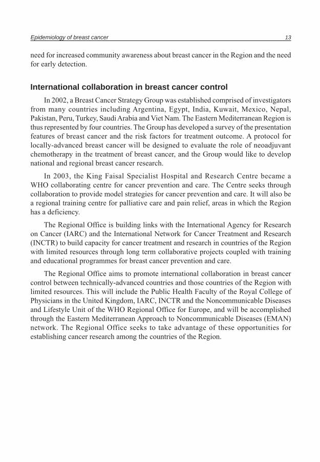
International collaboration in breast cancer control
In 2002, a Breast Cancer Strategy Group was established comprised of investigators from many countries including Argentina, Egypt, India, Kuwait, Mexico, Nepal, Pakistan, Peru, Turkey, Saudi Arabia and Viet Nam. The Eastern Mediterranean Region is thus represented by four countries. The Group has developed a survey of the presentation features of breast cancer and the risk factors for treatment outcome. A protocol for locally-advanced breast cancer will be designed to evaluate the role of neoadjuvant chemotherapy in the treatment of breast cancer, and the Group would like to develop national and regional breast cancer research.
In 2003, the King Faisal Specialist Hospital and Research Centre became a WHO collaborating centre for cancer prevention and care. The Centre seeks through collaboration to provide model strategies for cancer prevention and care. It will also be a regional training centre for palliative care and pain relief, areas in which the Region has a deficiency.
The Regional Office is building links with the International Agency for Research on Cancer (IARC) and the International Network for Cancer Treatment and Research (INCTR) to build capacity for cancer treatment and research in countries of the Region with limited resources through long term collaborative projects coupled with training and educational programmes for breast cancer prevention and care.
The Regional Office aims to promote international collaboration in breast cancer control between technically-advanced countries and those countries of the Region with limited resources. This will include the Public Health Faculty of the Royal College of Physicians in the United Kingdom, IARC, INCTR and the Noncommunicable Diseases and Lifestyle Unit of the WHO Regional Office for Europe, and will be accomplished through the Eastern Mediterranean Approach to Noncommunicable Diseases (EMAN) network. The Regional Office seeks to take advantage of these opportunities for establishing cancer research among the countries of the Region.
Natural history, etiology and risk factors
Natural historyBreast cancer appears to be a heterogeneous group of diseases. It was formerly
believed to be a localized disease originating and disseminating in a progressive fashion starting with benign disease, then atypia, progressing to carcinoma in situ, followed by invasive carcinoma, and finally metastasizing to regional axillary lymph nodes followed by distant metastases. As a consequence, radical surgery was advocated as the treatment of choice. The theory that breast cancer was a systemic disease from the day of diagnosis led to breast-conserving surgery and adjuvant therapy being heavily utilized. However, the current understanding is that the natural history of breast cancer is highly complex and many prognostic factors will play a role in determining the prognosis and outcome, and the natural history of the disease.
EtiologyThe etiology of breast cancer is also not fully understood. A variety of interrelated
factors, such as genetics, hormones, the environment, sociobiology and physiology can influence its development. Other risk factors such as proliferative breast disorders are also associated with breast cancer development, especially if the biopsy shows a typical hyperplasia [8]. However, in 70% of breast cancer patients no risk factors can be identified.
Genetic predispositionA positive family history increases the risk of breast cancer in first-line relatives
(mother, sister, or daughter). The risk is dependant upon whether the cancer was bilateral and whether it occurred in the pre- or postmenopausal period. Studies have shown that if the original cancer occurred during the premenopausal period, the risk of breast cancer in immediate relatives is approximately three times higher than in those who have no family history of breast cancer.
In those with a family history of breast cancer, 5%–10% of cases are attributed to inheritance of autosomal genes. The probability of genetic inheritance increases if there are multiple affected relatives and the cancer occurs at a younger age. Two genes,
Natural history, etiology and risk factors 15
BRCA 1 and BRCA 2 group, and p53, account for the majority of hereditary breast cancers. Ataxia telangiectasia (ATM gene) accounts for the majority of the rare cases of autosomally inherited cancers.
Hormonal factorsHormone regulation is important in the development of breast cancer. Early
pregnancy and early oophorectomy lower the incidence of breast neoplasm. In contrast, late menopause is associated with an increase in the incidence of breast cancer. Many of the hormonal risk factors such as long duration of reproductive life, multiparity and late age at the time of the birth of the first child imply increased exposure to estrogen peaks during menstrual cycles. Functioning ovarian tumours that elaborate estrogen are also associated with an increase in breast cancer in postmenopausal women. Among the factors that can also influence hormonal balance, resulting in the development of breast cancer, are the use of oral contraceptives and hormone therapy during menopause [9, 10].
A small increase in the risk of breast cancer has been noted in users of oral contraceptives. This risk, however, drops following the cessation of contraceptive use so that at ten years post-use, there is no significant increase in the risk of developing breast cancer. Use of oral contraceptives at an older age has also been linked to an increase in the number of breast cancer cases diagnosed.
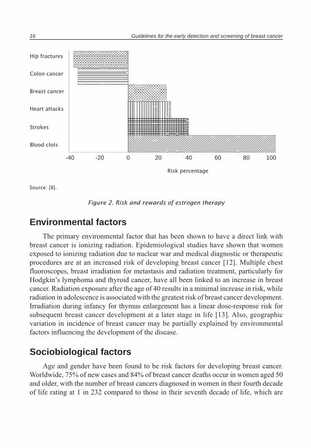
Current and recent users of hormone replacement therapy are at a higher risk of developing breast cancer than women who have never used hormone therapy. The risk increases with duration of hormone use, while it decreases significantly following cessation of the therapy. Thus, five years post-hormone therapy the risk of developing breast cancer as a result of the use of such hormones is nullified. A recent preliminary study of approximately 160 000 women conducted by investigators of the Women’s Health Initiative in the United States over a five-year period, assessed the major health benefits and risks of the most commonly-used combined hormonal preparations in the USA. As demonstrated in Figure 2, the study showed that the risk of breast cancer increases by 26% in those women who have used estrogen progesterone therapy compared with those who have not. The study concluded that the overall health risks of hormonal therapy exceeded the benefits for an average 5.2-year follow-up among postmenopausal women in the United States [11].
As a result of this and other studies, the United States Food and Drug Administration (FDA) recently ordered all products containing estrogen to include a prominent warning on their labels regarding the relationship between extended use of hormones and the risks of heart attack, breast cancer and potentially life-threatening blood clots.
16 Guidelines for the early detection and screening of breast cancer
Source: [8].
Figure 2. Risk and rewards of estrogen therapy
Environmental factorsThe primary environmental factor that has been shown to have a direct link with
breast cancer is ionizing radiation. Epidemiological studies have shown that women exposed to ionizing radiation due to nuclear war and medical diagnostic or therapeutic procedures are at an increased risk of developing breast cancer [12]. Multiple chest fluoroscopes, breast irradiation for metastasis and radiation treatment, particularly for Hodgkin’s lymphoma and thyroid cancer, have all been linked to an increase in breast cancer. Radiation exposure after the age of 40 results in a minimal increase in risk, while radiation in adolescence is associated with the greatest risk of breast cancer development. Irradiation during infancy for thymus enlargement has a linear dose-response risk for subsequent breast cancer development at a later stage in life [13]. Also, geographic variation in incidence of breast cancer may be partially explained by environmental factors influencing the development of the disease.
Sociobiological factorsAge and gender have been found to be risk factors for developing breast cancer.
Worldwide, 75% of new cases and 84% of breast cancer deaths occur in women aged 50 and older, with the number of breast cancers diagnosed in women in their fourth decade of life rating at 1 in 232 compared to those in their seventh decade of life, which are
Risk percentage
Hip fractures
Colon cancer
Breast cancer
Heart attacks
Strokes
Blood clots
-40 -20 0 20 40 60 80 100
Natural history, etiology and risk factors 17
rated at 1 in 29. This increase may be directly related to hormonal changes in women in this age group [14].
Nutritional intake and imbalances can also influence the risk of developing breast cancer. Consumption of fruits and vegetables may reduce the risk of developing breast cancer, while dietary intake of fat seems to increase the risk. In postmenopausal women, obesity increases the risk of breast cancer. This association is not observed in premenopausal women [14].
Physiological factorsPhysical activity levels can have an impact on the risk of breast cancer. Although
data in this area is not entirely consistent, moderate physical activity is associated with a lower risk of breast cancer. Studies have shown a 30% reduction in risk level associated with a few hours per week of vigorous activity compared to no exercise at all [15].
Pathology of the breast
Breast diseaseClinically, among 100 female patients aged 40–65 years presenting with breast
complaints, the following is likely: 30% have no breast lesion, 40% have fibrocystic changes, 7% have a benign tumour diagnosis and 10% have carcinoma. Breast disease can therefore be divided into the following groups [8,16].
Inflammatory lesions
These are rare breast lesions that can be acute or chronic and include acute mastitis, duct ectasia, post-traumatic lesions and granulomatous mastitis.
Benign fibrocystic lesions
Fibrocystic changes represent the single most common disorder of the breast and account for more than 40% of all surgical operations on the female breast [16]. It is diagnosed frequently between the ages of 20 and 40 years, and rarely develops after menopause. It is frequently influenced by hormonal imbalance.
Benign breast diseases
These are rare tumours, which include fibro adenomas, phyllodes tumours and large duct papilloma.
Proliferative breast disorder
Epidemiological studies have identified changes in the breast resulting in an increased risk of developing carcinoma. This risk is due to hyperplasia with or without atypia. These lesions are often accompanied by fibrocystic changes as well. They can be associated with mammographic abnormalities.
Carcinoma of the breast
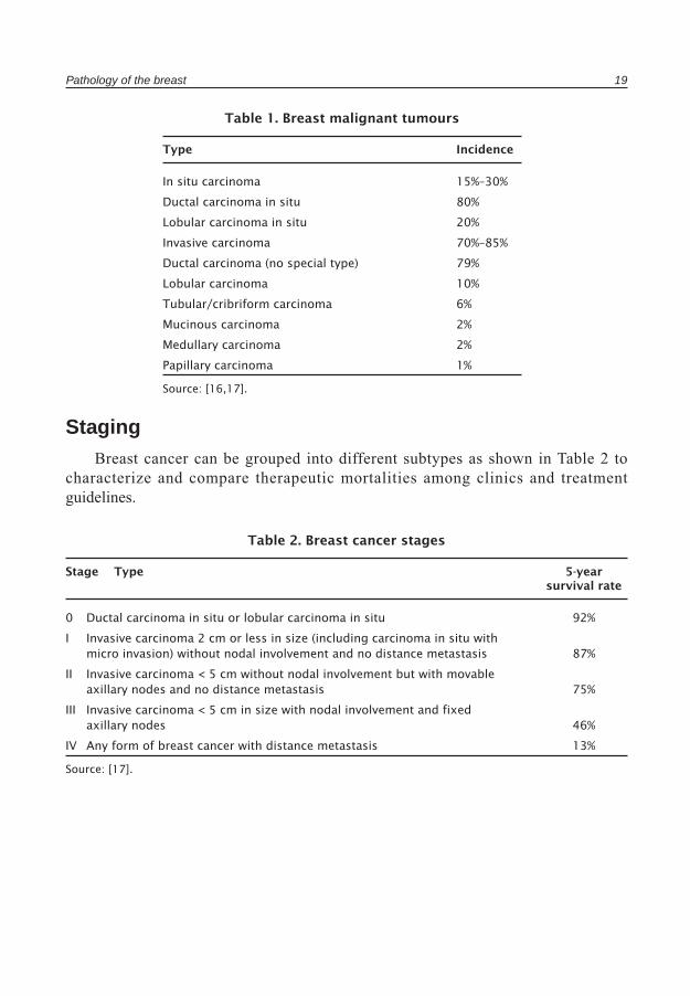
Breast cancer can be divided into two main groups: non-invasive or carcinoma in situ, and invasive carcinoma. Table 1 presents the incidence of various breast pathologies.
Pathology of the breast 19
Table 1. Breast malignant tumours
Type Incidence
In situ carcinoma 15%–30%
Ductal carcinoma in situ 80%
Lobular carcinoma in situ 20%
Invasive carcinoma 70%–85%
Ductal carcinoma (no special type) 79%
Lobular carcinoma 10%
Tubular/cribriform carcinoma 6%
Mucinous carcinoma 2%
Medullary carcinoma 2%
Papillary carcinoma 1%
Source: [16,17].
StagingBreast cancer can be grouped into different subtypes as shown in Table 2 to
characterize and compare therapeutic mortalities among clinics and treatment guidelines.
Table 2. Breast cancer stages
Stage Type 5-year survival rate
0 Ductal carcinoma in situ or lobular carcinoma in situ 92%
I Invasive carcinoma 2 cm or less in size (including carcinoma in situ with micro invasion) without nodal involvement and no distance metastasis 87%
II Invasive carcinoma < 5 cm without nodal involvement but with movable axillary nodes and no distance metastasis 75%
III Invasive carcinoma < 5 cm in size with nodal involvement and fixed axillary nodes 46%
IV Any form of breast cancer with distance metastasis 13%
Source: [17].

20 Guidelines for the early detection and screening of breast cancer
Clinical courseSeparate tumour characteristics that have important prognostic significance need to
be considered when designing an optimum treatment strategy for an individual patient. These include, but are not limited to, the following:
• Age of the patient (less than or equal to, or more than, 35 years).• Tumour size (less than or equal to, or more than, 2 cm).• Axillary lymph node status. This is the most important predictor of disease recurrence
and survival. Nearly 70%–80% of patients with negative node status survive 10 years; prognosis worsens as the number of positive nodes increase.
• Histological grade and nuclear grade. These have prognostic implications. • Estrogen and progesterone receptor status. Patients with receptor-positive primary
tumours have a lower rate of recurrence and longer survival, and a higher response to hormonal manipulation.
• Other biological markers including HER2/neu (c-erbB2), p53 and bcl-2 [18].
Managerial aspects of breast cancer detection
Managerial approachBreast cancer detection and prevention is a systemic and continuous management
process that includes planning, developing and evaluating breast cancer detection programmes, including policy formulation and the identification of priorities. Countries must develop comprehensive plans for screening and detection of breast cancer, including outreach and education with the general population, training for medical and technical staff, development of programmes and processes for accurate diagnosis of breast cancer, and facilities for timely and effective treatment. The responsibility for the development and implementation of a breast cancer detection programme rests with the Ministry of Health or other relevant organization. The overall aim should be to establish a mechanism for the political and technical support of the programme.
A successful managerial approach to breast cancer detection rests on the combined impact of several activities including surveillance, protection, continuing education and prevention, early detection and care.
SurveillanceSurveillance is key for identifying problems and developing appropriate and timely
interventions. The aims of surveillance activities include, but are not limited to, the following:
• estimating the burden of disease• identifying the risk factors that increase the incidence of breast cancer• building the basis for appropriate clinical interventions.
ProtectionCancer protection can be defined as the activities and processes associated with
protecting individuals from cancer or its recurrence, and affecting the burden of disease and disability. Protection includes a number of activities such as continuing education efforts, health promotion, prevention and early detection of disease (screening).
22 Guidelines for the early detection and screening of breast cancer
Continuing educationThe first step to initiate an effective continuing education programme is advocacy
on the urgency and importance of the programme to the government officials and policy-makers who can place breast cancer detection on the country’s national agenda. Public education programmes should focus on prevention, better understanding of the illness and the benefits of early detection. In addition, education programmes for health care recipients and their families should be developed to ensure that the benefits of health care services are maximized. These programmes should be developed to increase understanding of the needs of patients and the ability to cope with these needs. Finally, health promotion is the key strategy for controlling the risk factors for breast cancer through a collective and multisectoral policy.
PreventionAlthough breast cancer cannot be prevented, the risks of developing breast cancer
can be minimized through specific preventive activities. These include achieving changes in lifestyle, diet, overall physical characteristics and obesity, and interventions for women at high risk of developing breast cancer using tamoxifen and other anti-estrogen compounds.
Early detectionThe most important and beneficial area of protection activities is the early detection
of breast cancer (screening). Diagnosis of breast cancer during the early stages of disease has been positively linked to a decrease in the mortality and morbidity of the illness. There are a number of approaches to the screening of breast cancer.
• Breast self-examination has been endorsed and widely promoted by cancer organizations and authorities around the world. Its effectiveness, however, is dependent on education and outreach among women, and upon conscientious and regular self-examination.
• Clinical breast examination is one of the primary modes of screening for breast cancer. Its effectiveness is dependent upon the skills of the health worker and the facilities available. It is therefore important to use proven training strategies and standard techniques to ensure that health workers are fully and appropriately trained.
• Mammography is known to reduce breast cancer mortality among women, but its benefits are dependent upon several factors such as the equipment used, the skills of the technician taking the mammography and the expertise of the radiologist reading the mammogram.
Managerial aspects of breast cancer detection 23
Care/disease interventionCancer control programmes must ensure the diagnosis of the disease at the earliest
possible stage when treatment is most effective and cure is most likely. Beyond the initial early detection and diagnosis of breast cancer, improving the treatment and care provided to women with breast cancer is obviously an integral factor in decreasing overall mortality from breast cancer. Treatment of breast cancer should be expanded beyond surgery to include interventions such as drug therapy and radiation procedures. Additionally, adjuvant therapies should be used to prevent the recurrence of breast cancer. Finally, increasing the psychosocial support and the palliative care available can increase the quality of life for women with breast cancer and their families.
Cancer detection programmes
Early detection of cancerThe objective of an early detection programme is diagnosing cancer at its earliest
stages when it is localized to the organ of origin, without metastasis to other organs or the surrounding tissue. The early detection approach consists of identifying asymptomatic neoplastic lesions and understanding that cancer detection at the earliest stages promotes more successful treatment and cost-effective interventions. The major components of an early detection programme include public education and continuing education for professionals.
Public education seeks to educate the public regarding the risks and symptoms of cancer with the objective of promoting early diagnosis of the disease, and increasing appropriate access to diagnostic and treatment services. The continuing education of professionals focuses on the role of the professional as the initial point of contact between potential cancer patients and the health care system. These professionals must be aware of the early signs and symptoms of cancer, assisting in early detection. Similarly, continuing education programmes promote increased awareness of the burden of disease for government officials and policy-makers who are responsible for developing and implementing the national health care agenda and programmes.
Early detection programmes allow for a more favourable prognosis for patients, offer increased and less toxic treatment options, and enable the provision of services through more cost-effective modalities. It is important to note that a high proportion of cancers detected at the early stages in developed countries continue to be diagnosed at more advanced and often fatal stages in developing countries, thus increasing the associated burden of disease. It is therefore important that public and professional education services be combined with timely access to diagnostic and treatment facilities, effective treatment services and programmes, and ongoing follow-up services. With the anticipated increasing cost of cancer therapy, early detection will become even more cost saving. This is especially important in countries with limited health budgets.
Cancer screeningScreening (often used synonymously with “early detection”) programmes aim to
identify individuals during asymptomatic stages for possible detection of cancer during preclinical phases of the disease. Screening programmes enable early diagnosis, more effective treatment and increased possibility of a successful outcome. In developing and implementing screening programmes, three factors should be considered.
Cancer detection programmes 25
a) Characteristics of the cancerThe cancer that is screened should have significant and serious health and economic
consequences for the general population. Mortality is the most important consequence to be considered. In addition, it is important to understand the natural history and cellular development characteristics of the cancer being screened and whether it responds favourably to screening. Therefore, there must exist a detectable preclinical phase of some duration (lead time) when the cancer can be detected through testing well before actual symptoms develop. It should be noted that individual cancers have differing natural histories. Cancers with long natural histories and long lead times are most likely to be detected in a screening programme.
b) Screening tests The screening tests used should be acceptable and comfortable for the patient. They
should also be accurate, user-friendly for health workers and cost-effective.
c) Screening evaluation Evaluation of the programme should focus on measurable goals: early diagnosis of
the disease, patient benefit from treatment facilities and services, and reduced mortality to an acceptable rate. This evidence-based evaluation process should be utilized as an indicator of the programme’s efficacy and outcomes.
The success of screening programmes depends on a number of fundamental principles:
• the target disease should be a common form of cancer, of public health importance, and associated with high morbidity and mortality;
• there should be strong evidence that the screening tests adopted can result in reduced mortality and morbidity in the target population;
• test procedures should be acceptable, safe and relatively inexpensive;• there should be ethical, acceptable and effective procedures for detecting the disease
at an early stage to provide opportunity for intervention;• the benefits of screening should outweigh any adverse cost;• the implementation of screening, diagnostic and intervention activities should
strengthen the health system and social development;• a specific and sensitive test for the early detection of the disease must be
available; • there should be suitable facilities for the diagnosis and treatment of detected
abnormalities.
26 Guidelines for the early detection and screening of breast cancer
Screening for breast cancerBreast cancer is most easily and effectively treated in its early stages. Survival rates
drop dramatically when women present with advanced cases regardless of the setting; therefore, a primary strategy for reducing breast cancer mortality is increasing the proportion of cases that are detected during the early stages of the disease. Unfortunately, women in resource-poor countries generally present at a later stage of disease than women elsewhere, in part due to the absence of mass screening programmes in many such countries. Regular screening of all women aged fifty and over has the potential to sharply increase the proportion of cancer cases that are diagnosed in their earliest stages [19,20,21].
The goals of screening guidelines are two-fold:
• to provide guidance regarding the appropriate use of screening tools for breast cancer detection;
• to help physicians and patients make informed decisions regarding screening for breast cancer in asymptomatic women of all ages.
Screening approachThe main conceptual framework of a screening programme is to design a process that
will reduce mortality rates from breast cancer and increase the quality and longevity of life for the target population. This process should take place in a well-defined population at high risk in a cost-effective manner. The main approach should be a mechanism that can detect malignant disorders at their earliest stage of development. The outcome of this process will depend on two distinct conditions.
a) Screening and cellular stage of development
There is a period during the screening process in which there is no detectable disease, although early malignant changes may have already taken place. The point at which a tumour can be found by screening begins at the sojourn time or detectable preclinical phase. It is a joint outcome of the lesions and the screening test. Lead-time refers to the period between when a cancer is found by screening and when it would appear through clinical signs and symptoms. It can be affected by the frequency of screening. Neither the sojourn time nor the lead-time is directly observable for an individual, unless a screening test is repeated at frequent intervals [19,20]. In establishing screening frequency intervals breast cancer growth needs to be considered. The sojourn time for all cancer types is shorter in premenopause.
b) Intervention, practices and techniques
There are three methods of screening for breast cancer: breast self-examination, clinical breast examination and mammography. These methods are discussed in the following sections. For further information see IARC handbooks of cancer prevention, 7. Breast cancer-screening [21].
Breast self-examination
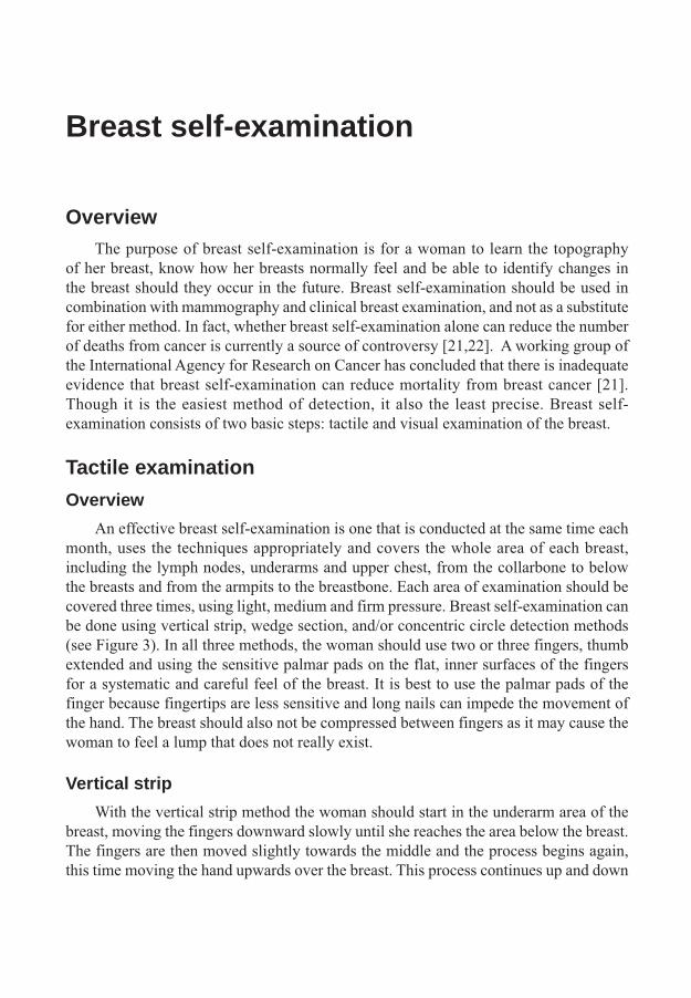
OverviewThe purpose of breast self-examination is for a woman to learn the topography
of her breast, know how her breasts normally feel and be able to identify changes in the breast should they occur in the future. Breast self-examination should be used in combination with mammography and clinical breast examination, and not as a substitute for either method. In fact, whether breast self-examination alone can reduce the number of deaths from cancer is currently a source of controversy [21,22]. A working group of the International Agency for Research on Cancer has concluded that there is inadequate evidence that breast self-examination can reduce mortality from breast cancer [21]. Though it is the easiest method of detection, it also the least precise. Breast self-examination consists of two basic steps: tactile and visual examination of the breast.
Tactile examination
Overview
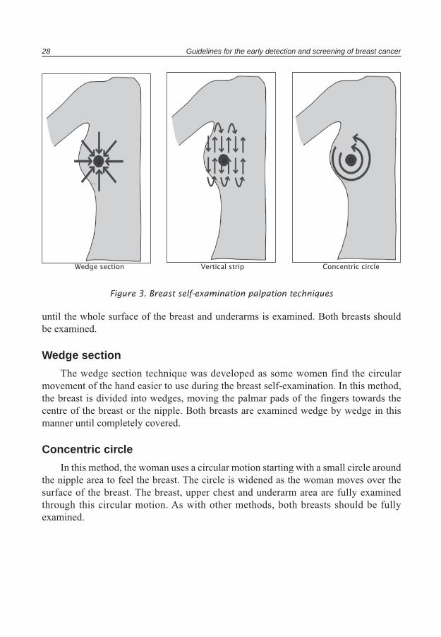
An effective breast self-examination is one that is conducted at the same time each month, uses the techniques appropriately and covers the whole area of each breast, including the lymph nodes, underarms and upper chest, from the collarbone to below the breasts and from the armpits to the breastbone. Each area of examination should be covered three times, using light, medium and firm pressure. Breast self-examination can be done using vertical strip, wedge section, and/or concentric circle detection methods (see Figure 3). In all three methods, the woman should use two or three fingers, thumb extended and using the sensitive palmar pads on the flat, inner surfaces of the fingers for a systematic and careful feel of the breast. It is best to use the palmar pads of the finger because fingertips are less sensitive and long nails can impede the movement of the hand. The breast should also not be compressed between fingers as it may cause the woman to feel a lump that does not really exist.
Vertical strip
With the vertical strip method the woman should start in the underarm area of the breast, moving the fingers downward slowly until she reaches the area below the breast. The fingers are then moved slightly towards the middle and the process begins again, this time moving the hand upwards over the breast. This process continues up and down
28 Guidelines for the early detection and screening of breast cancer
until the whole surface of the breast and underarms is examined. Both breasts should be examined.
Wedge section
The wedge section technique was developed as some women find the circular movement of the hand easier to use during the breast self-examination. In this method, the breast is divided into wedges, moving the palmar pads of the fingers towards the centre of the breast or the nipple. Both breasts are examined wedge by wedge in this manner until completely covered.
Concentric circle
In this method, the woman uses a circular motion starting with a small circle around the nipple area to feel the breast. The circle is widened as the woman moves over the surface of the breast. The breast, upper chest and underarm area are fully examined through this circular motion. As with other methods, both breasts should be fully examined.
Figure 3. Breast self-examination palpation techniques
Wedge section Vertical strip Concentric circle

Breast self-examination 29
a) Lying down b) Standing in the shower
Figure 4. Examination positions
Examination position
Tactile examination of the breast can be done lying down or standing up in the shower, primarily depending on the preference of the woman (see Figure 4).
a) Lying down
For this position, the woman should start by lying on the bed, placing a pillow or folded towel under the left shoulder and the left hand behind the head. The shoulder should be slightly raised, sufficiently for the breast to fall towards the centre of the top of the chest, and not towards the armpits. This allows the breast tissue to distribute evenly across the chest wall, making it easier to feel a lump in the outer upper quadrant of the breast, where the tissue is thickest and where most malignancies occur. Using the right hand, the woman should feel her breast using one of the examination techniques described above: vertical strip, wedge section or concentric circle. Once the left breast is examined with the left arm under the head, the entire examination should be repeated with the left arm in a relaxed position to the side. After this examination is complete, the woman should shift the pillow or towel to the right side, place the right arm under her head, and examine the right breast using the left hand. Once again, the examination should be repeated with the right hand in a relaxed position to the side.
b) In the shower
As many lumps are felt when the breast and fingers are wet and slippery with lather (decreasing the friction), tactile examination of the breast in the shower is the method of choice for many women. If the breasts are small, the woman should place one hand on her head and examine the breast on that side with the other arm, using the vertical strip method. If the breasts are larger, it is best for the woman to immobilize the breast with the palm of her hand (first supporting from below and then pressing down from the top) and examine it with the other hand, again first from above and then from below.

30 Guidelines for the early detection and screening of breast cancer
Changes to look out forIn examining the breast, the woman should feel for changes in the texture and feel
of the breast. Among the things that should be noted and reported to a physician are:
• any new lump or hard knot found in the breast or armpit;• any lump or thickening of the tissue that does not shrink or lessen after her next
period;• any change in the size, shape or symmetry of her breast;• a thickening or swelling of the breast; • any dimpling, puckering or indention in the breast;• dimpling, skin irritation or other change in the breast skin or nipple; • redness or scaliness of the nipple or breast skin;• discharge from the nipple (fluid coming from the nipples other than breast milk),
particularly if the discharge is clear and sticky, dark or occurs without squeezing the nipple;
• nipple tenderness or pain;• nipple retraction (turning or drawing inward or pointing in a new direction); • any breast changes that may cause concern.
Visual examinationThe visual examination of the breast is another tool in identifying possible breast
disease. It should be noted that no woman has two breasts that are exactly identical; however, once a woman knows what her breasts look like, she is able to identify any changes in the shape, form, colouring or structure of the breast more quickly and can discuss these with the appropriate health care provider.
In preparing for the visual examination, the woman should stand in front of a mirror with her upper body unclothed (see Figure 5). A good light should be placed to the side, rather than above, to better differentiate any irregularities. The woman should examine the breast with her arms relaxed and to the side; with her arms raised (see Figure 5: position 1); and with her palms flat on the sides of her hips and pressing down (see Figure 5: position 2). Two additional positions include clasping the hands in front of the forehead, palms squeezed together, to tighten the chest pectoral muscles and bending forward to examine the breasts.
When looking in the mirror, the woman must look for any changes in the contour or placement of the breasts, changes in colour and shape, discharge from the nipples and discoloration of the skin (see Figure 6). Redness, irritation or prominent veins in the breast can signal an increased supply of blood to the breast, a sign that often accompanies

Breast self-examination 31
Figure 5. Visual examination positions
What to look for1. Change in breast contour, such as swelling 3. Dimpling or puckering of the skin2. Inversion of the nipple 4. "Orange peel" appearance of the skin
Figure 6. Changes in breast surface
Position 1 Position 2
32 Guidelines for the early detection and screening of breast cancer
tumour growth. Whitish scale on the nipples, ulcers and sores that do not heal properly are other signs of possible breast disease. An “orange peel” skin (swollen and shiny with large deep pores) has been found to be associated with blocked lymph ducts. A nipple that is flat, inverted or retracted, especially if this is new development, or one that is not inverted when the woman is upright but inverts when she leans forward, can also be associated with breast disease.
Breast self-examination costsThe costs associated with the use of breast self-examination as a screening
intervention are easy to identify and conceptualize. The direct monitoring costs include health education and outreach activities associated with training the trainers, providing information to the target population, offering scientific and diagnostic information to health care providers, and educating the general public regarding the benefits of early detection and use of breast self-examination. The indirect costs are related to the diagnostic and treatment services provided by health care workers associated with any findings as a result of breast self-examination.
Mechanisms for improving breast self-examinationSurveys and studies have suggested the efficacy of breast self-examination as
an initial tool for the early detection of breast cancer. Provider encouragement and education, and a review of breast self-examination techniques during gynaecological and physical health visits are among the most effective factors in promoting the use of breast self-examination. Availability of brochures, pamphlets and shower cards, for example, are important as a reminder to women to use breast self-examination on a regular basis. Finally, providing information on the effectiveness and importance of breast self-examination as an early detection tool is important.

Clinical breast examination
OverviewClinical breast examination is an examination of the breast by a health care
professional, such as a physician, nurse, or physician’s assistant. It includes both inspection (looking) of the breast and palpation (feeling). The areas examined include the entire breast/chest area (including the lymph nodes), above and below the collarbone, and under each arm.
Clinical breast examination combined with mammography is considered essential to reducing mortality from breast cancer. Clinical breast examination is seen as an effective first step in determining the possible presence of the disease in women. However, it cannot be used alone, without the information provided through diagnostic mammography and find needle aspiration. The efficacy of clinical breast examination is dependent upon a number of factors: proper positioning of the patient, thoroughness of the search, use of a vertical strip technique, proper positioning and movement of the fingers, and an examination duration of at least 5 minutes per breast [23].
Clinical breast examination techniqueThe first component of the clinical breast examination is a visual examination of
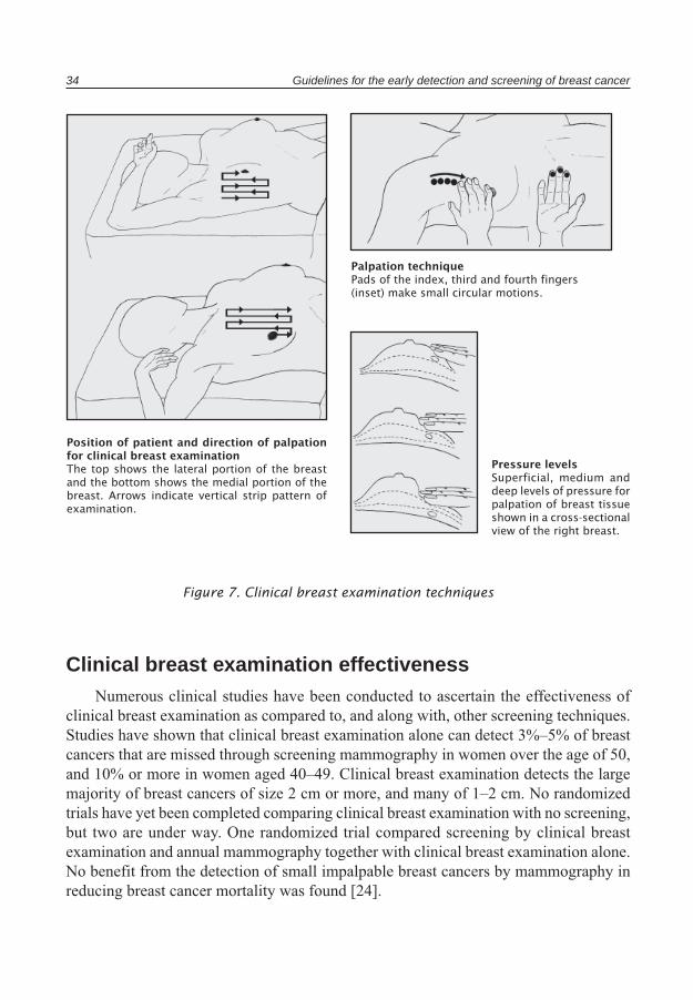
the breasts in three different standing positions: arms relaxed at the sides; hands pressed firmly on the waist and leaning forward; and arms over the head. During this process, the examiner identifies subtle asymmetries and changes in the appearance of the breasts. The entire area of the breast, including the area from the clavicle to the inframammary fold, and from the midaxillary line to the sternum, is examined in both the seated and supine positions. The application of three levels of pressure (superficial, medium and deep) at each palpation site is essential.
Palpation is done with the finger pads of the three middle fingers, and pressure is applied with circular motions at each site. For the lateral half of the breast, the torso is rotated in the medial direction; for the medial half of the breast, the torso is rotated laterally in order to spread out the breast tissue (Figure 7). When an abnormality is detected, the corresponding area of the other breast is examined. If the finding is not bilateral, further investigation is required [23].
A clinical breast examination should be a part of a woman’s routine check-up. Beginning at the age of 20, women should have a clinical examination every two or three years, increasing to once a year from the age of 40.
34 Guidelines for the early detection and screening of breast cancer
Clinical breast examination effectivenessNumerous clinical studies have been conducted to ascertain the effectiveness of
clinical breast examination as compared to, and along with, other screening techniques. Studies have shown that clinical breast examination alone can detect 3%–5% of breast cancers that are missed through screening mammography in women over the age of 50, and 10% or more in women aged 40–49. Clinical breast examination detects the large majority of breast cancers of size 2 cm or more, and many of 1–2 cm. No randomized trials have yet been completed comparing clinical breast examination with no screening, but two are under way. One randomized trial compared screening by clinical breast examination and annual mammography together with clinical breast examination alone. No benefit from the detection of small impalpable breast cancers by mammography in reducing breast cancer mortality was found [24].
Position of patient and direction of palpation for clinical breast examinationThe top shows the lateral portion of the breast and the bottom shows the medial portion of the breast. Arrows indicate vertical strip pattern of examination.
Palpation techniquePads of the index, third and fourth fingers (inset) make small circular motions.
Pressure levelsSuperficial, medium and deep levels of pressure for palpation of breast tissue shown in a cross-sectional view of the right breast.
Figure 7. Clinical breast examination techniques

Clinical breast examination 35
A working group of the International Agency for Research on Cancer has concluded there is inadequate evidence that breast screening with clinical breast examination alone or in addition to screening mammogram can reduce mortality from breast cancer [21].
Though clinical breast examination by itself does not rule out the presence of impalpable disease, consideration of its use as a screening technique in identifying certain abnormalities that can result in breast cancer is recommended.
Screening mammography
OverviewMammography is another screening tool utilized for detecting early breast cancer.
Screening mammography is defined as a standard two-view mammogram obtained of an asymptomatic woman with the purpose of early detection of breast cancer. The objective of population-based mammography screening is to reduce mortality and morbidity from breast cancer through the early detection and treatment of malignancies. There is ample evidence from a variety of well-documented sources that annual or biannual mammography is effective in reducing breast cancer mortality in women aged 50–69 years [21]. Women who have no family history of breast cancer or prior diagnosis of cancer and who are under the age of 50 are considered to be low risk, although they may benefit from regular mammograms at the discretion of their physician. Women with a family history of premenopausal breast cancer in a first-line relative, those with a history of breast or gynaecological cancer, and those who are over 50 years of age are considered to be at high risk and in developed countries are advised to have screening mammography every 1–3 years [21]. Despite all the cited benefits, it should be noted that mammography alone, with a false negative rate of 12%, is not an effective screening tool. This false-negative rate is higher among younger patients.
A working group of the International Agency for Research on Cancer has evaluated the efficacy of breast screening [21]. They concluded:
• There is sufficient evidence for the efficacy of screening women aged 50–69 years by mammography as the sole screening modality in reducing mortality from breast cancer. There is evidence of a 25% reduction in women aged 50–69 years.
• There is limited evidence for the efficacy of screening women aged 40–49 years by mammography as the sole screening modality in reducing mortality from breast cancer. When all valid trials were included there was evidence of a 11% reduction in women 40–49 years of age.
• No direct conclusion was possible for the efficacy of mammography in women younger than 40 or older than 69.
MammogramsMammography is an X-ray technique that was developed specifically for breast
lesion examination. Diagnosis, evaluation and determination of the results is based on the different absorption of X-rays between different types of breast tissue, such as

Screening mammography 37
fat, fibroglandular tissue, cysts, tumours and calcifications. During the procedures, the imaging system must be optimized to provide the minimum radiation dose as required. The level of radiation should be standardized based on national and international guidelines. The mean absorbed dose in the breast gland per mammographic film is in the order of 1.0–1.5 mGy for the average breast examined with modern equipment.
A few points to remember regarding mammographic density are that:
• breast parenchymal density as seen on a mammogram is a determinant of the sensitivity of mammography;
• breast parenchymal density decreases with age;• hormone replacement therapy of the combination type may result in increased breast
density; • tamoxifen may reduce breast density.
In reading mammographic images, most authors recommend a double reading, which increases the sensitivity of the reading by 10%–15% compared to single readings.
In recent years, digital mammography has also been used as an alternative to traditional mammography. In digital mammography, the image receptor used in conventional mammography is replaced by a digital receptor. The imaging techniques, however, remain the same in all other respects. From the point of view of the woman being screened, a digital mammogram is similar to a conventional mammogram, as breast compression and positioning are unchanged. Digital mammography has the potential to provide images with lower doses of radiation than screen-film mammography. Also, computer-aided detection can be incorporated into the workstation and the results of the computer analysis added into the image, thereby assisting the radiologist in detecting suspect lesions. Computer-aided detection has been assessed in several studies, which suggest an incremental value in terms of sensitivity, though the evidence on specificity is conflicting. Some data suggest that computer-aided detection could replace a second reader [25].
Many women recommended for this type of screening have voiced concerns regarding the risk of radiation from mammograms. Studies have shown that the average dose per examination (single view per breast) is approximately 2 mGy, the dose being dependent on breast thickness and exposure factors. The risk from radiation is cumulative, greatest for adolescent exposure and decreasing with an increase in age. In those over 50 years of age, the risk of cancer induction is approximately 1 in 100 000 per single view examination [12,25].
Mammograms should be performed by radiographers who have completed a postgraduate course in mammography and attended courses on mammographic techniques and procedures. In addition, radiologists with appropriate training, especially in breast diseases, should be part of the multidisciplinary team [25].
38 Guidelines for the early detection and screening breast cancer
Mammography readingIn performing a mammogram, two views of each breast are obtained: a view from
the top and a view from the side. To obtain accurate images, it is important that the breast is compressed to an even thickness, thus minimizing the radiation exposure while allowing for accurate visualization of possible abnormalities.
The ability to detect breast cancer is dependent upon being able to distinguish a mass or abnormality from normal breast tissue. Generally, the more fat there is in the breast, the easier it is to distinguish a mass or abnormality. When the breast is dense, the
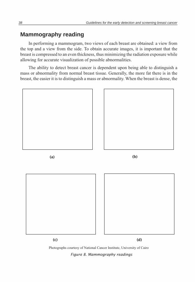
Figure 8. Mammography readings
(a) (b)
(c) (d)
Photographs courtesy of National Cancer Institute, University of Cairo
Screening mammography 39
abnormality may be hidden in the tissue and additional investigations may be needed, such as ultrasound if it is suspected to be a cystic mass or fine needle aspirate for cytologic examination. Figure 8a) shows a benign-looking mass with low density with a hollow sign surrounding the mass. Most probably it is a fibro-adenoma.
In general, when there are macrocalcifications they are usually not associated with cancer. Figure 8b) shows benign-looking macro calcifications overlying a low density, benign-looking, well defined mass with a hollow sign surrounding it. However, when there are microcalcifications, additional examination and investigations of the mass are required to differentiate between benign and malignant microcalcifications.
Figure 8c) shows a highly suspicious malignant mass with a lobulated margin, high density, microcalcifications and nipple retraction.
In Figure 8d), three masses of different sizes raise suspicion of malignancy as they are of high density, irregular speculated margin while the large one shows microcalcifications.
Studies have shown that the size of tumours identified through clinical breast examination versus those identified by mammography are significantly different, supporting the effectiveness of both as early detection tools for breast cancer.

Other breast imaging techniques
UltrasonographyUltrasound is an important complementary imaging technique to mammography and
physical examination. Breast ultrasound is used primarily to differentiate between a cyst and a solid mass, to explore a palpable abnormality not clearly visible on a mammogram or to obtain a better view of a lesion that cannot be mammogrammed. It is often the initial diagnostic exam in a young woman who presents with a well-circumscribed solid mass, often found to be a fibroadenoma. Ultrasound, however, is not accurate in detecting microcalcifications, though it can be used for guided aspiration or needle localization under specific guidelines.
Computed tomography (CT)The benefits of diagnostic computed tomography appear to be small, as it is of high
cost and has the potential for high exposure of radiation. Therefore, its indications are very limited.
Magnetic resonance imaging (MRI)The sensitivity of MRI is over 95% but its specificity is low, with a range of
53%–70%. False positive diagnoses of proliferative fibrocystic diseases, adenomatous fibroadenomas, radial scars, fat, necrosis, intramammary lymph nodes and breast parenchyma after surgery have demonstrated its inaccuracies for the detection of breast cancer. Additionally, microcalcifications are not visible on MRI scans, though dynamic contrast-enhanced MRIs can be helpful to rule out malignancy of non-palpable lesions. The best indication for breast MRI is the suspicion of breast cancer recurrence in patients treated conservatively (recurrence versus fibrosis). For these patients, the specificity of MRI has been found to be 98%.
Nuclear medicine breast imagingTechnetium-99 sestamibi has been found to concentrate in some breast cancers.
However, its role in breast cancer evolution has yet to be defined, because it cannot be used to differentiate benign lesions from malignant ones and its efficacy remains to be defined.
Other breast imaging techniques 41
Positron emission tomographic screening (PET)Early studies suggested that breast cancers have elevated metabolic activity, which
can be detected using fluorine 18-labeled glucose. PET may be a method for staging breast cancers and for assessing the possibility of recurrence after initial breast cancer treatment. The use of PET as a diagnostic technique to differentiate benign from malignant lesions and to reduce the need for needle aspiration biopsies is still under study.
Guided breast biopsyImaging modalities are very useful in guided biopsies of non-palpable lesions,
requiring an efficient collaboration between radiologists and pathologists. Two techniques may be employed: fine needle aspiration for cytologic analysis and core needle biopsy for histologic analysis. Further exploration is required in cases with negative results or where differing data between mammographic and histological findings have been obtained.
Breast mass evaluationWhen a mass is found as part of the screening process, the histological type of the
cancer should be determined. The first step is to determine whether the mass is solid or cystic, often accomplished through a needle aspiration biopsy, using a needle or syringe. If cystic fluid (non-bloody serous fluid) is found, the patient is recommended to return for a follow-up re-evaluation in four to six weeks. However, if the mass is not cystic, then a fine needle aspiration should be performed. If the fine needle aspiration is inconclusive or negative, an open biopsy must be performed.
Follow-up and cost-effectiveness
Physical health follow-upFollow-up services include those coping with the direct physical side effects
and problems associated with breast cancer treatment. Follow-up of patients after conservative treatment includes a periodic physical examination and mammography every six months during the first two years and every year thereafter. Local recurrences occur for approximately 1% of the target population per year. Therefore, continued screening for the early detection of local relapses improves long-term survival after breast cancer treatment, especially mastectomies.
Physical health follow-up is most successful when the patient has been clearly educated about the stages of treatment, the services offered and available, and the course of treatment and recuperation expected. As important, is educating the woman and her family in the early detection of any signs of recurrence or possible metastasis of the cancer to other areas of the body. Symptoms such as bone pain, shortness of breath, excessive tiredness, unexpected bleeding and the suchlike, should be pointed out as possible signs of concern. An educated woman is a better patient with a higher chance of survival.
Mental health follow-upFor women diagnosed with breast cancer, follow-up services should also include
interventions to deal with the emotional and psychological issues that are raised as a result of a diagnosis. The word cancer still brings with it a magnitude of fear and anxiety not associated with many other diseases. Breast cancer is an illness where the mortality and morbidity rate are clearly defined and where most women, especially those in developing countries, understand that their survival rate may not be strong. Women also continue to be the focal point of family life and an illness that threatens their life and ability to function significantly impacts the emotional well-being of the family unit.
Parents and siblings are devastated as they watch a child or sister go through the burden of a disease that is painful, difficult, and both emotionally and physically exhausting. Spouses are equally affected as they must take over a larger burden of daily activities, supporting the needs of their children in different ways than before the illness, and providing support to their wives as they go through breast cancer treatment, while coping with the emotional impact of possibly losing a loved one. Equally as significant in this process are the children, who are most affected by this illness. Many children, especially those who are young, do not understand the impact of cancer on their parents’
Follow-up and cost-effectiveness 43
daily lives. What they understand is that an illness is decreasing their mother’s ability to address their emotional and physical needs in the same manner as before. This is a cause of confusion, anger and frustration for most children who cope with the changes through outbursts and other negative behaviour. For children who are older and understand the ramifications of this illness, coping with their mother’s illness and possible death is a source of extreme emotional pain. Older children usually cope with this illness by going through the classic stages of grief: denial, bargaining, depression, anger and acceptance.
It is therefore of utmost importance that any treatment and follow-up programme for women with breast cancer include a strong psychosocial component that builds upon the strengths of the family, offers community and other normal forms of support during the most difficult stages of the illness, and assists the family in making the journey in a productive and positive manner.
Cost-effectivenessThe most important benefit of an effective breast cancer screening programme is
a reduction in breast cancer mortality and an increase in life years of good quality. The cost-effectiveness of a breast cancer screening programme should, therefore, take these issues into consideration and also be based on understanding that:
• The development and implementation of a screening programme is costly. It includes patient and provider training, early diagnosis resources and appropriate treatment services, all of which need adequate resources for their proper implementation and management.
• Screening can have profound and unexpected health, social, physiological and economic consequences, while also presenting unique ethical issues.
• The main goal of screening should be focused on detection of breast cancer at early stages of the disease and offering opportunities for timely treatment and a change in the overall course and outcome of the disease.
• Among the different screening techniques, mammography is probably the most internationally studied and utilized. There have been numerous studies that have analysed the cost-effectiveness of breast
cancer screening programmes, though there have been no studies that have looked at this issue from the perspective of a nationwide population-based cancer programme. In the studies conducted to date, breast cancer screening programme cost-effectiveness has been evaluated based on cost per life year saved (LYS) or cost per quality-adjusted life year saved (QALY). The studies have yielded very differing results and the majority have been performed in developed countries, so their results cannot be directly linked

44 Guidelines for the early detection and screening of breast cancer
to the Eastern Mediterranean Region. However, these studies provide a solid base for evaluating and understanding the potential cost-effectiveness of such programmes.
In a Finnish study conducted from 1987–1992, LYS were obtained for 89 893 women with and without breast cancer. The total unit cost for screening was between US$ 28 and US$ 51. The study also calculated associated costs for transportation, time and health care services. In reviewing the health care costs associated with every new case of breast cancer detected as a result of early treatment, it was estimated that the overall savings in health care costs for each case was US$ 2091, with an additional US$ 20 for transportation and US$ 325 for time costs, giving a total savings per new case of US$ 2436 [26].
Organization of screening programmesA screening programme is only cost-effective when basic steps in its development
and implementation are taken. These steps include: identifying the target population group, identifying the training and other needs of the health care providers, identifying and developing a list of technological, education and staffing resources needed for the implementation of the training programme, and ensuring the availability of appropriate treatment and follow-up services for the target population.
The role of primary health care providers, general practitioners and other health care workers is of utmost importance in the appropriate management of a screening programme. Not only do health care providers promote and encourage the use of screening techniques for the early detection of breast cancer, they also provide the required treatment and follow-up services. It is therefore important that educational, outreach, screening, and treatment services and resources be available to health care providers to allow them to conduct their jobs, and manage their roles and responsibilities, in an effective manner. This includes the development of facilities, such as breast clinics, that maintain an array of services spanning the continuum of care, and that allow women to obtain almost all services in one location.
Evaluation of screening programmes
Screening programmes should be evaluated continuously and systematically to ensure that they are meeting their targeted goals and objectives, that any areas forimprovement are identified and managed in a timely and effective manner, and to be able to define interventions and continuation of the process [27]. The goal of any breast cancer screening programme is to reduce mortality rates through early detection of the disease. As such, one of the factors in evaluating breast cancer screening programmes is whether the programme has affected the mortality rates within the target area. Obviously, ancillary issues, such as availability and access to treatment services, resources and follow-up services, should also be considered in evaluating the overall mortality rates. Additional components in evaluating the efficacy of breast cancer screening programmes are:
• compliance rate;• cancer detection rate;• rate of detection of advanced cancers, including tumour size and nodal position;• follow-up rates;• overall cost-effectiveness;• change in public perception and follow-through on screening recommendations;• change in provider and health care worker perception.
Framework for implementation of the guidelines
1. Each country in the Region should review its policy on breast cancer detection and screening in the light of their own national cancer control programme and the information contained in this Technical Publication.
2. The basis of an early detection programme for breast cancer is public education on breast cancer, supplemented by professional education. The education programme should concentrate on the importance of breast cancer in the country, its curability when detected early, and the lack of morbidity when modern treatment can be applied for a small invasive breast cancer detected as a result of heightened breast awareness and skilled professional breast examinations.
3. The second critical component of an early detection programme for breast cancer is adequate diagnosis. Diagnostic facilities for women suspected to have breast cancer should be available to all at a regional, or preferably district hospital, level. The facility should be designed for this purpose, be women-friendly and equipped with examination rooms, and materials and equipment, for performing fine needle aspiration cytology. There should be separate changing rooms within the facility. There should be ready access to ultrasonography and mammography for diagnosis, preferably within the same facility or adjacent to it. There should be no financial barriers to women using the facility. It should be adequately staffed with specially trained health personnel: the pathologist reading the slides from fine needle aspiration should have received special training in breast pathology; and the radiologist interpreting the imaging techniques used should have had special training in breast ultrasound and mammography.
4. The third key component of an early detection programme for breast cancer is adequate treatment. Treatment should be readily available to all in whom a diagnosis of breast cancer is confirmed, and should preferably be offered by a multidisciplinary team comprising a surgeon with expertise in breast cancer and a medical oncologist with interests in breast chemotherapy and hormone therapy. To the extent that facilities for radiotherapy are also available a radiation oncologist should be a member of this team. Guidelines for the diagnosis and treatment of breast cancer are further elaborated in Guidelines for management of breast cancer (2006)1.
1 Guidelines for management of breast cancer, Cairo, World Health Organization, Regional Office for the Eastern Mediterranean, (EMRO Technical Publication Series 31) 2006.
Framework for implementation of the guidelines 47
5. It is not currently known whether a programme based on education, diagnosis and treatment is adequate to produce the maximum benefit in a country. In general, it is believed that these essential baseline services should be supplemented by a population-based screening programme for breast cancer, though several of the details of such a programme are controversial, and subject to ongoing research. These guidelines are based on the current understanding of the evidence, coupled with a recognition that, in many of the countries in the Region, it is not currently practicable to introduce a population-based mammography screening programme. For a country that has the resources to undertake such a programme, the policy recommendations of an IARC Working Group (2002) [21] should be followed.
6. In the absence of the resources required to initiate a population-based mammography screening programme, a programme comprising physical (clinical) examinations of the breast combined with the teaching of breast self-examination should be considered. Such a programme should either be introduced initially as a demonstration project in one area, and only extended to other areas when it is fully functional in the initial area, or arrangements should be made through the Regional Office for the country to participate in one of the ongoing screening trials of these modalities to help collect evidence on their effectiveness.
7. If a country decides to initiate a demonstration project in population-based screening by clinical breast examination combined with the teaching of breast self-examination, it should be planned so that its effectiveness can be fully evaluated. In particular, potential problems in recruitment of women to the programme, the administration of the tests, the referral of women with abnormalities for diagnosis and ensuring adequate treatment of those diagnosed with breast cancer, will have to be fully evaluated in relation to the health care system of the country, and, if necessary, solutions found.
8. The age group of women participating in programmes requires careful consideration. Because of the population pyramid in the countries of the Region, the proportion of breast cancers diagnosed under the age of 50 is greater than in industrialized countries, though in terms of the rates of breast cancer in different age groups, the rates at younger ages are not higher than in industrialized countries. Further, the evidence, even for mammography screening, is of lower efficacy at ages 40–49 than over the age of 50, while there is no evidence of efficacy under the age of 40. Hence, caution is advised in terms of the age group included, and in most countries it will be preferable to concentrate on women age 40–69.
9. Screening clinical breast examinations should only be performed by specially trained health professionals, either female physicians or nurses. Many texts and
48 Guidelines for the early detection and screening of breast cancer
some publications provide details on the recommended techniques [23]. Sufficient time should be allowed in arranging appointments to provide the time for a careful, skilled examination to be performed. As it is desirable that breast self-examination is individually taught at the same time as the breast physical examination, 15 minutes for each appointment is recommended.
10. Experience within and outside the Region has confirmed that only an organized screening programme, as recommended by IARC [21], will be effective. Thus there has to be: a means to identify the target population; a mechanism to invite women comprising the target population to attend for screening; a mechanism in place to ensure that those women with a detected abnormality do attend for diagnosis, and if confirmed to have breast cancer, treatment; a mechanism to ensure that women offered treatment complete the treatment courses recommended; a mechanism to follow up such women; and a mechanism to invite women screened negative to return for re-screening at the appropriate time. These components of an organised system require a carefully designed information system. Such a system should be capable of producing the data necessary to evaluate the effectiveness of the programme.
11. It is currently not known whether it is necessary to repeat screening by breast physical examinations on an annual basis. If resources permit, annual re-screening of the target group is believed to be ideal, but screening approximately every 18 months to 2 years may be resorted to if the resources are such that annual screening cannot be offered.
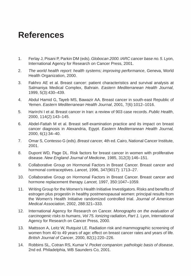
References
1. Ferlay J, Pisani P, Parkin DM (eds). Globocan 2000. IARC cancer base no. 5. Lyon, International Agency for Research on Cancer Press, 2001.
2. The world health report: health systems; improving performance, Geneva, World Health Organization, 2000.
3. Fakhro AE et al. Breast cancer: patient characteristics and survival analysis at Salmaniya Medical Complex, Bahrain. Eastern Mediterranean Health Journal, 1999, 5(3):430–439.
4. Abdul Hamid G, Tayeb MS, Bawazir AA. Breast cancer in south-east Republic of Yemen. Eastern Mediterranean Health Journal, 2001, 7(6):1012–1016.
5. Harirchi I et al. Breast cancer in Iran: a review of 903 case records. Public Health, 2000, 114(2):143–145.
6. Abdel-Fattah M et al. Breast self-examination practice and its impact on breast cancer diagnosis in Alexandria, Egypt. Eastern Mediterranean Health Journal, 2000, 6(1):34–40.
7. Omar S, Contesso G (eds). Breast cancer, 4th ed. Cairo, National Cancer Institute, 2001.
8. Dupont WD, Page DL. Risk factors for breast cancer in women with proliferative disease. New England Journal of Medicine, 1985, 312(3):146–151.
9. Collaborative Group on Hormonal Factors in Breast Cancer. Breast cancer and hormonal contraceptives. Lancet, 1996, 347(9017): 1713–27.
10. Collaborative Group on Hormonal Factors in Breast Cancer. Breast cancer and hormone replacement therapy. Lancet, 1997, 350:1047–1059.
11. Writing Group for the Women’s Health Initiative Investigators. Risks and benefits of estrogen plus progestin in healthy postmenopausal women: principal results from the Women’s Health Initiative randomized controlled trial. Journal of American Medical Association, 2002, 288:321–333.
12. International Agency for Research on Cancer. Monographs on the evaluation of carcinogenic risks to humans, Vol 75. Ionizing radiation, Part 1. Lyon, International Agency for Research on Cancer Press, 2000.
13. Mattsson A, Leitz W, Rutquist LE. Radiation risk and mammographic screening of women from 40 to 49 years of age: effect on breast cancer rates and years of life. British Journal of Cancer, 2000, 82(1):220–226.
14. Robbins SL, Cotran RS, Kumar V. Pocket companion: pathologic basis of disease, 2nd ed. Philadelphia, WB Saunders Co, 2001.
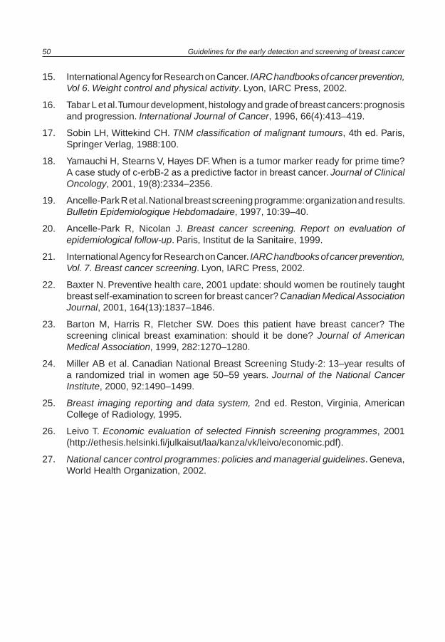
50 Guidelines for the early detection and screening of breast cancer
15. International Agency for Research on Cancer. IARC handbooks of cancer prevention, Vol 6. Weight control and physical activity. Lyon, IARC Press, 2002.
16. Tabar L et al. Tumour development, histology and grade of breast cancers: prognosis and progression. International Journal of Cancer, 1996, 66(4):413–419.
17. Sobin LH, Wittekind CH. TNM classification of malignant tumours, 4th ed. Paris, Springer Verlag, 1988:100.
18. Yamauchi H, Stearns V, Hayes DF. When is a tumor marker ready for prime time? A case study of c-erbB-2 as a predictive factor in breast cancer. Journal of Clinical Oncology, 2001, 19(8):2334–2356.
19. Ancelle-Park R et al. National breast screening programme: organization and results. Bulletin Epidemiologique Hebdomadaire, 1997, 10:39–40.
20. Ancelle-Park R, Nicolan J. Breast cancer screening. Report on evaluation of epidemiological follow-up. Paris, Institut de la Sanitaire, 1999.
21. International Agency for Research on Cancer. IARC handbooks of cancer prevention, Vol. 7. Breast cancer screening. Lyon, IARC Press, 2002.
22. Baxter N. Preventive health care, 2001 update: should women be routinely taught breast self-examination to screen for breast cancer? Canadian Medical Association Journal, 2001, 164(13):1837–1846.
23. Barton M, Harris R, Fletcher SW. Does this patient have breast cancer? The screening clinical breast examination: should it be done? Journal of American Medical Association, 1999, 282:1270–1280.
24. Miller AB et al. Canadian National Breast Screening Study-2: 13–year results of a randomized trial in women age 50–59 years. Journal of the National Cancer Institute, 2000, 92:1490–1499.
25. Breast imaging reporting and data system, 2nd ed. Reston, Virginia, American College of Radiology, 1995.
26. Leivo T. Economic evaluation of selected Finnish screening programmes, 2001 (http://ethesis.helsinki.fi/julkaisut/laa/kanza/vk/leivo/economic.pdf).
27. National cancer control programmes: policies and managerial guidelines. Geneva, World Health Organization, 2002.
Annex 1
Participants in the consultation on early detection and screening of breast cancerCairo, Egypt, 21–24 October 2002
Professor W Anwar, EgyptProfessor S Eissa, Egypt Professor A Hussain, QatarDr J Jabbour, LebanonDr M Jama, WHO Regional Office for the Eastern MediterraneanProfessor H Khaled, EgyptDr O Khatib, WHO Regional Office for the Eastern Mediterranean Professor A Modjtabai, United States of AmericaProfessor S Mostafa, EgyptProfessor S Omar, EgyptProfessor Y Omar, EgyptProfessor P Salem, United States of AmericaDr C Sepulveda, WHO headquartersDr A Verster, WHO Regional Office for the Eastern MediterraneanProfessor H Vianio, International Agency for Research on Cancer

Annex 2
Consensus opinion of the regional task force for developing breast cancer prevention, screening and management guidelines
The task force reviewed all available scientific literature on the subject including the findings of the Global Summit Early Detection Panel,1 which address early detection of breast cancer in countries with limited resources. The following conclusions and recommendations were made.
Conclusions• The Eastern Mediterranean Region has a huge area between its borders with an
ethnically diverse population, and significant differences in health care facilities and accessibility.
• It is important to have accurate and updated census data, as well as data on cancer in general and breast cancer in particular, including age-specific incidence and mortality data.
• These recommendations should be carefully studied and a set of guidelines developed that reflect them all. Guidelines that are applicable to one country, may not be applicable in another, therefore the feasibility and cost-effectiveness of guidelines should be carefully studied.
• There are three commonly-used diagnostic tools for breast cancer screening in resource-rich societies. These include mammography, clinical breast examination and breast self-examination.
• There is a key distinction between early detection and screening. Early detection is the identification of breast cancer at a stage in its natural history where the impact of therapy has the greatest chance of producing a cure. It may include programmes by which breast cancer that is causing symptoms can be diagnosed at an earlier stage. By comparison, screening for a disease means using a test on individuals or populations without any signs or symptoms of the disease of interest in order to identify those who are most likely to have the condition. It is a public health strategy that involves
1 Anderson BO et al. Early detection of breast cancer in countries with limited resources. The Breast Journal, 2003, 9(2):S51–S59.

Annex 2 53
administrating these tests to well, asymptomatic population groups. The purpose of screening is to identify a subgroup that warrants further evaluation. It is important to understand and carefully address false positive and false negative tests. A screening mammography is one tool for improving early detection.
• Because screening can take many forms, it is important to be clear about the forms under consideration. The task force reviewed screening evidence and balanced it against the available resources using the WHO framework. A distinction should be made between opportunistic screening and organized screening. Opportunistic screening occurs when a health care provider refers an individual who has no symptoms of breast cancer for a screening test. If the screening test shows an abnormality, then the woman and the provider take responsibility for coordinating and completing the diagnostic evaluation of that abnormality. Organized screening occurs as part of a programme that coordinates within a specific community, encouraging women to obtain a screening test at set time intervals. The screening programme must have the resources to perform a screening test and a complete diagnostic evaluation on those women who have abnormal results in their screening test.
Recommendations
Breast cancer screening in countries with limited resources
• The task force acknowledged the WHO approach of not advocating breast cancer screening in countries with low or medium level resources, based on the rationale that resources in such countries need to be reserved for cancer diagnosis and treatment, and possibly for screening of cervical cancer. In contrast, the task force supports the Global Summit Early Detection Panel, which suggests that screening can be implemented in countries with limited resources within a centralized cancer facility at which breast cancer treatment is available. Such programmes will not cover the country’s entire population and will only provide care to the portion of the population that has access to the facility. This recommendation is based on the premise that institution-based screening could be a pilot programme for more extensive programmes covering larger populations, and ultimately the entire population as more resources become available.
Early detection programmes without formal screening
• The task force supports and endorses the promotion of public education and awareness about breast cancer in most countries, regardless of their resource level, but recognizes that these efforts need to occur hand-in-hand with the establishment of diagnosis and treatment programmes.
54 Guidelines for the early detection and screening of breast cancer
Approach to education• The task force supports the rights of all women to be educated about breast cancer.
This education should be culturally appropriate and targeted towards, and tailored to, individual populations so that it can have the greatest impact. The points that should be stressed in public education programmes should include: – breast cancer can kill.– breast cancer can be effectively treated if detected early and need not kill.– the signs and symptoms of breast cancer.– most breast lumps are not cancer.– breast cancer is diagnosed by biopsy rather than mastectomy.
Breast self-examination• Breast self-examination screening should be distinguished from breast health
awareness. Health education activities could promote breast health awareness by urging women to become familiar with the feel and the appearance of their breast and to seek medical evaluation if they notice change in their breasts. There is hardly any evidence to support the promotion of the breast self-examination approach. Self-examination is not associated with increased detection rates or any reduction in breast cancer-specific mortality. In fact, in a Chinese randomized study of 13 000 healthy women there was an increased incidence of anxiety and surgical procedures with increased costs from unnecessary secondary investigations. However, we do not have evidence about the impact of breast self-examination in countries where women have greater breast cancer awareness and typically present with late stage breast cancer.2
Clinical breast examination • Clinical breast examination is a standardized procedure whereby a health care provider
examines a woman’s breast, chest wall and axilla. Like mammography, clinical breast examination can be either a screening test or a diagnostic test depending on the clinical setting in which it is applied. It is the consensus of the task force that clinical breast examination should be part of routine health examinations, whether used as a screening test in women who do not examine themselves, or as a diagnostic test in women who have found a change themselves. This practice will increase breast health awareness in both the women and the health care provider. Clinical breast examination has general qualities that suggest benefit through its routine use. The available data suggest that clinical breast examination does not have much benefit in women who are already being screened by mammography. The task force agrees that clinical breast examination alone is not recommended for screening as
2 Ibid.
Annex 2 55
a substitute for mammography. For countries that do not have the resources for a mammography, the use of clinical breast examination alone for screening might be a potentially interesting research topic.
Screening mammography• Mammography is the only screening modality that has evidence from randomized
trials showing reduced mortality in women who were offered screening. The strongest evidence supporting the efficacy of screening mammography is for women aged 50–74 years. Evidence also supports the value of mammography for women aged 40 –49 years and although mammography is less cost-effective in this age group, the potential for mortality reduction is similar to that observed in older women. In the United States, the Department of Health and Human Services has issued a statement formally recommending that all women aged 40 and older undergo screening mammography at 1–2 year intervals. There is no data to support its use in women under the age of 40.
Members of the task force
Professor H Abdel Azim, Medical Oncologist, EgyptDr D Ajarim, Medical Oncologist, Saudi ArabiaDr O Al Malik, Surgeon, Saudi ArabiaDr A Al Sayed, Medical Oncologist, Saudi ArabiaDr M Al Shabanah, Radiation Oncologist, Saudi ArabiaDr T Al Twegieri, Medical Oncologist, Saudi ArabiaDr A Andejani, Medical Oncologist, Saudi ArabiaDr Z Aziz, Medical Oncologist, PakistanDr S Bin Amer, Biomedical and Medical Research, Saudi ArabiaDr A Ezzat, Medical Oncologist, Saudi Arabia (Coordinator)Professor F Geara, Radiation Oncologist, LebanonProfessor H Khaled, Medical Oncologist, EgyptDr O Khatib, Regional Adviser, Noncommunicable diseases,WHO Regional Office for the Eastern MediterraneanProfessor A Modjtabai, Pathologist, United States of AmericaProfessor S Omar, Surgeon, EgyptDr R Sorbris, Surgeon, Saudi ArabiaDr L Temmim, Pathologist, KuwaitDr A Tulbah, Pathologist, Saudi Arabia