Embed Size (px)
Citation preview
EMRO Technical Publications Series 31
Guidelines for managementof breast cancer
WHO Library Cataloguing in Publication Data
Guidelines for management of breast cancer/by WHO Regional Office for the Eastern Mediterranean
p. (EMRO Technical Publications Series ; 31)
1. Breast neoplasms – Diagnosis 2. Breast neoplasms – Therapy3. Breast – Cancer 4. Breast – Cancer – GuidelinesI. Title II. WHO Regional Office for the Eastern MediterraneanIII. Series
ISBN : 978-92-9021-405-2 (NLM Classification: WP 870) ISSN : 1020-0428
© World Health Organization 2006
All rights reserved.
The designations employed and the presentation of the material in this publication do not imply the expression of any opinion whatsoever on the part of the World Health Organization concerning the legal status of any country, territory, city or area or of its authorities, or concerning the delimitation of its frontiers or boundaries. Dotted lines on maps represent approximate border lines for which there may not yet be full agreement.
The mention of specific companies or of certain manufacturers’ products does not imply that they are endorsed or recommended by the World Health Organization in preference to others of a similar nature that are not mentioned. Errors and omissions excepted, the names of proprietary products are distinguished by initial capital letters.
The World Health Organization does not warrant that the information contained in this publication is complete and correct and shall not be liable for any damages incurred as a result of its use.
Publications of the World Health Organization can be obtained from Distribution and Sales, World Health Organization, Regional Office for the Eastern Mediterranean, PO Box 7608, Nasr City, Cairo 11371, Egypt (tel: +202 670 2535, fax: +202 670 2492; email: [email protected]). Requests for permission to reproduce WHO EMRO publications, in part or in whole, or to translate them – whether for sale or for noncommercial distribution – should be addressed to the Regional Adviser, Health and Biomedical Information, at the above address (fax: +202 276 5400; email [email protected] ).
Cover design and layout by Ahmed Hassanein
Printed by Fikra Advertising Agency
Contents
Foreword ..........................................................................................................5
Preface .............................................................................................................7
Acknowledgements .........................................................................................9
List of abbreviations .....................................................................................10
Chapter 1. Diagnosis of breast cancer ........................................................11
Clinical examination ..................................................................................11Laboratory investigations ..........................................................................12Pathological diagnosis ..............................................................................13Clinical staging and risk assessment ........................................................13Prognostic factors .....................................................................................15
Chapter 2. Treatment policy ..........................................................................16
Adjuvant systemic treatment .....................................................................16Role of primary chemotherapy (neoadjuvant chemotherapy) in locallyadvanced breast cancer ............................................................................17Follow-up ...................................................................................................22
Chapter 3. Management of metastatic disease ...........................................24
Staging of metastatic or recurrent breast cancer ......................................24Local recurrence only ................................................................................24Systemic dissemination ............................................................................24Preferred chemotherapy regimens for recurrent or metastatic breastcancer .......................................................................................................25
Chapter 4. Surgical guidelines for breast cancer .......................................30
Surgical approach to the axilla including sentinel lymph node biopsy ......30Surgical technique ....................................................................................31Hospital stay ..............................................................................................32Guidelines .................................................................................................32
Chapter 5. Management of special problems in breast cancer .................35
Chapter 6. Pathological handling of breast cancer excision specimens .40
General considerations .............................................................................40Fine needle aspiration biopsy and core needle biopsy .............................41Excision specimen for a palpable mass ....................................................41
Tissue submitted for histological examination ...........................................42Mastectomy specimen ..............................................................................42Tissue submitted for histological examination ...........................................42Mammographically-directed excisions (wire localization specimens) .......43Tissue submitted for histological examination ...........................................43Frozen section diagnosis ..........................................................................44Surgical pathology report of breast cancer specimens .............................44
Chapter 7. Radiotherapy guidelines for breast cancer ..............................47
Radiotherapy for ductal carcinoma in situ .................................................47Criteria for breast conserving therapy .......................................................47Post-mastectomy radiotherapy ..................................................................49Radiotherapy after pre-operative systemic therapy ...................................51
Further reading ..............................................................................................52
Annex 1 Participants in the consultation on early detection and screening of breast cancer ...........................................................................55
Foreword
Cancer is an important factor in the global burden of disease. The estimated number of new cases each year is expected to rise from 10 million in 2002 to 15 million by 2025, with 60% of those cases occurring in developing countries. Breast cancer is the most common cancer in women in the Eastern Mediterranean Region and the leading cause of cancer mortality worldwide. There is geographic variation, with the standardized age-incidence rate being lower in developing than industrialized countries.
Although the etiology of breast cancer is unknown, numerous risk factors may influence the development of this disease including genetic, hormonal, environmental, sociobiological and physiological factors. Over the past few decades, while the risk of developing breast cancer has increased in both industrialized and developing countries by 1%–2% annually, the death rate from breast cancer has fallen slightly. Researchers believe that lifestyle changes and advances in technology, especially in detection and therapeutic measures, are in part responsible for this decrease.
Breast cancer does not strike an individual alone but the whole family unit. Despite considerable social changes, women continue to be the focus of family life. The impact of breast cancer is therefore profound on both the woman diagnosed with the disease and her family. Their fear and anxiety over the eventual outcome of the illness may manifest itself through behavioural changes.
The high incidence and mortality rates of breast cancer, as well as the high cost of treatment and limited resources available, require that it should continue to be a focus of attention for public health authorities and policy-makers. The costs and benefits of fighting breast cancer, including the positive impact that early detection and screening can have, need to be carefully weighed against other competing health needs. Ministry of Health officials need to formulate and implement plans that will effectively address the burden of the disease, including setting policies on the early detection and screening of breast cancer. Health care providers should also be involved in discussion of the issue and in developing programmes for the management of the disease. I hope these guidelines will support everyone involved in the battle against breast cancer in the Eastern Mediterranean Region.
Hussein A. Gezairy MD FRCS Regional Director for the Eastern Mediterranean
In the Name of God, the Compassionate, the Merciful
Preface
Breast cancer remains a common and frequently fatal disease, the most commonly diagnosed cancer in women and the second ranking cause of cancer death in the Eastern Mediterranean Region. More than 1.2 million women are diagnosed with breast cancer annually worldwide. In developed countries, most patients (> 80%) with breast cancer present with operable disease that can apparently be entirely resected surgically. About half of these patients eventually relapse, and when these are added to those initially presenting with primary advanced disease, this means that most patients with breast cancer ultimately require treatment for advanced disease. Clinical breast cancer research has focused on effective methods to detect breast cancer at its earliest stages and on standardized treatments to cure the disease after diagnosis. However, despite advances in these areas, one third of all women in North America who develop breast cancer will die of the disease.
Guidelines for breast cancer have been developed in many countries to assist clinicians and patients to make decisions about treatment and thus improve health outcomes. Observed differences in treatment outcome between populations suggest opportunities for improvement. Moreover, potentially important variations in clinical practice are well documented in many countries. Treatment practice that is informed by evidence, such as greater use of breast conserving surgery, has been observed more frequently among clinicians who regularly treat patients with breast cancer. Furthermore, congruence of treatment practice with published guidelines has been directly associated with improved patient survival. Improved treatment practice has the potential to improve survival by up to 15%. Therefore, enhanced implementation of soundly developed, evidence-based treatment guidelines is an important goal for health services and individual clinicians.
In many countries clinicians, scientists and patients involved in breast cancer diagnosis and treatment have formed cooperative groups to improve breast cancer treatment guidelines. These guidelines were prepared by the World Health Organization (WHO) Regional Office for the Eastern Mediterranean and the King Faisal Specialist Hospital and Research Centre, a WHO collaborating centre for cancer prevention and care. The idea was conceived at the Consultation on Early Detection and Screening of Breast Cancer, held at the Regional Office in Cairo in 2002, during which a framework for the guidelines was prepared by participants (see Annex 1). Subsequently, in January 2004, a Task Force for Developing Breast Cancer Prevention, Screening and Management Guidelines was established at a meeting at the King Faisal Specialist Hospital and Research Centre in Riyadh. The members of the task force developed the
8 Guidelines for management of breast cancer
guidelines with the consensus of all contributors. The task force took into consideration the cost of treatment and factors common to most countries in the Region, such as limited resources and a paucity of specialized cancer centres, without compromising the efficacy of the guidelines.
The guidelines are aimed at oncologists, internists, secondary and tertiary hospitals, ministries of health and other health decision-makers. The purpose is to provide answers to the practical questions involved in decision-making about day-to-day management of breast cancer. The paradigms that underpin them are also outlined, promoting evidence-based and cost-effective interventions. It is hoped that these guidelines will support the choices made by both health care providers and patients.
Chapter 1 outlines the elements involved in diagnosis of breast cancer, including clinical examination, laboratory investigation, pathologic diagnosis, staging and risk assessment, and prognostic factors. Treatment policy is addressed in Chapter 2 including adjuvant systemic treatment, an international overview of treatment outcomes, treatment of early stage invasive breast cancer including surgery, adjuvant therapy for node-negative and node-positive breast cancer, primary chemotherapy in locally-advanced breast cancer, the definitions used in response evaluation of primary systemic therapy, the treatment of locally-advanced invasive breast cancer, and follow-up. The diagnosis and management of metastatic disease, including preferred chemotherapy regimes and hormonal therapy, are covered in Chapter 3, while Chapter 4 contains surgical guidelines. Chapter 5 looks at special problems in breast cancer including bilateral breast cancer, cancer of the male breast, the unknown primary presenting with axillary lymphadenopathy, Paget’s disease of the nipple-areola complex and phyllodes tumour of the breast. Chapter 6 presents guidelines for the pathological handling of breast cancer excision specimens and Chapter 7 outlines radiotherapy guidelines for breast cancer.
Acknowledgements
The WHO Regional Office for the Eastern Mediterranean acknowledges with thanks the contributions of the participants at the Regional Consultation on Early Detection and Screening of Breast Cancer (Annex 1) held in Cairo, Egypt, 21–24 October 2002, whose discussions provided the impetus for this publication. WHO would like to thank the Task Force for Developing Breast Cancer Prevention, Screening and Management Guidelines whose members were as follows:
Professor H Abdel Azim, Medical Oncologist, EgyptDr D Ajarim, Medical Oncologist, Saudi ArabiaDr O Al Malik, Surgeon, Saudi ArabiaDr A Al Sayed, Medical Oncologist, Saudi ArabiaDr M Al Shabanah, Radiation Oncologist, Saudi ArabiaDr T Al Twegieri, Medical Oncologist, Saudi ArabiaDr A Andejani, Medical Oncologist, Saudi ArabiaDr Z Aziz, Medical Oncologist, PakistanDr S Bin Amer, Biomedical and Medical Researcher, Saudi ArabiaDr A Ezzat, Medical Oncologist, Saudi Arabia (Coordinator)Professor F Geara, Radiation Oncologist, LebanonDr O Khatib, Regional Adviser, Noncommunicable diseases,WHO Regional Office for the Eastern MediterraneanProfessor S Omar, Surgeon, EgyptDr R Sorbris, Surgeon, Saudi ArabiaDr L Temmim, Pathologist, KuwaitDr A Tulbah, Pathologist, Saudi Arabia
The draft publication was reviewed by Adnan Ezzat, Hussein Khaled, Oussama Khatib, Atord Modjtabai, Sherif Omar and Taher Al Twegieri.

List of abbreviations
5-FU fluorouracilAC doxorubicin and cyclophosphamideALND axillary lymph node dissectionBCS breast-conserving surgeryBCT breast-conserving treatmentcCR clinical complete responseCBC complete blood countCBCD complete blood count with differentialCMF cyclophosphamide, methotrexate and 5-fluorouracilCNB core needle biopsycPR clinical partial responseCSP cystosarcoma phyllodesCT computed tomographyCTX cyclophosphamideDCIS ductal carcinoma in situECG electrocardiogramER estrogen receptorFAC 5-fluorouracil, doxorubicin, cyclophosphamideFAD fibroadenomasFEC 5-fluorouracil, epirubicin and cyclophosphamide.FNA fine needle aspirationFNAB fine needle aspiration biopsyHER2 human epidermal growth factor receptor 2IHC immunohistochemistryLCIS lobular carcinoma in situLHRH luteinizing hormone-releasing hormoneMRI magnetic resonance breast imagingMRM modified radical mastectomyMUGA multiple gated acquisitionOA ovarian ablationpCR pathologic complete responsePCR polymerase chain reactionPET positron emission tomographyPR progesterone receptorSSM skin-sparing mastectomyTAC taxotere, doxorubicin and cyclophosphamideTNM tumour, nodes, metastasisTRAM transverse rectus abdominis muscleXRT radiation therapy
Chapter 1
Diagnosis of breast cancer
Clinical examination
History
This should include the following elements:
1. Presenting symptoms:• breast mass• breast pain• nipple discharge• nipple or skin retraction• axillary mass or pain• arm swelling• symptoms of possible metastatic spread• suspicious findings on routine mammography.
2. Past medical history of breast disease in detail.3. Family history of breast and other cancers with emphasis on gynaecological
cancers.4. Reproductive history:
• age at menarche• age at first delivery• number of pregnancies, children and miscarriages• age at onset of menopause• history of hormonal use including:
– contraceptive pills (type and duration)– hormonal replacement therapy (type and duration).
5. Past medical history.
Physical examination
Careful physical examination should cover the following:
1. Performance status.
12 Guidelines for management of breast cancer
2. Weight, height and surface area.3. General examination of other systems.4. Local examination:
• breast mass– size– location (specified by clock position and distance from the edge of the areola)– shape– consistency– fixation to skin, pectoral muscle and chest wall– multiplicity
• skin changes– erythema (location and extent)– oedema (location and extent)– dimpling– infiltration– ulceration– satellite nodules
• nipple changes– retraction– erythema– erosion and ulceration– discharge (specify)
• nodal status– axillary nodes on both sides (number, size, location and fixation to other nodes
or underlying structures)– supraclavicular nodes
• local examination of possible metastatic sites.
Laboratory investigationsThese include the following:
• Complete blood count with differential (CBCD), and renal and hepatic profile.• Bilateral mammography and/or ultrasound.• Chest X-ray ± computed tomography imaging (CT) of chest if needed.• Abdominal ultrasound ± CT of abdomen.
Diagnosis of breast cancer 13
• Bone scan if indicated.• Electrocardiogram (ECG) and echocardiogram or multiple gated acquisition (MUGA)
scan if age > 60.• Positron emission tomography (PET) scan optional.
Pathological diagnosisA pathological diagnosis should be obtained by core needle or fine needle biopsy
(depending on the availability of local expertise) prior to any surgical procedure. However, local excision biopsy or frozen section may be done if this is not possible. In the case of T1 or T2 lesions, a frozen section examination may provide better determination of surgical margin.
Final pathological diagnosis should be made according to the current pathological classification, analysing all tissue removed including axillary nodal status (number of nodes, capsular infiltration and level of nodes affected). Determination of estrogen receptor (ER) and progesterone receptor (PR) status is mandatory, and determination of HER2 receptor status should be considered.
Clinical staging and risk assessment
Tumour, nodes, metastasis (TNM) staging system
The tumour staging system provides information about extent of disease that can be used to guide treatment recommendations and to provide estimates of patient prognosis. In addition, it provides a framework for reporting treatment outcomes allowing the efficacy of new treatment to be assessed. The TNM staging system classification criteria are summarized below.
Primary tumour (T)
Tx Primary tumour cannot be assessed. T0 No evidence of primary tumour. Tis Carcinoma in situ: ductal (DCIS) or lobular (LCIS) carcinoma, or Paget’s
disease of the nipple with no tumour. T1 Tumour 2 cm or less in greatest dimension. T2 Tumour more than 2 cm, but not more than 5 cm in greatest dimension. T3 Tumour more than 5 cm in greatest dimension. T4 Tumour of any size with direct extension to the chest wall or skin
a. Extension to chest wall not including pectoral muscle
14 Guidelines for management of breast cancer
b. Oedema, including peau d’orange, ulceration of the skin or satellite skin nodules confined to the same breast
c. Both a and b d. Inflammatory carcinoma.
Regional lymph nodes (N)
Nx Regional lymph node cannot be assessed. N0 No regional lymph node metastasis. N1 Metastasis to movable ipsilateral axillary lymph node. N2 a. Metastasis to ipsilateral axillary lymph node fixed to one another or
matted to other structuresb. Clinically-apparent ipsilateral internal mammary lymph node in the absence of clinically-evident axillary lymph node metastasis.
N3 a. Metastasis to ipsilateral infraclavicular lymph node b. Clinically-apparent ipsilateral internal mammary lymph node in the presence of clinically-evident axillary lymph node metastasis c. Metastasis to ipsilateral supraclavicular lymph node with or without axillary or internal mammary lymph node involvement.
Pathological classification (pN)
pNx Regional lymph nodes cannot be assessed. pN0 No regional lymph node metastasis .pN1 a. Metastasis to 1 to 3 axillary lymph nodes
b. Metastasis to internal mammary lymph nodes with microscopic disease detected by sentinel lymph node dissection but not clinically apparentc. Metastasis to both a and b.
pN2 a. Metastasis to 4 to 9 axillary lymph nodes b. Metastasis to clinically-apparent internal mammary lymph node in the absence of axillary lymph node metastasis.
pN3 a. Metastasis to 10 or more axillary lymph nodes (at least 1 tumour deposit more than 2 mm) or to infraclavicular lymph node b. Metastasis to clinically-apparent ipsilateral internal mammary lymph node in the presence of 1 or more positive axillary lymph nodes or to more than 3 axillary lymph nodes and to internal mammary lymph node with microscopic disease detected by sentinel lymph node dissection but not clinically-apparent.c. Metastasis to ipsilateral supraclavicular lymph node.
Diagnosis of breast cancer 15
Prognostic factorsSeveral tumour characteristics that have important prognostic significance need to
be considered when designing an optimal treatment strategy for the individual patient. These are:
• Age of patient.• Tumour size.• Axillary lymph node status. This is the most important predictor of disease recurrence
and survival: 70%–80% of patients with node-negative status survive 10 years; prognosis worsens as the number of positive lymph nodes increases. About 40%–50% of patients with 1 to 3 positive nodes survive 10 years, whereas only 15% of those with more than 4 nodes survive with surgical treatment alone.
• Histological grade.• Estrogen receptor (ER) and progesterone receptor (PR) status. These are cellular
proteins present in hormone-responsive target tissues. Patients with receptor-positive primary tumours have lower rates of recurrence and longer survival than those with receptor-negative tumours.
• HER2-neu (C-erb B2).• Tumour suppressor genes p53 and bcl-2 are presently considered research tools.
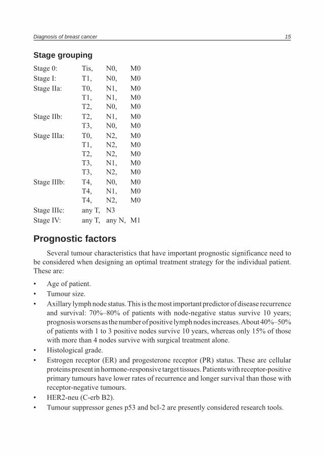
Stage grouping
Stage 0: Tis, N0, M0 Stage I: T1, N0, M0 Stage IIa: T0, N1, M0 T1, N1, M0 T2, N0, M0 Stage IIb: T2, N1, M0 T3, N0, M0 Stage IIIa: T0, N2, M0 T1, N2, M0 T2, N2, M0 T3, N1, M0 T3, N2, M0 Stage IIIb: T4, N0, M0 T4, N1, M0 T4, N2, M0Stage IIIc: any T, N3Stage IV: any T, any N, M1
Chapter 2
Treatment policy
Adjuvant systemic treatment
Introduction
Despite optimal local treatment, virtually all patients with invasive breast cancer have some risk of systemic relapse. This risk varies with numerous patient and disease-related factors. Therefore, all women with invasive breast cancer stand to benefit from systemic treatment to try and reduce this risk. However, because all of these treatments have side effects and potential risks, the need for systemic treatment must be assessed on an individual basis.
Adjuvant chemotherapy has been defined as the administration of chemotherapy to kill or inhibit clinically undetectable micrometastasis after primary surgery. Such an approach is prudent, as adjuvant systemic chemotherapy with or without hormonal therapy has been demonstrated to improve survival in both node-negative and node-positive disease. Adjuvant chemotherapy may increase 10-year survival by 7%–11% in premenopausal women with early stage disease and by 2%–3% in women aged over 50.
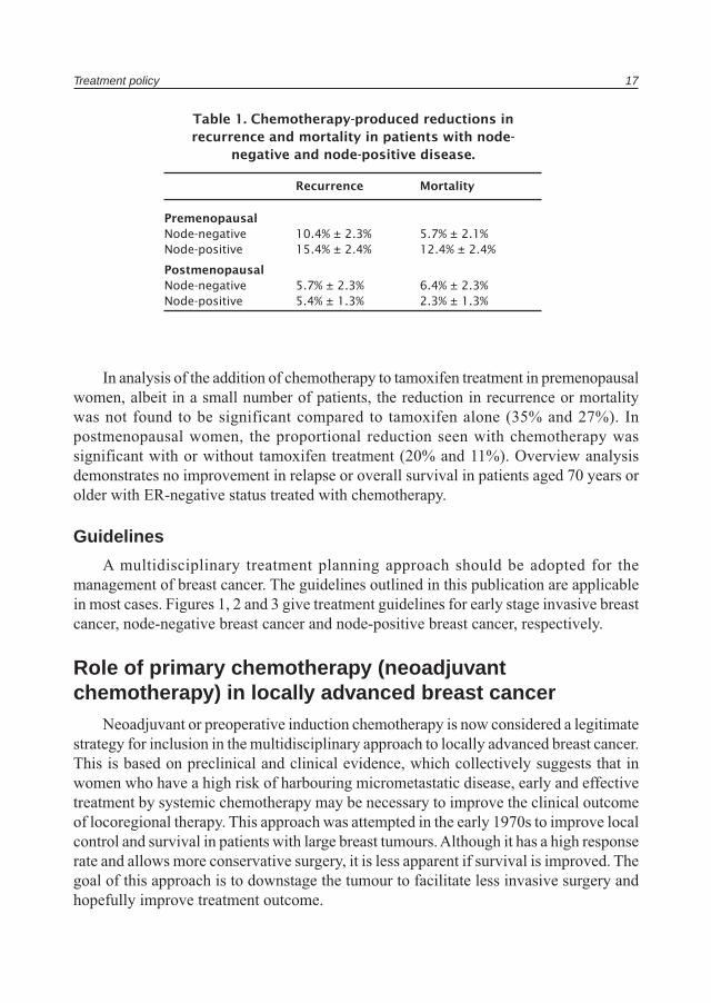
International overview
The meta-analysis by the Early Breast Cancer Trialists’ Collaborative Group regarding polychemotherapy, consisted of a total of 69 trials involving about 30 000 women. Comparison of prolonged versus no chemotherapy has also been analysed in 18 000 women in 47 trials.
Chemotherapy-produced reduction in recurrence and increased survival was found in all groups analysed (reduction in recurrence = 23.5% ± 2% and reduction in mortality = 15.3% ± 2%). This was more prominent in premenopausal women and those with ER-negative status. Survival benefit was seen in the first 5 years with additional benefit during the second 5 years. A significant reduction in recurrence and mortality was seen in both pre and postmenopausal patients. When nodal involvement was considered, the proportional reductions in recurrence and mortality were similar in node-negative or node-positive disease (see Table 1).
Treatment policy 17
In analysis of the addition of chemotherapy to tamoxifen treatment in premenopausal women, albeit in a small number of patients, the reduction in recurrence or mortality was not found to be significant compared to tamoxifen alone (35% and 27%). In postmenopausal women, the proportional reduction seen with chemotherapy was significant with or without tamoxifen treatment (20% and 11%). Overview analysis demonstrates no improvement in relapse or overall survival in patients aged 70 years or older with ER-negative status treated with chemotherapy.
Guidelines
A multidisciplinary treatment planning approach should be adopted for the management of breast cancer. The guidelines outlined in this publication are applicable in most cases. Figures 1, 2 and 3 give treatment guidelines for early stage invasive breast cancer, node-negative breast cancer and node-positive breast cancer, respectively.
Role of primary chemotherapy (neoadjuvant chemotherapy) in locally advanced breast cancer
Neoadjuvant or preoperative induction chemotherapy is now considered a legitimate strategy for inclusion in the multidisciplinary approach to locally advanced breast cancer. This is based on preclinical and clinical evidence, which collectively suggests that in women who have a high risk of harbouring micrometastatic disease, early and effective treatment by systemic chemotherapy may be necessary to improve the clinical outcome of locoregional therapy. This approach was attempted in the early 1970s to improve local control and survival in patients with large breast tumours. Although it has a high response rate and allows more conservative surgery, it is less apparent if survival is improved. The goal of this approach is to downstage the tumour to facilitate less invasive surgery and hopefully improve treatment outcome.
Table 1. Chemotherapy-produced reductions in recurrence and mortality in patients with node-
negative and node-positive disease.
Recurrence Mortality
PremenopausalNode-negative 10.4% ± 2.3% 5.7% ± 2.1%Node-positive 15.4% ± 2.4% 12.4% ± 2.4%
PostmenopausalNode-negative 5.7% ± 2.3% 6.4% ± 2.3%Node-positive 5.4% ± 1.3% 2.3% ± 1.3%
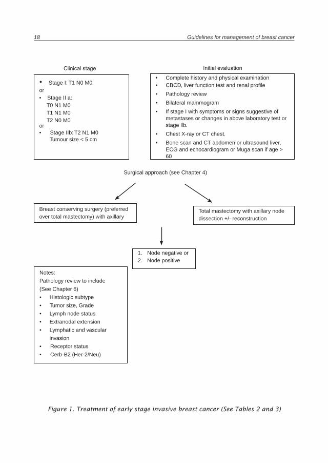
18 Guidelines for management of breast cancer
• Complete history and physical examination• CBCD, liver function test and renal profile
• Pathology review
• Bilateral mammogram
• If stage I with symptoms or signs suggestive of metastases or changes in above laboratory test or stage llb.
• Chest X-ray or CT chest.
• Bone scan and CT abdomen or ultrasound liver, ECG and echocardiogram or Muga scan if age > 60
• Stage I: T1 N0 M0 or• Stage II a: T0 N1 M0 T1 N1 M0 T2 N0 M0 or• Stage IIb: T2 N1 M0 Tumour size < 5 cm
Notes:
Pathology review to include
(See Chapter 6)
• Histologic subtype
• Tumor size, Grade
• Lymph node status
• Extranodal extension
• Lymphatic and vascular
invasion
• Receptor status
• Cerb-B2 (Her-2/Neu)
Clinical stage Initial evaluation
Surgical approach (see Chapter 4)
Figure 1. Treatment of early stage invasive breast cancer (See Tables 2 and 3)
Total mastectomy with axillary node dissection +/- reconstruction
Breast conserving surgery (preferred over total mastectomy) with axillary
1. Node negative or2. Node positive
Treatment policy 19
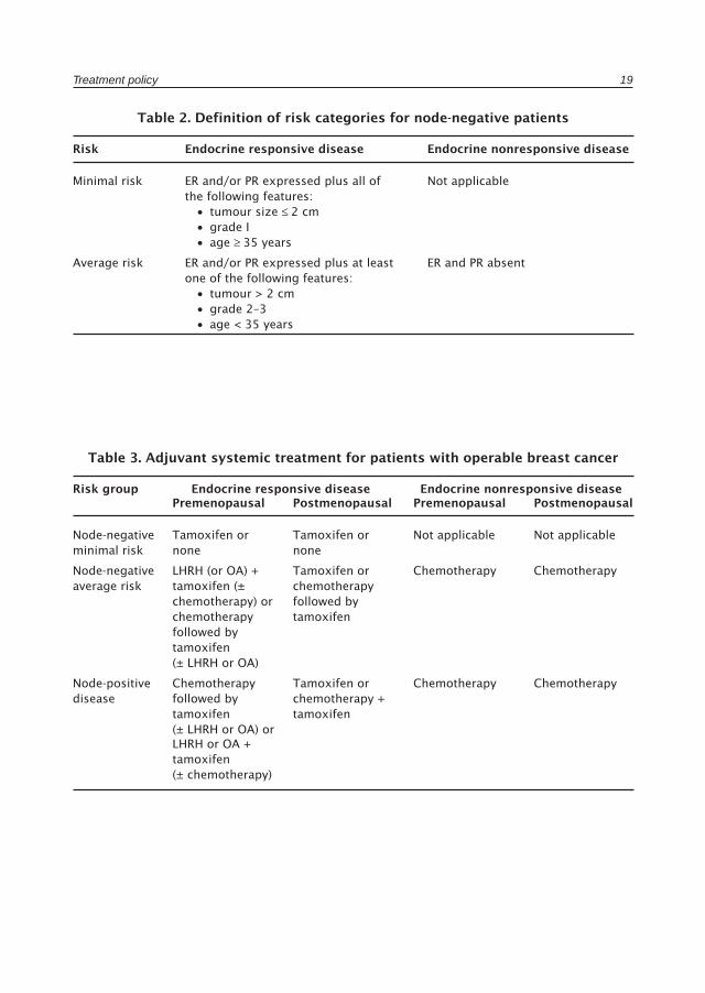
Table 2. Definition of risk categories for node-negative patients
Risk Endocrine responsive disease Endocrine nonresponsive disease
Minimal risk ER and/or PR expressed plus all of Not applicable the following features: • tumour size ≤ 2 cm • grade I • age ≥ 35 years
Average risk ER and/or PR expressed plus at least ER and PR absent one of the following features: • tumour > 2 cm • grade 2–3 • age < 35 years
Table 3. Adjuvant systemic treatment for patients with operable breast cancer
Risk group Endocrine responsive disease Endocrine nonresponsive disease Premenopausal Postmenopausal Premenopausal Postmenopausal
Node-negative Tamoxifen or Tamoxifen or Not applicable Not applicableminimal risk none none
Node-negative LHRH (or OA) + Tamoxifen or Chemotherapy Chemotherapyaverage risk tamoxifen (± chemotherapy chemotherapy) or followed by chemotherapy tamoxifen followed by tamoxifen (± LHRH or OA)
Node-positive Chemotherapy Tamoxifen or Chemotherapy Chemotherapydisease followed by chemotherapy + tamoxifen tamoxifen (± LHRH or OA) or
LHRH or OA + tamoxifen (± chemotherapy)
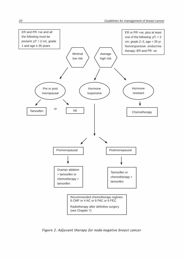
20 Guidelines for management of breast cancer
Figure 2. Adjuvant therapy for node-negative breast cancer
ER and PR +ve and all
the following must be
present: pT ≤ 2 cm, grade
1 and age ≥ 35 years
ER or PR +ve, plus at least
one of the following: pT, > 2
cm, grade 2–3, age < 35 yr.
Nonresponsive endocrine
therapy: ER and PR -veMinimal
low risk
Average
high risk
Pre or post
menopausal
Hormone
responsive
Hormone
resistant
Tamoxifen Nil Chemotherapy
Premenopausal
Ovarian ablation
+ tamoxifen or
chemotherapy +
tamoxifen
Recommended chemotherapy regimes6 CMF or 4 AC or 6 FAC or 6 FEC
Radiotherapy after definitive surgery (see Chapter 7)
Postmenopausal
or
Tamoxifen or
chemotherapy +
tamoxifen
Treatment policy 21
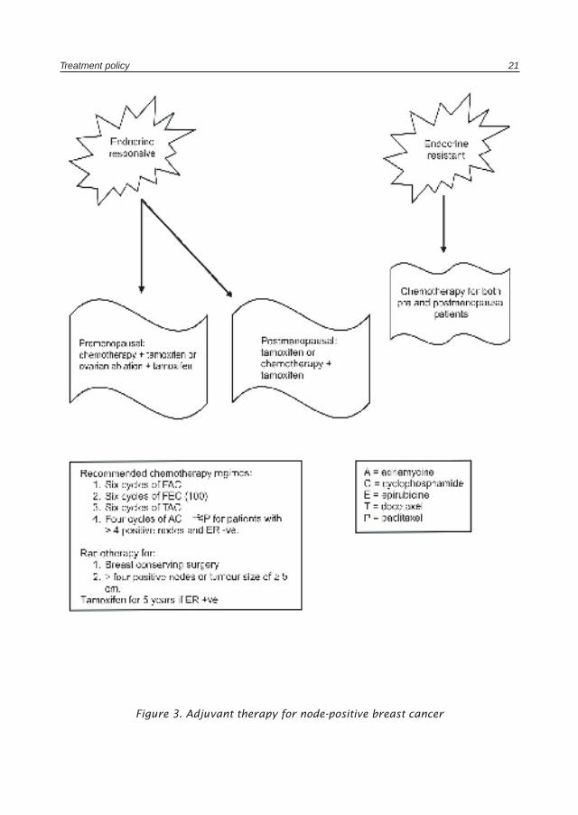
Figure 3. Adjuvant therapy for node-positive breast cancer
22 Guidelines for management of breast cancer
Definitions for response evaluation of primary systemic therapy
Clinical definition• Complete: no palpable mass detectable (cCR)• Partial: reduction of tumour area to < 50% (cPR)
Imaging definition• No tumour visible by mammogram and/or ultrasound and/or MRI
Pathological definition• Only focal invasive tumour residuals in the removed breast tissue• Only in situ tumour residuals in the removed breast tissue (pCR inv)• No invasive or in situ tumour cells (pCR)• No malignant tumour cells in breast and lymph nodes (pCR breast and nodes).
Guidelines
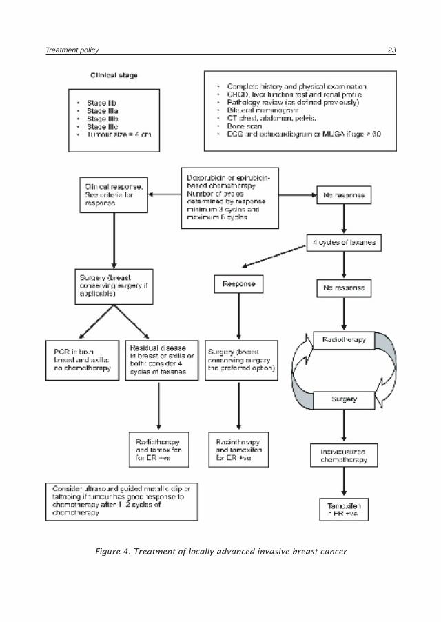
Figure 4 gives the treatment guideline for locally advanced invasive breast cancer. The best treatment option for this type of cancer is participation in a clinical trial if available.
Follow-upHistory taking and physical examination is recommended every 3–6 months for 3
years, then every 6–12 months for the next 2 years and annually after that with attention paid to long-term side effects such as osteoporosis.
Ipsilateral (after breast-conserving surgery) and contralateral mammography is to be done every 1–2 years. Blood counts, chemistry, chest X-rays, bone scans, liver ultrasound, CT scans of chest and abdomen, and monitoring of tumour markers such as CA15.3 and CEA are not routinely recommended for asymptomatic patients. Because of the risk of tamoxifen-assisted endometrial cancer, a yearly pelvic examination coupled with evaluation of vaginal spotting is essential. The performance of endometrial biopsy or ultrasound is not recommended.
Treatment policy 23
Figure 4. Treatment of locally advanced invasive breast cancer
Chapter 3
Management of metastatic disease
Staging of metastatic or recurrent breast cancerThe staging evaluation of women presenting with metastatic or recurrent breast
cancer includes the performance of a CBC, platelet count, liver function tests, chest X-ray, bone scan, X-rays of symptomatic bones or bones that appear abnormal on bone scan, CT or MRI of symptomatic areas, and biopsy documentation of first recurrence, if possible (see Figure 4).
Local recurrence onlyPatients with local recurrence only are divided into those who have initially been
treated by mastectomy and those who have received breast-conserving therapy.
Mastectomy-treated patients should undergo surgical resection of the local recurrence, if it can be accomplished without extensive surgery, and involved-field radiotherapy (if the chest wall was not previously treated or if additional radiotherapy may be safely administered). The use of surgical resection in this setting implies the use of limited excision of disease with the goal of obtaining clear margins of resection.
Women whose disease recurs locally following initial breast-conserving therapy should undergo a total mastectomy.
Following local treatment, women with local recurrences should be considered for systemic chemotherapy or hormonal therapy, as is the case for those with systemic recurrences.
Systemic disseminationThe treatment of systemic recurrence of breast cancer prolongs survival and
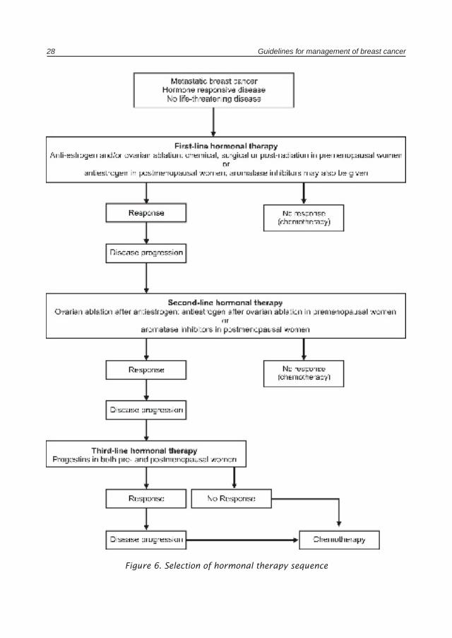
enhances quality of life but is not curative, and, therefore, treatments associated with minimal toxicity are preferred. Thus, the use of the minimally toxic hormonal therapies is preferred to the use of cytotoxic therapy whenever reasonable.
Women with osteolytic bone lesions may be given zoledronate or pamidronate if expected survival is 3 months or greater and there is normal renal function. Bisphosphonates can be given in addition to chemotherapy or hormonal therapy.

Management of metastatic disease 25
Women considered to be appropriate candidates for initial hormonal therapy for treatment of recurrent or metastatic disease include those whose tumours are estrogen-and/or progesterone-positive, those with bone or soft-tissue disease only, or those with limited, asymptomatic visceral disease.
In women without prior exposure to an antiestrogen, antiestrogen therapy is the preferred first hormonal therapy unless there are contraindications to tamoxifen therapy.
In women with prior antiestrogen exposure, recommended second-line hormonal therapies include, preferably, selective aromatase inhibitors (anastrozole, letrozole or exemestine) in postmenopausal women, progestins (megestrol acetate), and in premenopausal women, luteinizing hormone-releasing hormone (LHRH) agonists and surgical or radiotherapeutic oophorectomy. Women who respond to a hormonal manoeuvre with either shrinkage of the tumour or long term stabilization of their disease should receive additional but different hormonal therapy at the time of progression.
Women with estrogen and progesterone receptor-negative tumours, symptomatic visceral metastasis, or hormone-refractory disease should receive chemotherapy. A wide variety of chemotherapy regimens are felt to be appropriate, as outlined below.
Preferred chemotherapy regimens for recurrent or metastatic breast cancer
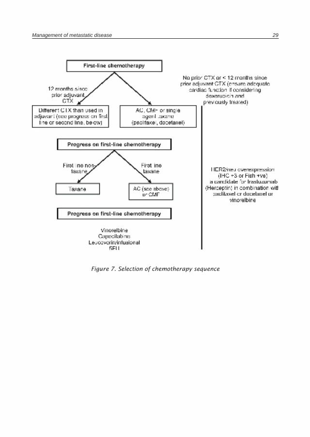
Preferred first-line chemotherapy
• Anthracycline-based.• Taxanes.• Cyclophosphamide, methotrexate and 5-fluorouracil (CMF).
Preferred second-line chemotherapy• If first-line was anthracycline-based or CMF, then a taxane.• If first-line was a taxane, then anthracycline-based or CMF.• Other active regimens include capecitabine, 5-fluorouracil (via infusion), vinorelbine,
and mitoxantrone.
In patients whose tumours overexpress HER2/neu, consideration may be given to using trastuzumab in combination with paclilaxel, docetaxel or vinorelbine. Trastuzumab has also been given in combination with doxorubicin and cyclophosphamide (AC), but the use of trastuzumab plus AC is associated with significant cardiac toxicity.
26 Guidelines for management of breast cancer
Failure to achieve a tumour response to two sequential chemotherapy regimens or an Eastern Cooperative Oncology Group performance status of 3 or greater was felt to be an indication for supportive therapy only.
Patients with metastatic breast cancer frequently develop a number of anatomically localized problems that may benefit from local irradiation, surgery, or regional chemotherapy e.g. intrathecal methotrexate for leptomeningeal carcinomatosis.
Guidelines
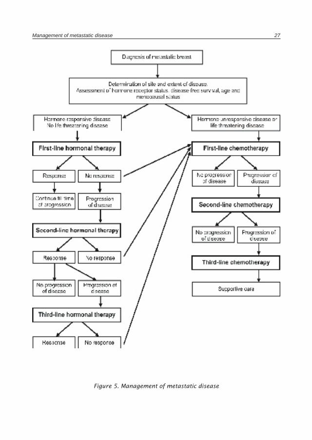
The guidelines for selection of systemic hormonal therapy or chemotherapy are summarized in Figures 5, 6 and 7.
Management of metastatic disease 27
Figure 5. Management of metastatic disease
28 Guidelines for management of breast cancer
Figure 6. Selection of hormonal therapy sequence
Management of metastatic disease 29
Figure 7. Selection of chemotherapy sequence

Chapter 4
Surgical guidelines for breast cancer
Surgical approach to the axilla including sentinel lymph node biopsy
There are several options for the surgical approach to the axilla:
• limited level one axillary dissection• level one axillary dissection• axillary sampling• level one and two axillary dissection• complete axillary dissection: level one, two and three• selective axillary dissection with sentinel lymphadenectomy• selective axillary dissection based on tumour size and other characteristics.
The standard of care would be a level one and two axillary dissection for all stages of breast cancer and a complete axillary dissection if lymphadenopathy involves level three. Axillary dissection is not indicated in pure ductal carcinoma in situ (DCIS). Patients undergoing a simple mastectomy for DCIS will have inadvertently a level one dissection because the axillary tail is usually included within the mastectomy specimen. The minimum number of lymph nodes extracted should not be less than ten and a proper level one and two dissection would yield more than 10 lymph nodes.
The patient population is an important factor in determining the type of surgical approach to the axilla. Axillary dissection can be avoided in those with a frequency of lymph node metastasis < 5 %. This would include DCIS with microinvasion, T1a and T1b mucinous and tubular carcinomas, T1a papillary carcinomas, and T1a grade 1 carcinomas. The frequency of involved lymph nodes varies from one institution to another. The ideal situation would be that each institution avoids axillary dissection based upon their frequency of involved lymph nodes for each pathological stage. The other option would be to extrapolate the experience from other centres. Each patient that is a candidate for avoidance of axillary dissection should be informed of the pros and cons of such an approach.
Sentinel lymphadenectomy should be performed only within the correct set-up. A combined team formed of a surgeon, a pathologist and a nuclear medicine physician should receive proper training and must test the procedure in their hospital setting. The
Surgical guidelines for breast cancer 31
most important factor determining the results of this procedure is the patient population. The majority of the studies evaluating axillary disection were done on tumours less than 2.5 cm. The most important factor affecting its acceptance is the false negative rate. Studies need long-term follow-up to validate the results and safety of the procedure before its acceptance as a standard of care. This technique should be studied in all stages of breast cancer before it can be recommended. The advantages and disadvantages of the technique are as follows.
Advantages• Diagnostic. The TNM staging system is based on the accurate knowledge of the involved
lymph nodes. Neither clinical nor radiological assessment of the axilla is reliable; only pathological evaluation of the excised axillary contents is diagnostic.
• Prognostic. It is the single most important predictor of survival.• Therapeutic. It provides local-regional control by reducing axillary recurrence. It
may be a prophylactic measure by preventing further systemic spread and distant metastasis. The impact on overall survival is controversial; there may be a small but significant survival benefit of axillary dissection.
• Dictates adjuvant therapy. The number of involved lymph nodes dictates the type of systemic therapy and the need for radiation therapy to the axilla.
Disadvantages• Morbidity. Axillary dissection is associated with a small but significant amount of
morbidity. The frequency of lymphedema varies between 3%–30%. Intercostobrachial nerve injury causes numbness in the arm. Wound infections and seromas are also other possible complications. Injury to the thoracodorsal and the long thoracic nerves should not occur. If they are encased with tumour they are sacrificed.
• In certain patients it does not dictate adjuvant therapy.• It does not affect survival in the majority of patients.
Surgical techniqueA U-shaped or transverse incision should be used for better cosmesis and exposure.
A radial or a continuous incision at the excision site is not recommended. Both incisions cause deformity of the breast. The incision is made in the axillary hairline between the pectoralis major muscle medially and the latissmus dorsi muscle laterally. No axillary lymph nodes exist above the axillary fascia; therefore, thin flaps are to be avoided. Creation of thin flaps exaggerates the axillary hollow and causes deformity.
The clavipectoral fascia once encountered is incised transversely. The lateral border of the pectoralis major is identified and the neurovascular bundle (lateral pectoral nerve) is identified, isolated and preserved. The axillary vein is then identified. The axillary sheath should not be incised. The long thoracic nerve is then identified, isolated and preserved.
32 Guidelines for management of breast cancer
The thoracodorsal neurovascular bundle is identified underneath the axillary vein and is carefully preserved. A level one and two would be included in the specimen if the axillary contents in between the two nerves (long thoracic and the thoracodorsal nerves) is excised. All palpable diseased lymph nodes should be excised. The pectoralis minor muscle should not be excised. Preservation of the intercostobrachial nerves is preferred if possible. Metallic clips are inserted at the end of the procedure to guide radiation therapy. A Jackson-Pratt drain is inserted at the end of the procedure. The drain is removed when the drainage is less than 30 cm3/24 hours or less than 15 cm3/12 hours. Antibiotic prophylaxis is indicated if a repeat dissection is done. Patients are started on exercise the next day. Patients are taught how to take care of the drain before and after surgery.
Hospital stayPatients undergoing a lumpectomy and axillary dissection can be discharged the
next day. Those undergoing a modified radical mastectomy can be discharged on the second postoperative day. They should be seen daily in the surgical nursing clinic. They should also be seen in the surgical office within the first or second postoperative week. Once the wound has healed they should be referred for adjuvant systemic therapy and adjuvant radiation therapy.
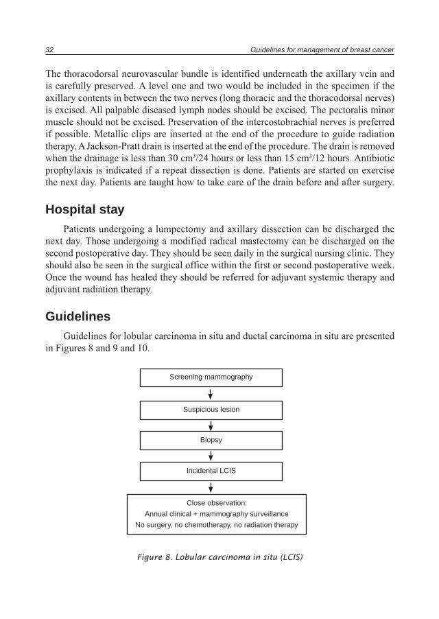
GuidelinesGuidelines for lobular carcinoma in situ and ductal carcinoma in situ are presented
in Figures 8 and 9 and 10.
Figure 8. Lobular carcinoma in situ (LCIS)
Screening mammography
Suspicious lesion
Biopsy
Incidental LCIS
Close observation:
Annual clinical + mammography surveillance
No surgery, no chemotherapy, no radiation therapy
Surgical guidelines for breast cancer 33
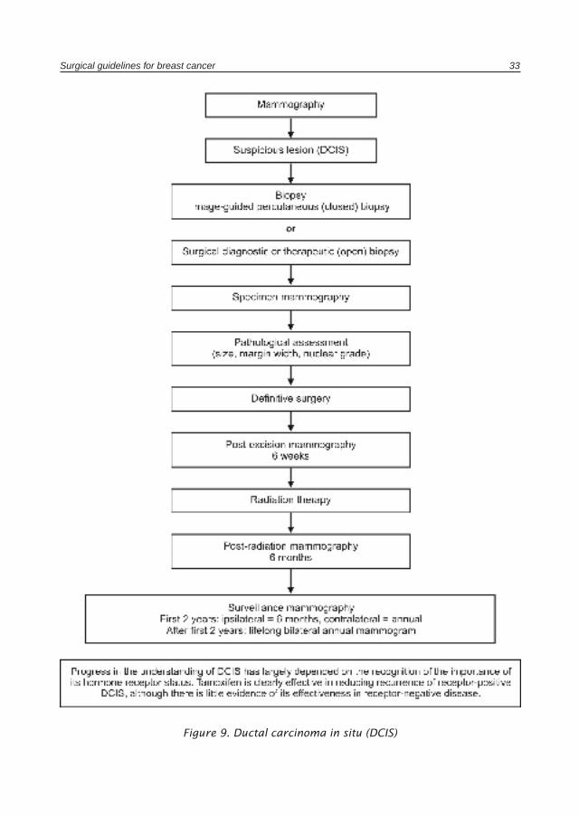
Figure 9. Ductal carcinoma in situ (DCIS)
34 Guidelines for management of breast cancer
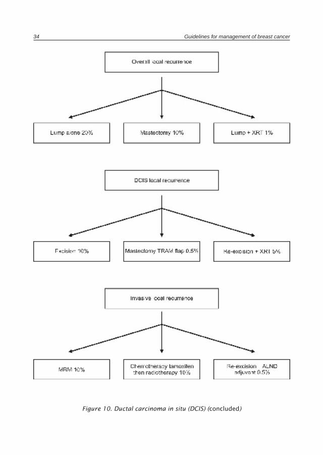
Figure 10. Ductal carcinoma in situ (DCIS) (concluded)
Chapter 5
Management of special problems in breast cancer
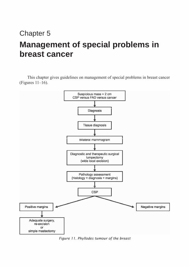
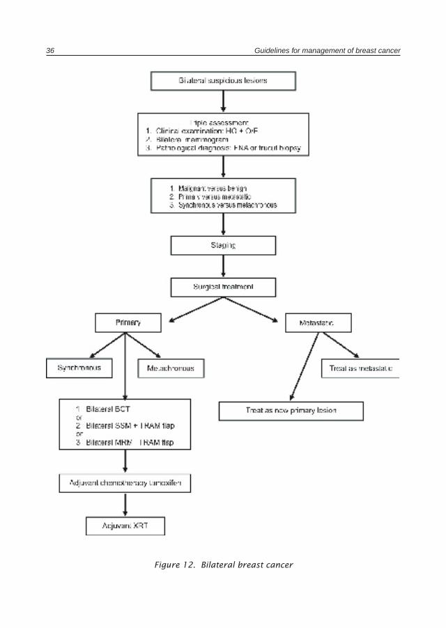
This chapter gives guidelines on management of special problems in breast cancer (Figures 11–16).
Figure 11. Phyllodes tumour of the breast
36 Guidelines for management of breast cancer
Figure 12. Bilateral breast cancer
Management of special problems in breast cancer 37
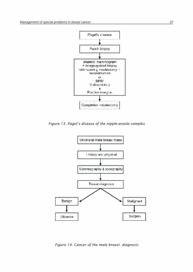
Figure 13. Paget’s disease of the nipple-areola complex
Figure 14. Cancer of the male breast: diagnosis
38 Guidelines for management of breast cancer
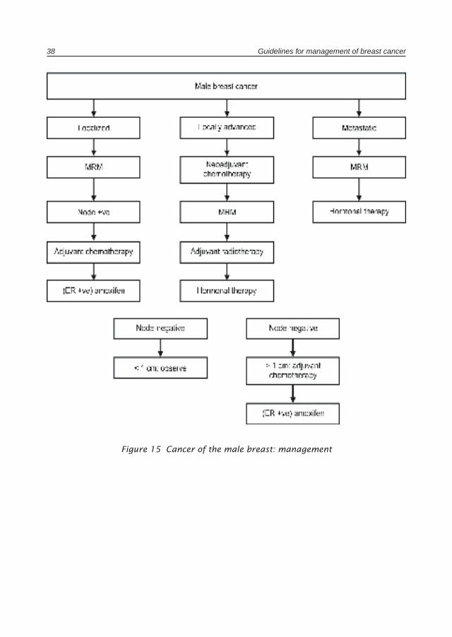
Figure 15 Cancer of the male breast: management
Management of special problems in breast cancer 39
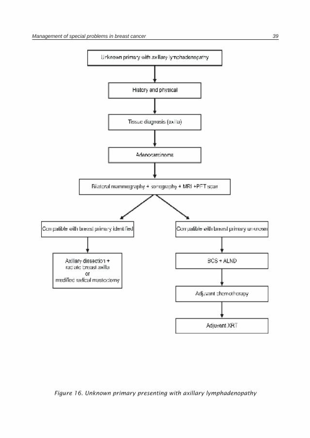
Figure 16. Unknown primary presenting with axillary lymphadenopathy
Chapter 6
Pathological handling of breast cancer excision specimens
General considerationsWith the increasing use of conservative surgery and radiation therapy for the
treatment of breast cancer, it is no longer sufficient for a pathologist to just determine whether a breast mass is or is not carcinoma. Successful management requires careful pathological evaluation to determine the adequacy of excision and histological features of potential prognostic significance.
A variety of breast specimens are encountered by the pathologist on a daily basis. These include cytological material, biopsies (core or incisional), excision specimens, re-excision specimens and mastectomies, in addition to axillary lymph nodes for evaluation.
Regardless of the type of the specimen, some general comments are applicable:
• Every breast excision specimen should be delivered to the pathologist intact. If the specimen has been incised previously or fragmented, evaluation of the surgical margins will not be possible.
• The specimen should be oriented and marked by the surgeon using sutures. These suture marks should be documented clearly for the pathologist in the pathology request form.
• Ideally, specimens should be transported fresh (in a plastic bag or container on ice) to the laboratory, to enable the pathologist to examine and mark the margins, and slice the specimen prior to fixation. However, immediate transport is not practical for many laboratories. In such cases, the specimen should be immediately placed in formalin (at least twice the specimen volume). The container should be large enough to ensure that no distortion of the specimen occurs. As a guide, a specimen left at room temperature for 30 minutes may commence autolysis and compromise hormone receptors results as well as pathology assessment. Adequate fixation is essential to allow accurate grading of invasive carcinoma and to help in the evaluation of borderline lesions.
• Surgeons who prefer to use diathermy should be aware that the thermal injury could produce cautery artifact, which may compromise accurate pathology interpretation and assessment of margins.
Pathological handling of breast cancer excision specimens 41
• Before incising the specimen, the surface should be painted with a dye that will be visible on permanent sections. India ink is a very satisfactory dye. Drying the specimen and/or dipping it in Bouin’s solution or alcohol for a brief period of time (i.e. 30 seconds) may assist with the adherence of the marking solution.
Fine needle aspiration biopsy and core needle biopsyFine needle aspiration biopsy (FNAB) and core needle biopsy (CNB) have almost
replaced open surgical biopsies for diagnosis of breast cancer.
FNAB is a minimally invasive, simple, cost effective and rapid technique, with an extremely low frequency of complications. If FNAB is used as a diagnostic procedure, the accuracy of interpretation should approach that of diagnosis by frozen section. However, an accurate interpretation requires proficiency in specimen acquisition, staining and preparation. It is also essential to have a pathologist experienced in the cytomorphologic features to reach an accurate diagnosis. The terminology used for reporting FNAB results should approach that of the surgical biopsy. Both the physician and the pathologist should be aware of the inherent limitations and pitfalls of FNAB, such as sampling error and difficult interpretation in a number of cases with certain lesions such as papillary lesions, low grade carcinoma and fibroepithelial lesions. FNAB cannot reliably distinguish between invasive and non-invasive carcinoma. Use of clinico-radiological-pathological correlation provides a high degree of diagnostic accuracy.
CNB provides tissue for histological evaluation. It is the preferred method for sampling of non-palpable breast lesions. However, like FNAB, its success depends on the technique and the availability of experienced pathologists and the appropriate correlation of the pathology findings with the clinical and imaging information. CNB has the same limitation as FNAB with respect to small sample size and difficulty of interpretation of indeterminate lesions.
Excision specimen for a palpable mass• The specimen should be oriented by the surgeon using sutures.• This orientation should be noted by the pathologist while marking and examining the
margins, so that any re-excision that may be required can be directed to a specific site.
• The specimen should be measured in three dimensions.• The specimen is serially sectioned and any gross lesion should be described and its
maximum dimensions should be recorded (in three dimensions).• The distance of the lesion to the nearest margin or margins is measured.
42 Guidelines for management of breast cancer
Tissue submitted for histological examination• Adequate sampling of the tumour and adjacent breast tissue. The number of blocks
is determined according to tumour size; 1 block/1 cm of the tumour is usually sufficiently representative.
• Relationship of tumour and closest margin or margins.• All surgical margins.• Non-neoplastic breast tissue.
The pathology report should include adequate gross information of the specimen and specific identification of each block of tissue taken.
Mastectomy specimen• The specimen should be oriented so that the quadrants are identified.• The following features should be recorded:
– the overall dimensions– descriptions and measurements of the skin, nipple and any incisions or scars– presence of fascia or muscle at the deep margin– description of axillary tail (if present)– location and size of the tumour and its relationship to the overlying skin and
deep margin.• The posterior/ deep margin and the closest margin should be marked with ink.
Tissue submitted for histological examination• Tumour with adjacent breast tissue, or areas around previous biopsy cavity if no
residual tumour is grossly identified.• Tumour and closest margin if possible.• Any other suspicious area.• Nipple and areola.• One section from each quadrant.• Deep margin and close margin/s to the tumour.• The overlying skin including scars.• Axillary lymph nodes. Grossly negative nodes should be submitted in entirety;
with grossly positive nodes, a representative section is required with surrounding extranodal area.
Pathological handling of breast cancer excision specimens 43
Mammographically-directed excisions (wire localization specimens)• The most frequent mammographic abnormalities prompting biopsy are
microcalcifications, a soft tissue density or a combination of the two.• Radiography of the intact specimen is an essential part of the processing of these
specimens. This is to ensure that the lesion is contained in the specimen. A specimen X-ray should be sent to the pathologist along with the specimen.
• Examine intact specimen and measure (three dimensions).• Apply ink to the surface before cutting.• Serially slice the specimen at 3 mm–4 mm intervals, maintaining specimen slice
order.• If there is a grossly evident tumour, measure and record distance between tumour
and margin.• An alternate procedure is to lay out the slices in sequence and X-ray them. This
allows accurate localization of the lesion.
Tissue submitted for histological examination• When small, breast biopsies should be embedded in total, if possible.• It is essential that the area of mammographic abnormality should be identified and
embedded totally.• The lesion and close margins and all other suspicious areas.• If X-ray of the sliced tissue specimen is available, all abnormal areas seen should
be submitted and labelled on the radiograph.• Random sections from the rest of the tissue.
If the mammographic abnormality reveals microcalcification, the pathologist should make every effort to identify them in the histological sections.
If these are not identified in the sections, the following manoeuvres may be helpful
• The microcalcifications may represent calcium oxalate crystals. These require polarization lenses to visualize, as they are transparent in a haematoxylin and eosin (H&E) section.
• X-ray of the paraffin blocks and any remaining wet tissue, if any. Multiple level sections can be made of the blocks containing the calcification.
• Calcification can be leached out by acidic fixatives or shattered out by the microtome knives. The pH of the fixative should be checked regularly and if there is tear in the sections, deep recuts of the block is needed.
44 Guidelines for management of breast cancer
Frozen section diagnosis• Frozen section has a limited role in the diagnosis of carcinoma nowadays with the
availability of fine needle aspiration (FNA) and needle biopsy as preoperative diagnostic procedures.
• Frozen section should not be performed on small lesions (< 1cm), where the pathologist believes that freezing will distort subsequent tissue morphology.
• Frozen section should be discouraged for evaluation of resection margins that are grossly free of tumour. If there is a specific area of concern to the surgeon, frozen section of that particular area may be indicated.
• Frozen sections should not be performed on a breast excision specimen removed because of mammographic calcifications.
Surgical pathology report of breast cancer specimensThe final pathology report should include certain information of prognostic
importance required for therapy.
Invasive carcinoma
• Laterality of the breast and procedure.• Histological type:
– ductal (usual, no special type)– lobular (specify type: classic or other)– tubular– medullary– mucinous– papillary– secretory– adenoid cystic– metaplastic– other (specify)
• Histological grade. All invasive carcinomas with the exception of medullary carcinoma should be graded. The Elston and Ellis modification of Scarff-Bloom-Richardson grading system is recommended. It evaluates the following three parameters:
– tubule formation:score 1. ≥ 75% of the tumour is composed of tubules.score 2. 10%–75% of the tumour is composed of tubules.score 3. < 10% of the tumour is composed of tubules.

Pathological handling of breast cancer excision specimens 45
– nuclear pleomorphism:score 1. small and regular nucleus.score 2. moderate variability in size and shape.score 3. marked increase in size and marked irregularity.
– mitotic indexscore 1. ≤ 10 mf/10 hpf.score 2. 11–20 mf/10 hpf.score 3. > 20/10 hpf.
– The histological grade is determined by summing the points of these parameters:grade I = total score 3–5grade II = total score 6–7grade III = total score 8–9
• Margins of resection. There is no standard definition of what constitutes a positive or negative margin. However, when reporting margins state:– if tumour is at the margin (grossly or microscopically)– if tumour is not at the margin, the distance from the margin should be specified
e.g. within 5 mm of the nearest margin or more than 5 mm.
• Lymph node status. The number of nodes involved and the total number of nodes removed should be mentioned. The presence or absence of perinodal extension of the carcinoma cells into axillary fat should be recorded. If metastases size is ≤ 2 mm, this should be recorded.
• Lymphovascular invasion. Peritumoural vessel invasion is assessed; if the lymphovascular spaces in the skin are involved, this should be mentioned separately.
• Size of the carcinoma. It is preferred to mention this in the diagnosis, even though this is recorded in the gross description (maximum diameter).
• Extent of in situ carcinoma. The presence or absence of in situ component should be recorded. If it is present, its extent should be approximated. If it is more than 25% of the tumour mass or the tumour is primarily intraductal with only focal microscopic invasion, the in situ component is considered to be “extensive”. If only focal microscopic invasion is present, the size of this focus/foci should be measured on the slide and recorded, in addition to the maximum diameter of the DCIS.
• Microcalcifications. If seen on the mammogram their presence or absence and location should be stated, to be sure a calcified lesion was not missed.
• Other significant disease such as papillomas, Paget’s disease of the nipple, etc.• If information required for therapy or prognosis is not available or cannot be
adequately assessed (e.g. no nodes submitted with a mastectomy specimen, margins
46 Guidelines for management of breast cancer
not assessable because specimen was cut before inking, etc.) this should be stated specifically in the report.
• Prognostic factors, if available, should be included in the report (e.g. ER/PR receptor studies, HER2, etc.). Hormone receptor (estrogen and progesterone) investigation is now almost exclusively performed by immunohistochemical means, performed on paraffin embedded material. It should be performed on all cases of invasive carcinoma. Only nuclear staining is considered to indicate a positive result. An estimate of the percentage of nuclei stained should be included in the report. There is no consensus on the lower cut-off point for a positive assay, but a practical cut-off point is greater than 10% of nuclei staining.
Ductal carcinoma in situ
• Laterality of the breast and procedure.• Nuclear grade. Should be reported as low, intermediate, or high grade (grade 1, 2,3)
using the same criteria advocated for invasive carcinoma.• Necrosis:
– present (central duct necrosis i.e. comedo necrosis)– absent or minimal (no central duct necrosis, but focal)
• Architecture type (many tumours show more than one type):– cribriform– micropapillary– solid– comedo (high nuclear grade; necrosis usually present)– papillary (includes intracystic)– mixed
• Margins of resection. Distance of DCIS from the closest margin should be recorded in millimetres. If DCIS is present at the margin, this should be specified.
• Size. If a mass is present, state the size from the gross. If not, then an estimate of the extent of the tumour may be attempted:– estimate the percent of the breast units affected by DCIS or– estimate the size of the lesion based on the sections by counting the number of
specimen slices in which the lesion occurs and multiplying by the average slice thickness (3mm–4mm) or
– if the lesion is small, measurement of the size of the tumour should be obtained directly from the slide.
• Presence and location of microcalcification.• Other significant disease (atypical hyperplasia or papilloma, etc.).
Chapter 7
Radiotherapy guidelines for breast cancer
Radiotherapy for ductal carcinoma in situ
Group 1: Unicentric, negative resection margins
• Breast-conserving surgery and radiation therapy.• Total mastectomy without lymph node dissection.• Breast-conserving surgery without radiation therapy (highly selected group: new
Van Nuys Index).
Group 2: Multicentric, widespread
• Mastectomy.• Radiation therapy is given to the whole breast 5000 cGy in 25 fractions over 5
weeks.
Criteria for breast conserving therapy
Objectives
• Local tumour control.• Preservation of a cosmetically acceptable breast.
Absolute contraindications• ≥ 2 primary tumours in separate quadrants.• Diffuse malignant appearing microcalcifications.• History of previous radiotherapy, which combined with proposed radiotherapy would
result in excessively high total dose to a significant volume.• Pregnancy, because of the possible teratogenic and carcinogenic effects of radiotherapy
on the fetus.• Persistent, positive margins after “reasonable” surgical attempts.
48 Guidelines for management of breast cancer
Relative contraindications
• History of collagen vascular disease (scleroderma and active lupus erythematous are absolute contraindications).
• Multiple gross tumours in the same quadrant and indeterminate calcifications.• Large tumour in a small breast.• Breast size. Reproducibility of set-up and dose homogeneity must be considered.
Radiotherapy technique
• A variety of different treatment techniques exist. Many technique details are aimed at avoiding unnecessary irradiation to tissues outside the target volume and hence reducing treatment morbidity.
Target volume• The whole breast. Partial breast irradiation is still considered investigational and
should not be used unless patients are on a protocol.• Regional nodal irradiation. There are limited data on regional nodal failures in patients
treated with breast-conserving therapy. In studies that evaluated nodal recurrence patterns, there is little justification for regional nodal irradiation in patients with three or fewer involved nodes if nodal recurrence is utilized as an end point. In patients with four or more positive axillary lymph nodes, irradiation of the supraclavicular fossa is indicated. This recommendation is based largely on the incidence of locoregional recurrences in patients with four or more involved nodes who were treated with mastectomy alone.
Patient positioning and set-up• The patient should be positioned in a comfortable, stable and reproducible position
for treatment with the adjacent arm placed outside the radiation beam. A method verifying patient position reproducibility from day-to-day should be practiced.
Simulation• The patient is set up in the treatment position for simulation. The stability and
reproducibility of the treatment position should be assessed at this time.
Treatment fields• Most commonly opposed tangential fields are used. The field size is sufficient to
cover the whole breast. The desired maximum lung depth within the treated volume is in the range 1 cm–3 cm. Some lung must be seen to verify that the deep surface of the breast is within the treatment path.
Radiotherapy guidelines for breast cancer 49
Treatment modality: beam energy• Super voltage equipment (4, 6, or 8 MV or cobalt-60) is preferred.• In the patient where the field separation is greater than 22 cm, higher beam energy
(10 MV) may give a more homogenous dose distribution.
Dosimetry• The aim is to create a homogeneous dose distribution throughout this irregular shaped
treatment volume (± 5% variation).
Dose/fractionation• A whole breast dose of 45 Gy–50 Gy is prescribed.• This is given in 1.8 Gy–2.0 Gy fractions per day, 5 days per week.
Boost• In patients with negative margins, randomized trials demonstrate that the use of a
boost is effective, but 10-year results are needed to fully assess the effects of a boost on local control and survival.
• This effect was clearer in patients < 50 years of age.• Via the boost, the dose at the tumour bed is increased to 60 Gy–66 Gy.• A boost may be give by electron beam (10 Gy in 5 fractions or 16 Gy/8f) or interstitial
implant.• Photons can be used as well.
Post-mastectomy radiotherapy
Objectives
• Post-mastectomy radiotherapy reduces the risk of locoregional failure and increases the long-term survival rate for a substantial proportion of women with positive axillary nodes treated with systemic therapy.
Indications
• Four or more positive axillary lymph nodes.• Tumour > 5 cm in size.• Close or positive margins.• Inadequate axillary surgery (the removal of less than 10 nodes).
50 Guidelines for management of breast cancer
Consideration
• 1 to 3 positive axillary lymph nodes.
Radiotherapy technique
Target volume• The target volume must include the chest wall.• Supraclavicular lymph node group for patients with 4 or more positive axillary lymph
nodes.• Axilla if inadequate axillary surgery was done.
In addition, the following areas may be included even though there is insufficient evidence to made recommendations:
• Drain sites.• Internal mammary chain (IMC).
Patient positioning and set-up• The patient should be positioned in a comfortable, stable and reproducible position
for treatment with the adjacent arm placed outside the radiation beam. A method verifying patient position reproducibility from day-to-day should be practiced.
Simulation• The patient is set up in the treatment position for simulation. The stability and
reproducibility of the treatment position should be assessed at this time.
Treatment fieldsChest wall. Most commonly opposed tangential fields are used. The field’s size
must be sufficient to cover the extent of the breast prior to mastectomy and in general include the mastectomy scar. If the lateral end of the mastectomy scar extends posteriorly, then a smaller electron beam field may need to be added on. The lower edge of the extent of the breast can be determined from the contralateral breast if it is still in place.
Approximate position of field edges:
• Medial edge: approximately mid-line.• Upper level: approximately a line joining the sternal notch to the base of the
axilla.• Lower level: approximately 2 cm below the inferior margin of the breast prior to
mastectomy.• Lateral edge: approximately the mid-axillary line.

Radiotherapy guidelines for breast cancer 51
The thickness of lung with the treated volume should lie between 1 cm and 3 cm. Some lung must be seen to verify that the deep surface of the chest wall is within the treatment volume.
Axilla and supraclavicular nodal areas. The depth required to treat the supraclavicular area should be considered. The depth in the axilla must also be determined. The potential for overlapping fields to produce areas of overdose or underdose at the junctions should be addressed.
Internal mammary lymph node chain. At present, no uniformly acceptable method to localize the position and depth of the internal mammary lymph nodes is available. No optimal technique exists for internal mammary (IM) nodal irradiation. After localization of the desired target volume, the dose of the heart, lungs and contralateral breast should be considered.
Treatment modality: beam energy• Super voltage equipment (4, 6, or 8 MV or cobalt-60) is preferred.• In the patient where the field separation is greater than 22 cm, higher beam energy
(10 MV) may give a more homogenous dose distribution.
Dosimetry• The aim is to create a homogeneous dose distribution throughout this irregular shaped
treatment volume (± 5% variation).
Dose-fractionation• A dose of 45 Gy–50 Gy is prescribed.• This is given in 1.8 Gy–2 Gy fractions, 1 fraction per day, 5 days per week.• All fields should be treated each day, although the posterior axillary boost (if used)
is generally given in fewer fractions.
Radiotherapy after pre-operative systemic therapyThere is insufficient evidence to make recommendations on whether all patients who
receive pre-operative systemic therapy should receive radiotherapy.

Further reading
Arthur DW et al. Internal mammary node coverage: an investigation of presently accepted techniques. International Journal of Radiation Oncology, Biology, Physics, 2000, 48:139–146.
Association of Directors of Anatomic and Surgical Pathology. Immediate management of mammographically detected breast lesions. American Journal of Surgical Pathology, 1993, 17(8):850–1.
Association of Directors of Anatomic and Surgical Pathology. Recommendation for the reporting of breast carcinoma. Human Pathology, 1996, 27(3):220–4.
Bentel GC et al. Variability of the location of internal mammary vessel and glandular breast tissue in breast cancer patients undergoing routine CT-based treatment planning. International Journal of Radiation Oncology, Biology, Physics, 1999, 44:1017–1025.
Bentel GC et al. Variability of the depth of supraclavicular and axillary lymph nodes in patients with breast cancer: is a posterior axillary boost field necessary? International Journal of Radiation Oncology, Biology, Physics, 2000, 47:755–758.
Carlson RW et al. Treatment of breast cancer in countries with limited resources. The Breast Journal, 2003, 9(Suppl. 2):S67–74.
Drummond R, Kenny L. Guidelines for the technical aspects of therapeutic radiation treatment. Radiation Oncology Advisory Group of the National Breast Cancer Centre, Australia and the Faculty of Radiation Oncology of the Royal Australian and New Zealand College of Radiologists, www.nbcc.org.au/resources, accessed 2005.
Early Breast Cancer Trialists’ Collaborative Group. Effects of adjuvant tamoxifen and cytotoxic therapy on mortality in early breast cancer: an overview of 61 randomized trials among 28896 women. New England Journal of Medicine, 1988, 319:1681–1692.
Early Breast Cancer Trialists’ Collaborative Group. Effects of radiotherapy and surgery in early breast cancer: an overview of the randomized trials. New England Journal of Medicine, 1995, 333:1444–1455.
Early Breast Cancer Trialists’ Collaborative Group. Tamoxifen for early breast cancer: an overview of randomized trials. Lancet, 1998, 351:1451–1467.
Early Breast Cancer Trialists’ Collaborative Group. Polychemotherapy for early breast cancer: an overview of randomized trials. Lancet, 1998, 352:930–942.
Ellis MJ et al., eds. Treatment of metastatic breast cancer: disease of the breast. Philadelphia, Lippincott William & Wilkins, 2000.
Further reading 53
Fitzgibbons P, Connolly J, Page D. Updated protocol for the examination of specimens from patients with carcinomas of the breast. Archives of Pathology and Laboratory Medicine, 2000, 124:1026–33.
Fowble B et al. Internal mammary node irradiation neither decreases distant metastases nor improves survival in stage I and II breast cancer. International Journal of Radiation Oncology, Biology, Physics, 2000, 47:883–894.
Gagliardi G et al. Radiation pneumonitis after breast cancer irradiation: analysis of the complication probability using the relative seriality model. International Journal of Radiation Oncology, Biology, Physics, 2000, 46:373–381.
Gelber RD et al. Features that predict responsiveness to chemotherapy and endocrine therapies. The Breast 2001, 10 (Suppl. 1):S11.
Goldhirsch A et al. Meeting highlights: updated international expert consensus on the primary therapy of early breast cancer. Journal of Clinical Oncology, 2003, 21(17):3357–3365.
Goodman RL et al. The relationship between radiation fields and regional lymph nodes in carcinoma of the breast. International Journal of Radiation Oncology, Biology, Physics, 2001, 50:99–105.
Grimshaw JM, Russell IT. Effect of clinical guidelines on medical practice: a systematic review of rigorous evaluation. Lancet, 1993, 342:1317–1322.
Harris J, Kurtz J, Vicini FA. Multidisciplinary breast cancer management: Part 3: radiation issues. Fairfax, VA, American Society for Therapeutic Radiology and Oncology, 2002.
Henderson C et al. Improved outcomes from adding sequential paclitaxel but not from escalating doxorubicin dose in an adjuvant chemotherapy regimen for patients with node-positive primary breast cancer. Journal of Clinical Oncology, 2003, 21(6):1–9.
Hortobagyi GN. Treatment of breast cancer. New England Journal of Medicine 1998, 339:974–984.
Hurkmans CW et al. An improved technique for breast cancer irradiation including the locoregional lymph nodes. International Journal of Radiation Oncology, Biology, Physics, 2000, 47:1421–1429.
Kaufmann M et al. International expert panel on the use of primary (neo-adjuvant) systemic treatment of operable breast cancer review and recommendations. Journal of Clinical Oncology, 2003, 21(13):2600–2608.
Liljegren G et al. Risk factors for local recurrence after conservative treatment in stage I breast cancer: definition of a subgroup not requiring radiotherapy. Annals of Oncology, 1997, 8:235–241.
Mehta K, Haffty BG. Long term outcome in patients with four or more positive lymph nodes treated with breast-conserving therapy. International Journal of Radiation Oncology, Biology, Physics, 1996, 35:679–685.
54 Guidelines for management of breast cancer
National Cancer Institute Sponsored Conference. The uniform approach to breast fine-needle aspiration biopsy. Diagnostic Cytopathology, 1997, 16:295–311.
Nemoto T et al. Factors affecting recurrence in lumpectomy without irradiation for breast cancer. Cancer, 1991, 67:2079–2082.
Obedian E, Haffty BG. Internal mammary nodal irradiation in conservatively-managed breast cancer patients: is there a benefit? International Journal of Radiation Oncology, Biology, Physics, 2000, 44:951–957.
Ooi GC et al. Pulmonary sequelae of treatment for breast cancer: a prospective study. International Journal of Radiation Oncology, Biology, Physics, 2001, 50:411–419.
Peto R. Early Breast Cancer Trialists’ Collaborative Group: updated results from September 2000 worldwide overview. European Journal of Cancer, 2000, 36(Suppl. 5):S47.
Pierce SM et al. Long term radiation complications following conservative surgery (CS) and radiation therapy (RT) in patients with early stage breast cancer. International Journal of Radiation Oncology, Biology, Physics, 1992, 23:915–923.
Recht A et al. Regional nodal failure after conservative surgery and radiotherapy for early-stage breast carcinoma. Journal of Clinical Oncology, 1991, 9:988–996.
Singletary SE et al. Revision of the American Joint Committee on cancer staging system for breast cancer. Journal of Clinical Oncology, 2002, 20(17):3628–3636.
Sledge GW (ed). Highlights of 2000 NIH consensus conference on adjuvant breast cancer. Philadelphia, PA, The Phillips Group Oncology Communication Co., 2000:1–8.
Sobin LH, Wittekind CH (eds). TNM classification of malignant tumour. 6th edition. New York, John Wiley and Sons, 2002.
Takeda A et al. The modified tangential irradiation technique for breast cancer: how to cover the entire axillary region. International Journal of Radiation Oncology, Biology, Physics, 2000, 46:815–822.
Vicini FA et al. The role of regional nodal irradiation in the management of patients with early-stage breast cancer treated with breast-conserving therapy. International Journal of Radiation Oncology, Biology, Physics, 1997, 39:1069–1076.
Wong JS et al. The relationship between lymphatic vessel invasion, tumour size and pathologic nodal status: can we predict who can avoid a third field in the absence of axillary dissection? International Journal of Radiation Oncology, Biology, Physics, 2000, 48:133–137.
Woolf SH et al. Potential benefits, limitations, and harms of clinical guidelines. British Medical Journal, 1999, 318:527–530.
Annex 1
Participants in the consultation on early detection and screening of breast cancerCairo, Egypt, 21–24 October 2002
Professor W Anwar, EgyptProfessor S Eissa, EgyptProfessor A Hussain, QatarDr J Jabbour, LebanonDr M Jama, WHO Regional Office for the Eastern MediterraneanProfessor H Khaled, EgyptDr O Khatib, WHO Regional Office for the Eastern MediterraneanProfessor A Modjtabai, United States of AmericaProfessor S Mostafa, EgyptProfessor S Omar, EgyptProfessor Y Omar, EgyptProfessor P Salem, United States of AmericaDr C Sepulveda, WHO headquartersDr A Verster, WHO Regional Office for the Eastern MediterraneanProfessor H Vianio, International Agency for Research on Cancer

58 Guidelines for management of breast cancer