Embed Size (px)
Citation preview
2749
Encrypting messages with artificial bacterial receptorsPragati Kishore Prasad, Naama Lahav-Mankovski, Leila Motiei* and David Margulies*
Full Research Paper Open Access
Address:Department of Organic Chemistry, Weizmann Institute of Science,Rehovot 7610001, Israel
Email:Leila Motiei* - [email protected]; David Margulies* [email protected]
* Corresponding author
Keywords:artificial receptors; cell surface modification; fluorescent probes;molecular cryptography
Beilstein J. Org. Chem. 2020, 16, 2749–2756.https://doi.org/10.3762/bjoc.16.225
Received: 17 September 2020Accepted: 30 October 2020Published: 12 November 2020
This article is part of the thematic issue "Molecular recognition" and isdedicated to the memory of Carsten Schmuck.
Guest Editor: J. Niemeyer
© 2020 Kishore Prasad et al.; licensee Beilstein-Institut.License and terms: see end of document.
AbstractA method for encrypting messages using engineered bacteria and different fluorescently labeled synthetic receptors is described.We show that the binding of DNA-based artificial receptors to E. coli expressing His-tagged outer membrane protein C(His-OmpC) induces a Förster resonance energy transfer (FRET) between the dyes, which results in the generation of a unique fluo-rescence fingerprint. Because the bacteria continuously divide, the emission pattern generated by the modified bacteria dynami-cally changes, enabling the system to produce encryption keys that change with time. Thus, this development indicates the poten-tial contribution of live-cell-based encryption systems to the emerging area of information protection at the molecular level.
2749
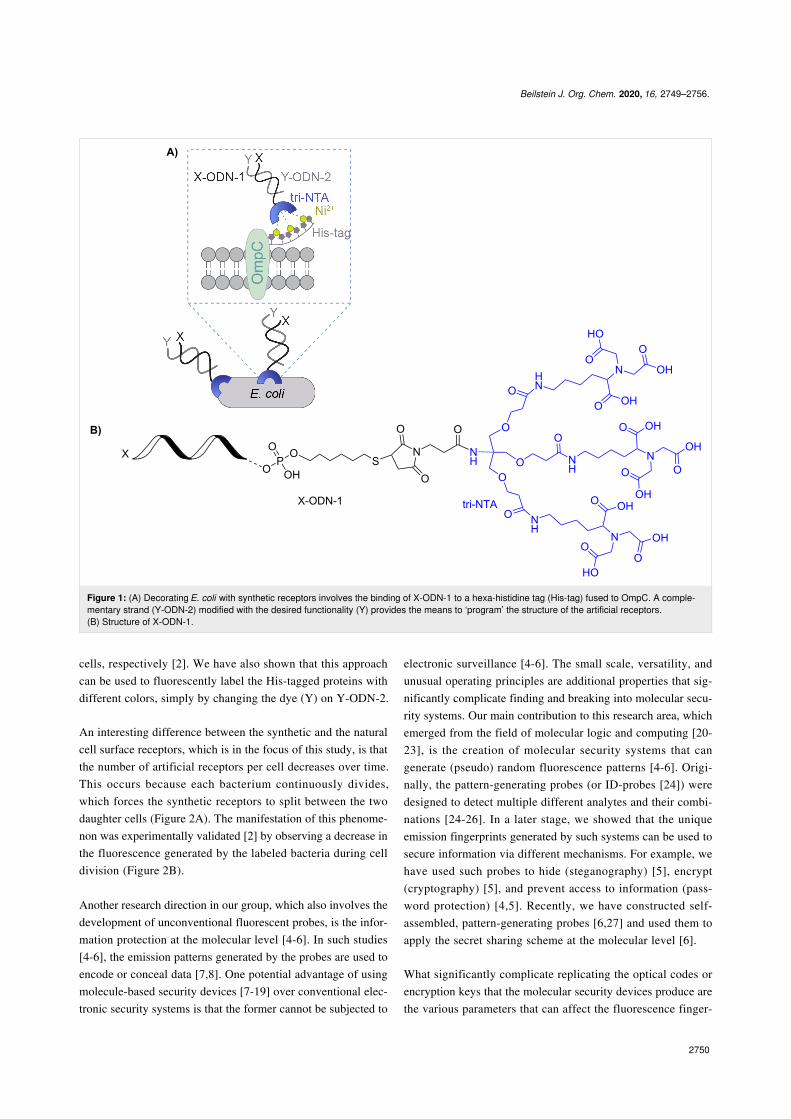
IntroductionIn living cells, information is processed and transferred via aseries of recognition and signaling events, which normallybegin by the binding of cell-surface receptors to extracellularsignals, such as small molecules or proteins. In recent years,there has been considerable interest in modifying cells with arti-ficial receptors, as a means to provide them with new proper-ties [1]. We have recently reported a method for decorating His-tagged cell surface proteins with self-assembled synthetic re-ceptors based on modified DNA duplexes [2] (Figure 1A). Oneof the oligodeoxynucleotides (ODNs) constituting the artificialreceptors (ODN-1) is appended with a trinitrilotriacetic acidgroup (tri-NTA) that was developed by our group [3] and canselectively bind a hexa-histidine tag (His-tag). ODN-1 can also
be modified with a second functional group (X), such as a fluo-rescent dye, to afford X-ODN-1 (Figure 1A and Figure 1B). Inthis way, the binding of X-ODN-1 to the bacteria will lead tothe presentation of X on the cell surface (Figure 1A). A simplerway to modify the bacterial membrane is by adding toX-ODN-1 a complementary strand (Y-ODN-2) that is modifiedwith the desired functionality (Y) (Figure 1A). In this way, thestructure of the artificial receptors can be ‘programmed’ by asimple self-assembly process, which provides the means to re-versibly change the properties of the cell. For example, we haveshown that synthetic receptors appended with a thiol or a folategroup enable bacteria expressing the His-tagged outer mem-brane protein C (His-OmpC) to bind to gold surfaces or cancer
Beilstein J. Org. Chem. 2020, 16, 2749–2756.
2750
Figure 1: (A) Decorating E. coli with synthetic receptors involves the binding of X-ODN-1 to a hexa-histidine tag (His-tag) fused to OmpC. A comple-mentary strand (Y-ODN-2) modified with the desired functionality (Y) provides the means to ‘program’ the structure of the artificial receptors.(B) Structure of X-ODN-1.
cells, respectively [2]. We have also shown that this approachcan be used to fluorescently label the His-tagged proteins withdifferent colors, simply by changing the dye (Y) on Y-ODN-2.
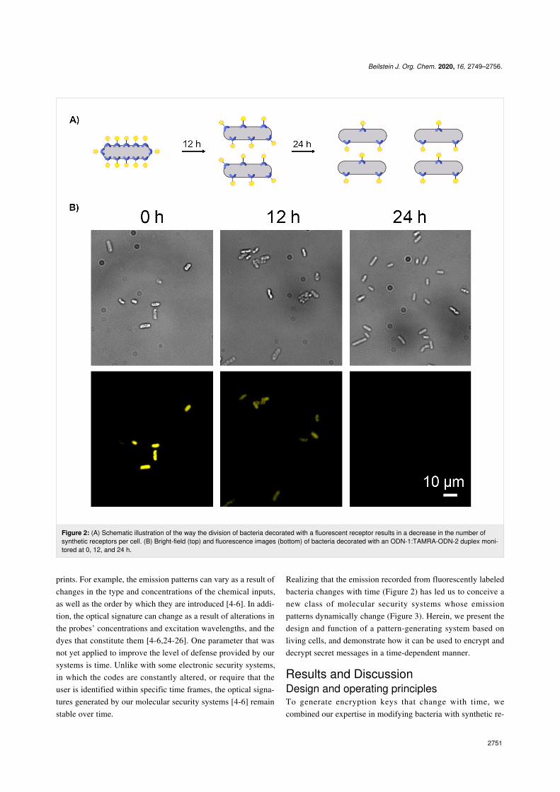
An interesting difference between the synthetic and the naturalcell surface receptors, which is in the focus of this study, is thatthe number of artificial receptors per cell decreases over time.This occurs because each bacterium continuously divides,which forces the synthetic receptors to split between the twodaughter cells (Figure 2A). The manifestation of this phenome-non was experimentally validated [2] by observing a decrease inthe fluorescence generated by the labeled bacteria during celldivision (Figure 2B).
Another research direction in our group, which also involves thedevelopment of unconventional fluorescent probes, is the infor-mation protection at the molecular level [4-6]. In such studies[4-6], the emission patterns generated by the probes are used toencode or conceal data [7,8]. One potential advantage of usingmolecule-based security devices [7-19] over conventional elec-tronic security systems is that the former cannot be subjected to
electronic surveillance [4-6]. The small scale, versatility, andunusual operating principles are additional properties that sig-nificantly complicate finding and breaking into molecular secu-rity systems. Our main contribution to this research area, whichemerged from the field of molecular logic and computing [20-23], is the creation of molecular security systems that cangenerate (pseudo) random fluorescence patterns [4-6]. Origi-nally, the pattern-generating probes (or ID-probes [24]) weredesigned to detect multiple different analytes and their combi-nations [24-26]. In a later stage, we showed that the uniqueemission fingerprints generated by such systems can be used tosecure information via different mechanisms. For example, wehave used such probes to hide (steganography) [5], encrypt(cryptography) [5], and prevent access to information (pass-word protection) [4,5]. Recently, we have constructed self-assembled, pattern-generating probes [6,27] and used them toapply the secret sharing scheme at the molecular level [6].
What significantly complicate replicating the optical codes orencryption keys that the molecular security devices produce arethe various parameters that can affect the fluorescence finger-
Beilstein J. Org. Chem. 2020, 16, 2749–2756.
2751
Figure 2: (A) Schematic illustration of the way the division of bacteria decorated with a fluorescent receptor results in a decrease in the number ofsynthetic receptors per cell. (B) Bright-field (top) and fluorescence images (bottom) of bacteria decorated with an ODN-1:TAMRA-ODN-2 duplex moni-tored at 0, 12, and 24 h.
prints. For example, the emission patterns can vary as a result ofchanges in the type and concentrations of the chemical inputs,as well as the order by which they are introduced [4-6]. In addi-tion, the optical signature can change as a result of alterations inthe probes’ concentrations and excitation wavelengths, and thedyes that constitute them [4-6,24-26]. One parameter that wasnot yet applied to improve the level of defense provided by oursystems is time. Unlike with some electronic security systems,in which the codes are constantly altered, or require that theuser is identified within specific time frames, the optical signa-tures generated by our molecular security systems [4-6] remainstable over time.
Realizing that the emission recorded from fluorescently labeledbacteria changes with time (Figure 2) has led us to conceive anew class of molecular security systems whose emissionpatterns dynamically change (Figure 3). Herein, we present thedesign and function of a pattern-generating system based onliving cells, and demonstrate how it can be used to encrypt anddecrypt secret messages in a time-dependent manner.
Results and DiscussionDesign and operating principlesTo generate encryption keys that change with time, wecombined our expertise in modifying bacteria with synthetic re-
Beilstein J. Org. Chem. 2020, 16, 2749–2756.
2752
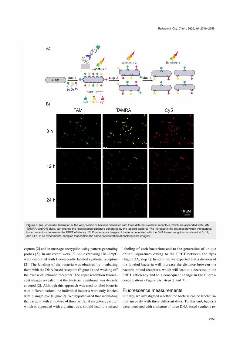
Figure 3: (A) Schematic illustration of the way division of bacteria decorated with three different synthetic receptors, which are appended with FAM,TAMRA, and Cy5 dyes, can change the fluorescence signature generated by the labeled bacteria. The increase in the distance between the bacteria-bound receptors decreases the FRET efficiency. (B) Fluorescence images of bacteria decorated with the DNA-based receptors monitored at 0, 12,and 24 h. In all experiments, samples that contain the same concentration of bacteria were imaged.
ceptors [2] and in message encryption using pattern-generatingprobes [5]. In our recent work, E. coli-expressing His-OmpCwere decorated with fluorescently labeled synthetic receptors[2]. The labeling of the bacteria was obtained by incubatingthem with the DNA-based receptors (Figure 1) and washing offthe excess of unbound receptors. The super resolution fluores-cent images revealed that the bacterial membrane was denselycovered [2]. Although this approach was used to label bacteriawith different colors, the individual bacteria were only labeledwith a single dye (Figure 2). We hypothesized that incubatingthe bacteria with a mixture of three artificial receptors, each ofwhich is appended with a distinct dye, should lead to a mixed
labeling of each bacterium and to the generation of uniqueoptical signatures owing to the FRET between the dyes(Figure 3A, step 1). In addition, we expected that a division ofthe labeled bacteria will increase the distance between thebacteria-bound receptors, which will lead to a decrease in theFRET efficiency and to a consequent change in the fluores-cence pattern (Figure 3A, steps 2 and 3).
Fluorescence measurementsInitially, we investigated whether the bacteria can be labeled si-multaneously with three different dyes. To this end, bacteriawere incubated with a mixture of three DNA-based synthetic re-
Beilstein J. Org. Chem. 2020, 16, 2749–2756.
2753
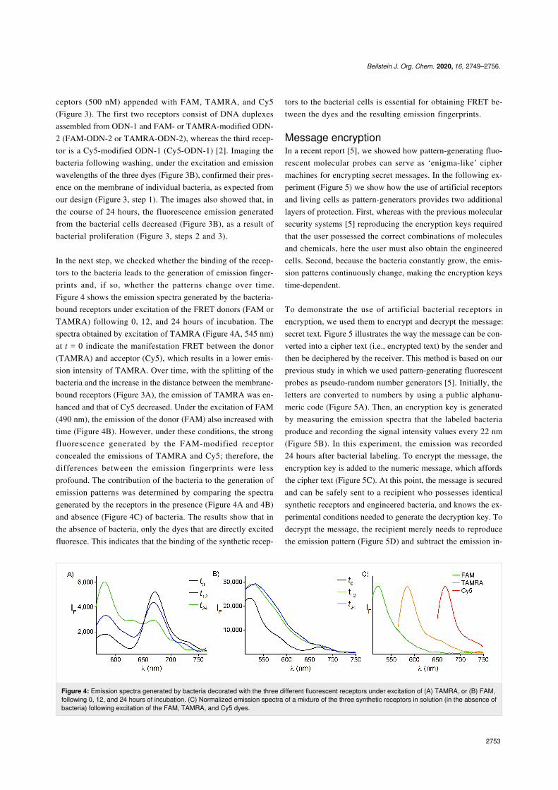
Figure 4: Emission spectra generated by bacteria decorated with the three different fluorescent receptors under excitation of (A) TAMRA, or (B) FAM,following 0, 12, and 24 hours of incubation. (C) Normalized emission spectra of a mixture of the three synthetic receptors in solution (in the absence ofbacteria) following excitation of the FAM, TAMRA, and Cy5 dyes.
ceptors (500 nM) appended with FAM, TAMRA, and Cy5(Figure 3). The first two receptors consist of DNA duplexesassembled from ODN-1 and FAM- or TAMRA-modified ODN-2 (FAM-ODN-2 or TAMRA-ODN-2), whereas the third recep-tor is a Cy5-modified ODN-1 (Cy5-ODN-1) [2]. Imaging thebacteria following washing, under the excitation and emissionwavelengths of the three dyes (Figure 3B), confirmed their pres-ence on the membrane of individual bacteria, as expected fromour design (Figure 3, step 1). The images also showed that, inthe course of 24 hours, the fluorescence emission generatedfrom the bacterial cells decreased (Figure 3B), as a result ofbacterial proliferation (Figure 3, steps 2 and 3).
In the next step, we checked whether the binding of the recep-tors to the bacteria leads to the generation of emission finger-prints and, if so, whether the patterns change over time.Figure 4 shows the emission spectra generated by the bacteria-bound receptors under excitation of the FRET donors (FAM orTAMRA) following 0, 12, and 24 hours of incubation. Thespectra obtained by excitation of TAMRA (Figure 4A, 545 nm)at t = 0 indicate the manifestation FRET between the donor(TAMRA) and acceptor (Cy5), which results in a lower emis-sion intensity of TAMRA. Over time, with the splitting of thebacteria and the increase in the distance between the membrane-bound receptors (Figure 3A), the emission of TAMRA was en-hanced and that of Cy5 decreased. Under the excitation of FAM(490 nm), the emission of the donor (FAM) also increased withtime (Figure 4B). However, under these conditions, the strongfluorescence generated by the FAM-modified receptorconcealed the emissions of TAMRA and Cy5; therefore, thedifferences between the emission fingerprints were lessprofound. The contribution of the bacteria to the generation ofemission patterns was determined by comparing the spectragenerated by the receptors in the presence (Figure 4A and 4B)and absence (Figure 4C) of bacteria. The results show that inthe absence of bacteria, only the dyes that are directly excitedfluoresce. This indicates that the binding of the synthetic recep-
tors to the bacterial cells is essential for obtaining FRET be-tween the dyes and the resulting emission fingerprints.
Message encryptionIn a recent report [5], we showed how pattern-generating fluo-rescent molecular probes can serve as ‘enigma-like’ ciphermachines for encrypting secret messages. In the following ex-periment (Figure 5) we show how the use of artificial receptorsand living cells as pattern-generators provides two additionallayers of protection. First, whereas with the previous molecularsecurity systems [5] reproducing the encryption keys requiredthat the user possessed the correct combinations of moleculesand chemicals, here the user must also obtain the engineeredcells. Second, because the bacteria constantly grow, the emis-sion patterns continuously change, making the encryption keystime-dependent.
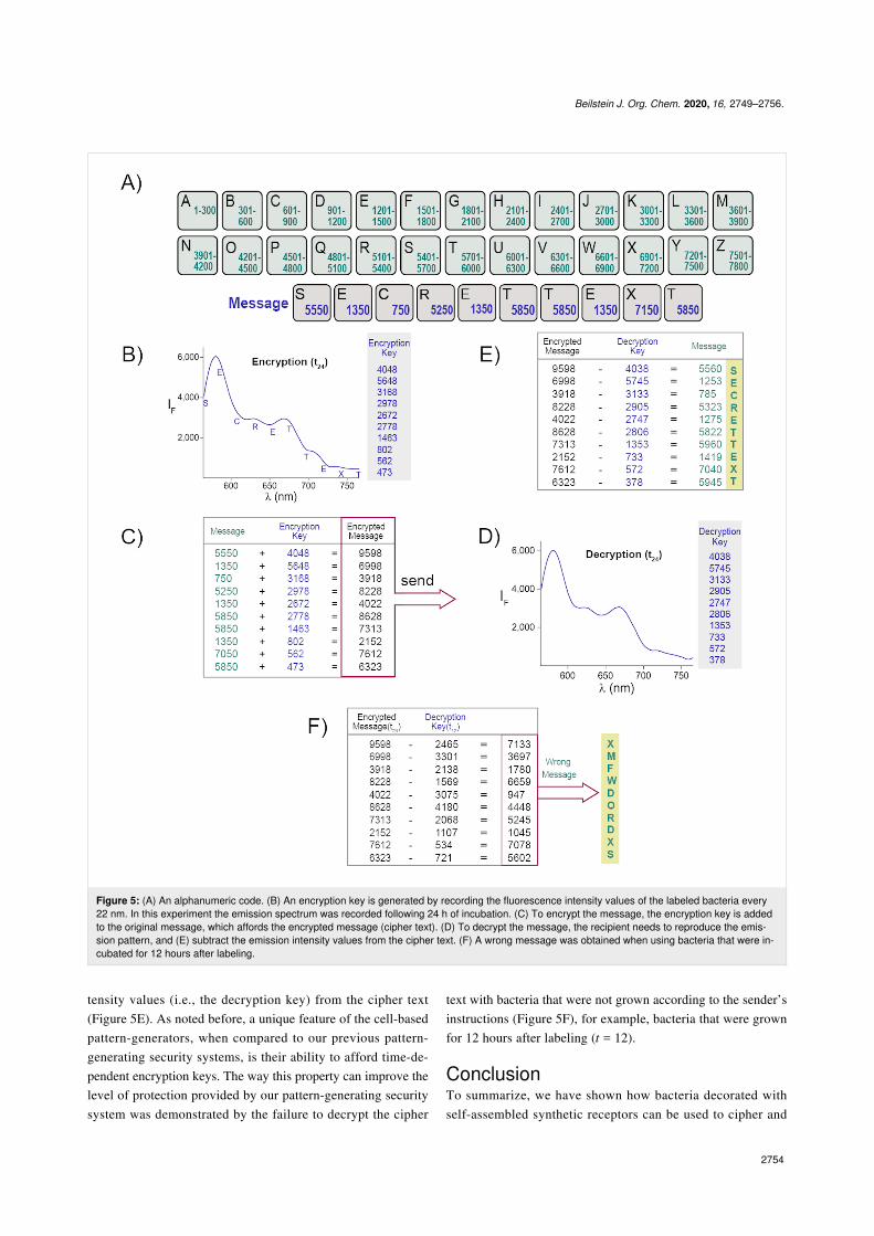
To demonstrate the use of artificial bacterial receptors inencryption, we used them to encrypt and decrypt the message:secret text. Figure 5 illustrates the way the message can be con-verted into a cipher text (i.e., encrypted text) by the sender andthen be deciphered by the receiver. This method is based on ourprevious study in which we used pattern-generating fluorescentprobes as pseudo-random number generators [5]. Initially, theletters are converted to numbers by using a public alphanu-meric code (Figure 5A). Then, an encryption key is generatedby measuring the emission spectra that the labeled bacteriaproduce and recording the signal intensity values every 22 nm(Figure 5B). In this experiment, the emission was recorded24 hours after bacterial labeling. To encrypt the message, theencryption key is added to the numeric message, which affordsthe cipher text (Figure 5C). At this point, the message is securedand can be safely sent to a recipient who possesses identicalsynthetic receptors and engineered bacteria, and knows the ex-perimental conditions needed to generate the decryption key. Todecrypt the message, the recipient merely needs to reproducethe emission pattern (Figure 5D) and subtract the emission in-
Beilstein J. Org. Chem. 2020, 16, 2749–2756.
2754
Figure 5: (A) An alphanumeric code. (B) An encryption key is generated by recording the fluorescence intensity values of the labeled bacteria every22 nm. In this experiment the emission spectrum was recorded following 24 h of incubation. (C) To encrypt the message, the encryption key is addedto the original message, which affords the encrypted message (cipher text). (D) To decrypt the message, the recipient needs to reproduce the emis-sion pattern, and (E) subtract the emission intensity values from the cipher text. (F) A wrong message was obtained when using bacteria that were in-cubated for 12 hours after labeling.
tensity values (i.e., the decryption key) from the cipher text(Figure 5E). As noted before, a unique feature of the cell-basedpattern-generators, when compared to our previous pattern-generating security systems, is their ability to afford time-de-pendent encryption keys. The way this property can improve thelevel of protection provided by our pattern-generating securitysystem was demonstrated by the failure to decrypt the cipher
text with bacteria that were not grown according to the sender’sinstructions (Figure 5F), for example, bacteria that were grownfor 12 hours after labeling (t = 12).
ConclusionTo summarize, we have shown how bacteria decorated withself-assembled synthetic receptors can be used to cipher and
Beilstein J. Org. Chem. 2020, 16, 2749–2756.
2755
decipher messages. Two important roles bacteria play in theencryption process have been demonstrated: First, we haveshown that the FRET patterns required for encryption can begenerated only in the presence of the engineered bacteria. In ad-dition, we have shown that the bacterial growth makes theencryption key change with time. These properties significantlycomplicate the decryption of secret messages by unauthorizedpersonnel.
An ultimate challenge of artificial receptors is imitating the waynatural cell surface receptors process and transfer informationinto the cell [2]. Although it will take some time until artificialreceptors will be able to engage in cell signaling pathways, thiswork shows an alternative way by which artificial cell surfacereceptors can process information. Specifically, it shows thatfluorescence signals generated by such systems can be used toencrypt and decrypt messages. The use of modified living cellsas pseudo-random number generators further demonstrates thepotential contribution of such systems to the emerging area ofinformation protection at the molecular level [7,8].
ExperimentalBacterial strains and growth conditions. The K-12 strainKRX (Promega) was used for OmpC expression in E. coli. Theexpression of 3 copies of hexahistidine-tag at the 7th loop of theOmpC was described in our previously published paper [2]. Thetransformed bacteria with a His-tagged OmpC construct werecultured to saturation in LB medium supplemented with100 μg/mL of ampicillin at 30 °C. Then, the pre-cultured cellswere diluted 1:100 in fresh LB medium supplemented with thesame concentration of ampicillin, and incubated until the OD600reached ≈0.6. In order to induce protein expression, 0.1% rham-nose and 20 μM isopropyl-β-ᴅ-1-thiogalactopyranoside (IPTG)were added to the culture, and then the cells were allowed togrow for 12 h at 30 °C.
Decorating bacteria with modified oligonucleotides. Thestructures of the ODNs are reported in our previously publishedpaper [2]. Samples of ODN-1:FAM-ODN-2 (duplex), ODN-1:TAMRA-ODN-2 (duplex), and CY5-ODN-1 were incubated(50 μM each) with NiCl2·6H2O (2.5 mM) in Milli-Q water for30 min. Meanwhile, the bacterial samples were collected bycentrifugation at 6000g for 4 min. The pellets were washedtwice with M9 medium (supplemented with 2% glucose) andresuspended in 99 μL of the medium to an OD600 of 0.3. Thesebacterial cells were then incubated at room temperature for 1 hwith 1 μL of a preincubated mixture of three DNA-based recep-tors (500 nM final concentration of each). After the incubation,the samples of the bacterial suspension were washed twice withthe M9 medium and then allowed to grow in the same mediumon a shaking incubator at 30 °C.
Fluorescent imaging to study labeling during bacterialgrowth. The bacterial sample to be imaged was normalized toan OD600 = 0.3, suspended in 100 µL PBS, and placed on aglass-bottom dish (P35G-1.5-14-C; MatTek) precoated withpoly-ʟ-lysine (Sigma-Aldrich) and left to adhere for 1 h.Finally, the wells were washed vigorously with PBS three timesand imaged using an Olympus IX51 fluorescent microscope.The samples were imaged using 100× objective lenses at timepoints of 0, 12, and 24 h.
Fluorescence measurements and message encryption duringbacterial growth. The emission spectra of the bacterial sam-ples labeled with the three DNA-based receptors were recordedusing black flat-bottom 384-well microplates (Corning) and aBioTek synergy H4 hybrid multiwell plate reader. The fluores-cence responses were measured using excitation wavelengths of490 nm, 545 nm, and 630 nm. The experiments were per-formed in duplicate for bacterial samples and recorded at timepoints of 0, 12, and 24 h. With this procedure the encryption/decryption key was successfully reproduced four times.
FundingThis work was supported by the Israel Science Foundation (No.519/18).
ORCID® iDsPragati Kishore Prasad - https://orcid.org/0000-0003-1337-1639David Margulies - https://orcid.org/0000-0002-8151-733X
References1. Zelikin, A. N.; Städler, B. Small 2020, 16, 2003442.
doi:10.1002/smll.2020034422. Lahav-Mankovski, N.; Prasad, P. K.; Oppenheimer-Low, N.; Raviv, G.;
Dadosh, T.; Unger, T.; Salame, T. M.; Motiei, L.; Margulies, D.Nat. Commun. 2020, 11, 1299. doi:10.1038/s41467-020-14336-7
3. Nissinkorn, Y.; Lahav-Mankovski, N.; Rabinkov, A.; Albeck, S.;Motiei, L.; Margulies, D. Chem. – Eur. J. 2015, 21, 15981–15987.doi:10.1002/chem.201502069
4. Rout, B.; Milko, P.; Iron, M. A.; Motiei, L.; Margulies, D.J. Am. Chem. Soc. 2013, 135, 15330–15333. doi:10.1021/ja4081748
5. Sarkar, T.; Selvakumar, K.; Motiei, L.; Margulies, D. Nat. Commun.2016, 7, 11374. doi:10.1038/ncomms11374
6. Lustgarten, O.; Carmieli, R.; Motiei, L.; Margulies, D.Angew. Chem., Int. Ed. 2019, 58, 184–188.doi:10.1002/anie.201809855
7. Lustgarten, O.; Motiei, L.; Margulies, D. ChemPhysChem 2017, 18,1678–1687. doi:10.1002/cphc.201700506
8. Andréasson, J.; Pischel, U. Chem. Soc. Rev. 2018, 47, 2266–2279.doi:10.1039/c7cs00287d
9. Margulies, D.; Felder, C. E.; Melman, G.; Shanzer, A.J. Am. Chem. Soc. 2007, 129, 347–354. doi:10.1021/ja065317z
10. Strack, G.; Ornatska, M.; Pita, M.; Katz, E. J. Am. Chem. Soc. 2008,130, 4234–4235. doi:10.1021/ja7114713
Beilstein J. Org. Chem. 2020, 16, 2749–2756.
2756
11. Andréasson, J.; Straight, S. D.; Moore, T. A.; Moore, A. L.; Gust, D.Chem. – Eur. J. 2009, 15, 3936–3939. doi:10.1002/chem.200900043
12. Carvalho, C. P.; Domínguez, Z.; Da Silva, J. P.; Pischel, U.Chem. Commun. 2015, 51, 2698–2701. doi:10.1039/c4cc09336d
13. Chen, J.; Zhou, S.; Wen, J. Angew. Chem., Int. Ed. 2015, 54, 446–450.doi:10.1002/anie.201408334
14. La Clair, J. J. Chem. Commun. 2018, 54, 2611–2614.doi:10.1039/c8cc00080h
15. Arcadia, C. E.; Kennedy, E.; Geiser, J.; Dombroski, A.; Oakley, K.;Chen, S.-L.; Sprague, L.; Ozmen, M.; Sello, J.; Weber, P. M.; Reda, S.;Rose, C.; Kim, E.; Rubenstein, B. M.; Rosenstein, J. K. Nat. Commun.2020, 11, 691. doi:10.1038/s41467-020-14455-1
16. Qin, M.; Xu, Y.; Gao, H.; Han, G.; Cao, R.; Guo, P.; Feng, W.; Chen, L.ACS Appl. Mater. Interfaces 2019, 11, 35255–35263.doi:10.1021/acsami.9b12421
17. McGoldrick, L. K.; Weiss, E. A.; Halámek, J. ACS Synth. Biol. 2019, 8,1655–1662. doi:10.1021/acssynbio.9b00164
18. Hsu, C.-W.; Sauvée, C.; Sundén, H.; Andréasson, J. Chem. Sci. 2018,9, 8019–8023. doi:10.1039/c8sc03127d
19. Boukis, A. C.; Reiter, K.; Frölich, M.; Hofheinz, D.; Meier, M. A. R.Nat. Commun. 2018, 9, 1439. doi:10.1038/s41467-018-03784-x
20. Ling, J.; Daly, B.; Silverson, V. A. D.; de Silva, A. P. Chem. Commun.2015, 51, 8403–8409. doi:10.1039/c4cc10000j
21. Andréasson, J.; Pischel, U. Chem. Soc. Rev. 2015, 44, 1053–1069.doi:10.1039/c4cs00342j
22. Erbas-Cakmak, S.; Kolemen, S.; Sedgwick, A. C.; Gunnlaugsson, T.;James, T. D.; Yoon, J.; Akkaya, E. U. Chem. Soc. Rev. 2018, 47,2228–2248. doi:10.1039/c7cs00491e
23. Yao, C.-Y.; Lin, H.-Y.; Crory, H. S. N.; de Silva, A. P.Mol. Syst. Des. Eng. 2020, 5, 1325–1353. doi:10.1039/d0me00082e
24. Pode, Z.; Peri-Naor, R.; Georgeson, J. M.; Ilani, T.; Kiss, V.; Unger, T.;Markus, B.; Barr, H. M.; Motiei, L.; Margulies, D. Nat. Nanotechnol.2017, 12, 1161–1168. doi:10.1038/nnano.2017.175
25. Rout, B.; Unger, L.; Armony, G.; Iron, M. A.; Margulies, D.Angew. Chem., Int. Ed. 2012, 51, 12477–12481.doi:10.1002/anie.201206374
26. Hatai, J.; Motiei, L.; Margulies, D. J. Am. Chem. Soc. 2017, 139,2136–2139. doi:10.1021/jacs.6b10809
27. Peri-Naor, R.; Pode, Z.; Lahav-Mankovski, N.; Rabinkov, A.; Motiei, L.;Margulies, D. J. Am. Chem. Soc. 2020, 142, 15790–15798.doi:10.1021/jacs.0c05644
License and TermsThis is an Open Access article under the terms of theCreative Commons Attribution License(https://creativecommons.org/licenses/by/4.0). Please notethat the reuse, redistribution and reproduction in particularrequires that the authors and source are credited.
The license is subject to the Beilstein Journal of OrganicChemistry terms and conditions:(https://www.beilstein-journals.org/bjoc)
The definitive version of this article is the electronic onewhich can be found at:https://doi.org/10.3762/bjoc.16.225