Embed Size (px)
DESCRIPTION
endo perio lesions seminar
Citation preview
CONTENTS
INTRODUCTION
INTER COMMUNICATION BETWEEN PULPAL AND
PERIODONTAL TISSUE
EFFECT OF PULPAL DISEASES AND ENDODONTIC
PROCEDURES ON PERIODONTIUM
EFFECT OF PERIODONTAL DISEASES AND PROCEDURES ON
PULP
PERIODONTAL EVALUATION
CLASSIFICATION OF PULPO-PERIO LESIONS
o COHEN
o GROSSMAN
o WEINE
CLINICAL FEATURES, DIAGNOSIS, TREATMENT OF
DIFFERENT TYPES OF PULPO – PERIO LESIONS
INFLUENCE OF EXTERNAL ROOT RESORPTION
ALTERNATIVE TREATMENT MODALITIES ;
ENDODONTIC STABILIZERS
DIFFERENTIAL DIAGNOSIS OF PULPAL PERIAPICAL AND
PERIODONTAL LESIONS
ROLE OF ANTIBIOTICS IN THE MANAGEMENT OF ENDO
PERIO LESIONS
REFERENCES
CONCLUSION

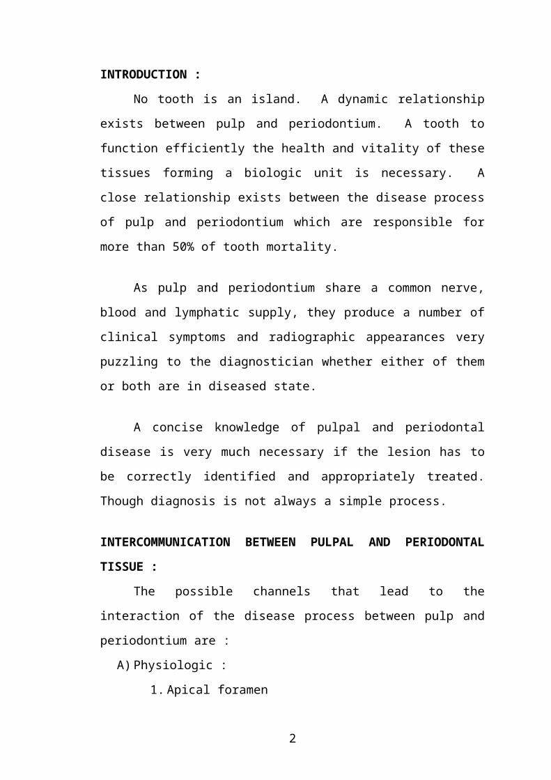
INTRODUCTION :
No tooth is an island. A dynamic relationship exists between pulp and
periodontium. A tooth to function efficiently the health and vitality of these
tissues forming a biologic unit is necessary. A close relationship exists
between the disease process of pulp and periodontium which are responsible
for more than 50% of tooth mortality.
As pulp and periodontium share a common nerve, blood and lymphatic
supply, they produce a number of clinical symptoms and radiographic
appearances very puzzling to the diagnostician whether either of them or both
are in diseased state.
A concise knowledge of pulpal and periodontal disease is very much
necessary if the lesion has to be correctly identified and appropriately treated.
Though diagnosis is not always a simple process.
INTERCOMMUNICATION BETWEEN PULPAL AND
PERIODONTAL TISSUE :
The possible channels that lead to the interaction of the disease process
between pulp and periodontium are :
A) Physiologic :
1. Apical foramen
2. Lateral canals
3. Dentinal tubules
4. Periodontal ligament
5. Alveolar bone
6. Neural pathways
7. Vasculolymphatic pathway
B) Iatrogenic :
1. Palatogingival grooves
2. Vertical root fractures
3. Perforations
1
APICAL FORAMEN AND LATERAL CANALS :
Apical foramen is the most direct route of communication to the
periodontium. Egress of irritants form necrotic pulps via apical foramen into
periapical tissues initiates an inflammatory destruction of apical PDL and
resorption of bone, cementum and even dentin.
In the event of retro infection where in plaque covers the entire length of
the root, the periodontal disease may lead to pulp necrosis through apical
foramen which is a rare condition.
Lateral and accessory canals mainly in the apical area and in the
furcation of molars also connect the dental pulp with periodontium, which can
carry toxic substances from pulp to periodontium or vice-versa to induce
pathologic changes such as atrophy, calcification, irreversible inflammation
and necrosis of pulp.
DENTINAL TUBULES :
The dentinal tubules with their odontoblastic processes extend from the
pulp-dentin border to the CDJ. They can communicate pulp chamber with
external root surface especially when the cementum is denuded, thus
channelising toxic metabolites during pulp or periodontal disease in both
direction.
PALATOGINGIVAL GROOVES :
These are developmental anomalies of the maxillary incisors with more
incidence among lateral incisors, they usually begin in the central fossa, cross
cingulum and extend apically with varying distances.
Everett and Krammer reported that 0.5% extends till the root apex
contributing to an endodontic pathologic condition.
2
PERFORATIONS :
Perforation of root creates a communication between root canal system
and PDL. They occur as a result of over instrumentation of root canal, internal
and external root resorption or caries invading through floor of pulp chamber.
Perforations in the middle or apical 1/3rd of root have greater chance of
healing, closer the perforation to the gingival sulcus particularly in the coronal
3rd of the root or furcation region, the likelihood of apical migration of gingiva
in initiation of periodontal lesion.
VERTICAL ROOT FRACTURE (VRF) :
A VRF can produce a “halo” effect around the tooth radiographically.
Deep periodontal pocket and localized destruction of alveolar bone are often
related to long standing root fractures.
Radiographically mimics profile of occlusal trauma with localized loss
of lamina dura, altered trabecular pattern and widened PDL. The fracture site
provides portal of entry for irritants from root canal to the surrounding PDL.
Vertical root fractures have contributed to progressive periodontal destruction
in the presence of apparently successful endodontic therapy and over all
periodontal site stability.
Effects of Pulpal Disease and Endodontic Procedures on the
Periodontium:
Pulp pathology as a cause of periodontal disease has received much
attention during the last decade. It is important to understand that vital
inflammed pulp will not cause damage to the periodontium; only pulp necrosis
will result in necrotic debris, bacterial by products and other toxic irritants that
exit through apical foramen causing periodontal tissue destruction apically and
potentially migrating towards gingival margin. Simring and Goldberg termed
this “retrograde periodontitis” to differentiate from “marginal periodontitis” in
which the disease proceeds physically from gingival margin toward root apex.
3
This inflammation often results in dysfunction of the PDL, resorption of
alveolar bone, cementum and even dentin. The endodontic infection has been
regarded as local modifying risk factor for periodontitis progression if left
untreated.
In a periodontally involved tooth the endodontic pathogen growth,
infectious products and root canal medicaments may aggravate periodontal
pocket formation, bone loss and impair wound healing to further accelerate
periodontal disease development and progression. However nature and extent
of periodontal destruction depends on the virulence of the organism, duration
of disease and host defense mechanism.
Manifestations of Endolesions in Marginal Periodontium from Lateral
Canals :
Inflammatory lesions may develop at the lateral aspects of root from
root canal infection, where the bacterial byproduct reach periodontium through
lateral canals along the lateral aspects of root and into furcation area. This type
of lesion is very rare as the lateral canals, most of them are either blocked and
narrowed by dentin deposition or covered by cementum deposition.
Manifestation of Acute Endodontic Lesions in the Marginal Periodontium
(fig.)
Acute endodontic lesions may destroy and expand into the periodontium
to an extent that apical marginal communication may emerge.
This drainage of endodontic lesion follows one of the two routes.
1. Along the PDL space (PDL fistulation), this results into narrow opening
of the fistula into the gingival sulcus/ pocket which can be readily
probed down to the apex of the tooth where no increased probing depth
exists around the other surfaces of the tooth. In multirooted teeth the
fistulation can drain off into furcation area resembling a thorough and
through furcation defect form periodontal disease.
4
2. The lesion can perforate the cortical bone close to the apex and elevate
the soft tissue including periostuem from the bone surface and drains
into gingival sulcus / pocket resulting in wide opening of fistula into
pocket and is most often seen in the buccal aspect of the tooth. Since
the type of fistula is not associated with loss of bone tissue at the inner
walls of the alveolus, as periodontal probe cannot penetrate into the PDL
space.
These acute manifestations of root canal infections can result in rapid
and extensive destruction of attachment apparatus. These lesions heal
following proper endo therapy.
The periodontium regeneration on a pulpless teeth is questionable,
according to Saunders, whereas Diem et al reported that all tissues of
periodontium regenerate after periodontal therapy irrespective of its pulpal
status. It has been suggested that endodontic treatment should occur before
periodontal therapy for successful results.
Endodontic Procedures :
During root canal therapy iatrogenic alterations of periodontium can
occur leading to the development of periodontal lesion.
For Example :
Pulp extirpation may develop an acute inflammatory reaction in
periodontium.
Pushing of root canal debris beyond apex during canal instrumentation.
Over instrumentation and over obturation
Perforation of pulp chamber floor and root during access, cleaning and
shaping and post space preparation
Vertical root fracture during obturation and post placement.
5
Effect of Periodontal Disease and Procedures on the Pulp :
Clinically its not uncommon to observe a tooth with advanced
periodontal lesion without any signs of decay being non-vital. Disease form
periodontal pocket can spread to the pulp most commonly through accessory
canals at the furcation and apex of the teeth.
Teeth with large canals and periodontal breakdown extending to the
apex are the teeth more affected.
The pulpal reaction is not only influenced by the stages of periodontal
disease but also by periodontal treatment procedures such as scaling, root
planning and administration of medication.
During deep curettage the blood vessels supplying the pulp via
accessory canals may be damaged. Scaling and root planning removes
cementum resulting in exposing dentinal tubules and sometimes also lateral
canals. These procedures lead to easy entry of irritants through exposed
dentinal tubules and lateral canals on the root surface endangering pulp.
It has been reported that canals of teeth with longstanding periodontal
disease develop fibrosis and became narrower, which suggests reparative
process than inflammatory response.
Although consensus supports the influence that a degenerating or
inflamed effects of periodontium on pulp, not all researchers are in agreement.
Another school of thought says when pathologic changes occur in the
pulp as a result of periodontal disease, the pulp usually does not degenerate as
long as the main canal is not involved, as long as the blood supply through the
apical foramen remains intact, the pulp is capable of withstanding
physiologic insults induced by periodontal disease.
6
Periodontal Evaluation :
No dental examination is complete without careful evaluation of teeths
periodontal support. Periodontal probing and recording pocket depths provide
information with respect to possible etiology and prognosis. There is little
question that pulpal necrosis can lead to loss of periodontal support. Whether
periodontal disease can cause pulpal degeneration is a question not clearly
answered. However there is an agreement that potential interaction exists
between pulp and periodontium.
Diagnosis :
For endodontic purpose of a single tooth, probing maybe limited to the
involved tooth and atleast the adjacent teeth.
Periodontal Disease :
Periodontal stability is a basic requirement for any tooth being
considered for endodontic therapy. This stability is determined by the amount
of bony support, health of the support and health of the overlying soft tissue.
Examination alone cannot guarantee the future health of these tissues,
but usually it can determine the existing disease.
Examination :
As part of the examination, probe the sulcus of the tooth in question and
record pocket depth.
Record the mobility of the tooth using a system of 0 to 3.
Grade 0 : Normal mobility (Physiologic)
Grade 1 : Slight mobility
Grade 2 : Marked mobility
Grade 3 : Mobility with depressibility
7
Record for
Bleeding on probing
Palatal grooves in single rooted teeth.
Furcas in multirooted teeth.
Other anomalies like enamel projections as they may aggravate gingival
conditions and make for unstable future periodontal health.
Interpretation :
3-5 mm pocket and Grade I mobility – “Moderate Periodontitis’.
When this is found, the entire mouth should be examined of periodontal
disease.
More than 5 mm pocket with or without Grade II/III mobility indicates
“Severe periodontitis”.
Referral to a periodontist must be considered.
Periodontal Pocket :
Presence and distribution on each tooth surface.
Depth
Level of attachment on root
Type of pocket (suprabony / infrabony) should be examined for
periodontal pocket evaluation.
Signs and Symptoms :
Although probing is the only accurate method for detecting pockets.
Clinical signs such as ;
Bluish red marginal gingiva / vertical zone extending form marginal to
attached gingiva.
“Rolled” edge separating gingival margin form tooth surface.
Enlarged edematous gingiva may suggest their presence.
Bleeding, suppuration, loose extruded teeth.
Symptoms :
8
Usually painless, but sometimes
Localized or radiating pain or sensation of pressure after eating which
gradually diminishes.
Foul taste in localized areas.
Sensitivity hot and cold
Tooth ache in absence of caries are present
Detection of Pockets :
Gutta percha points or calibrated silver points can be used with
radiographs to determine the level of attachment of periodontal pockets.
Their use in localized cases is feasible when used in generalized cases it
would be cumbersome.
Clinical Probing is more efficient
Two different pocket depths are ;
1) Biologic depth / histologic depth
2) Clinical / probing depth
“Biologic depth” is ascertained in histologic sections, it’s the depth form
gingival margin to base of pocket (coronal end of junctional epithelium).
“Probing depth” is the depth to which an adhoc instrument (probe) penetrates
into the pocket.
Technique :
Probe is held parallel to the long axis of tooth and walled
circumferentially around each surface of each tooth to detect areas of deepest
penetration (Fig.).
Force : 0.75 N.
To detect interdental craters probe must be placed obliquely form both
facial and lingual surfaces (Fig.).
9
Bleeding on Probing :
Insertion of probe to the bottom of the pocket elicits bleeding if the
gingiva is inflamed and the pocket epithelium atrophic / ulcerated.
Bleeding on probing is early sign of inflammation than colour changes.
Test :
Probe is carefully inserted to the bottom of the pocket and gently moved
laterally along pocket wall. Sometimes bleeding appears immediately after
probing, where as sometimes it may take few seconds (30 to 60 seconds after
probing).
When to Probe :
In moderate or advanced cases probing is done twice as profuse bleeding
impairs clinical examination.
Initial Probing : is done to determine whether the tooth can be saved or
extracted along with clinical and radiographic examination.
After adequate plaque control measures.
Second probing is done to determine the level of attachment, degree of
involvement of roots and furcation.
Probing around Implants :
To prevent scratching of implant surface, plastic periodontal probes
should be used instead of steel probes.
Amount of Attached Gingiva :
Width of attached gingiva is the distance between the mucogingival line
and the projection on the external surface of the bottom of gingival sulcus.
Specially designed Naber’s probe allows easier and accurate exploration of
horizontal component of furcation lesions.
“Pocket Depth” is the distance between base of the pocket and gingival
margin. It keeps changing with the position of gingival margin and maybe
unrelated to the existing attachment of the tooth.
10
Level of Attachment :
It is the distance between the base of the pocket and a fixed point on the
crown such a CEJ.
Level of attachment affords a better indication of the degree of
periodontal destruction.
Shallow pockets attached at the level of apical third of root indicate
more severe destruction than deep pockets attached at the coronal third of the
roots.
Determining the Level of attachment :
When gingival margin is on the anatomic crown :
Depth of pocket - Distance between gingival margin to CEJ = Level of
attachment.
When gingival margin is at CEJ
Loss of attachment = Pocket depth
Gingival margin apical to CEJ.
Because loss of attachment is greater than pocket depth. So distance
between CEJ and gingival margin is added to pocket depth. Width of attached
gingiva is determined by substracting the sulcus or pocket depth form the total
width of gingiva (gingival margin to mucogingival line). This done by
stretching the lip to demarcate mucogingival line.
When width of attached gingiva is less, the free gingival margin moves
on stretching of lip / cheek.
Degree of Gingival Recession :
Distance between CEJ and gingival margin.
PERIODONTAL PORTRAIT :
11
During the course of treatment of Class I Endo-Perio Lesion (Weine) the
rapid healing that immediately follows the canal preparation appointment, the
dentist may confuse when seeing the patient at the succeeding visit.
Where was the pocket before?
How deep was it
Was it on buccal or lingual of the root.
Attempting to reprobe incorrectly may damage the sensitive healing
tissue or injure the normal areas. Therefore prior to initiation of therapy an
accurate recording of preoperative conditions must be made. An easy and
useful accurate one is the periodontal portrait (by Melton) (Fig.).
CLASSIFICATION OF ENDO-PERIO LESIONS :
According to SIMON GLICK FRANK (Cohen)
Primary endodontal lesions
Primary endodontal lesion with secondary periodontal involvement
Primary periodontal lesion
Primary periodontal lesion with secondary endodontal involvement
True combined lesion
According to Oliet, Pollock (Grossman) :
1) Lesions that require endodontic treatment procedures only : -
- Any tooth with necrotic pulp and apical granulomatous tissue
replacing periodontium and bone with or without sinus tract.
- Chronic periapical abscess with a sinus tract draining through the
gingival crevice thus passing through a section of the attachment
apparatus in its entire length along the side of the root.
- Longitudinal and horizontal root fractures.
- Pathologic and iatrogenic root perforations
- Teeth with incomplete apical root development
- Endodontic implants
- Teeth requiring hemisection / radisectomy
12
- Root submergence.
II) Lesions that require periodontal procedures only :
- Occlusal trauma causing reversible pulpitis
- Occlusal trauma plus gingival inflammation resulting in pocket
formation and reversible pulpitis.
- Suprabony or infrabony pocket formation treated with
overzealous root planning and curettage leading to pulpal
sensitivity.
- Extensive infrabony pocket formation extending beyond the root
apex and sometimes coupled with lateral or apical resorption yet
with pulp that responds within normal limits to clinical testing.
III) Lesions that require combined endodontic – periodontic treatment
procedures :
- Any lesion in group I that results in irreversible reactions in the
attachment apparatus and requires periodontal treatment.
- Any lesion in group II that results in irreversible reactions in pulp
tissue and also requires endodontic treatment.
According to Weine :
Class I Tooth in which symptoms clinically and radiographically
simulate periodontal disease but are infact due to pulpal
inflammation and/or necrosis.
Class II Tooth that has both pulpal or periapical disease and periodontal
disease concomitantly.
Class III Tooth that has no pulpal problem but require endodontic therapy
plus root amputation to gain periodontal healing.
Class IV Tooth that clinically and radiographically simulated pulpal or
periapical disease but infact has periodontal disease.
PRIMARY ENDODONTAL LESIONS :
13
These are the lesions, which arise form the diseased pulp. These lesions
sometimes may show as drainage form the gingival sulcus or swelling of the
facial attached gingiva. So initially when you see the lesion, it may appear as
periodontal in origin but in reality it is fistula formed as a result of periapical
infection that instead of opening on the buccal or lingual mucosa, drains along
the periodontal ligament into the sulcus.
Clinical Features :
- Drainage may be evident in sulcus area.
- Swelling may be present especially in bifurcation area simulating
periodontal abscess.
- Vitality test will reveal necrotic pulp or atleast an altered response in case
of multi rooted teeth indicating that at least one canal is necrotic.
- Periodontal probing usually shows normal sulci around the tooth except in
one area with a narrow defect. Placement of silverpoint, gutta percha cone
or periodontal probe in this sinus tract shows that the defect is deep and
will go towards the source of irritation generally the root apex or the
lateral canal.
- Pain is usually not present though the patient may have some minor
discomfort.
Radiographic Appearance :
Radiograph may show deep dental caries, deep filling or fractured
restoration. The drainage through the sulcus often appears as a radiolucency
along the mesial or distal root surface or in the bifurcation area.
Treatment :
Because this lesion is an endodontic problem that has merely fistulated
through the periodontal ligament, complete healing is usually anticipated after
routine endodontic therapy.
Periodontal curettes are used to plane the root surface to be sure it is free
of plaque and deposits.
14
PRIMARY ENDODONTAL LESION WITH SECONDARY
PERIODONTAL INVOLVEMENT :
This is merely an extension of the endodontic lesion when the patient
ignores the draining fistulas. As the drainage persists through the gingival
sulcus, super imposition of the plaque and the calculus into the pocket like
defect occurs resulting in periodontal pocket and apical migration of the
attachment.
To differentiate these lesions form the lesions of primary periodontal
origin you should consider the following factors.
If the problem is primarily endodontic, there must be an etiologic factor
for the pulp necrosis like presence of caries, extensive restorations,
fractured restorations or teeth, discolored crowns, severe attrition etc.
Periodontal disease usually has generalized nature. Usually periodontal
lesions are not isolated to a single tooth (but this also may occur
sometimes. Additional tests and findings are needed).
Vitality Tests : Vitality tests reveal necrotic pulp.
Treatment :
Localized periodontitis complicates the prognosis and the tooth now
requires both endodontic and periodontal therapy the bone loss of endodontic
origin will heal if appropriate root canal treatment is performed but not the
secondary pocket, it requires curettage and root planning.
PRIMARY PERIODONTAL LESION :
Unchecked or untreated periodontitis progresses along the root surface
until the Periapex is reached. Important diagnostic finding is to see whether the
cervical lesion is isolated to a single tooth or is generalized. Periodontal
disease is usually generalized. It may be localized because of traumatic
occlusion. Teeth show periodontitis as a result of accumulation of plaque
and/or calculus formation.
15
Vitality tests : Shows positive indicating vital pulp.
Mobility : Teeth may be associated with various degrees of mobility.
Clinical examination : We may find plaque, calculus and soft tissue
inflammation associated with a purulent exudates.
Probing : Clinically probing detects broad based (infrabony) pocket formation
and causes bleeding of the tissue.
Treatment :
Treatment is only periodontal therapy and depends on the extent of the
periodontal disease and the patients ability to comply with possible long term
treatment and maintenance therapy. Prognosis depends on the outcome of the
periodontal therapy and care must be taken not to devitalize the pulp during
deep therapy.
PRIMARY PERIODONTAL LESIONS WITH SECONDARY
ENDODONTAL INVOLVEMENT :
- Progression of the periodontal disease may involve lateral or accessory
canals or may extend to the apex leading to the pulp. When the lesion
involves the primary pulp vessels at the apex retroinfection takes place
and the pulp becomes inflamed and necrotic.
- Progressive periodontal disease results in the apical migration of the
attachment and root surface exposure to the oral cavity and to irritants
such as bacterial plaque. “Seltzer” states that oral flora can transmit
toxic products into the pulp via the lateral canals or dentinal tubules to
cause atrophic, degenerative, inflammatory and resorptive alterations.
- Periodontal therapy may also lead to pulp death if neurovascular bundle
entering a mid root lateral canal is severed during deep periodontal
therapy.
Clinical Features :
16
- The tooth with primary periodontal and secondary endodontic
disease will have deep pockets and history of extensive
periodontal disease.
- When the pulp becomes involved the patient reports accentuated
pain and clinical signs of pulpal disease.
Vitality Tests : Once the pulp becomes involved, pulp tests will confirm pulp
neurosis.
Mobility : Various degrees of mobility.
Clinical Examination : Plaque, calculus and soft tissue inflammation is seen.
Treatment :
Both endodontic and periodontal therapy will be necessary to provide a
successful result. The endodontic treatment should be completed first because
the toxic reservoir in the root canal will continue to abort the desired
periodontal healing.
Prognosis in these cases of primary periodontal etiology is not as good
as when the primary lesion is endodontal because all periodontal therapy has a
much more guarded prognosis than endodontal therapy.
TRUE COMBINED LESIONS :
These lesions occur where a pulpally induced periapical lesion exists on
a tooth that is also periodontally involved. These consists of two concurrent
lesions, one is an independent periapical lesion originating from a necrotic
pulp and other is an independent periodontal lesion that has progressed apically
towards the periapical lesion. Depending on the stage of their development, the
lesions may or may not communicate. The infrabony defect occurs when the
two lesions meet and merge. A new term “concomitant pulpal-periodontal
lesion” has given to those teeth which have both disease processes occurring
but do not communicate.
Clinical Features :
17
In a combined lesion,
Periodontal Examination : Probing of tooth shows presence of plaque, calculus,
periodontitis and a wide and conical periodontal pocket characteristic of
periodontal defect of periodontal disease origin.
Vitality tests : Confirm pulp necrosis.
Radiographic Examination : Shows periodontal infra bony pocket
communicating the periapical lesion and degrees of crestal bone loss.
The character of the combined lesion may mimic the lesion of
endodontic origin.
Treatment :
Treatment of combined lesions consists of endodontic and periodontal
therapy. The overall prognosis depends on the prognosis of each individual
factor. In cases in which periapical and periodontal lesions communicate,
complete cleaning and obturation of the root canal system prevents egress of
the irritants form the periapical lesion into the periodontal defect. Then the
prognosis of the affected tooth then depends totally on the outcome of the
periodontal therapy.
If a combined lesion is found in a mouth otherwise free of periodontal
disease, we can suspect vertical root fracture, particularly if the lesion does not
respond to combined therapy (it may be necessary to lay a flap to find the
defect which has hopeless prognosis).
COMBINED LESION TREATMENT :
These patients in contradiction to Class I (Weine) exhibit periodontal
disease in a number of areas of the mouth.
3 distinctly different types of lesions occur :
1. Two separate lesions (Pulpo-periapical and periodontal)
with no communication between them (concomitant pulpo periapical
lesions).
18
2. Single lesion that involves both endodontic and
periapical pathoses.
3. Separate endodontic and periodontal lesions that later
communicate.
1. Separate unrelated lesions :
First it must be determined if periodontal condition is treatable.
Performing endodontic therapy on teeth with hopeless periodontal lesions is
disastrous. Endodontic therapy is performed first and then periodontal therapy.
2. Single lesion with both pulpal and periodontal pathoses.
This is most difficult to treat successfully.
Clinical Features :
Involved teeth has single lesion radiographically.
Probes as a routine deep periodontal pocket
Patient has periodontal condition involving other teeth
Endo therapy should be initiated followed by periodontal therapy.
Results are not predictable and patients must be forewarned concerning
doubtful prognosis.
3. Periodontal and Endodontic Lesion that have merged :
A periapical endo lesion merges with periodontal pocket eventhough
separate lesions are present. Portion of lesion extending form apical area
towards the crestal bone is periapical due to pulpal damage. Whereas the
lesion from the sulcus extending apically is periodontal breakdown.
Treatment :
If endodontics is only performed the periapical lesion heals to the site
where periodontal lesion begins.
If periodontal therapy only is performed, the crestal bone may heal upto
where the periapical lesion begins.
Both pulpal therapy followed by periodontal should be performed.
19
ALTERNATIVE TREATMENT MODALITIES :
When traditional endodontic and periodontal treatments prove
insufficient to stabilize an affected teeth, the clinician must consider other
treatment alternatives like root amputation and hemisection.
Root amputation procedures are a logical way to eliminate a weak,
diseased root and allow the strongest to survive, where as retained together
they would collectively fail. Hemisection is the most common method of
removing a pathologically involved root. Before carrying out any such
procedures the occlusal forces, restorability and the value of the remaining
roots must be examined.
Root amputation or radisectomy denotes the removal of one or more
roots of a molar. Hemisection refers to sectioning of the crown of a molar
tooth, with either the removal of half the crown and its supporting root
structures or retention of both the halves to be used after reshaping and
splinting as two premolars. Radisectomy and Hemisection are often desirable
for periodontal reasons.
At times, a multi rooted tooth has an untreatable periodontal lesion on
one or more of its roots but the remaining root/roots are well supported and
treatable. For example, one root may have an extensive infrabony pocket with
concomitant bone loss and the other root may be surrounded by normal gingiva
and supporting bone. To retain a portion of this strategic tooth and to avoid
extraction of the entire tooth, hemisection or radisectomy can be performed.
Whenever possible, Endodontic treatment should precede root removal
because it is difficult to treat a tooth properly when it has been sectioned
20

through the pulp chamber because asepsis is impossible and anatomic
guidelines used in the treatment are destroyed.
The role of root amputation procedures is gradually decreasing because
of the problems associated with long term success of the teeth treated with
amputational therapy, now guided tissue regeneration has come which provided
the regeneration of the lost periodontal structures without any root amputation.
GTR appears to be an excellent alternative choice as it is less destructive
regenerative and requires less time.
HEMISECTION AND ROOT AMPUTATION :
First attempted by G.V.Black in 1880. However until late 1950s interest
was not shown by dental profession. Teeth formerly considered hopeless now
may be retained.
With Hemisection and root amputation endodontics should be
completed before surgery to avail easy isolation and prevent contamination.
Indications :
Hemisection and root amputations are indicated when one or two roots
of multirooted teeth becomes untreatable because of ;
1. Endodontic reasons (separated instrument, root preparations (resorption)
obstructed canals).
2. Periodontal reasons (furcation involvement, severe bone loss around one
root).
3. Restorative reasons (caries destruction, erosion of large part of crown
and root, perforations during posterior preparation / #).
4. Combination of these.
Hemisection :
This is a procedure to retain half of the tooth in an essence of converting
molar into two premolars (Bicuspidization) or one premolar.
21
Root canals are obturated and pulp chamber filled with amalgam before
surgery.
Flap may not be necessary all the time. In case of periodontal
involvement of furca.
Sectioning can be done with a pencil diamond or long shanked fissure
bur with initial cut at the extent of the root to be removed, so that there is
enough tooth structure to aid in preparing the crown for post crown restoration,
part of overdentures, bridge abutment.
When the defect periodontally is in the furcation both the roots can be
retained with opening up the furcation and restored to form 2 premolars –
Bicuspidization is the procedure termed as;
SURGICAL CONSIDERATIONS :
Periodontal evaluation plays a very important role in surgical
endodontics.
Flap design will dictate health of periodontal attachment and gingiva.
If sufficient attached gingiva is present envelop type flaps can be given.
If little or no attached gingiva exists a full flap must be raised and
repositioned apically to create new attached gingiva.
Flap edges should be sutured over round bone to promote healing and to
avoid post surgical recession or soft tissue defects.
When a full flap is raised in the presence of periodontal disease,
necessary scaling, curettage or osseous contouring procedures should be
performed at the same time.
Both these procedures are excellent if used when indicated and if used
properly. If observed the techniques can lead to predictable failure both
periodontally and restoratively.
In 10 years study all the failures in teeth which had undergone
hemisection and root amputation were attributed to endodontic or
restorative failures, not periodontal disease.
22
Endodontic Implant :
This is indicated in cases of less crown root ratio and stabilization in
advanced periodontally involved teeth or secondary to eliminate secondary
occlusal trauma. However endodontic implant does not cure periodontal
disease.
Currently ADA classifies the endo implants as an experimental
procedure.
Root Amputation :
After the root canal system is obturated with gutta percha, a No.4 – No.6
slow speed, long shanked round bur is used to drill 2 to 3 mm into root/s
to be amputated. The roots prepared and entire pulp chamber are filled
thoroughly with well condensed amalgam to ensure complete seal of the
pulp chamber after too is removed.
In some cases flap may not be required when the extent of bone loss and
periodontal involvement is severe. Usually a small triangular flap is
raised.
In Maxillary Molars :
One or even two roots may be amputated.
Procedure is similar to hemsection except the contouring of remaining
crown is complex.
The crown portion supported by the root to be amputated should be
recontoured to accept new occlusal demands and ensure good oral
hygiene.
The crown can be contoured first and then underlying root removed or
crown and root cut off together.
23
ROLE OF ANTIBIOTICS IN ENDO-PERIO LESIONS :
In Endodontic Therapy :
The treatment of acute and chronic infections of endodontic origin is
primarily by operative intervention.
Now a days endodontic therapy emphasizes the importance of
debridement procedures there by eliminating or reducing the micro organisms.
Severe infections rarely occur. Even if it occurs, establishing the surgical
drainage is the primary treatment in order to remove the cause of infection.
Antibiotics should be given :
Only when the drainage becomes difficult to obtain.
If there is a diffuse spread of infection (Ex. In case of acute dento
alveolar abscess).
When the host resistance is low (as in case of medically compromised
patients).
When the virulence of the microorganism is very high.
The most commonly prescribed antibiotics are erythromycin,
amoxicillin, penicillin and metronidazole.
In Periodontal Therapy :
The use of antibiotics in the treatment of periodontal disease is base don
the infectious nature of the disease. Many micro-organisms are present in the
oral cavity which cause various periodontal disorders. In the early stages of
periodontal disease gram positive bacteria will be seen where as in the later
stages, gram negative bacteria and spirochaetes lay an important role. The
bacterial cultures and antibiotic sensitivity test should be performed before
selecting an antibiotics.
No single antibiotic at concentrations achieved in body fluids inhibits all
periodontal pathogens. A combination of antibiotics may be necessary to
24
eliminate all the pathogens from periodontal pockets. Both systemic and
topical antibiotics have been evaluated.
Topical application has advantages like, the antibiotics can be directed
to their specific target areas, reduced drug dosages and increased
concentrations and reduces side effects.
Antibiotics for Endo Perio Lesions :
There are no authorative studies to support the use of systemic
antibiotics in the management of endo-perio lesions. The treatment of
combined lesions is based upon the basic principles of endodontic and
periodontal therapy and is dependent upon the etiology of the condition.
Endodontic treatment usually involves RCT and sometimes root resection and
repair of the perforation. Periodontal treatment includes curettage, scaling and
root planning procedures and sometimes flap surgery and bone grafting.
Systemic antibiotics are not a substitute for the effective mechanical
debridement of root canal system and the root surface. Topical antibiotics such
as tetracyclines or metronidazole may be applied to the periodontal ligament as
an adjunct to root planning. Tetracycline’s appear to be more useful for both
endodontics and periodontal infections. Tetracyclines are highly effective
against many periodontal pathogens because higher concentration of drug can
be achieved in gingival crevice 2-10 times more than in serum. Systemic
antibiotics can be used prophylactically for patients with a history of rheumatic
heart disease and other systemic conditions. They can be used as an adjunct to
the surgical procedures involving root resection, osseous grafting etc.
Tetracycline 250 mg (4 times a day)
Doxycycline 100 mg (2 times on first day and once thereafter)
Metronidazole 250 mg (3 times a day for 7 days)
Now recently Chlorhexidine is gaining increasing popularity as a
microbial agent. Chlorhexidine is incorporated in the small strip which is
25
placed directly in the periodontal defect and left in place. This is called perio
chip.
CONCLUSION :
A concise knowledge of both pulpal and periodontal diseases is
necessary for proper identification of the lesion. Proper diagnosis, followed by
the removal of the etiological factor and rendering appropriate treatment
utilizing GTR techniques combined with osseous grafting where ever necessary
will restore the tooth damaged by the endo-perio lesion to health and function.
REFERENCES :
1. Pathways of the pulp, 8th edition. – Stephen Cohen.
1. Clinical Implantology, 3rd Edition – Jan Lindhe.
2. Periodontology, 9th Edition – Glickman.
26
COLLEGE OF DENTAL SCIENCES
DEPARTMENT OF CONSERVATIVE DENTISTRY AND
ENDODONTICS
SEMINAR ON
“ENDODONTIC – PERIODONTIC
INTERRELATIONSHIP”
Presented By : -
Dr. GAURAV PATRI