Embed Size (px)
Citation preview
Joseph Rabban MD MPHPathology Department
Endocervical Adenocarcinoma
Challenges in Classification, Differential Diagnosis and Reporting
Outline of Talk
Treatment Decisions for Endocervical Adenocarcinoma
New 2014 WHO Classification system
Update on Mucinous Adenocarcinoma variants
Common Problems in Usual type Endocervical Adenocarcinoma
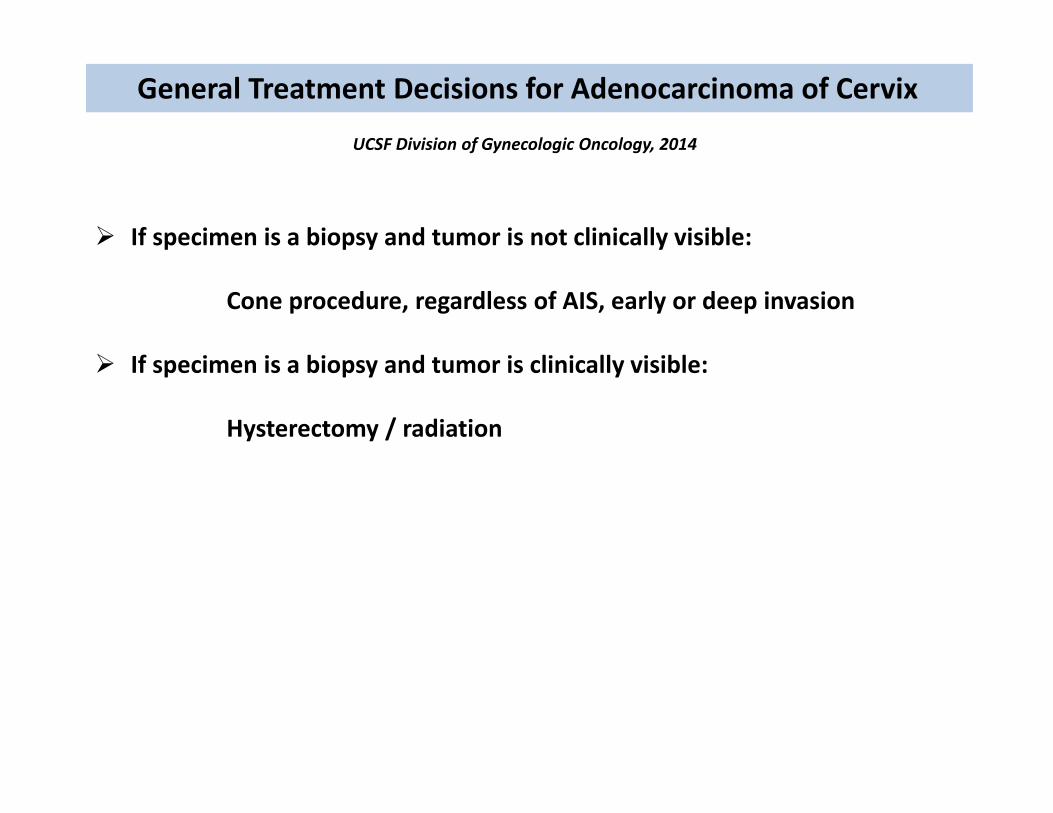
General Treatment Decisions for Adenocarcinoma of Cervix
If specimen is a biopsy and tumor is not clinically visible:
Cone procedure, regardless of AIS, early or deep invasion
If specimen is a biopsy and tumor is clinically visible:
Hysterectomy / radiation
UCSF Division of Gynecologic Oncology, 2014
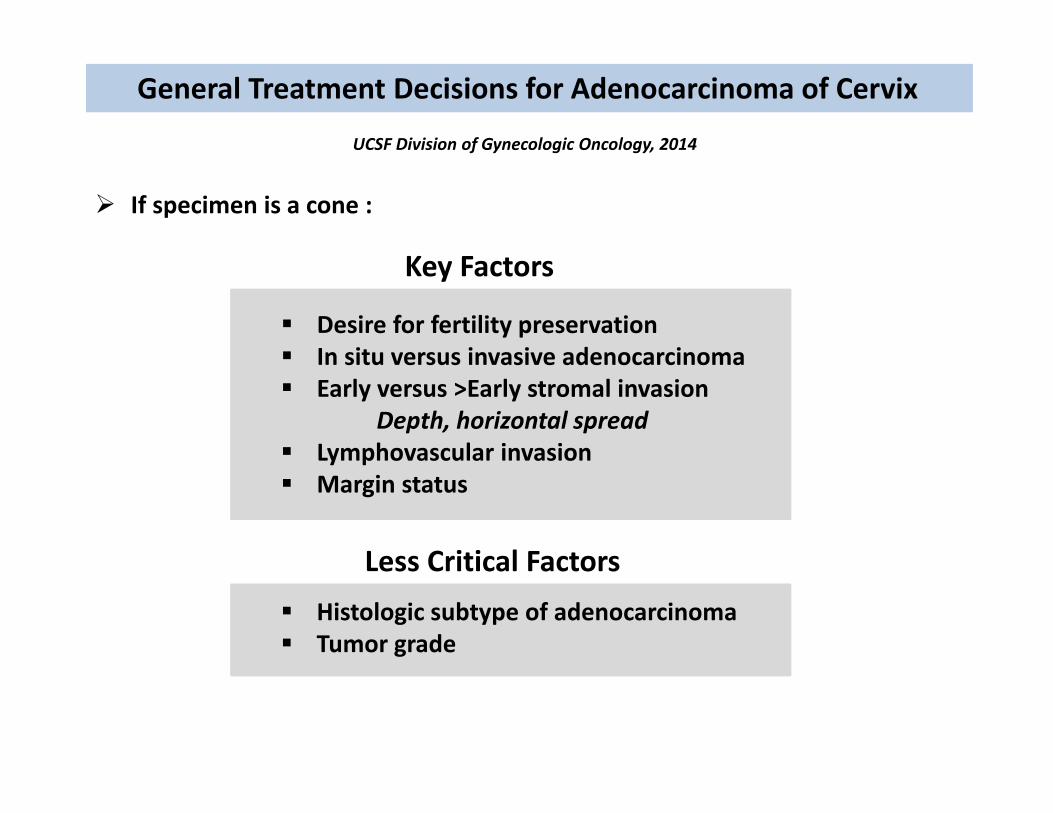
General Treatment Decisions for Adenocarcinoma of Cervix
If specimen is a cone :
UCSF Division of Gynecologic Oncology, 2014
Desire for fertility preservation In situ versus invasive adenocarcinoma Early versus >Early stromal invasion
Depth, horizontal spread Lymphovascular invasion Margin status
Histologic subtype of adenocarcinoma Tumor grade
Key Factors
Less Critical Factors
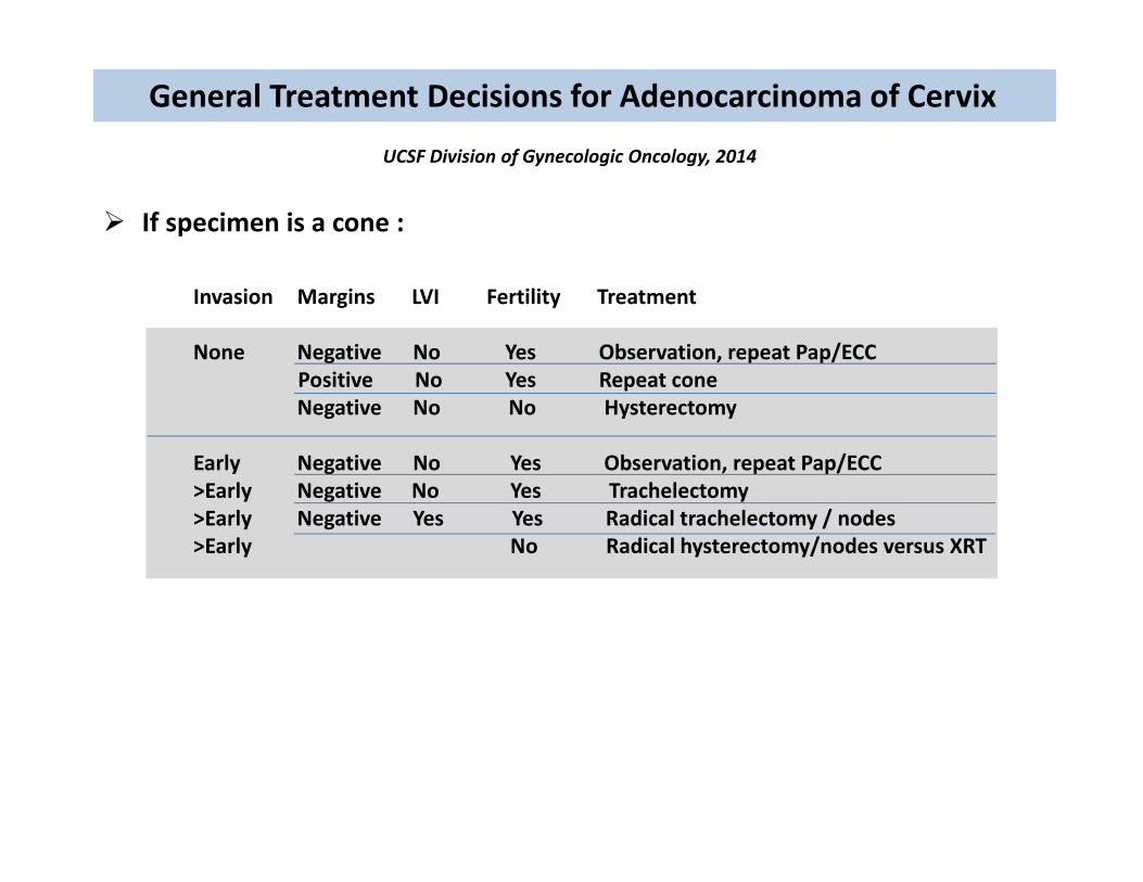
General Treatment Decisions for Adenocarcinoma of Cervix
If specimen is a cone :
UCSF Division of Gynecologic Oncology, 2014
Invasion Margins LVI Fertility Treatment
None Negative No Yes Observation, repeat Pap/ECCPositive No Yes Repeat coneNegative No No Hysterectomy
Early Negative No Yes Observation, repeat Pap/ECC>Early Negative No Yes Trachelectomy>Early Negative Yes Yes Radical trachelectomy / nodes>Early No Radical hysterectomy/nodes versus XRT
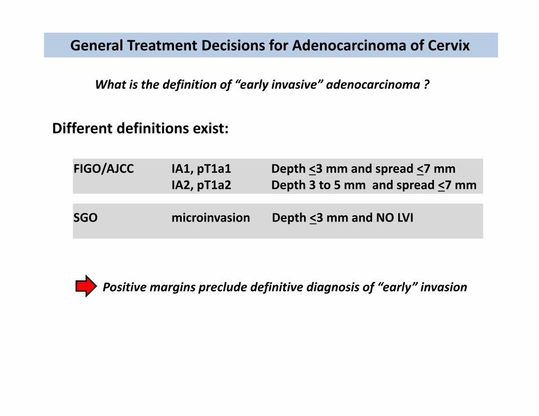
General Treatment Decisions for Adenocarcinoma of Cervix
What is the definition of “early invasive” adenocarcinoma ?
Different definitions exist:
FIGO/AJCC IA1, pT1a1 Depth <3 mm and spread <7 mm IA2, pT1a2 Depth 3 to 5 mm and spread <7 mm
SGO microinvasion Depth <3 mm and NO LVI
Positive margins preclude definitive diagnosis of “early” invasion
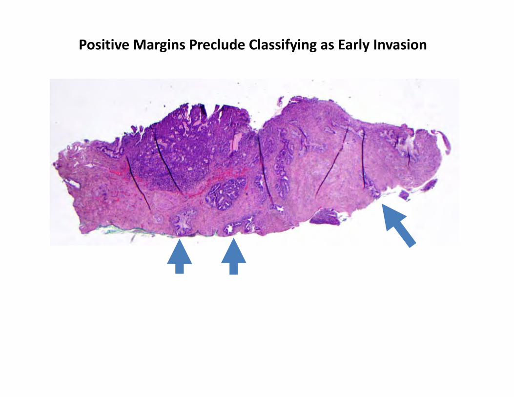
Positive Margins Preclude Classifying as Early Invasion

General Treatment Decisions for Adenocarcinoma of Cervix
UCSF Pathology Report Template
Invasive tumor type Invasive tumor grade Depth of invasion (mm) Horizontal spread of invasion (mm) LVI Margin for invasive tumor Margin for in situ tumor Margin for HSIL
Outline of Talk
Treatment Decisions for Endocervical Adenocarcinoma
New 2014 WHO Classification system
Update on Mucinous Adenocarcinoma variants
Common Problems in Usual type Endocervical Adenocarcinoma
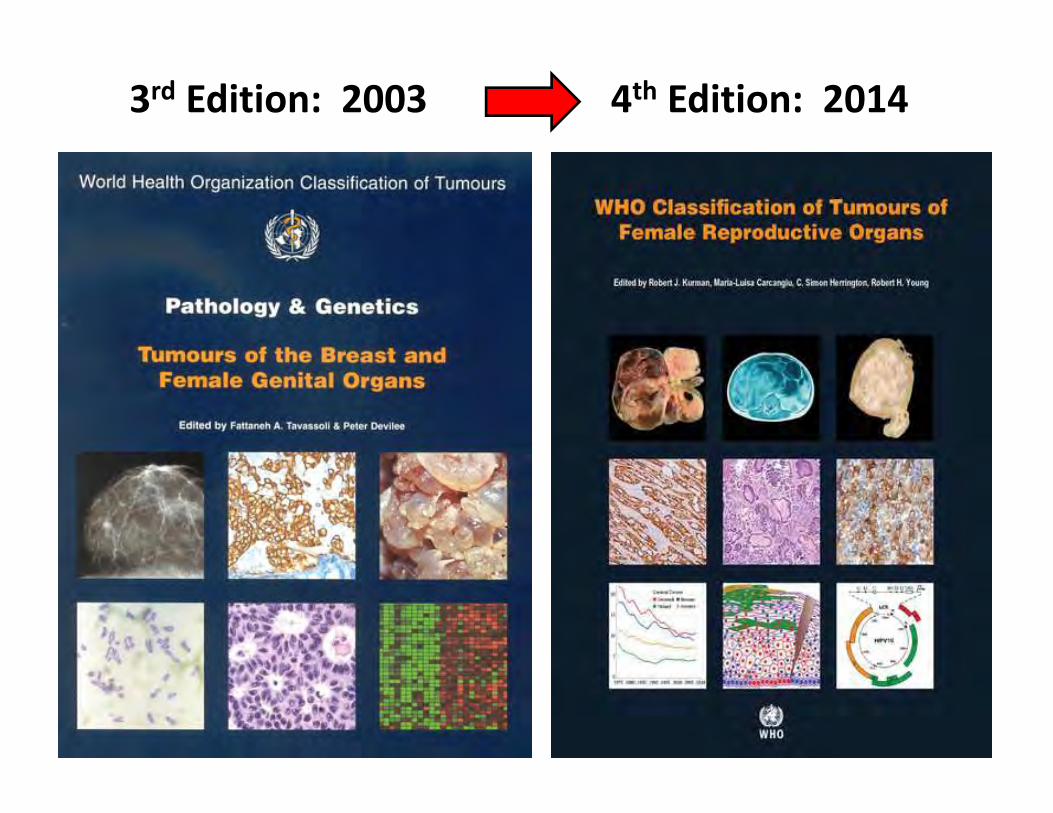
3rd Edition: 2003 4th Edition: 2014
3rd Edition: 2003 4th Edition: 2014
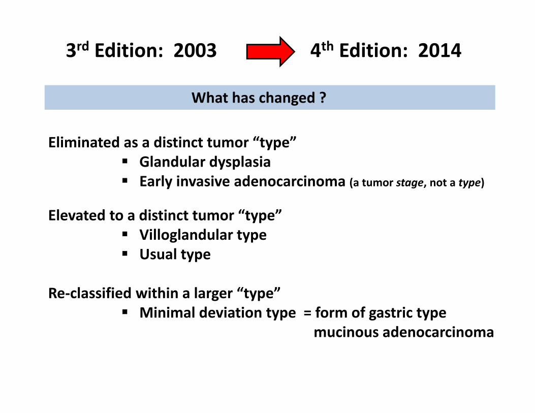
What has changed ?
Eliminated as a distinct tumor “type” Glandular dysplasia Early invasive adenocarcinoma (a tumor stage, not a type)
Elevated to a distinct tumor “type” Villoglandular type Usual type
Re‐classified within a larger “type” Minimal deviation type = form of gastric type
mucinous adenocarcinoma
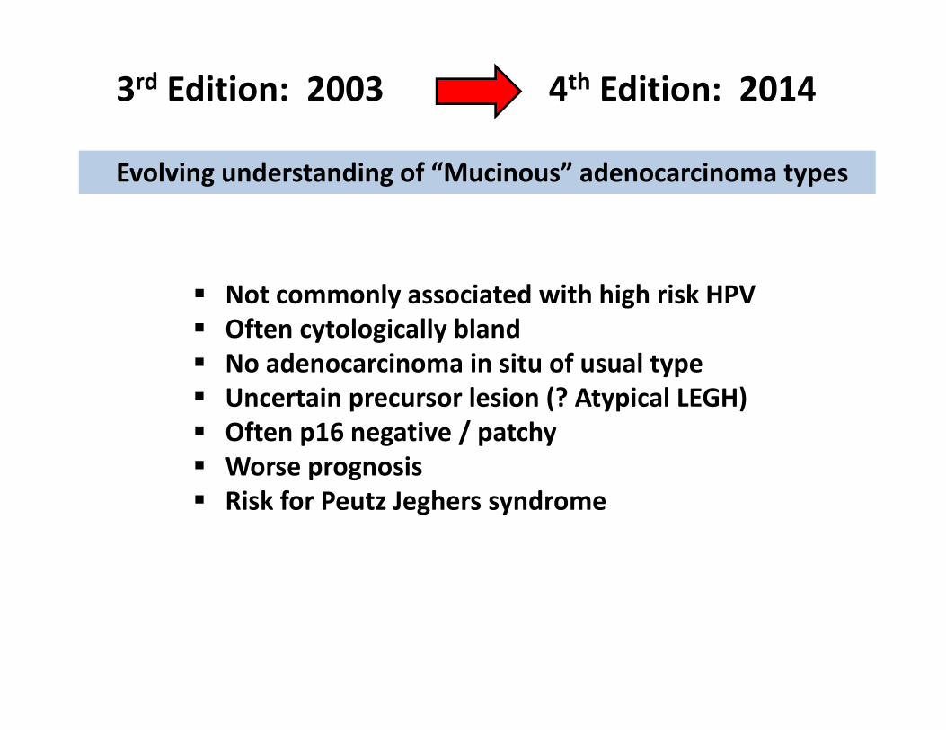
3rd Edition: 2003 4th Edition: 2014
Evolving understanding of “Mucinous” adenocarcinoma types
Not commonly associated with high risk HPV Often cytologically bland No adenocarcinoma in situ of usual type Uncertain precursor lesion (? Atypical LEGH) Often p16 negative / patchy Worse prognosis Risk for Peutz Jeghers syndrome
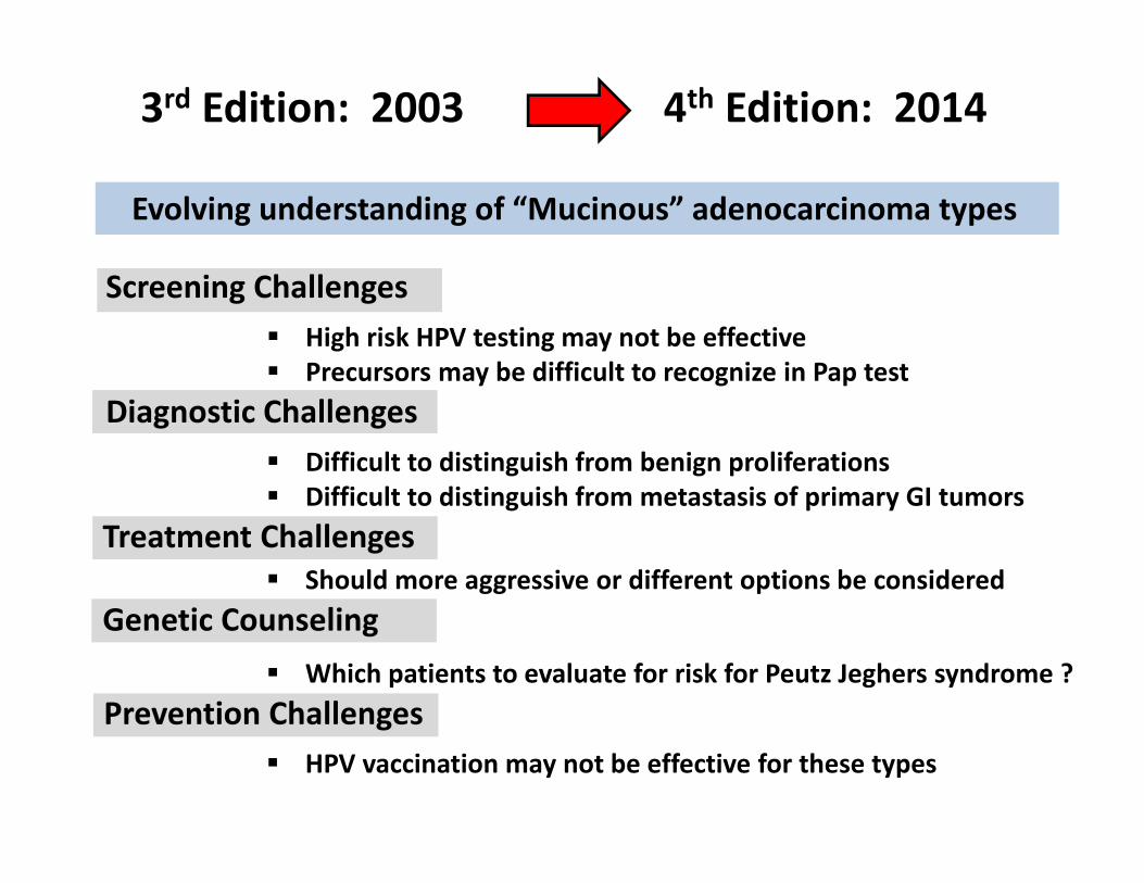
3rd Edition: 2003 4th Edition: 2014
Evolving understanding of “Mucinous” adenocarcinoma types
Screening Challenges
Diagnostic Challenges
High risk HPV testing may not be effective Precursors may be difficult to recognize in Pap test
Difficult to distinguish from benign proliferations Difficult to distinguish from metastasis of primary GI tumors
Should more aggressive or different options be considered
Which patients to evaluate for risk for Peutz Jeghers syndrome ?
HPV vaccination may not be effective for these types
Treatment Challenges
Genetic Counseling
Prevention Challenges
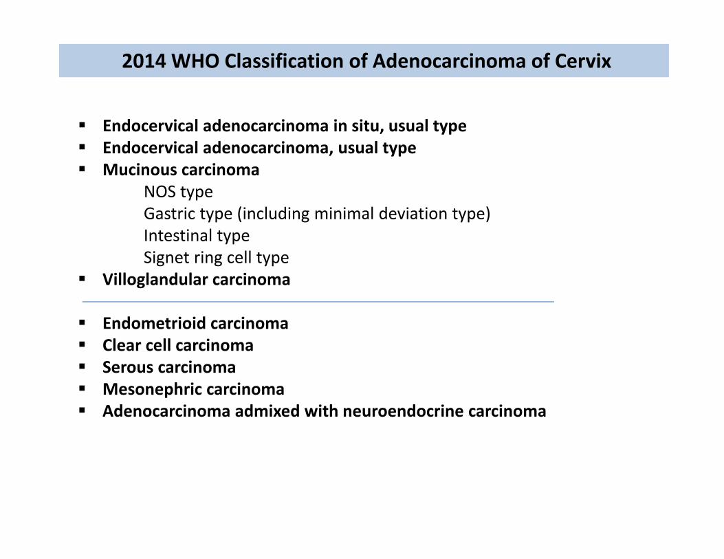
2014 WHO Classification of Adenocarcinoma of Cervix
Endocervical adenocarcinoma in situ, usual type Endocervical adenocarcinoma, usual type Mucinous carcinoma
NOS typeGastric type (including minimal deviation type)Intestinal typeSignet ring cell type
Villoglandular carcinoma
Endometrioid carcinoma Clear cell carcinoma Serous carcinoma Mesonephric carcinoma Adenocarcinoma admixed with neuroendocrine carcinoma
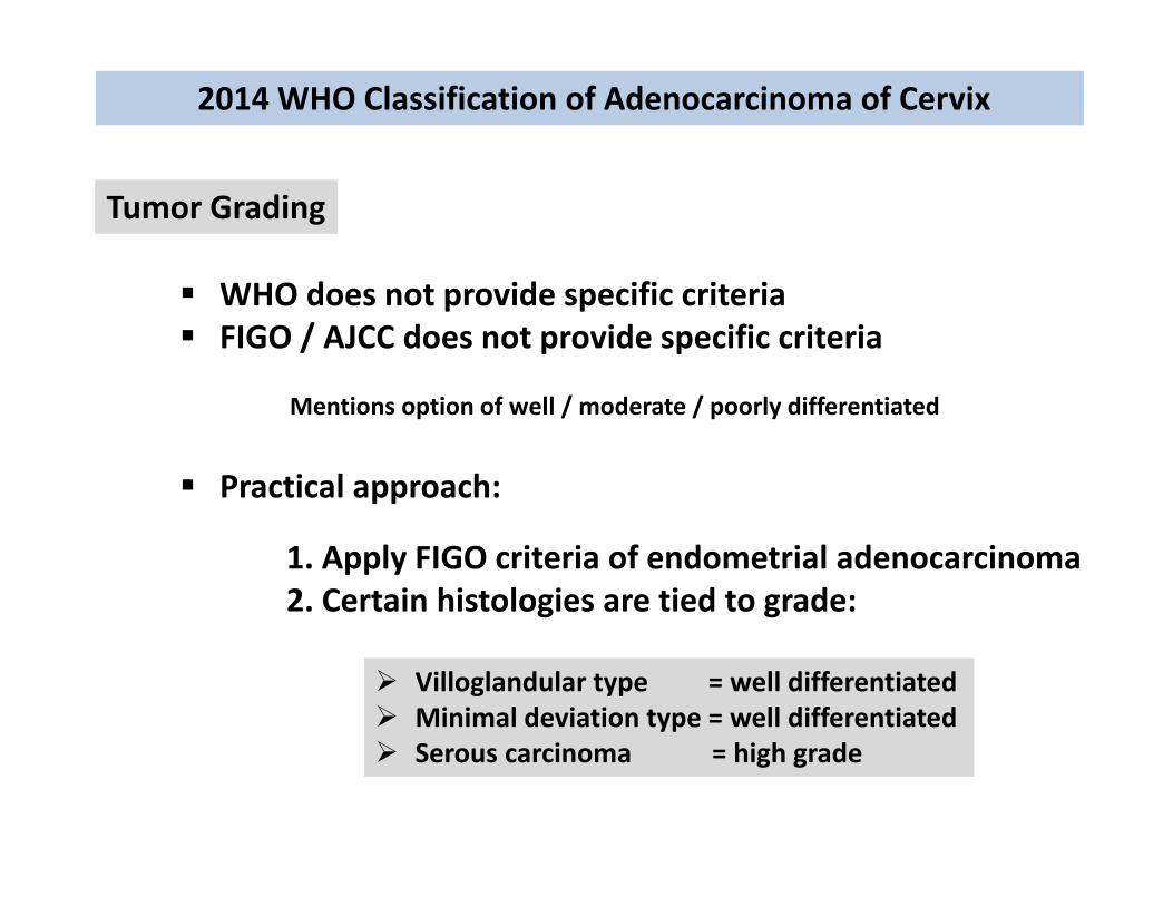
2014 WHO Classification of Adenocarcinoma of Cervix
Tumor Grading
WHO does not provide specific criteria FIGO / AJCC does not provide specific criteria
Mentions option of well / moderate / poorly differentiated
Practical approach:
1. Apply FIGO criteria of endometrial adenocarcinoma2. Certain histologies are tied to grade:
Villoglandular type = well differentiated Minimal deviation type = well differentiated Serous carcinoma = high grade
2014 WHO Classification of Adenocarcinoma of Cervix
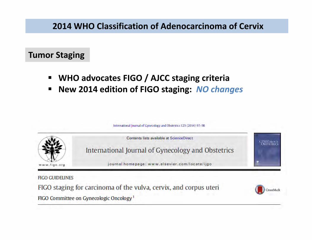
Tumor Staging
WHO advocates FIGO / AJCC staging criteria New 2014 edition of FIGO staging: NO changes
2014 WHO Classification of Adenocarcinoma of Cervix
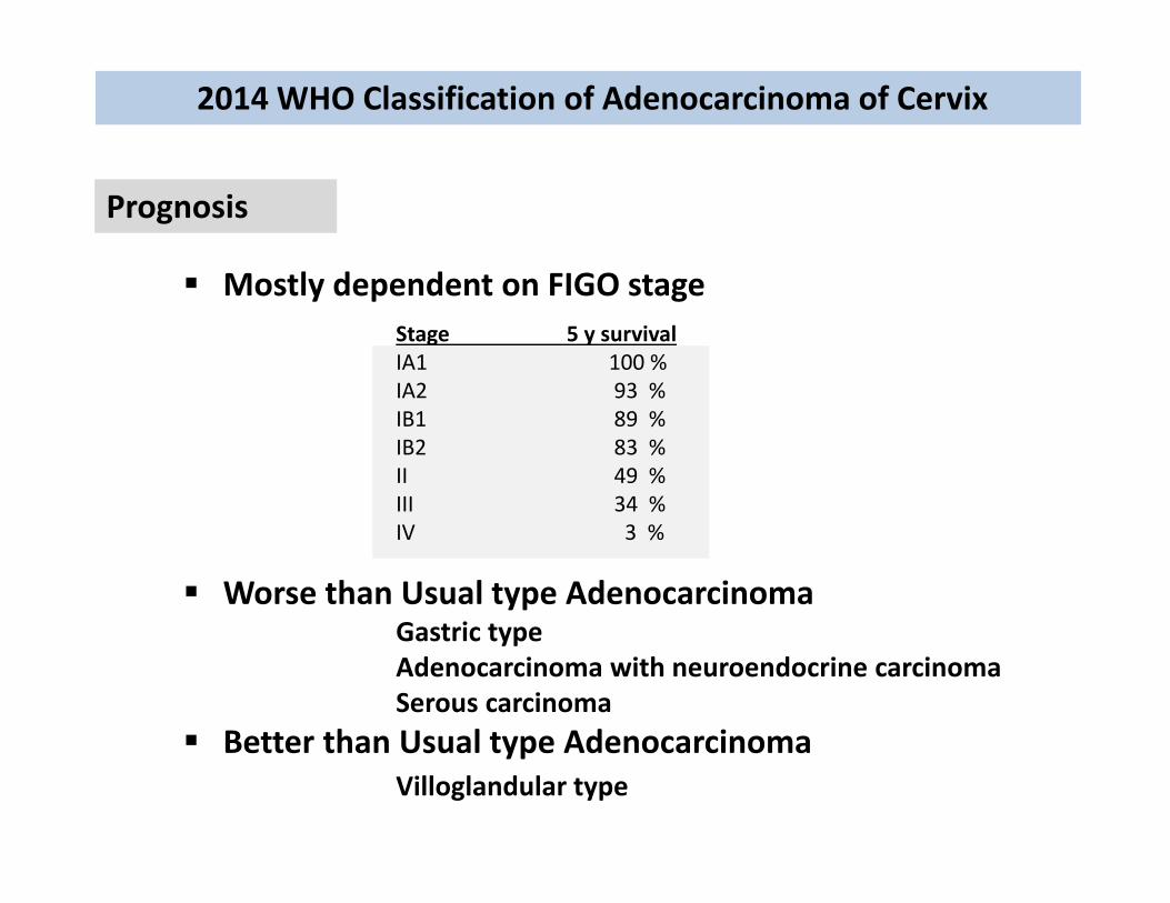
Prognosis
Mostly dependent on FIGO stageStage 5 y survivalIA1 100 %IA2 93 %IB1 89 %IB2 83 %II 49 %III 34 %IV 3 %
Worse than Usual type AdenocarcinomaGastric typeAdenocarcinoma with neuroendocrine carcinomaSerous carcinoma
Better than Usual type AdenocarcinomaVilloglandular type
Outline of Talk
Treatment Decisions for Endocervical Adenocarcinoma
New 2014 WHO Classification system
Update on Mucinous Adenocarcinoma variants
Common Problems in Usual type Endocervical Adenocarcinoma
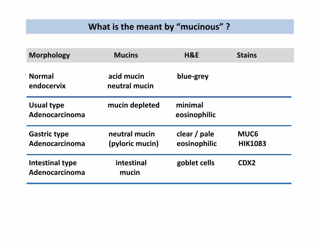
What is the meant by “mucinous” ?
Morphology Mucins H&E Stains
Normal acid mucin blue‐grey endocervix neutral mucin
Usual type mucin depleted minimal Adenocarcinoma eosinophilic
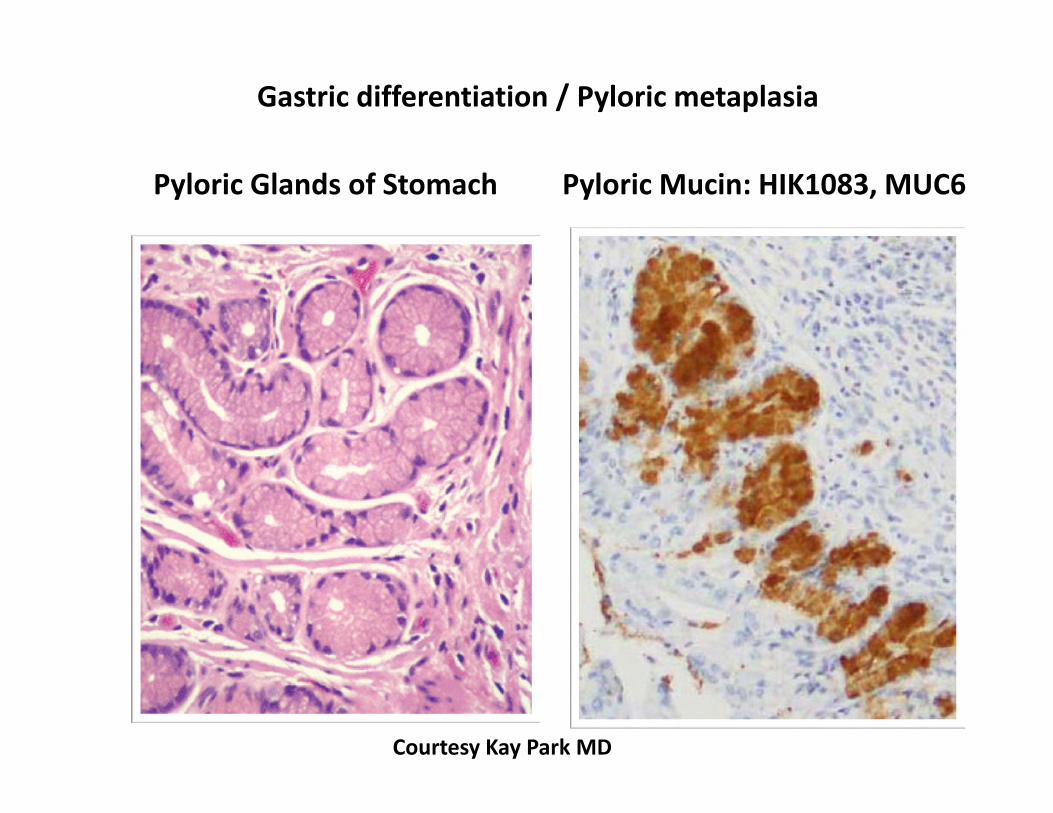
Gastric type neutral mucin clear / pale MUC6Adenocarcinoma (pyloric mucin) eosinophilic HIK1083
Intestinal type intestinal goblet cells CDX2Adenocarcinoma mucin
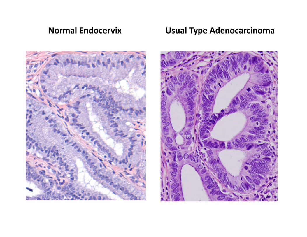
Normal Endocervix Usual Type Adenocarcinoma
Courtesy Kay Park MD
Pyloric Glands of Stomach Pyloric Mucin: HIK1083, MUC6
Gastric differentiation / Pyloric metaplasia
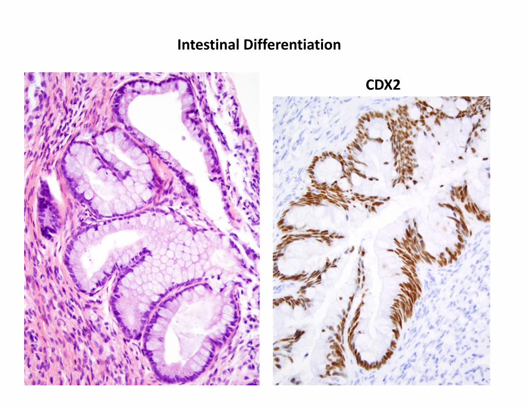
Intestinal Differentiation
CDX2
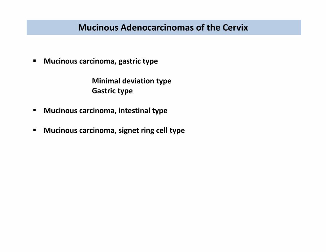
Mucinous Adenocarcinomas of the Cervix
Mucinous carcinoma, gastric type
Minimal deviation typeGastric type
Mucinous carcinoma, intestinal type
Mucinous carcinoma, signet ring cell type
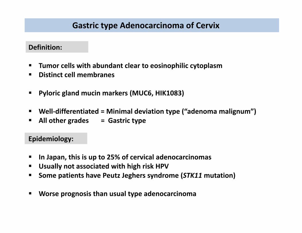
Gastric type Adenocarcinoma of Cervix
Definition:
Tumor cells with abundant clear to eosinophilic cytoplasm Distinct cell membranes
Pyloric gland mucin markers (MUC6, HIK1083)
Well‐differentiated = Minimal deviation type (“adenoma malignum”) All other grades = Gastric type
Epidemiology:
In Japan, this is up to 25% of cervical adenocarcinomas Usually not associated with high risk HPV Some patients have Peutz Jeghers syndrome (STK11mutation)
Worse prognosis than usual type adenocarcinoma
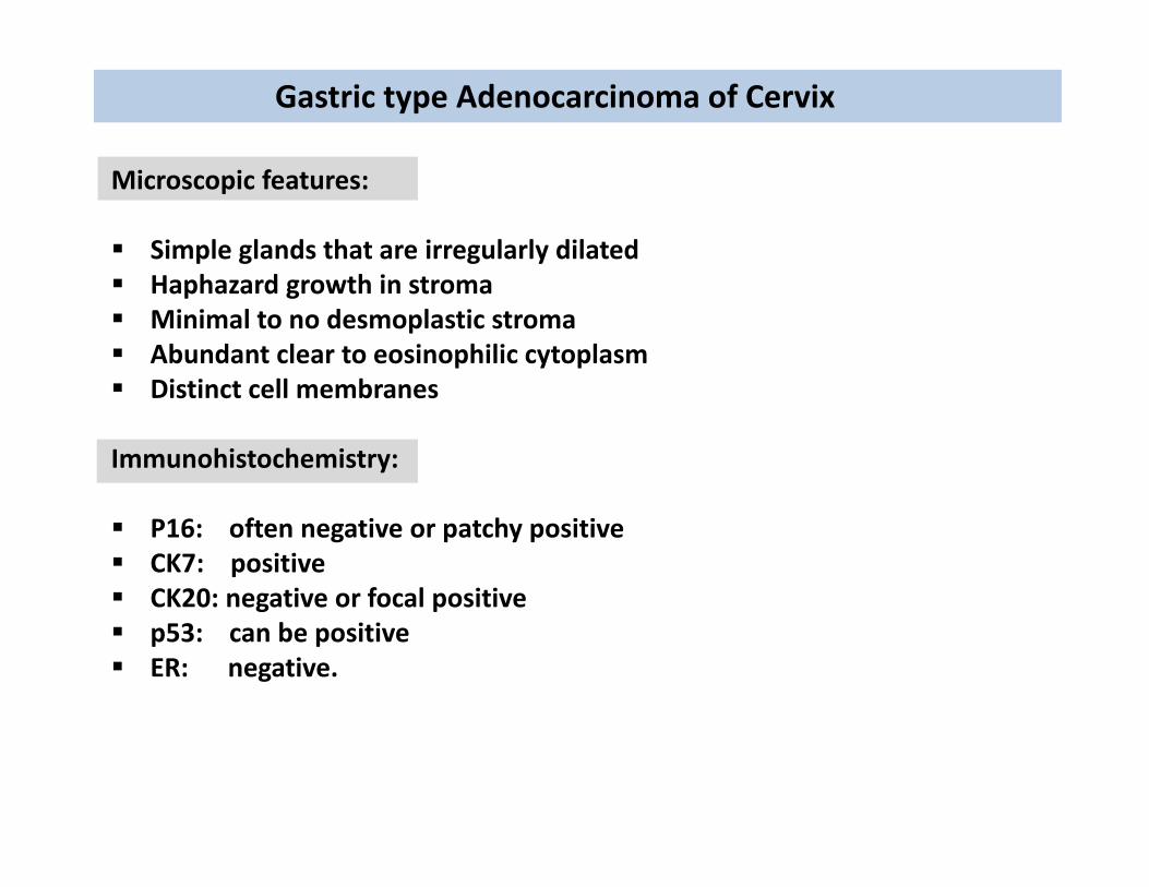
Gastric type Adenocarcinoma of Cervix
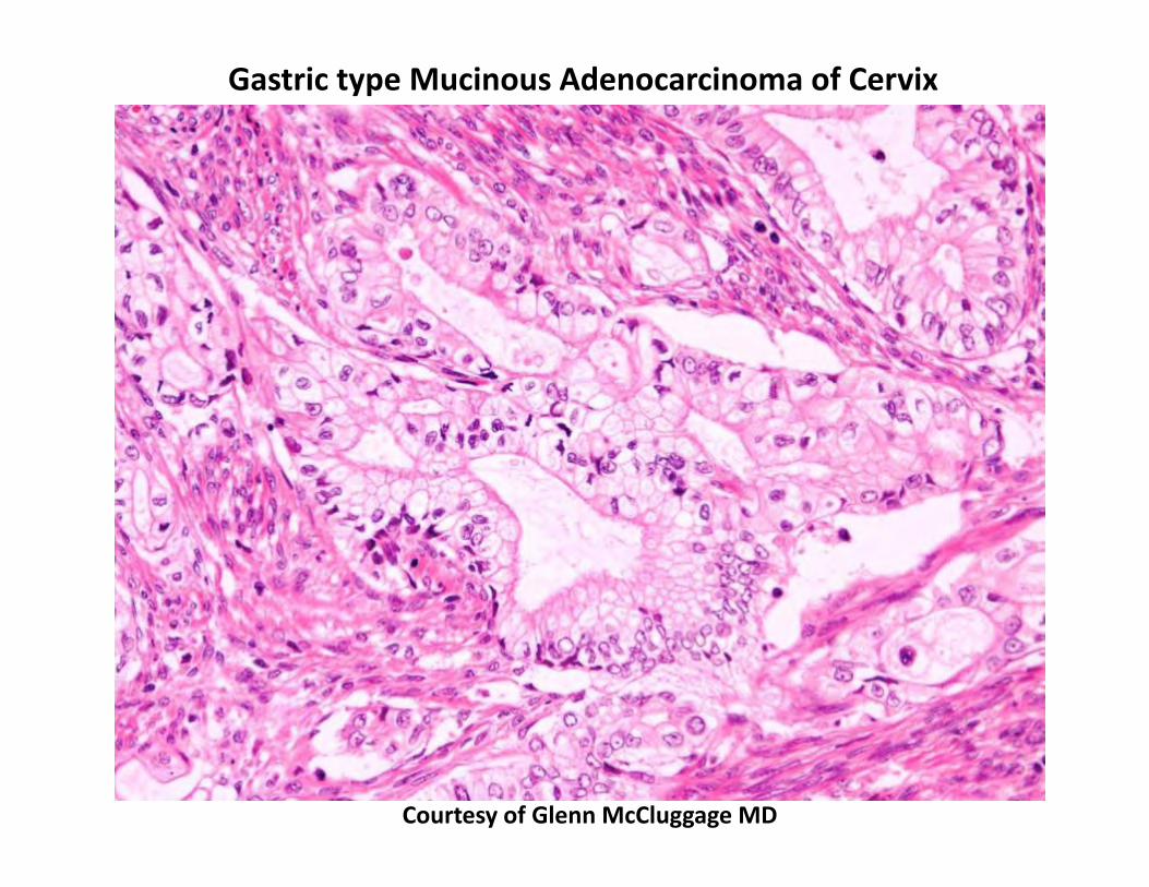
Microscopic features:
Simple glands that are irregularly dilated Haphazard growth in stroma Minimal to no desmoplastic stroma Abundant clear to eosinophilic cytoplasm Distinct cell membranes
Immunohistochemistry:
P16: often negative or patchy positive CK7: positive CK20: negative or focal positive p53: can be positive ER: negative.
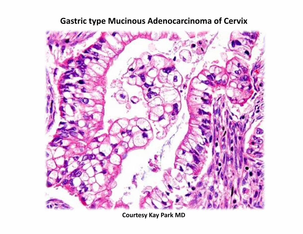
Courtesy Kay Park MD
Gastric type Mucinous Adenocarcinoma of Cervix
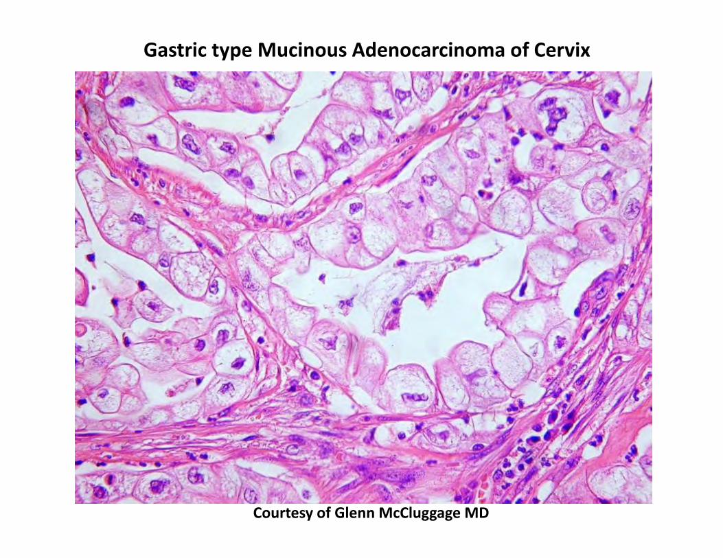
Courtesy of Glenn McCluggage MD
Gastric type Mucinous Adenocarcinoma of Cervix
Courtesy of Glenn McCluggage MD
Gastric type Mucinous Adenocarcinoma of Cervix
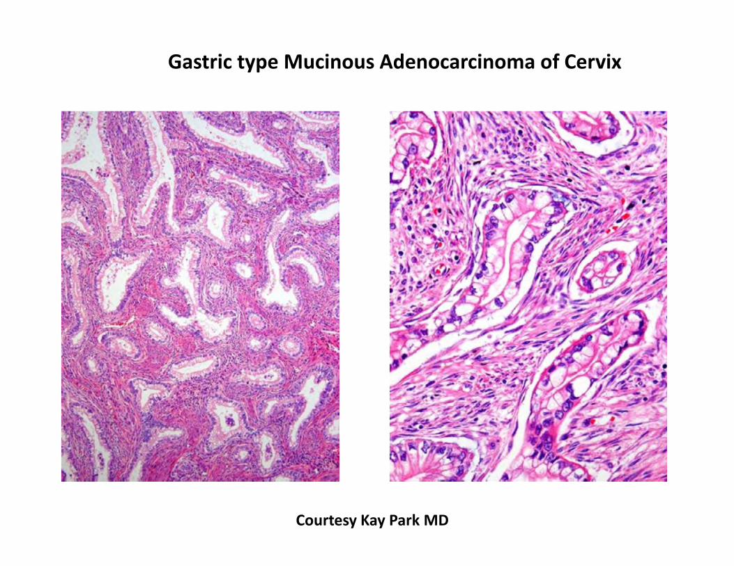
Gastric type Mucinous Adenocarcinoma of Cervix
Courtesy Kay Park MD
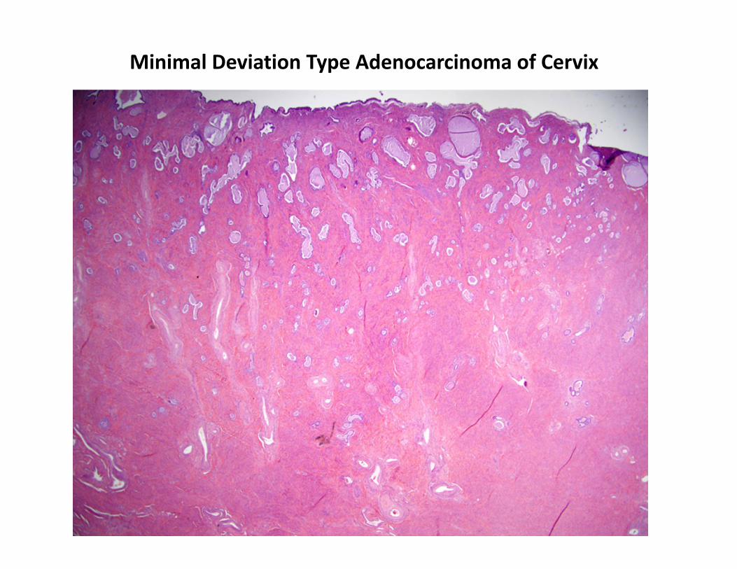
Minimal Deviation Type Adenocarcinoma of Cervix
Minimal Deviation Type Adenocarcinoma of Cervix
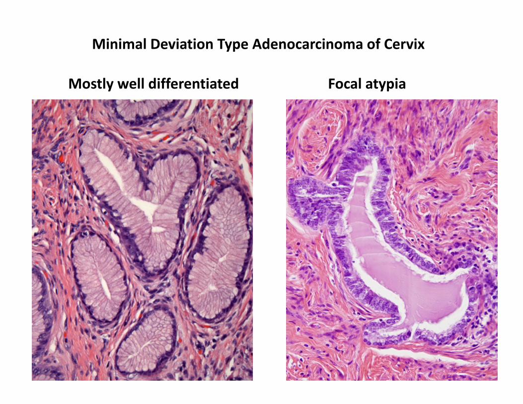
Minimal Deviation Type Adenocarcinoma of Cervix
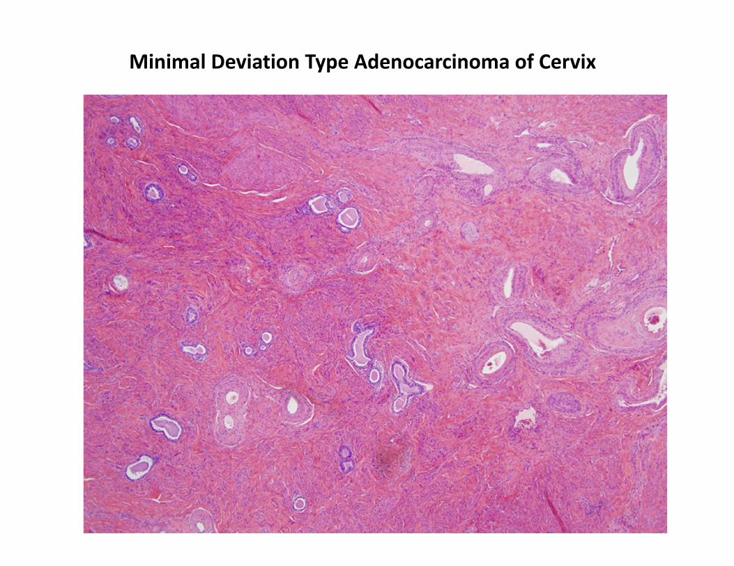
Mostly well differentiated Focal atypia
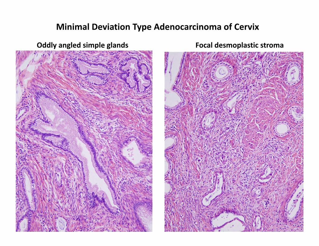
Minimal Deviation Type Adenocarcinoma of Cervix
Oddly angled simple glands Focal desmoplastic stroma
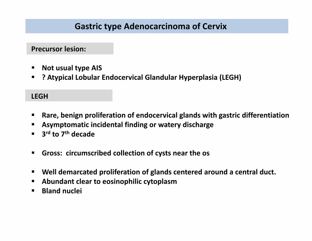
Gastric type Adenocarcinoma of Cervix
Precursor lesion:
Not usual type AIS ? Atypical Lobular Endocervical Glandular Hyperplasia (LEGH)
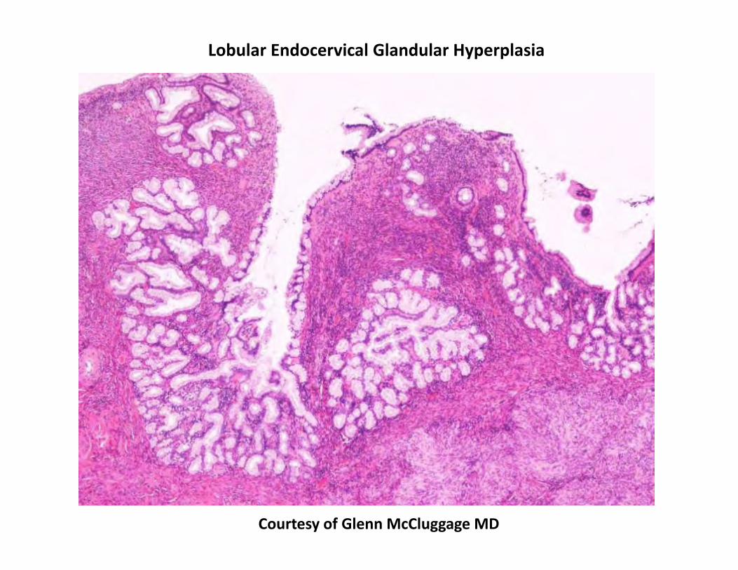
LEGH
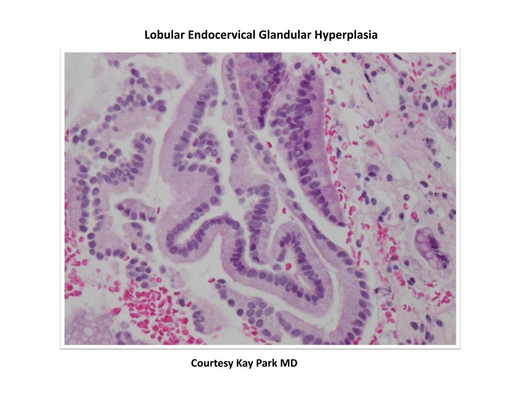
Rare, benign proliferation of endocervical glands with gastric differentiation Asymptomatic incidental finding or watery discharge 3rd to 7th decade
Gross: circumscribed collection of cysts near the os
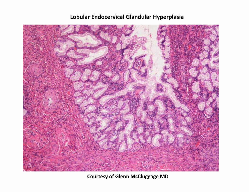
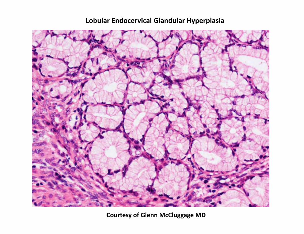
Well demarcated proliferation of glands centered around a central duct. Abundant clear to eosinophilic cytoplasm Bland nuclei
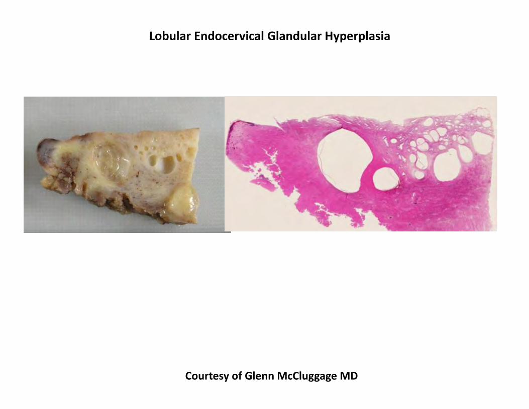
Courtesy of Glenn McCluggage MD
Lobular Endocervical Glandular Hyperplasia
Lobular Endocervical Glandular Hyperplasia
Courtesy of Glenn McCluggage MD
Lobular Endocervical Glandular Hyperplasia
Courtesy of Glenn McCluggage MD
Lobular Endocervical Glandular Hyperplasia
Courtesy of Glenn McCluggage MD
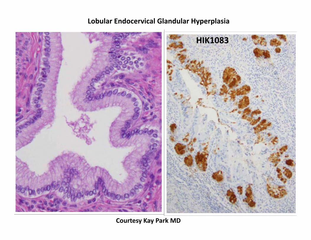
Courtesy Kay Park MD
Lobular Endocervical Glandular Hyperplasia
HIK1083
Courtesy Kay Park MD
Lobular Endocervical Glandular Hyperplasia
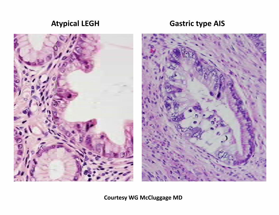
Atypical LEGH Gastric type AIS
Courtesy WG McCluggage MD
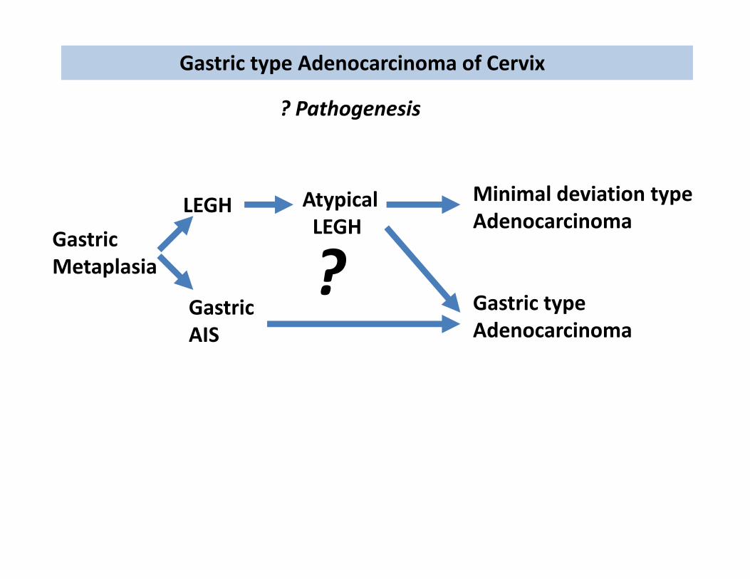
Gastric type Adenocarcinoma of Cervix
AtypicalLEGH
Minimal deviation typeAdenocarcinoma
Gastric typeAdenocarcinoma
LEGH
? Pathogenesis
GastricMetaplasia
GastricAIS
?
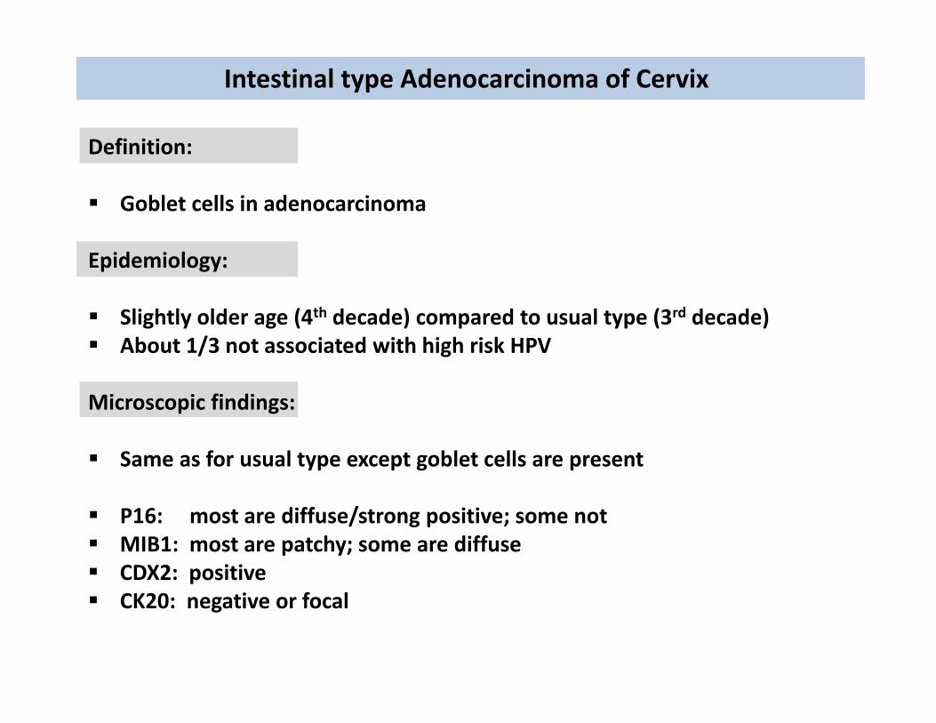
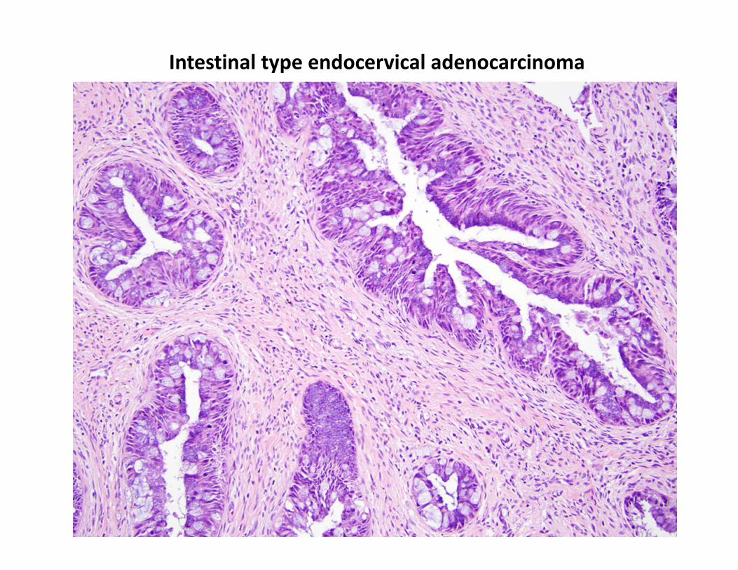
Intestinal type Adenocarcinoma of Cervix
Definition:
Goblet cells in adenocarcinoma
Epidemiology:
Slightly older age (4th decade) compared to usual type (3rd decade) About 1/3 not associated with high risk HPV
Microscopic findings:
Same as for usual type except goblet cells are present
P16: most are diffuse/strong positive; some not MIB1: most are patchy; some are diffuse CDX2: positive CK20: negative or focal
Intestinal type endocervical adenocarcinoma
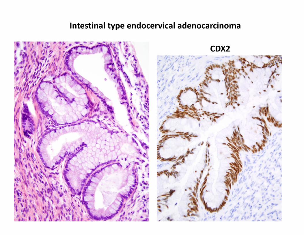
CDX2
Intestinal type endocervical adenocarcinoma
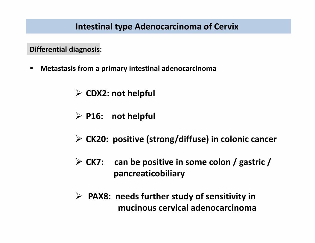
Intestinal type Adenocarcinoma of Cervix
Differential diagnosis:
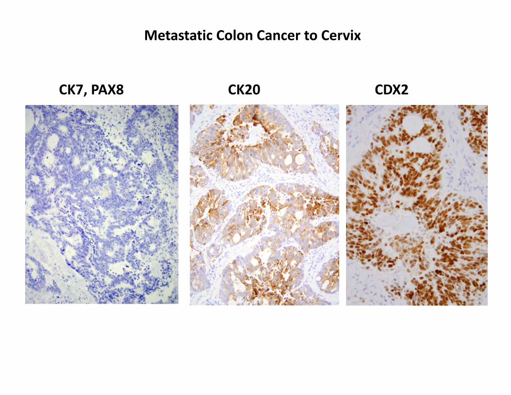
Metastasis from a primary intestinal adenocarcinoma
CDX2: not helpful
P16: not helpful
CK20: positive (strong/diffuse) in colonic cancer
CK7: can be positive in some colon / gastric / pancreaticobiliary
PAX8: needs further study of sensitivity in mucinous cervical adenocarcinoma
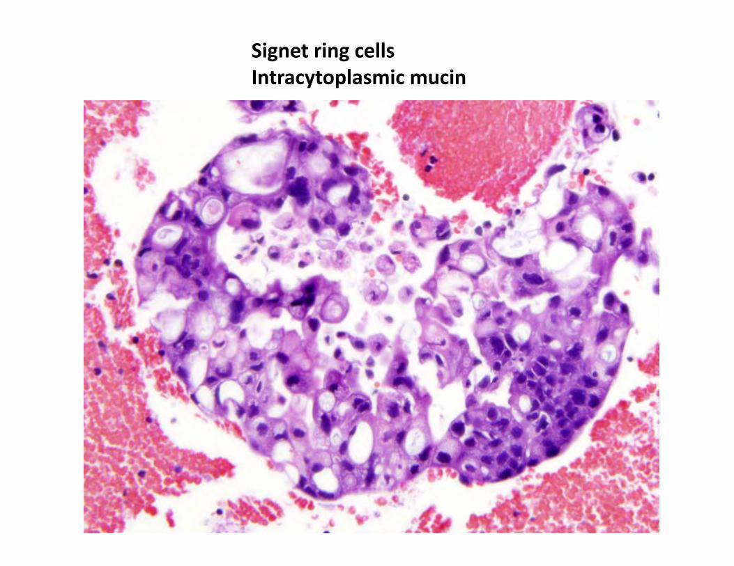
Signet ring cellsIntracytoplasmic mucin
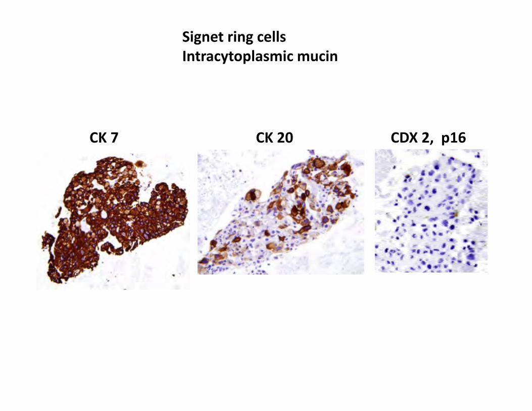
Signet ring cellsIntracytoplasmic mucin
CK 7 CK 20 CDX 2, p16
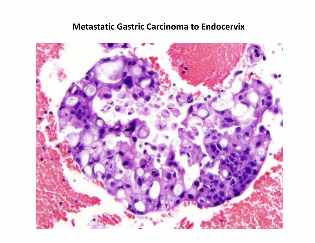
Metastatic Gastric Carcinoma to Endocervix
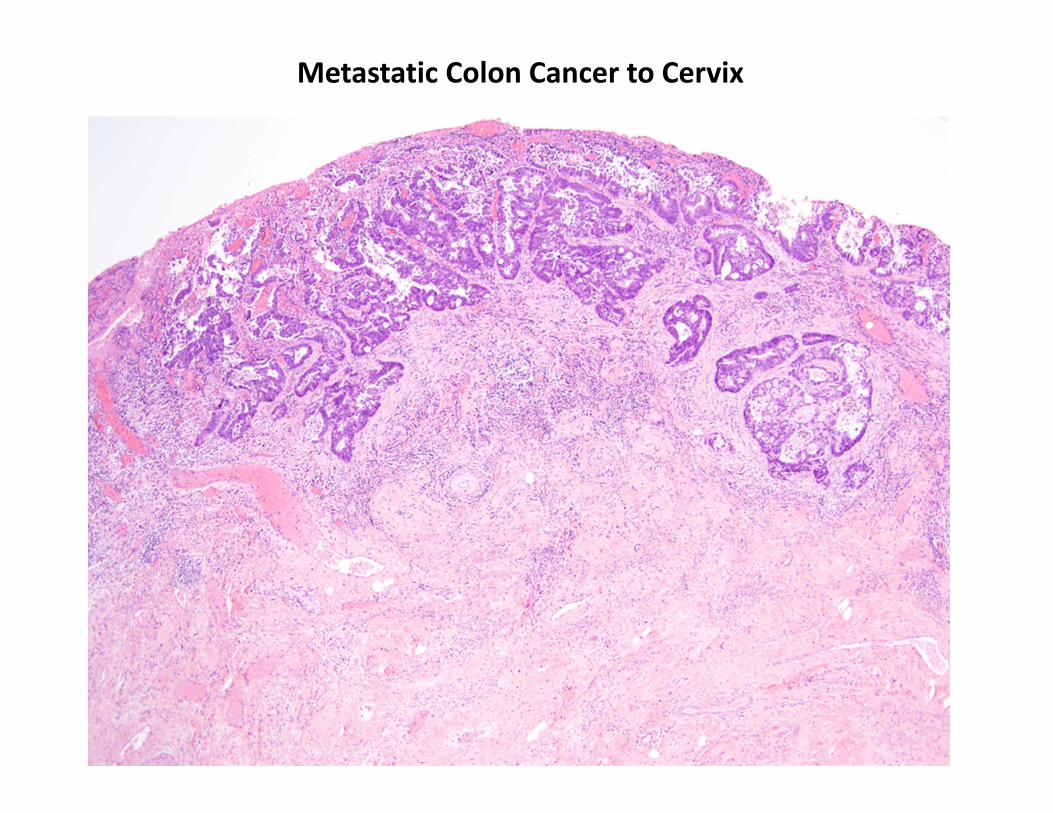
Metastatic Colon Cancer to Cervix
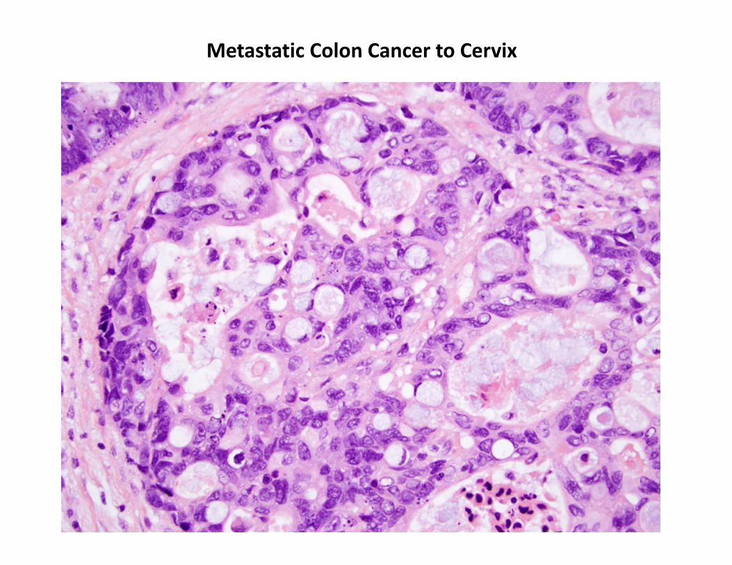
Metastatic Colon Cancer to Cervix
Metastatic Colon Cancer to Cervix
CK7, PAX8 CK20 CDX2
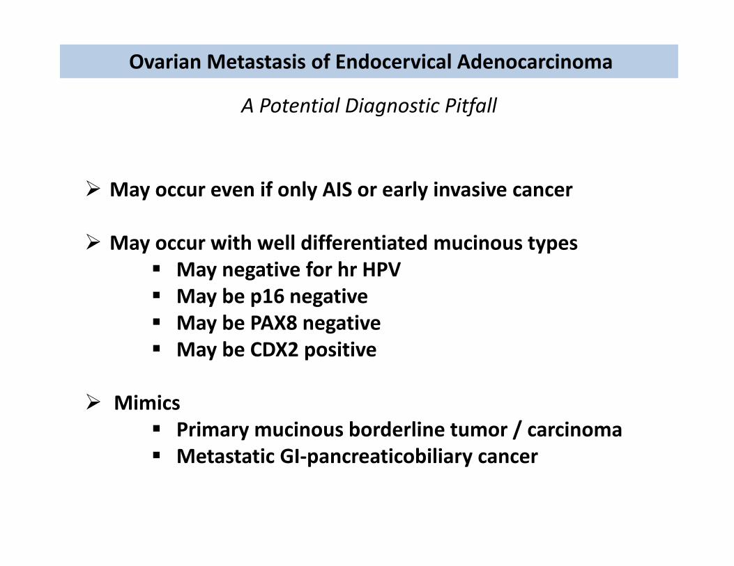
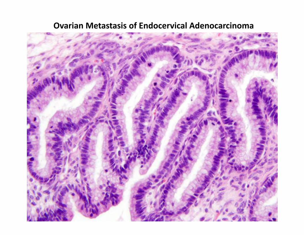
Ovarian Metastasis of Endocervical Adenocarcinoma
A Potential Diagnostic Pitfall
May occur even if only AIS or early invasive cancer
May occur with well differentiated mucinous types May negative for hr HPV May be p16 negative May be PAX8 negative May be CDX2 positive
Mimics Primary mucinous borderline tumor / carcinoma Metastatic GI‐pancreaticobiliary cancer
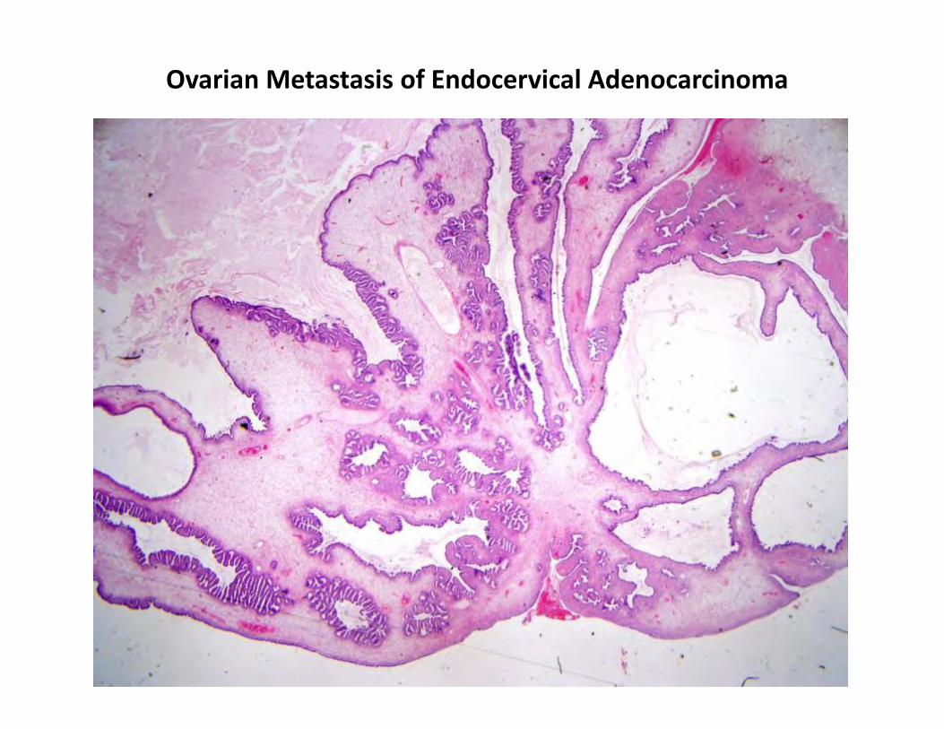
Ovarian Metastasis of Endocervical Adenocarcinoma
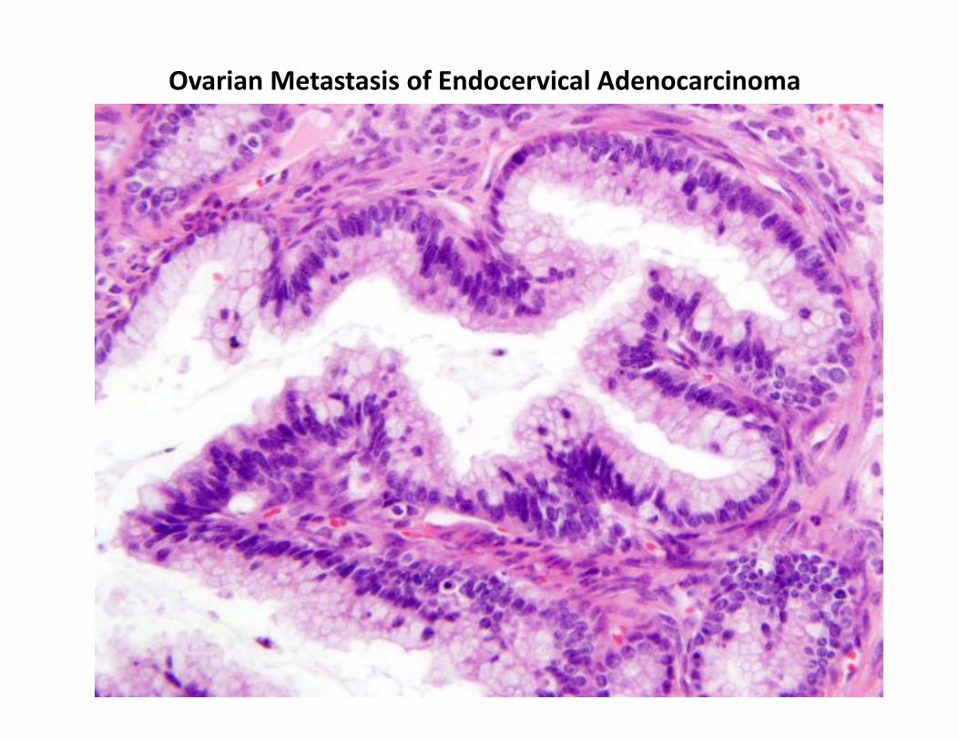
Ovarian Metastasis of Endocervical Adenocarcinoma
Ovarian Metastasis of Endocervical Adenocarcinoma
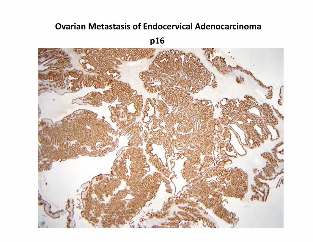
Ovarian Metastasis of Endocervical Adenocarcinomap16
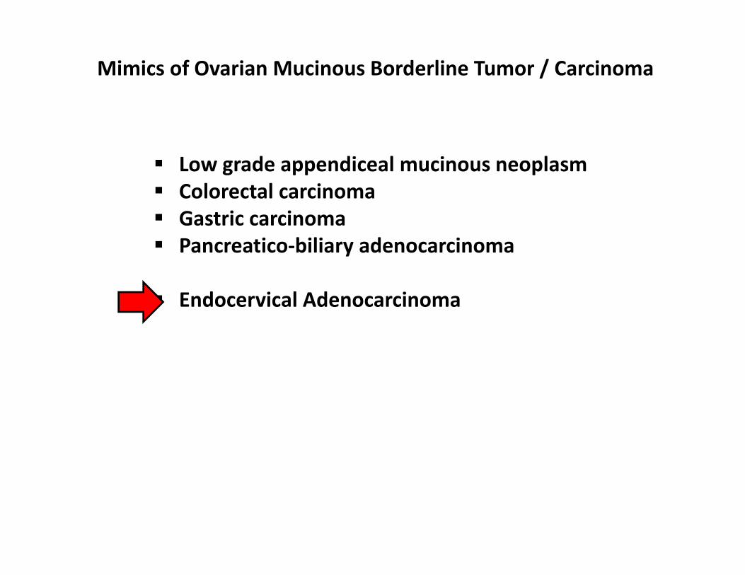
Mimics of Ovarian Mucinous Borderline Tumor / Carcinoma
Low grade appendiceal mucinous neoplasm Colorectal carcinoma Gastric carcinoma Pancreatico‐biliary adenocarcinoma
Endocervical Adenocarcinoma
Outline of Talk
Treatment Decisions for Endocervical Adenocarcinoma
New 2014 WHO Classification system
Update on Mucinous Adenocarcinoma variants
Common Problems in Usual type Endocervical Adenocarcinoma
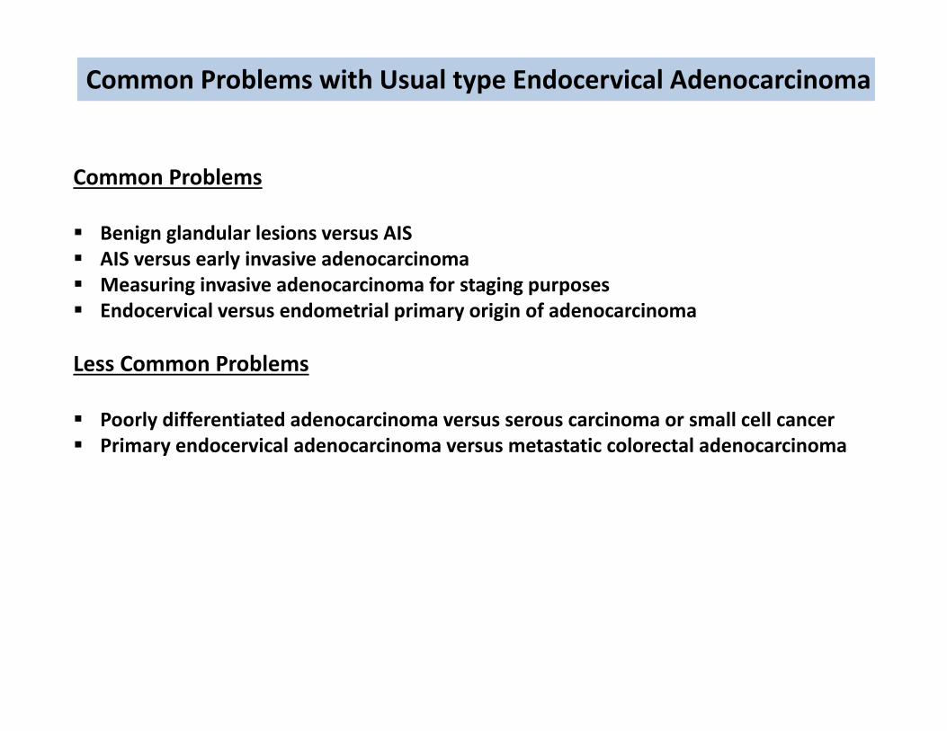
Common Problems with Usual type Endocervical Adenocarcinoma
Common Problems
Benign glandular lesions versus AIS AIS versus early invasive adenocarcinoma Measuring invasive adenocarcinoma for staging purposes Endocervical versus endometrial primary origin of adenocarcinoma
Less Common Problems
Poorly differentiated adenocarcinoma versus serous carcinoma or small cell cancer Primary endocervical adenocarcinoma versus metastatic colorectal adenocarcinoma
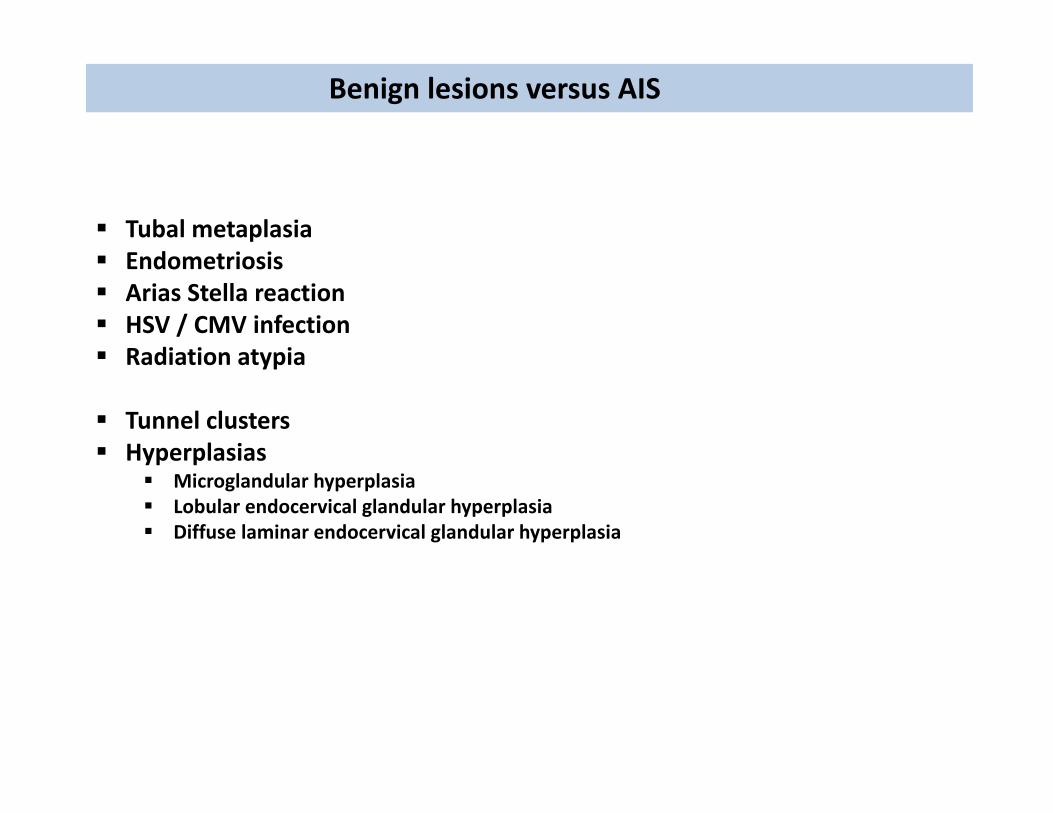
Benign lesions versus AIS
Tubal metaplasia Endometriosis Arias Stella reaction HSV / CMV infection Radiation atypia
Tunnel clusters Hyperplasias
Microglandular hyperplasia Lobular endocervical glandular hyperplasia Diffuse laminar endocervical glandular hyperplasia
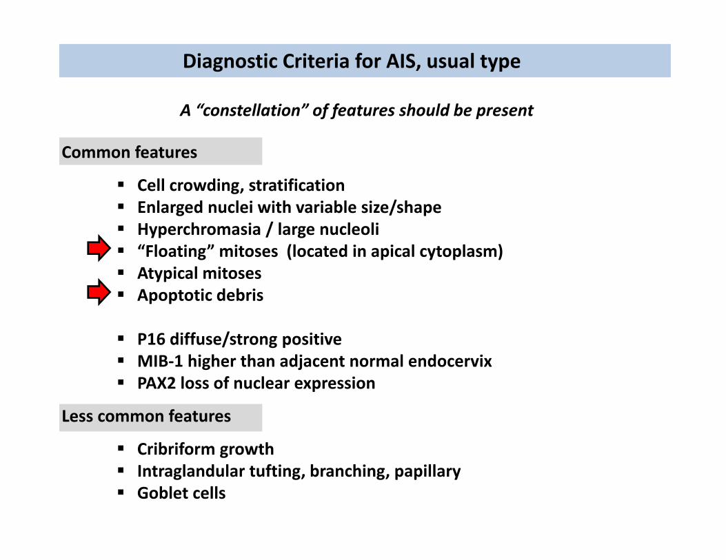
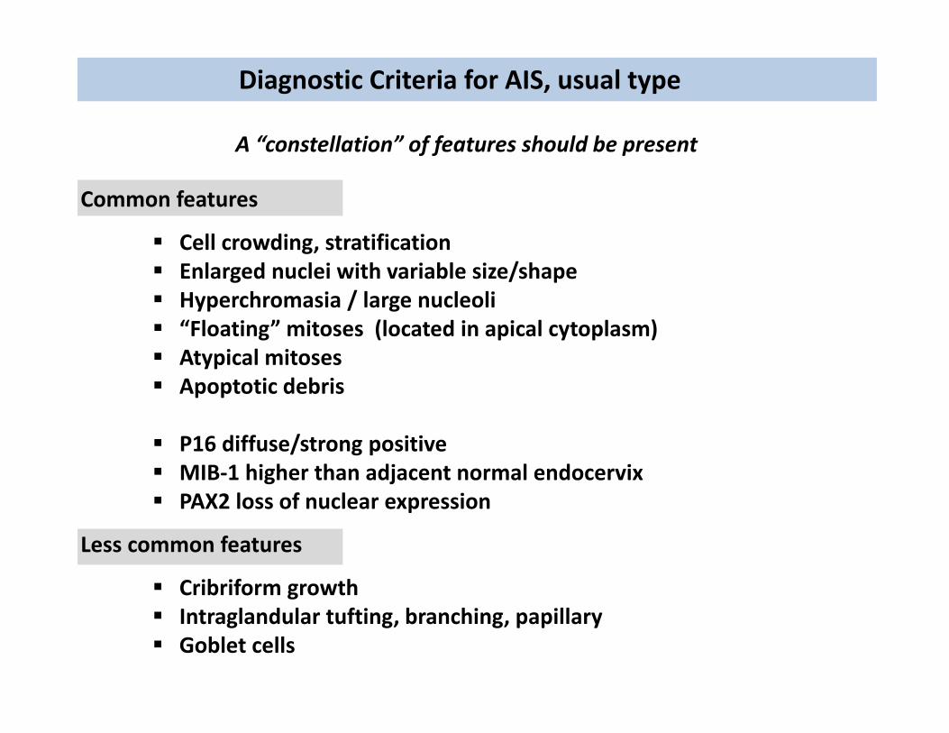
Diagnostic Criteria for AIS, usual type
Cell crowding, stratification Enlarged nuclei with variable size/shape Hyperchromasia / large nucleoli “Floating” mitoses (located in apical cytoplasm) Atypical mitoses Apoptotic debris
P16 diffuse/strong positive MIB‐1 higher than adjacent normal endocervix PAX2 loss of nuclear expression
Cribriform growth Intraglandular tufting, branching, papillary Goblet cells
A “constellation” of features should be present
Common features
Less common features
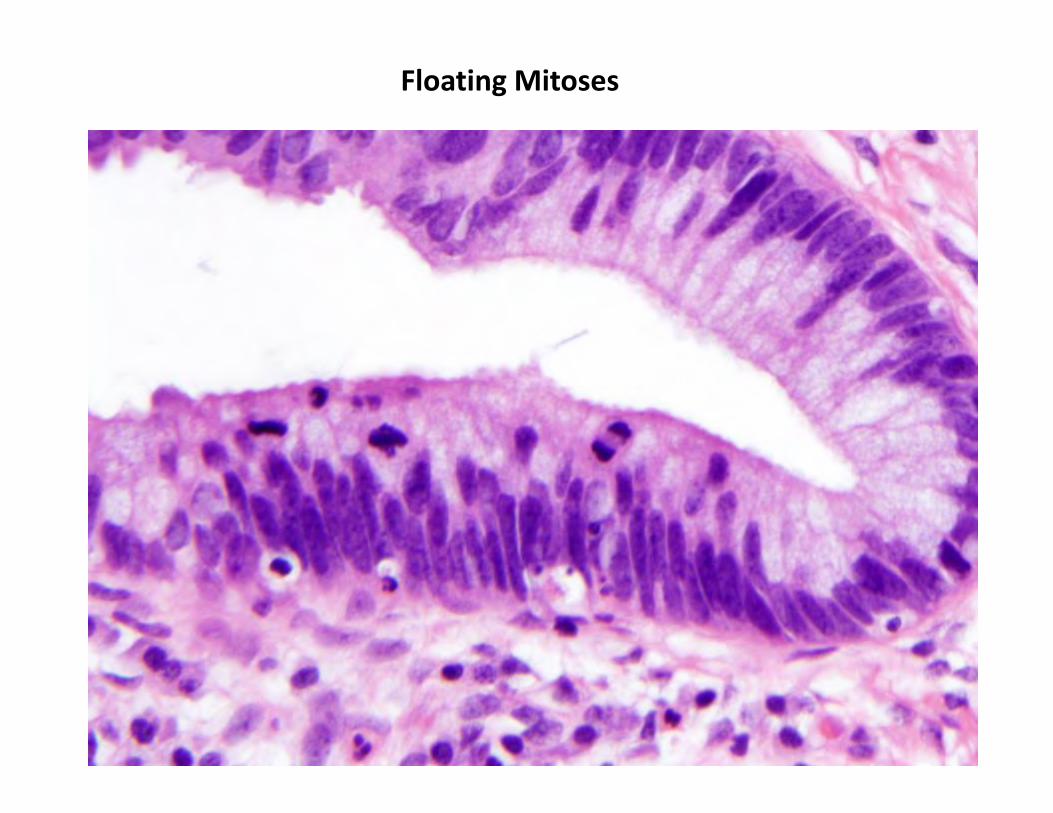
Floating Mitoses
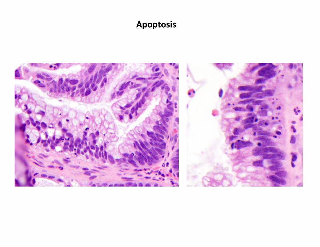
Apoptosis
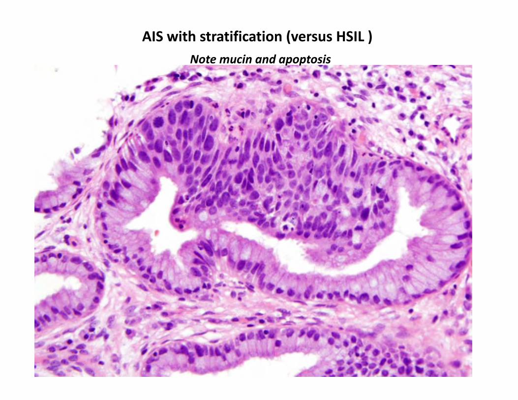
AIS with stratification (versus HSIL )Note mucin and apoptosis
Diagnostic Criteria for AIS, usual type
Are mitoses in endocervical glands pathognomonic of AIS ?
No.
Rare mitoses (in absence of other abnormalities) can be seen in:
Normal endocervix Endometriosis of cervix Hyperplasias
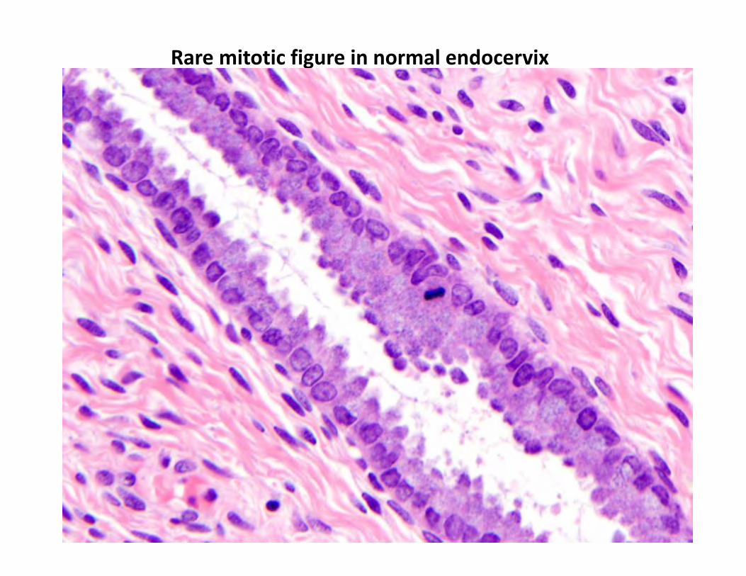
Rare mitotic figure in normal endocervix
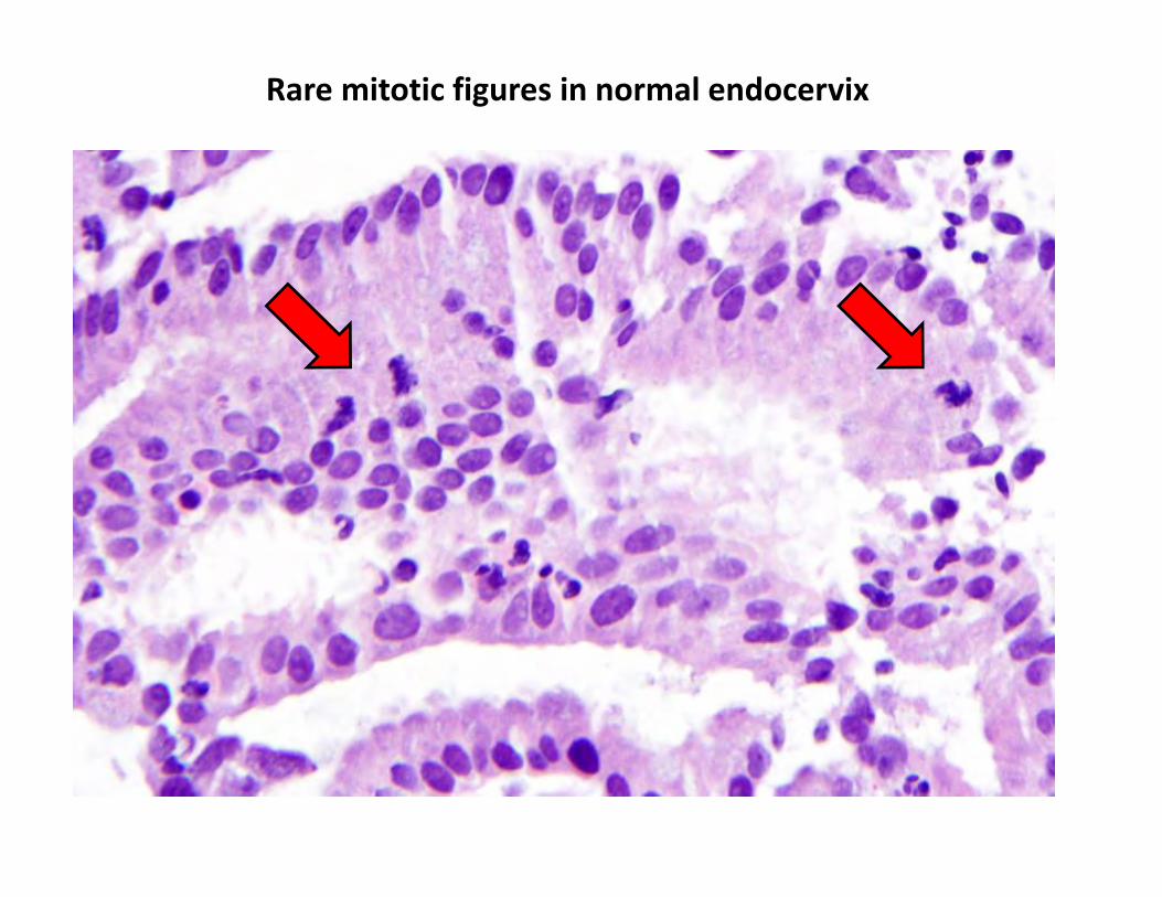
Rare mitotic figures in normal endocervix
Diagnostic Criteria for AIS, usual type
Are “atypical” nuclei in endocervical glands pathognomonic of AIS ?
No.
Abnormal nuclear size/shape (in absence of other abnormalities) can be seen in:
Radiation atypia HSV, CMV infection Arias Stella reaction Reactive inflammatory changes
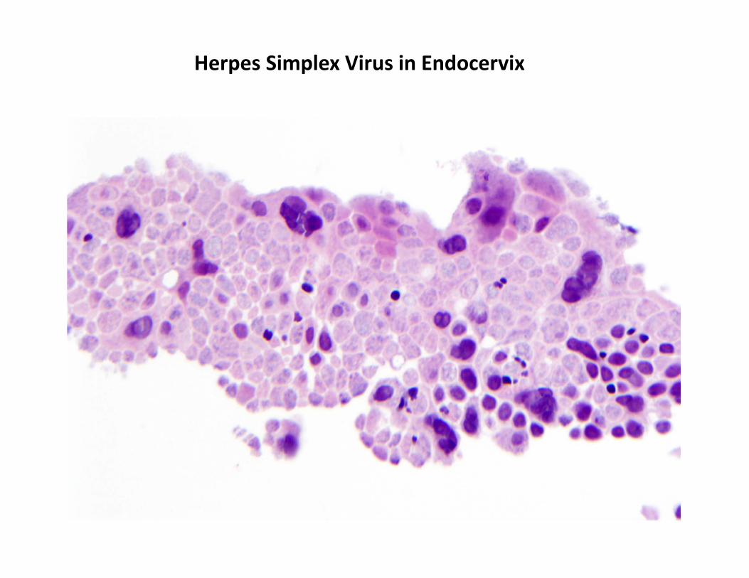
Herpes Simplex Virus in Endocervix
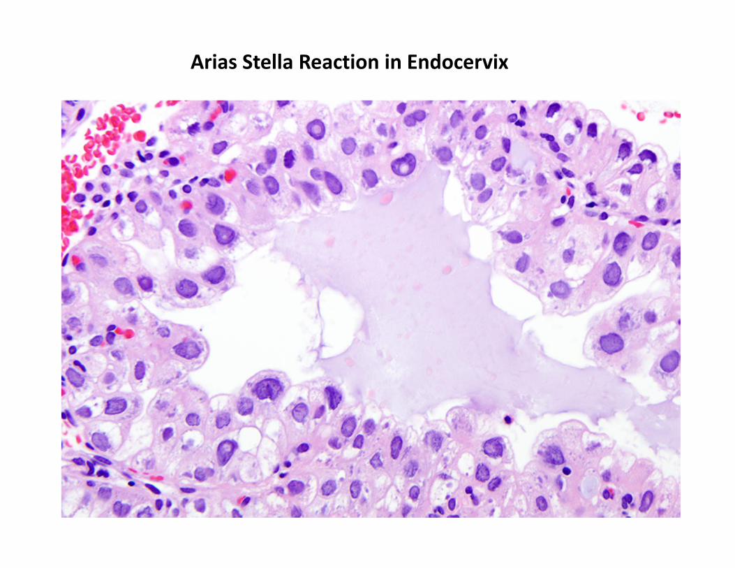
Arias Stella Reaction in Endocervix
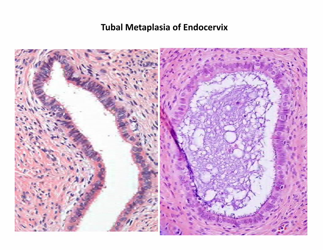
Tubal Metaplasia of Endocervix

Superficial Endometriosis of Cervix
Mitoses, crowding, stratification Endometrial stroma
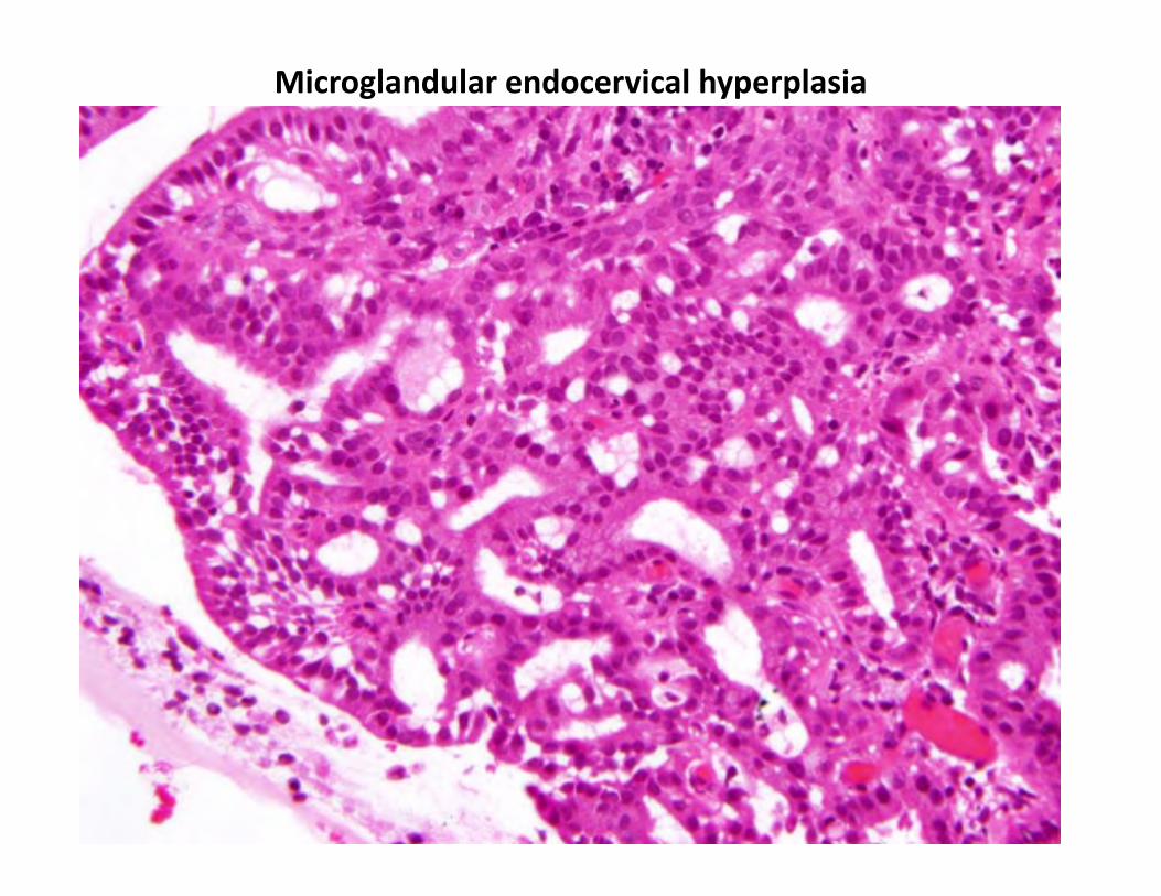
Microglandular endocervical hyperplasia
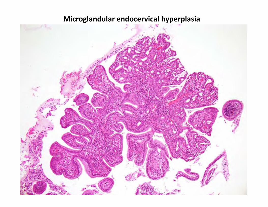
Microglandular endocervical hyperplasia
Diagnostic Criteria for AIS, usual type
Cell crowding, stratification Enlarged nuclei with variable size/shape Hyperchromasia / large nucleoli “Floating” mitoses (located in apical cytoplasm) Atypical mitoses Apoptotic debris
P16 diffuse/strong positive MIB‐1 higher than adjacent normal endocervix PAX2 loss of nuclear expression
Cribriform growth Intraglandular tufting, branching, papillary Goblet cells
A “constellation” of features should be present
Common features
Less common features
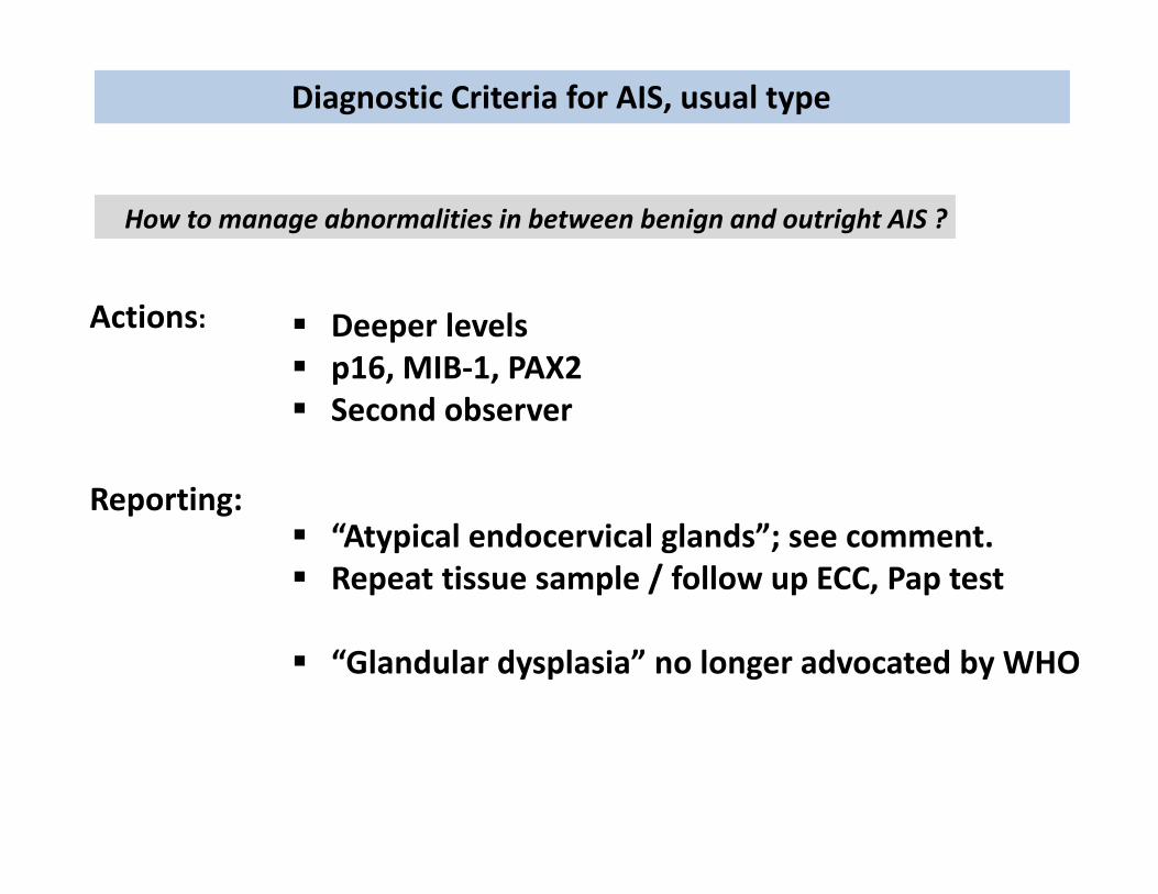
Diagnostic Criteria for AIS, usual type
How to manage abnormalities in between benign and outright AIS ?
Actions:
Reporting:
Deeper levels p16, MIB‐1, PAX2 Second observer
“Atypical endocervical glands”; see comment. Repeat tissue sample / follow up ECC, Pap test
“Glandular dysplasia” no longer advocated by WHO
Common Problems with Usual type Endocervical Adenocarcinoma
Benign glandular lesions versus AIS AIS versus early invasive adenocarcinoma Measuring invasive adenocarcinoma for staging purposes Endocervical versus endometrial primary origin of adenocarcinoma
Types of Invasion in Endocervical Adenocarcinoma
Destructive stromal invasion
Expansile invasion
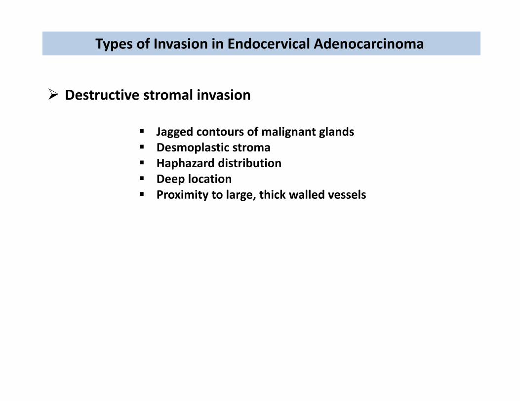
Types of Invasion in Endocervical Adenocarcinoma
Destructive stromal invasion
Jagged contours of malignant glands Desmoplastic stroma Haphazard distribution Deep location Proximity to large, thick walled vessels
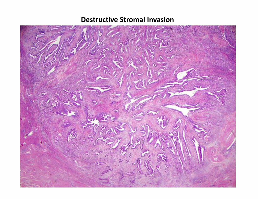
Destructive Stromal Invasion
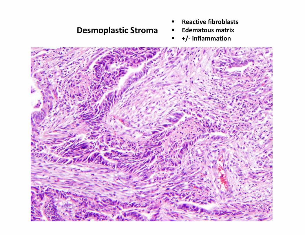
Desmoplastic Stroma Reactive fibroblasts Edematous matrix +/‐ inflammation
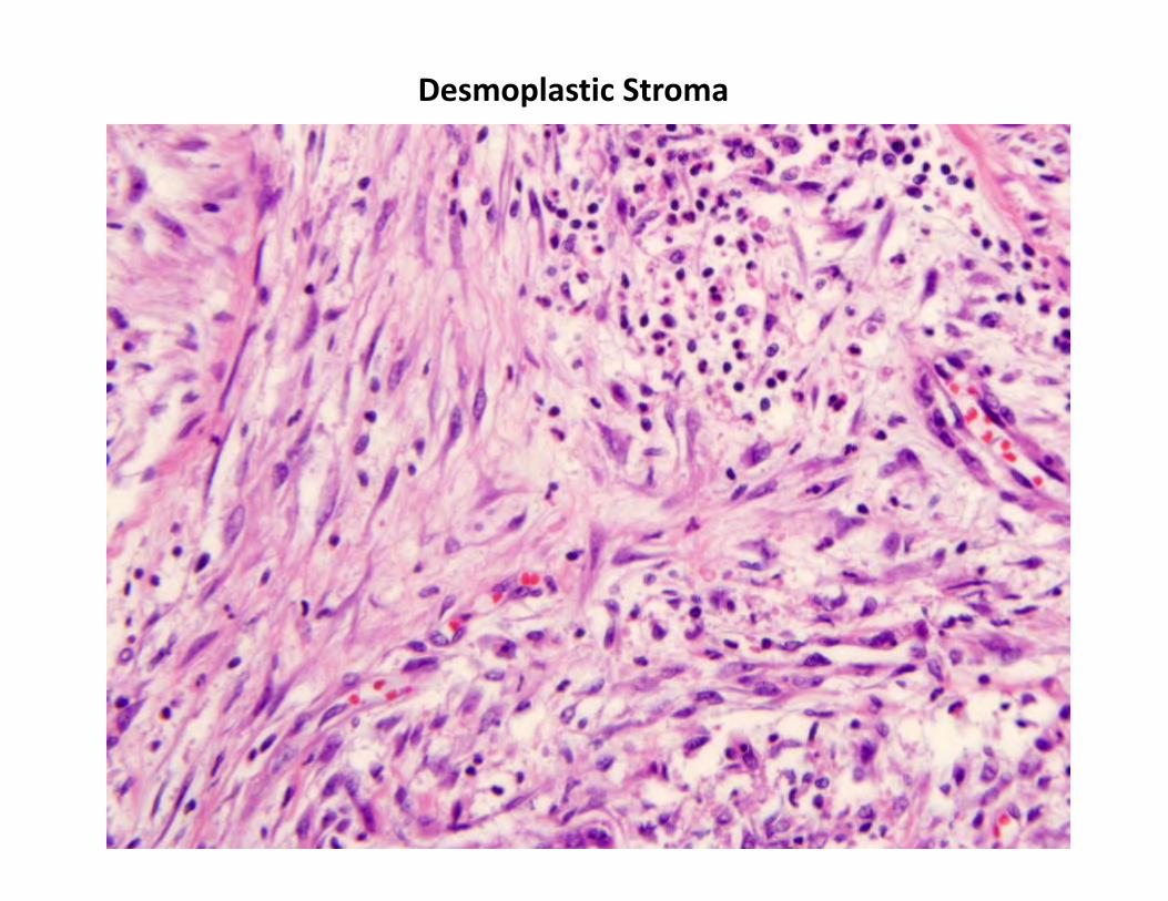
Desmoplastic Stroma
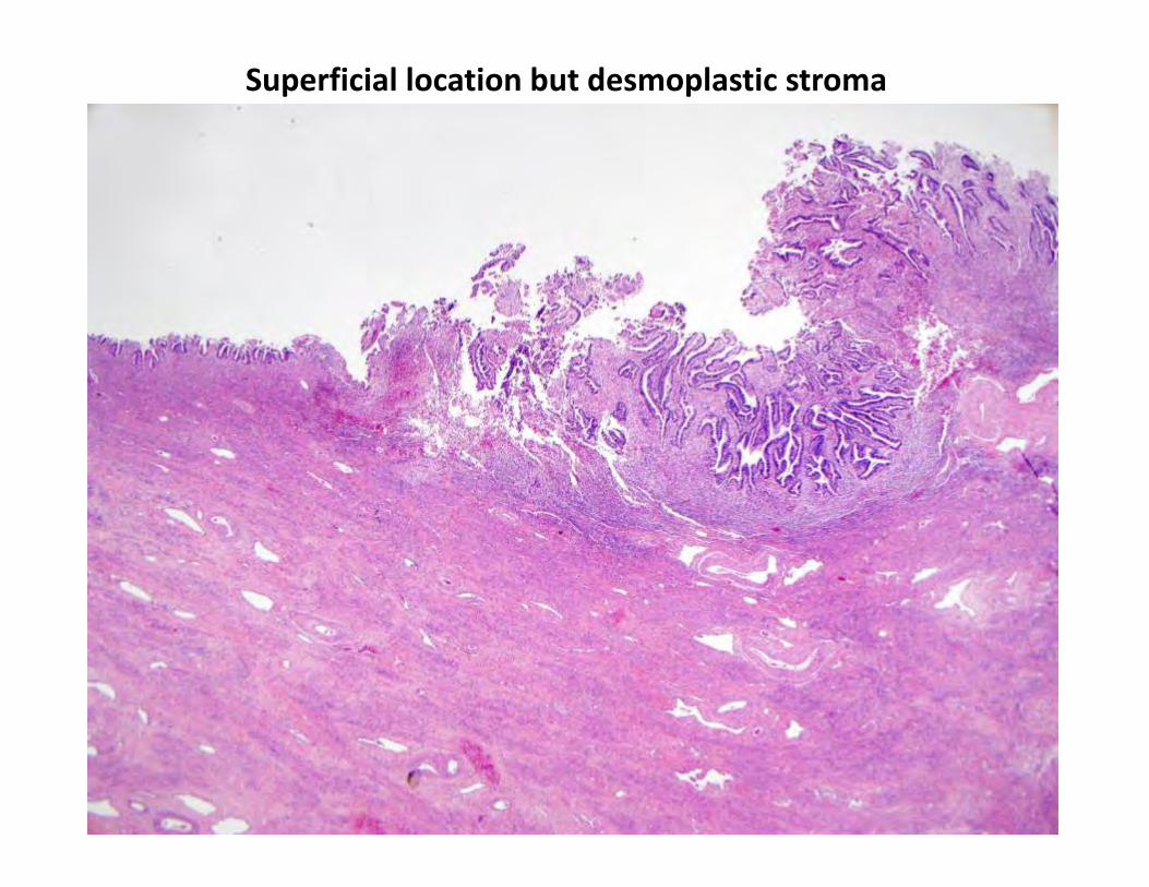
Superficial location but desmoplastic stroma
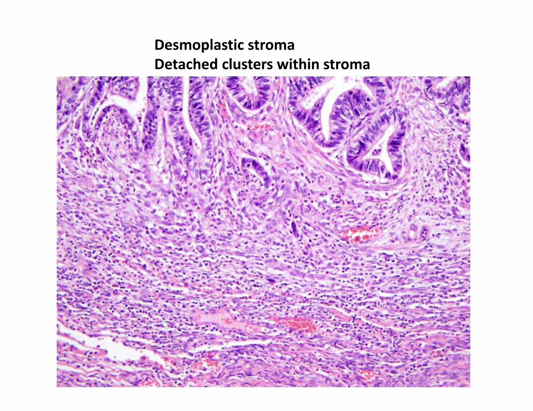
Desmoplastic stroma Detached clusters within stroma
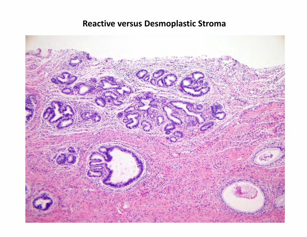
Reactive versus Desmoplastic Stroma
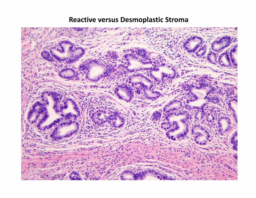
Reactive versus Desmoplastic Stroma
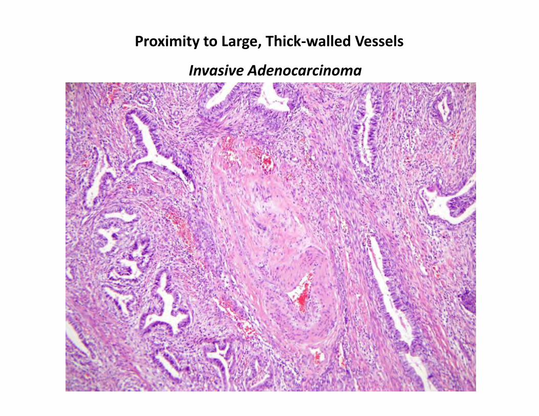
Proximity to Large, Thick‐walled Vessels
Invasive Adenocarcinoma
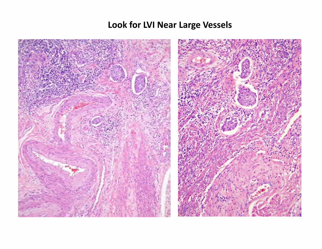
Look for LVI Near Large Vessels
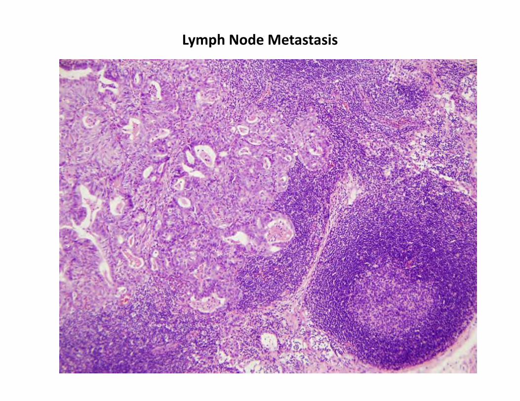
Lymph Node Metastasis
Types of Invasion in Endocervical Adenocarcinoma
Expansile invasion
WHO: Architecture too complex compared to normal endocervix
Proliferation of small malignant glands Complex tubulo‐glandular, papillary formation Pushing growth
Features of destructive stromal invasion may not be present
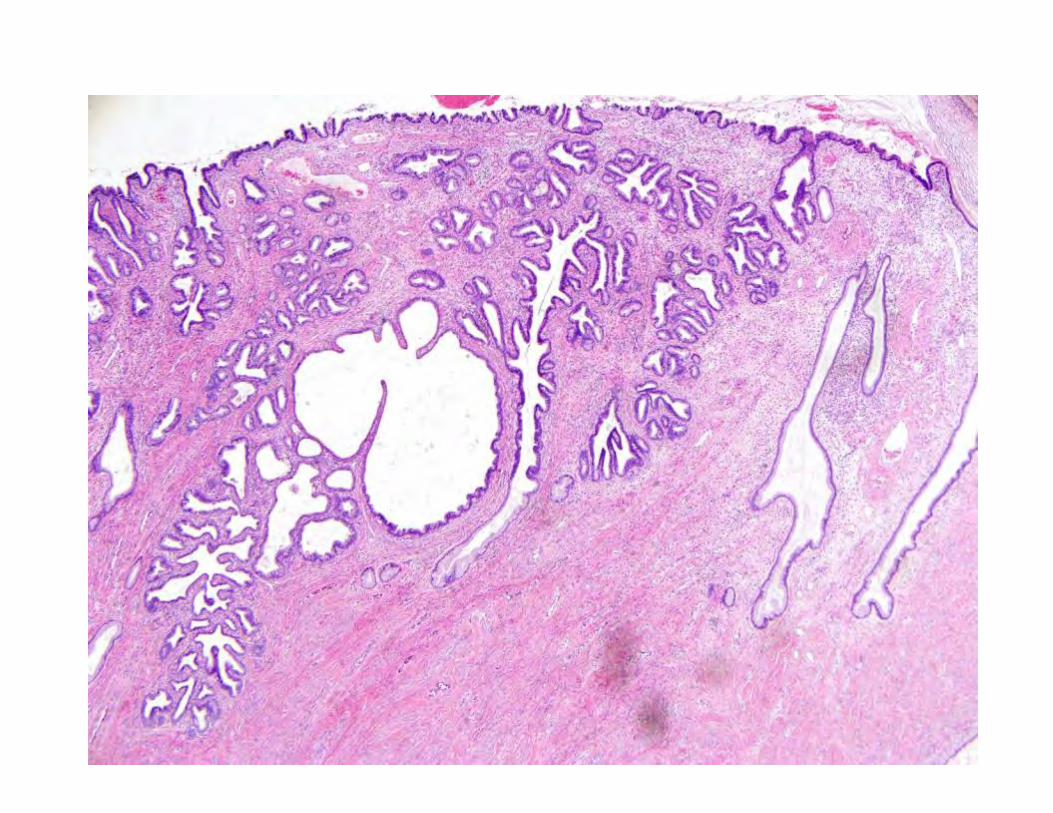
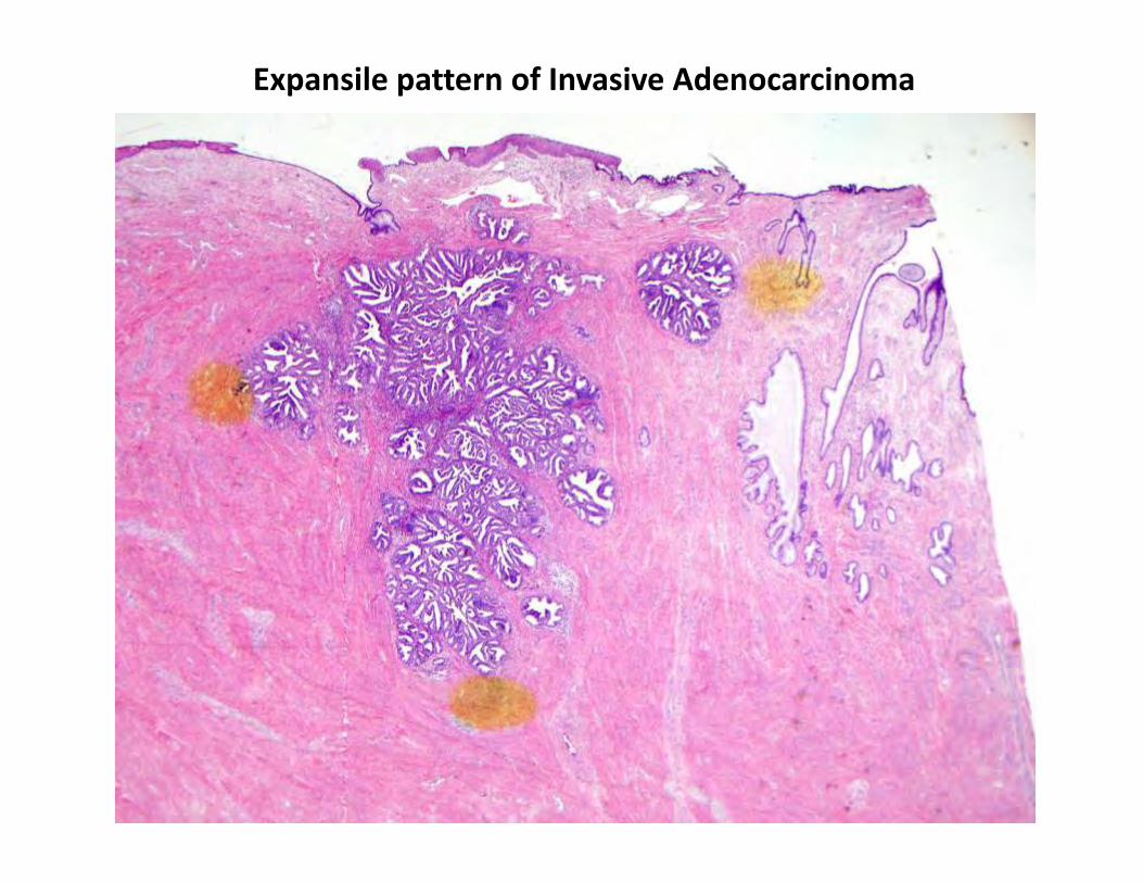
Expansile pattern of Invasive Adenocarcinoma
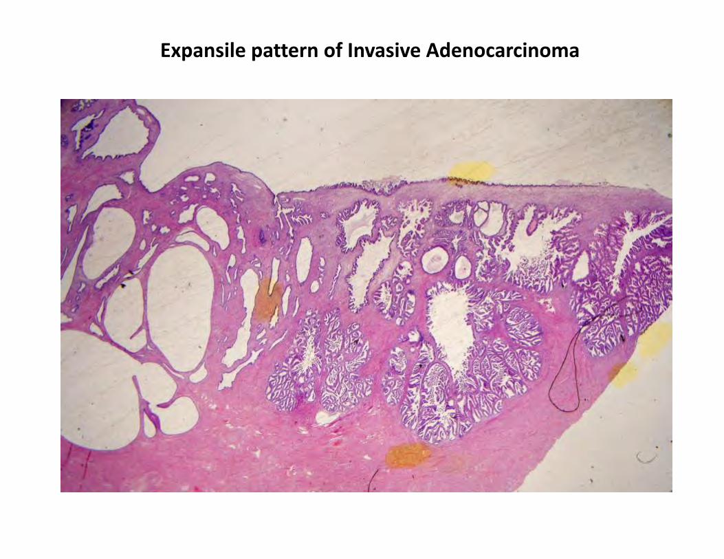
Expansile pattern of Invasive Adenocarcinoma
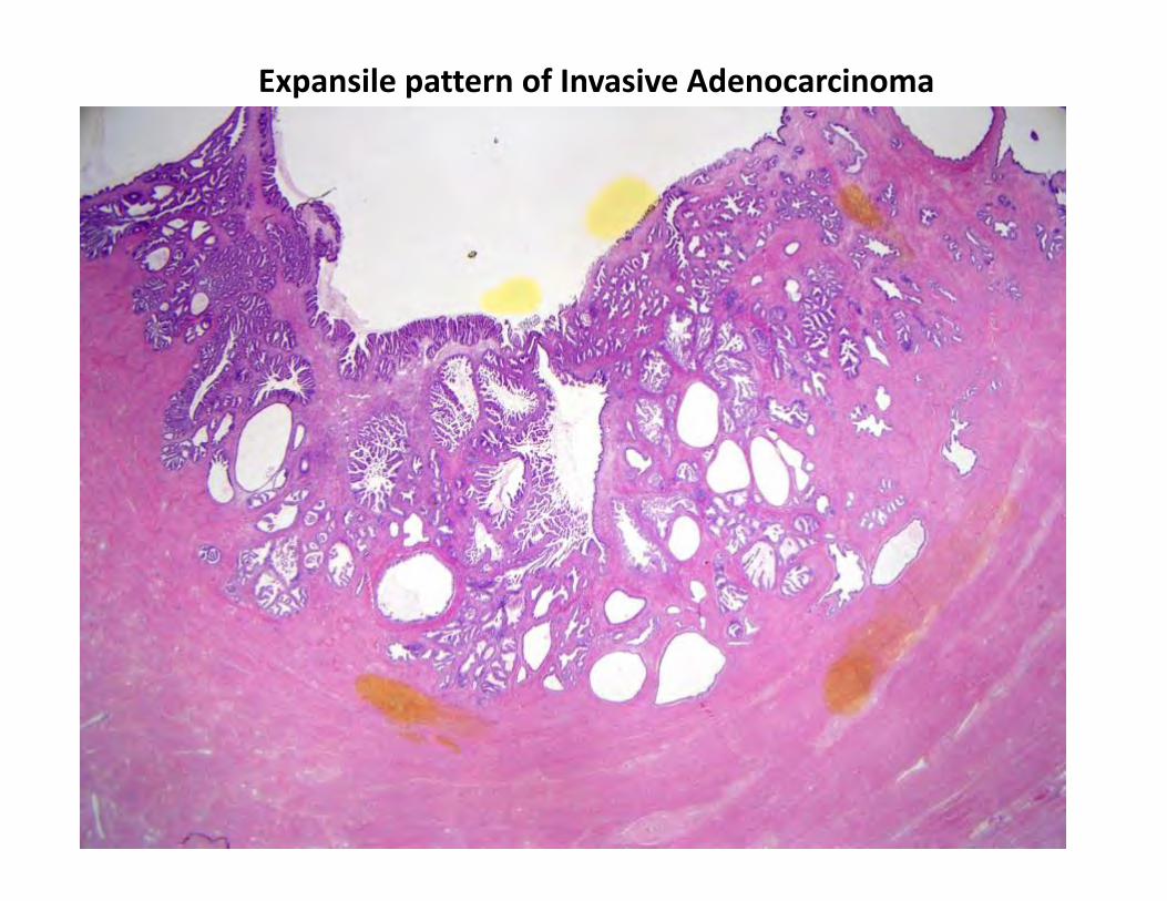
Expansile pattern of Invasive Adenocarcinoma
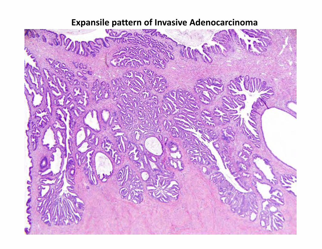
Expansile pattern of Invasive Adenocarcinoma
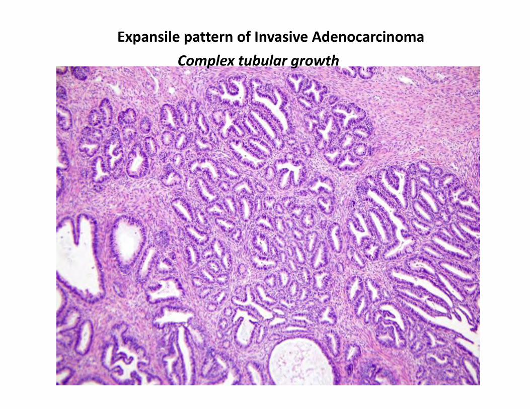
Expansile pattern of Invasive Adenocarcinoma Complex tubular growth
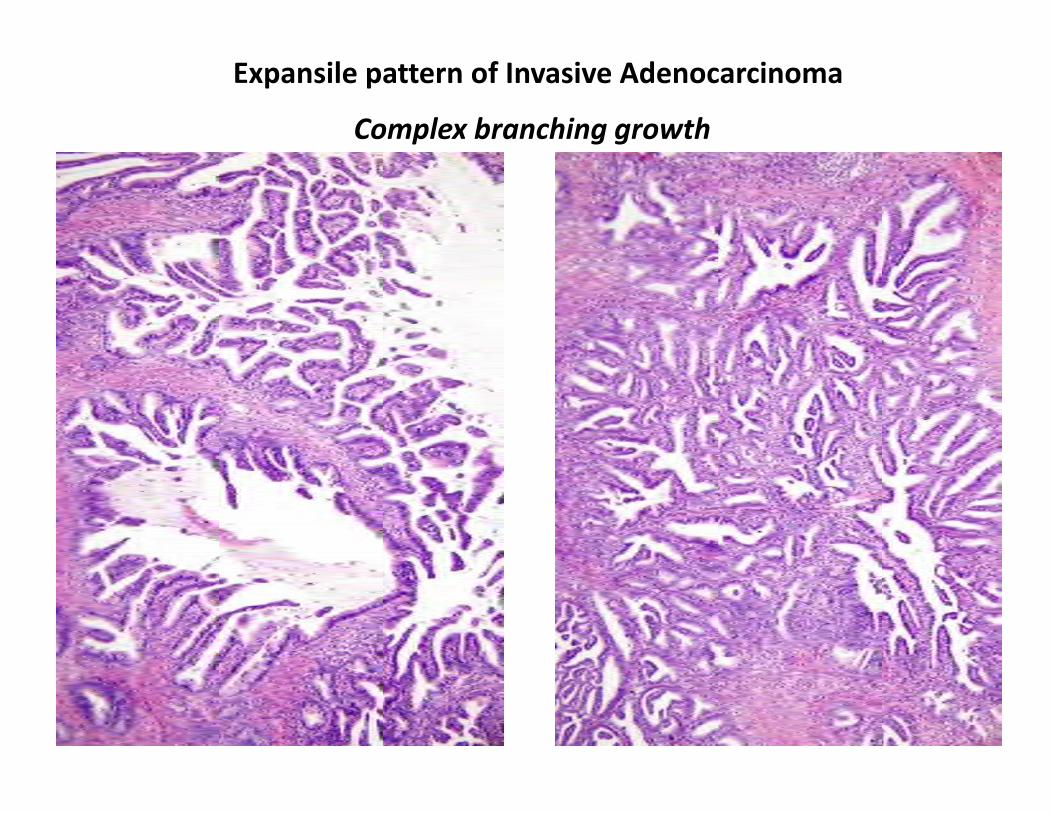
Expansile pattern of Invasive Adenocarcinoma
Complex branching growth
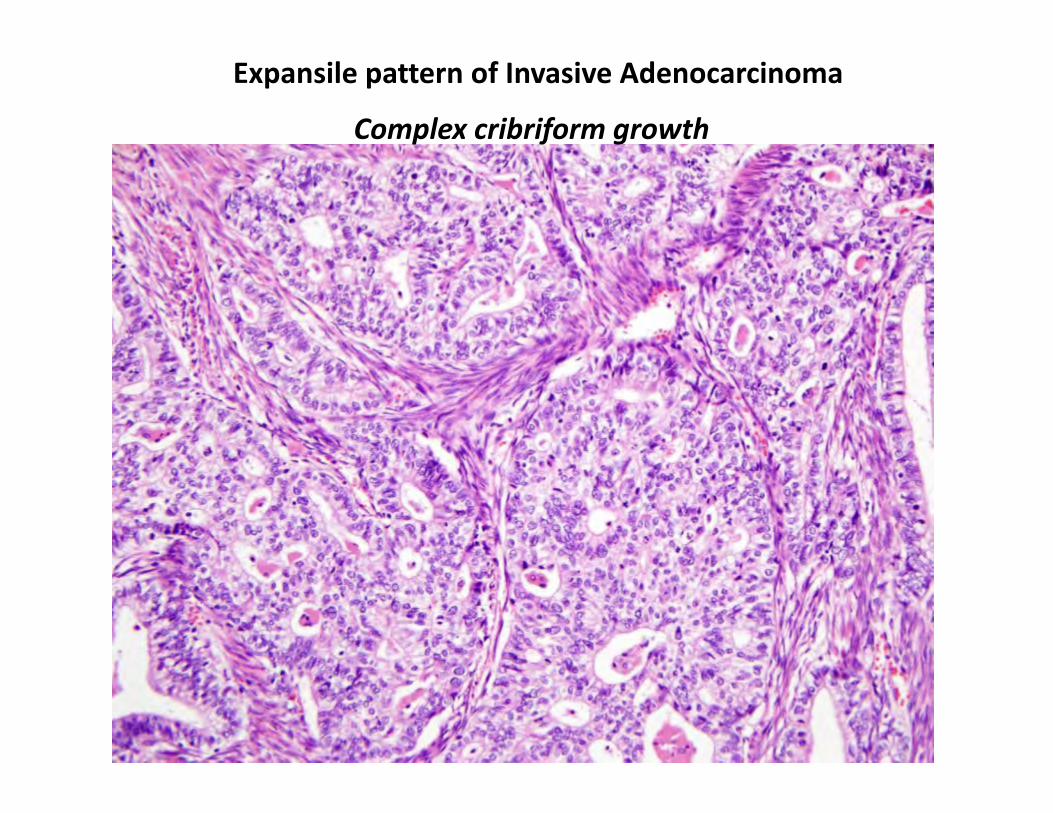
Complex cribriform growth
Expansile pattern of Invasive Adenocarcinoma
Findings at the Cusp between AIS and Invasive cancer
Deeper levels of all blocks with AIS
Also hunt for LVI (strong clue for stromal invasion)
Second observer
Common Problems with Usual type Endocervical Adenocarcinoma
Benign glandular lesions versus AIS AIS versus early invasive adenocarcinoma Measuring invasive adenocarcinoma for staging purposes Endocervical versus endometrial primary origin of adenocarcinoma
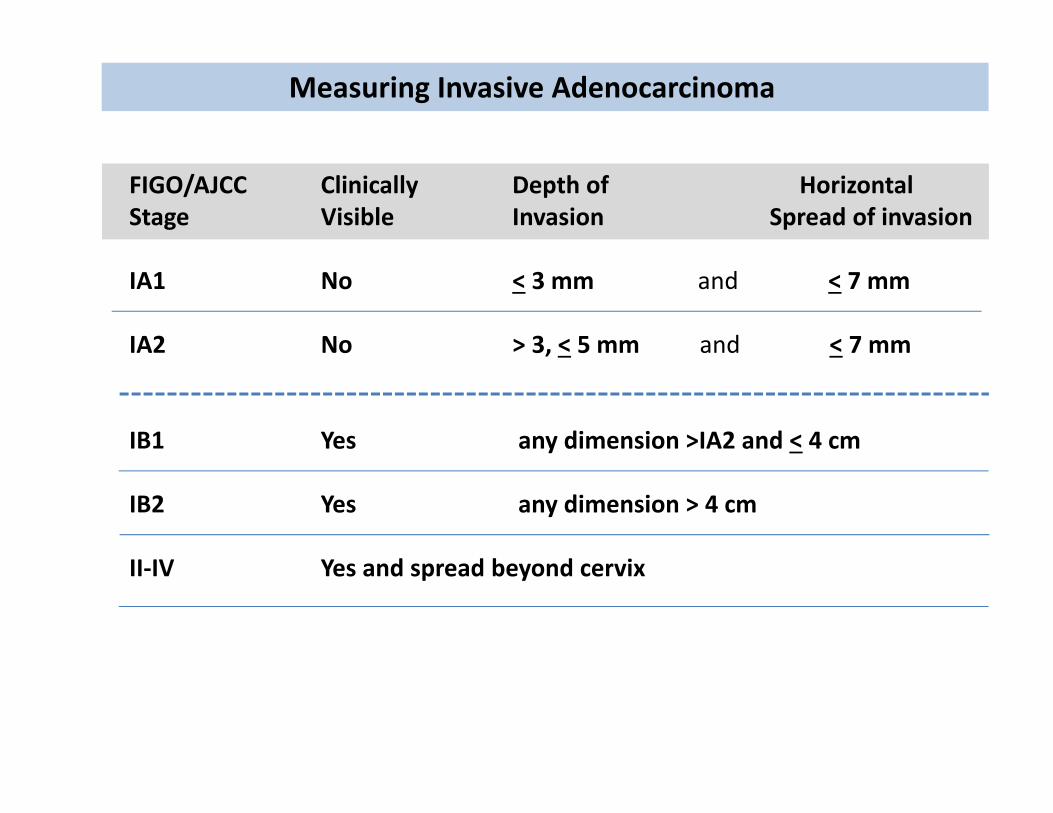
Measuring Invasive Adenocarcinoma
FIGO/AJCC Clinically Depth of Horizontal Stage Visible Invasion Spread of invasion
IA1 No < 3 mm and < 7 mm
IA2 No > 3, < 5 mm and < 7 mm
IB1 Yes any dimension >IA2 and < 4 cm
IB2 Yes any dimension > 4 cm
II‐IV Yes and spread beyond cervix
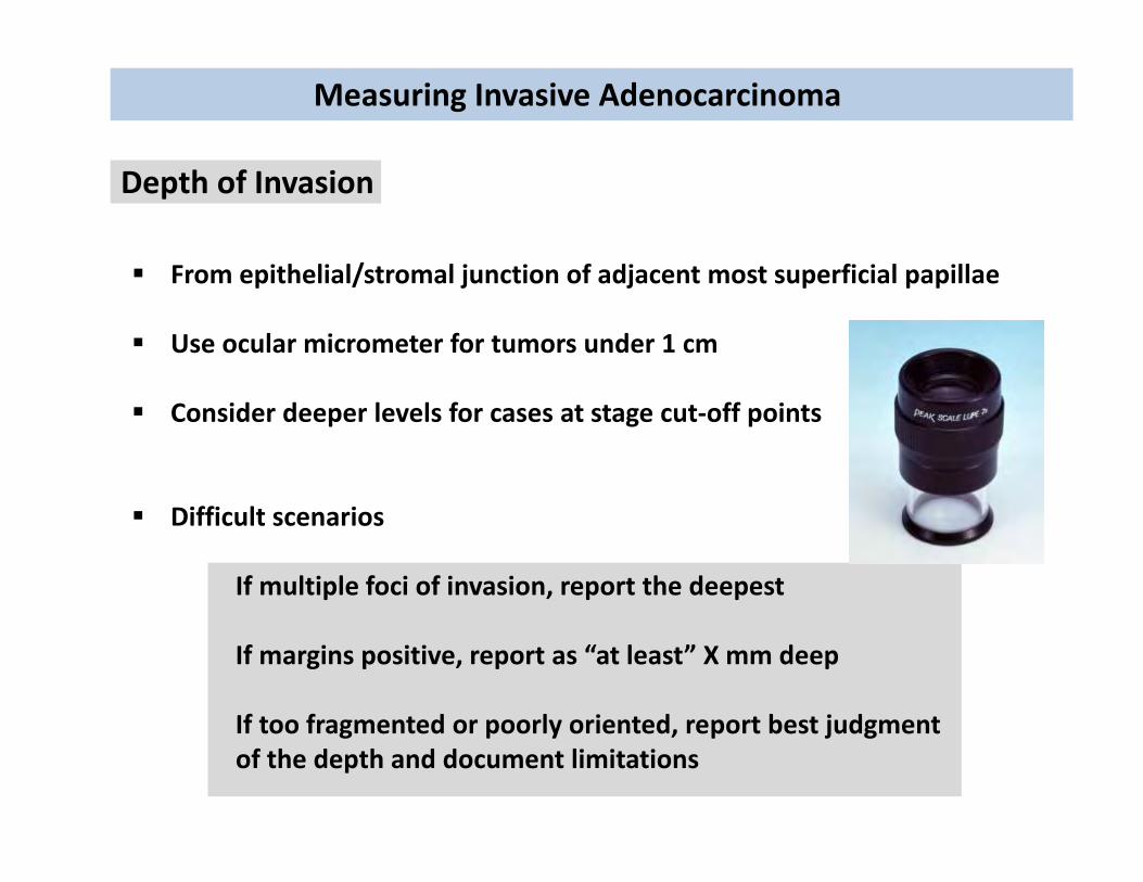
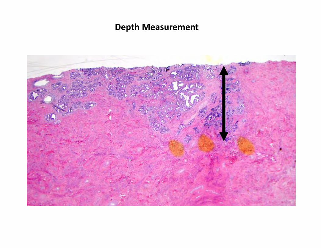
Measuring Invasive Adenocarcinoma
Depth of Invasion
From epithelial/stromal junction of adjacent most superficial papillae
Use ocular micrometer for tumors under 1 cm
Consider deeper levels for cases at stage cut‐off points
Difficult scenarios
If multiple foci of invasion, report the deepest
If margins positive, report as “at least” X mm deep
If too fragmented or poorly oriented, report best judgment of the depth and document limitations
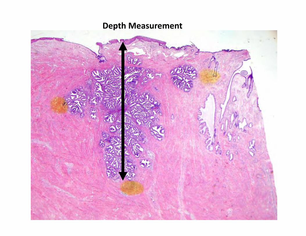
Depth Measurement
Depth Measurement
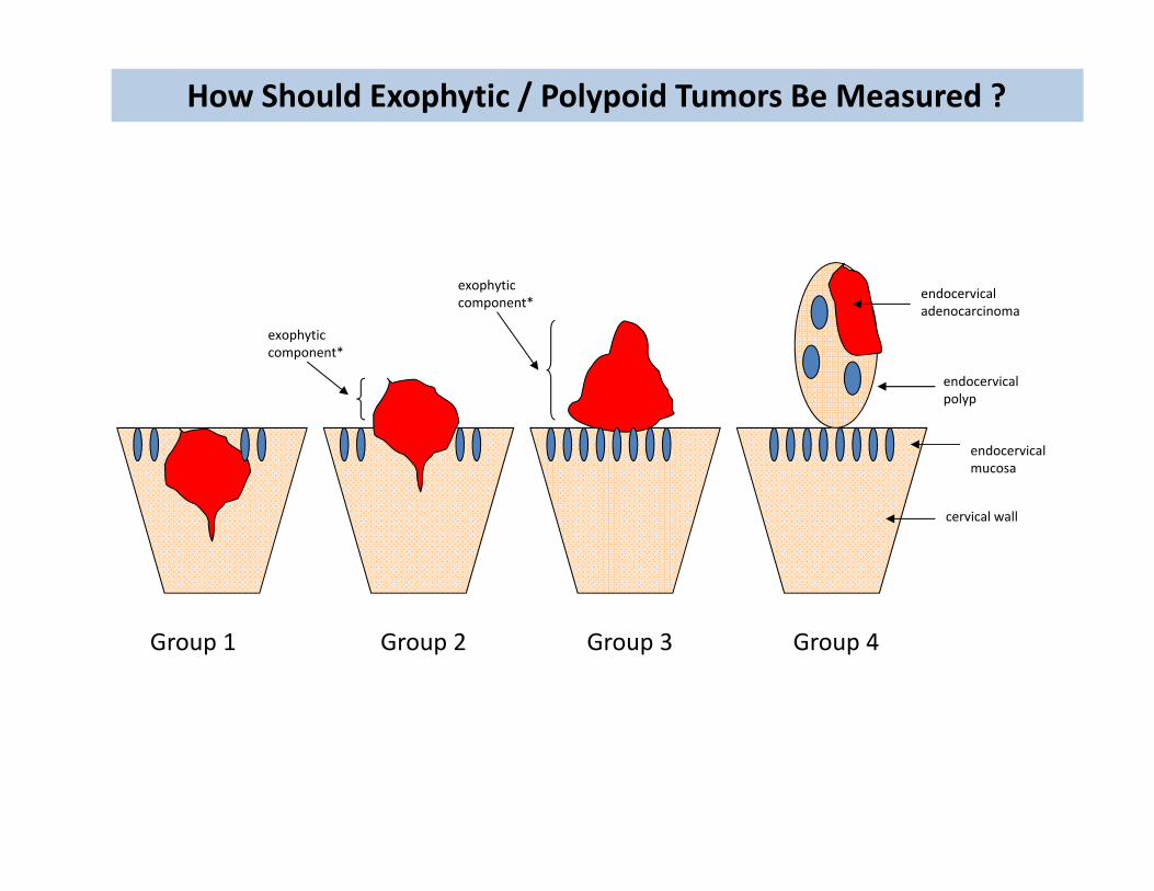
endocervicalpolyp
endocervicalmucosa
cervical wall
Group 1 Group 2 Group 3 Group 4
endocervicaladenocarcinoma
exophyticcomponent*
exophyticcomponent*
How Should Exophytic / Polypoid Tumors Be Measured ?
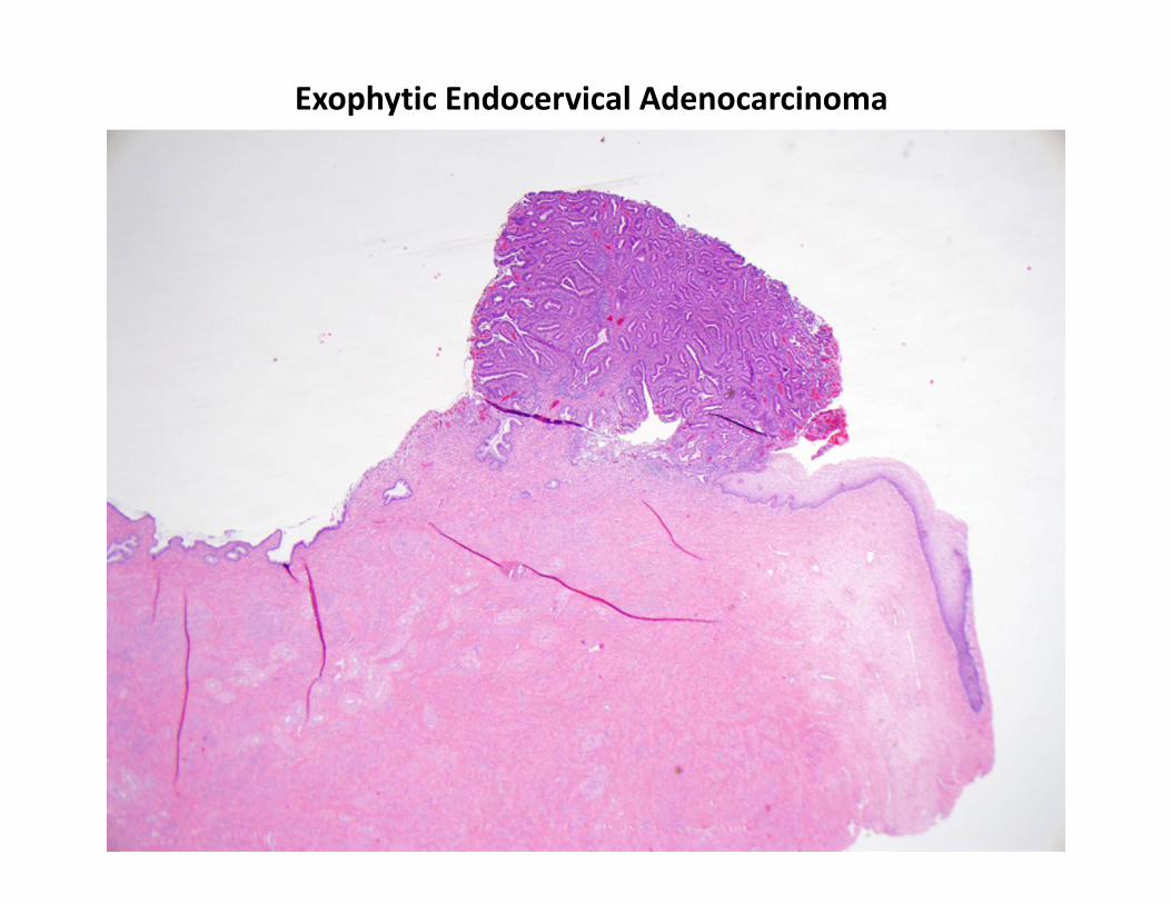
Exophytic Endocervical Adenocarcinoma
How Should Exophytic / Polypoid Tumors Be Measured ?
If cervical wall landmarks are not present in biopsy:
Document that cervical wall is not present
Discuss possibility that tumor could be eitherinvading the wall or growing as exophytic tumor
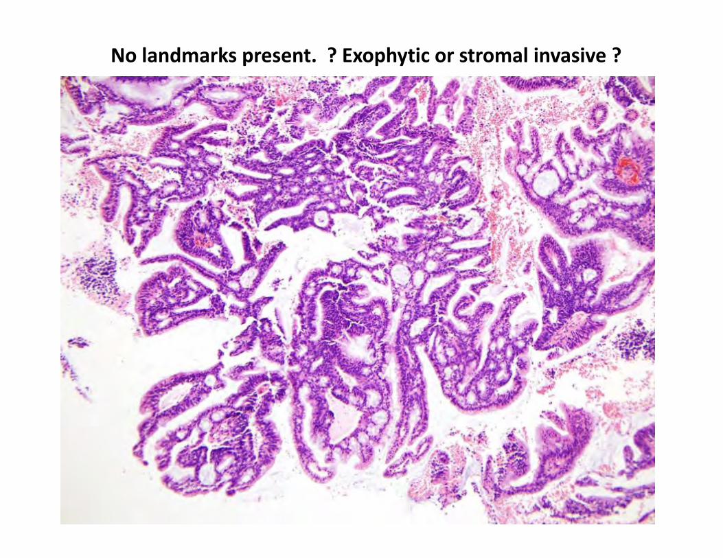
No landmarks present. ? Exophytic or stromal invasive ?
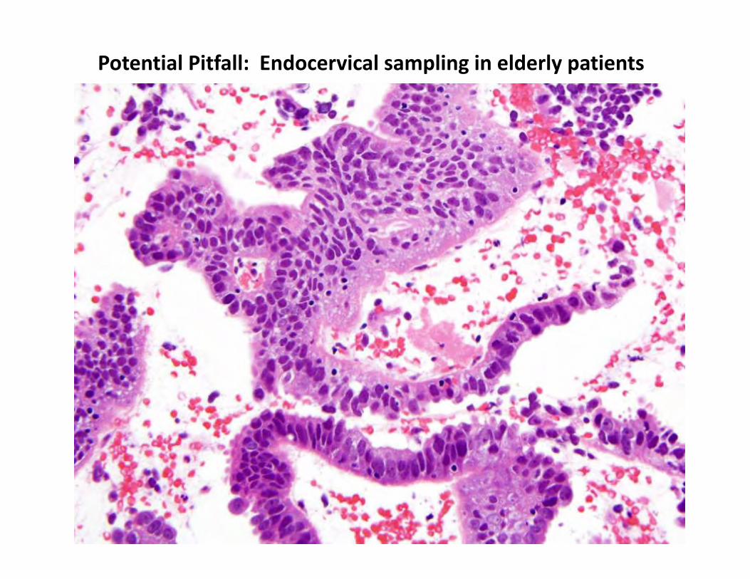
Potential Pitfall: Endocervical sampling in elderly patients
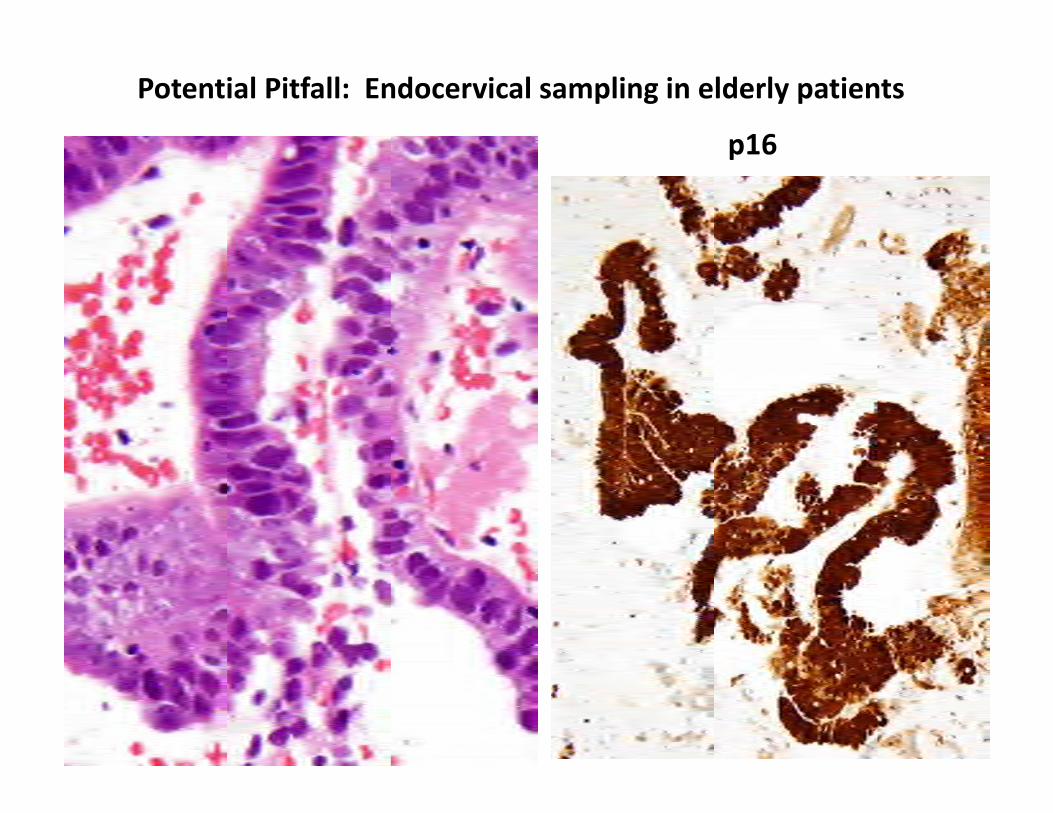
Potential Pitfall: Endocervical sampling in elderly patients
p16
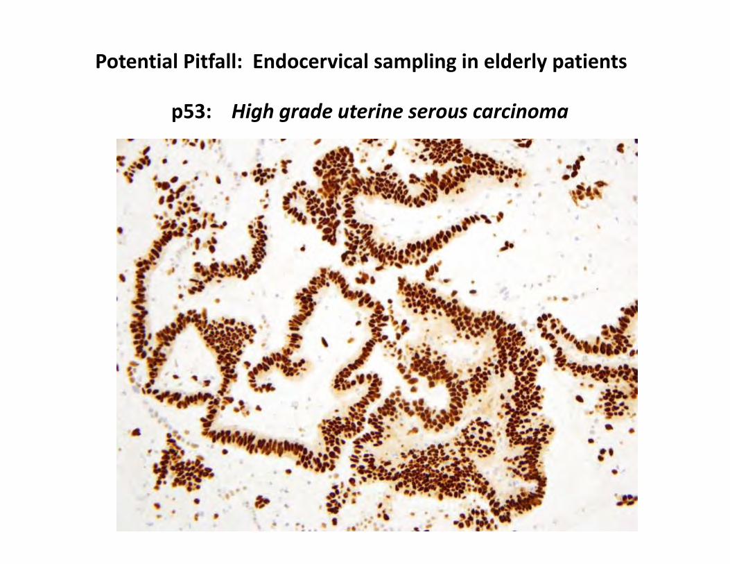
Potential Pitfall: Endocervical sampling in elderly patients
p53: High grade uterine serous carcinoma

Measuring Invasive Adenocarcinoma
Horizontal spread of invasive tumor
Maximal distance between peripheral edges of invasive tumor
Use ocular micrometer for tumors under 1 cm
Consider deeper levels for cases at stage cut‐off points
No rules are provided for measuring:
Multiple contiguous slides with invasion
Multiple non‐contiguous foci of invasion in a single slide
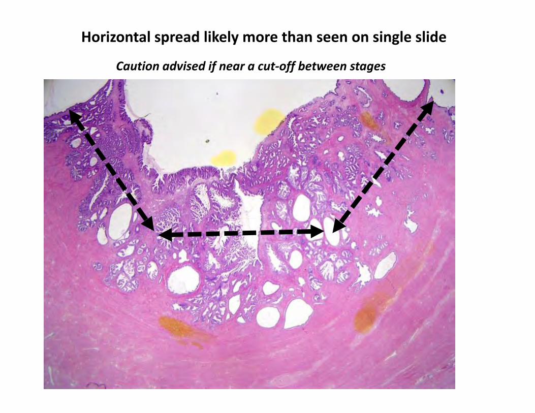
Horizontal spread likely more than seen on single slide
Caution advised if near a cut‐off between stages
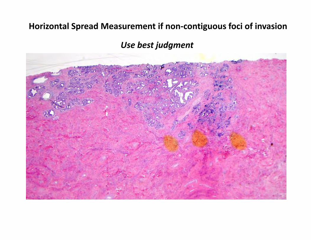
Horizontal Spread Measurement if non‐contiguous foci of invasion
Use best judgment
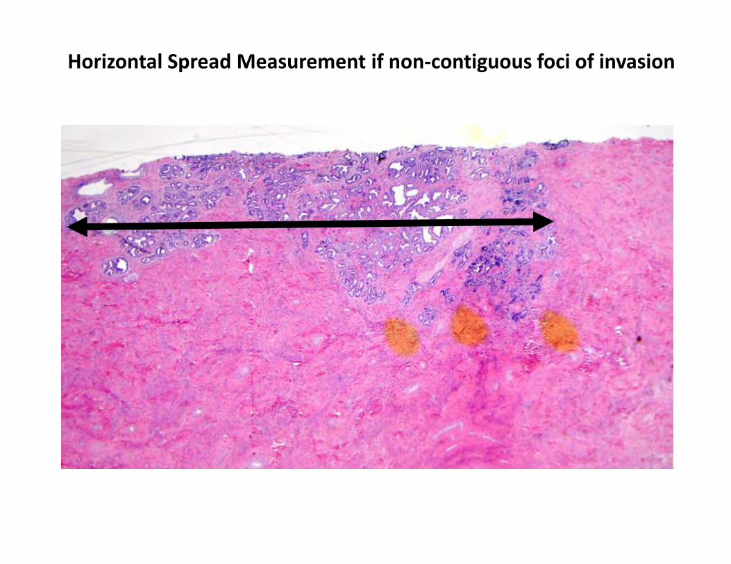
Horizontal Spread Measurement if non‐contiguous foci of invasion
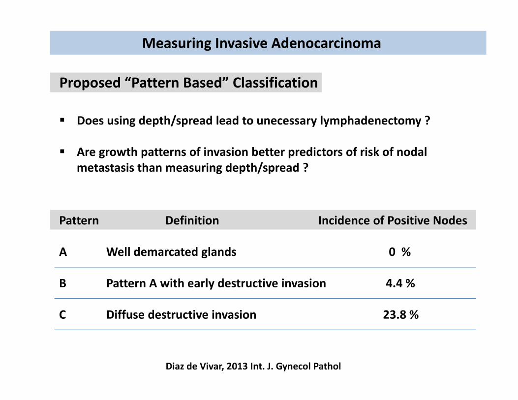
Measuring Invasive Adenocarcinoma
Proposed “Pattern Based” Classification
Does using depth/spread lead to unecessary lymphadenectomy ?
Are growth patterns of invasion better predictors of risk of nodal metastasis than measuring depth/spread ?
Pattern Definition Incidence of Positive Nodes
A Well demarcated glands 0 %
B Pattern A with early destructive invasion 4.4 %
C Diffuse destructive invasion 23.8 %
Diaz de Vivar, 2013 Int. J. Gynecol Pathol
Common Problems with Usual type Endocervical Adenocarcinoma
Benign glandular lesions versus AIS AIS versus early invasive adenocarcinoma Measuring invasive adenocarcinoma for staging purposes Endocervical versus endometrial primary origin of adenocarcinoma
Endocervical vs endometrial primary origin of adenocarcinoma
What is the gold standard to define origin ?
Gross findings in the hysterectomy specimen ? HPV status ? Immunophenotype ? Molecular definition: PTEN ? ARID1A ?
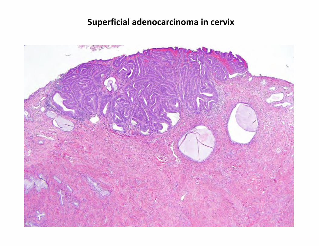
Superficial adenocarcinoma in cervix
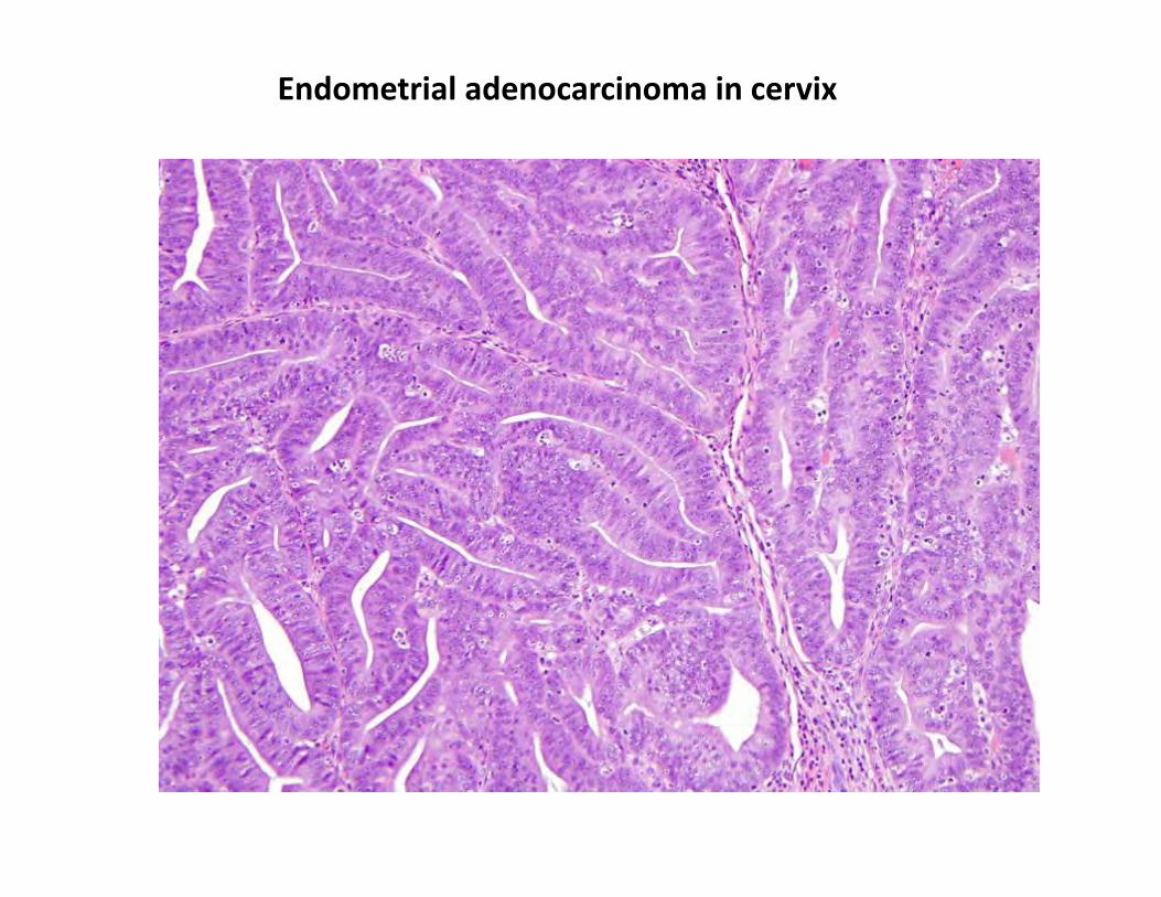
Endometrial adenocarcinoma in cervix
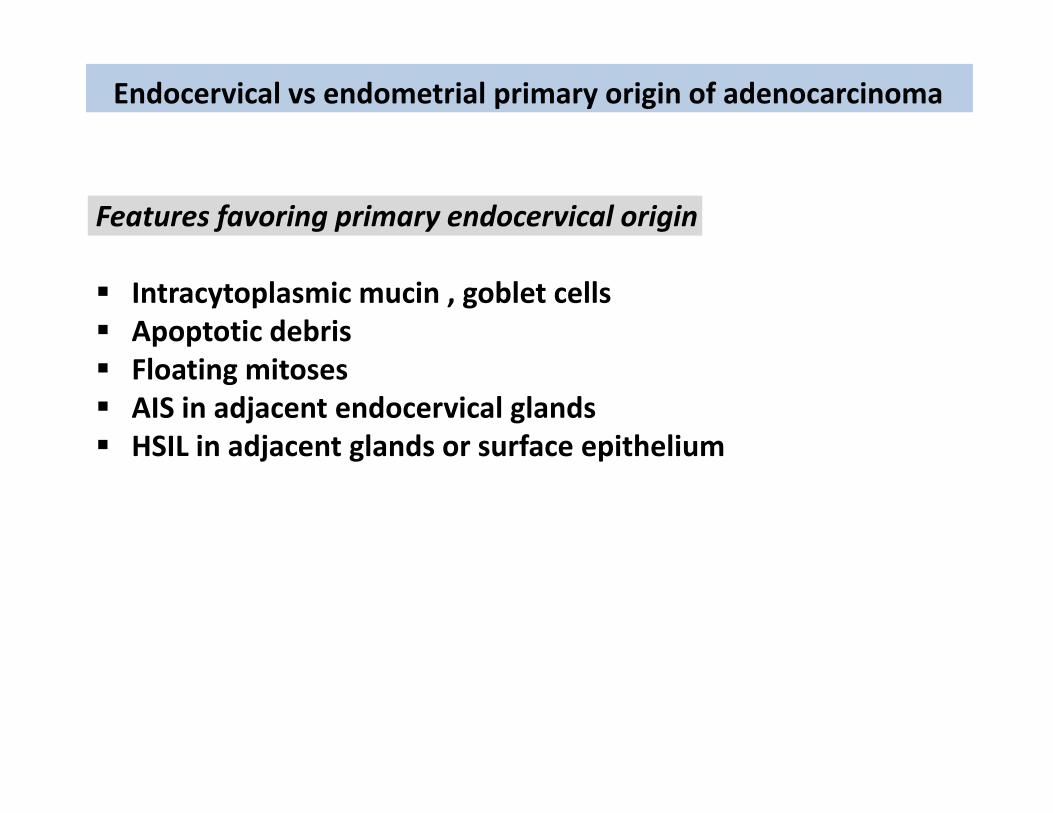
Endocervical vs endometrial primary origin of adenocarcinoma
Features favoring primary endocervical origin
Intracytoplasmic mucin , goblet cells Apoptotic debris Floating mitoses AIS in adjacent endocervical glands HSIL in adjacent glands or surface epithelium
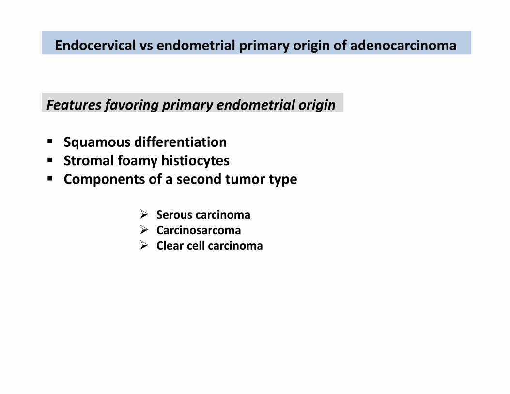
Endocervical vs endometrial primary origin of adenocarcinoma
Features favoring primary endometrial origin
Squamous differentiation Stromal foamy histiocytes Components of a second tumor type
Serous carcinoma Carcinosarcoma Clear cell carcinoma
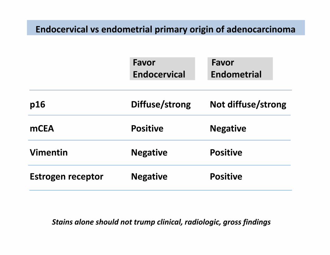
Endocervical vs endometrial primary origin of adenocarcinoma
p16 Diffuse/strong Not diffuse/strong
mCEA Positive Negative
Vimentin Negative Positive
Estrogen receptor Negative Positive
Favor FavorEndocervical Endometrial
Stains alone should not trump clinical, radiologic, gross findings
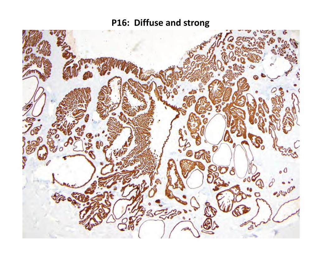
P16: Diffuse and strong
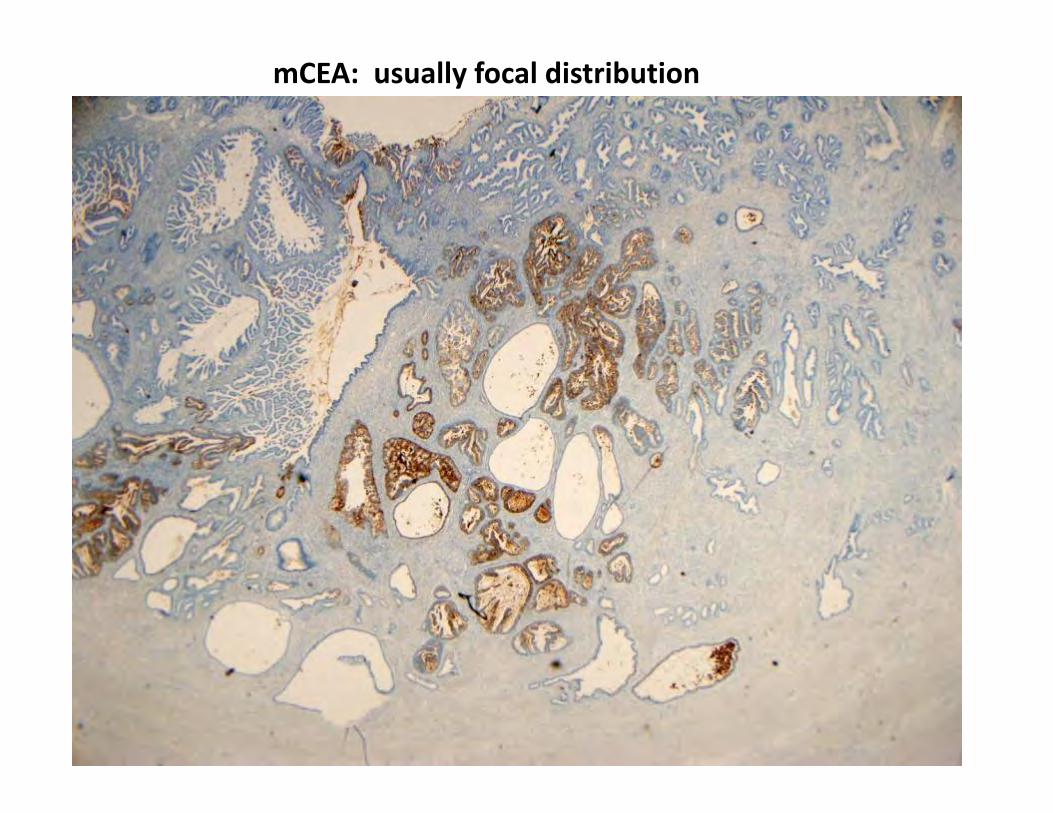
mCEA: usually focal distribution
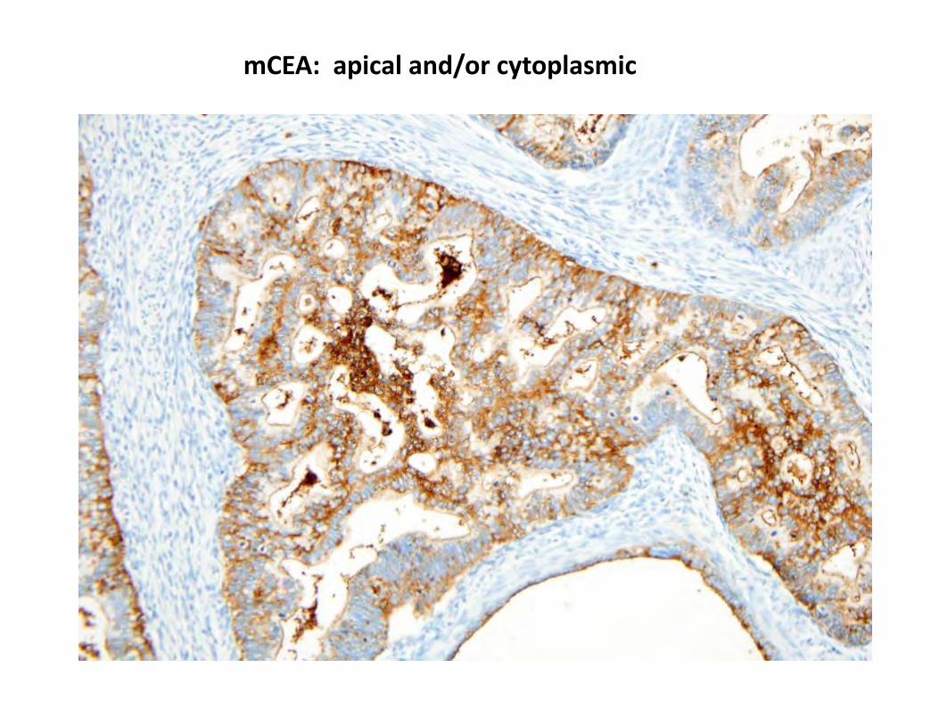
mCEA: apical and/or cytoplasmic
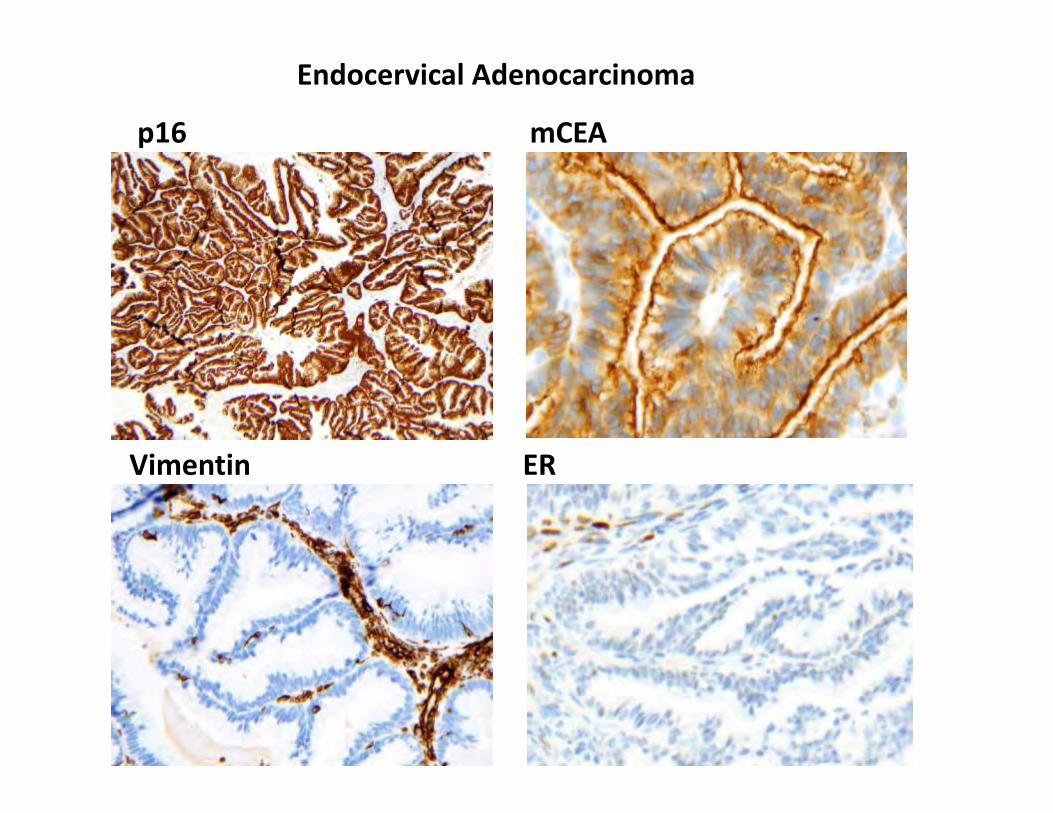
Endocervical Adenocarcinoma
p16 mCEA
Vimentin ER
Outline of Talk
Treatment Decisions for Endocervical Adenocarcinoma
New 2014 WHO Classification system
Update on Mucinous Adenocarcinoma variants
Common Problems in Usual type Endocervical Adenocarcinoma