Embed Size (px)
Citation preview
Role of Endogenous Secretinin Acid-induced Inhibition of HumanGastric FunctionJ. H. Kloibeuker, V. E. Eysselein, V. E. Maxwell,and J. H. WalshCenter for Ulcer Research and Education,Veterans Administration Wadsworth Hospital Center,Los Angeles California 90073; University of California,Los Angeles, California 90024
A bstract. The role of secretin in the inhibitionof gastric acid secretion that occurs during acidificationof the gastric lumen was studied in nine healthy men.Gastric acid secretion was stimulated by 500-ml mealsof 8% peptone solution, and the pH of the stomach wasmaintained at 5.5, 2.5, or 2.0 by intragastric titration.The increase in plasma secretin was measured, after ex-traction, by a new secretin radioimmunoassay. After de-termining the intravenous dose of secretin required toreproduce plasma secretin concentrations achieved duringpH 2.5 and 2.0 meals, similar doses were given duringadministration of a pH 5.5 peptone meal. The doses ofsecretin led to plasma secretin concentrations that av-eraged 3.4 pM, not different from the 3.2 and 3.9 pMconcentrations achieved during acidified meals. However,exogenous secretin infusion failed to inhibit acid secretionor gastrin response to peptone, although significant in-hibitions occurred in both during peptone meals givenat pH 2.5 or 2.0. When secretin infusions were given atfivefold higher rates, plasma gastrin responses again failedto demonstrate significant inhibition. Gastric emptyingwas inhibited significantly by both acidified peptone mealsbut only slightly (P = 0.053) during exogenous infusionof physiologic secretin doses. The decrease in acid secre-tion could be explained by decreased gastrin release, butneither of these findings could be explained by circulatingsecretin concentrations. These results cast strong doubton a physiological role of secretin in inhibition of acidsecretion in man.
Address reprint request to Dr. John H. Walsh, Veterans AdministrationWadsworth/CURE, Veterans Administration Wadsworth Hospital Cen-ter, Bldg. 115, Room 217, Los Angeles, CA 90073.
Received for publication 23 August 1983 and in revised form 24October 1983.
The Journal of Clinical Investigation, Inc.Volume 73, February 1984, 526-532
Introduction
Secretin is released by endocrine cells in the mucosa of theduodenum and upper jejunum when the intraduodenal pH fallsbelow 3 to 4 (1-3). The amount of secretin released probablyis dependent on the total amount of titratable acid delivered tothe duodenum. It is now generally accepted that secretin is aphysiologic stimulant of pancreatic water and bicarbonate se-cretion (4, 5). Whether it has any other function has not yetbeen established. Exogenous secretin in supraphysiologic dosesinhibits acid secretion and gastrin release, basally as well as inresponse to a variety of stimuli (6-10).
Recent studies in the dog (1 1, 12) indicate that secretin isa physiologic inhibitor of gastric acid secretion and gastrin release.So far, this has not been reported in man. A recent study (13)showed that secretin in low doses inhibits the gastric emptyingof liquid meals in humans. Wehave found that inhibition ofpeptone-stimulated acid secretion at low intragastric pH wasdue entirely to inhibition of gastrin release (14).
Weused a newly developed radioimmunoassay for secretinto study whether circulating secretin is a mediator of this acid-induced inhibition of gastrin release and acid secretion. In ad-dition, we studied the gastric emptying rate.
Methods
Nine healthy male subjects (mean age 38 yr, range 20-69 yr) withoutprevious gastric or duodenal disorders were studied. All gave writteninformed consent. The study was approved by the HumanStudies Com-mittee at Veterans Administration Wadsworth Center. After an overnightfast a nasogastric tube (AN 10, Andersen Samplers Inc., Atlanta GA)was passed into the stomach. The position of the tube was checked bya water-recovery (15) test and, if necessary, by fluoroscopy. After completeemptying of the stomach by aspiration, 500 ml of a liquid meal, containing8% peptone (wt/vol) (Bacto Peptone, Difco Laboratories, Inc., Detroit,MI), was instilled intragastrically and acid secretion was measured duringI h by automated intragastric titration (16). During the test the volumeof the intragastric contents was measured at 5, 15, 30, 45, and 60 min,using the dye-dilution technique (17). Phenol red was used as marker.Its concentration was measured spectrophotometrically at pH 1 1, at a
wavelength of 560 nm(Model DU, Beckman Instruments, Inc., Fullerton,
526 J. H. Kleibeuker, V. E. Eysselein, V. E. Maxwell, and J. H. Walsh
CA). The duodenal acid load during meals at low pH was calculatedaccording to Gross et al. ( 18). Titratable acid of the gastric contents wasmeasured by titration to pH 7.
Before and during tests blood samples were taken through a needlein the antecubital vein for measurement of plasma gastrin and secretinconcentrations. Samples were collected in standard EDTA-containingtubes (10.5 mg/tube) to which 200 Ml Trasylol (aprotinin), 10,000 KIU/ml (Bayer AG, Bayerwerk, FRG) per 5 ml blood was added. Sampleswere refrigerated immediately after collection, centrifuged within 30min, and the plasma was stored at -20'C until radioimmunoassayswere performed.
For secretin infusion pure natural porcine secretin (manufacturedby Kabi Vitrum AB, Stockholm, Sweden) dissolved in 0.15 MNaCl,containing 0.25% human serum albumin (wt/vol), was used. The infusionwas administered through a needle in the other arm. A small portionof each infusion solution was stored at -20'C for measurement of thesecretin concentration. The biologic activity of the secretin was testedby infusing increasing doses intravenously in a dog with a chronic pan-creatic fistula after an overnight fast, and measuring pancreatic secretionin respect to volume and bicarbonate contents. Gastrin plasma con-centrations were measured with a specific radioimmunoassay as pre-viously described (19), using antibody 161 1.
Secretin plasma levels were measured with a newly developed ra-dioimmunoassay.
Antibody. Secretin antibodies were raised in rabbits, using syntheticsecretin (E. R. Squibb & Sons, Princeton, NJ) as antigen. The secretinwas conjugated to bovine serum albumin with carbodiimide as describedearlier (19). The conjugation product was emulsified with completeFreund's adjuvant for the initial immunization, and with incompleteFreund's adjuvant for subsequent booster immunizations. Booster im-munizations were given at 4-8-wk intervals after the previous immu-nization. Blood was obtained 7 d after an immunization. The antiserumused in this assay, 7842, was obtained after the fourth immunization.
Cross-reactivity of the antiserum was tested for glucagon, gastricinhibitory peptide, and vasoactive intestinal peptide and further forbombesin, cholecystokinin octapeptide, gastrin, insulin, motilin, neu-rotensin, pancreatic polypeptide, and somatostatin at concentrations upto at least 10 pmol/ml.
The sensitivity was tested by determining the ID50,' defined as theconcentration of antigen required to reduce the binding of labeled antigenby 50%, and by determining the detection limit, defined as the smallestconcentration of added antigen, which produces a significant inhibitionof binding (P < 0.05 by t test), when samples are tested in four replications.
Labeled antigen. Secretin was labeled by the chloramine-T method.Synthetic secretin was purchased from Research Plus, NaI251 (100 mCi/ml) from Amersham Corp. I; chloramine-T from Eastman Kodak Co.,Rochester, NY; sodium metabisulphite from J. T. Baker Chemical Co.,Phillipsburg, NJ; Sephadex G-10 and SP-Sephadex C-25 from PharmaciaFine Chemicals, Piscataway, NJ. Secretin, chloramine-T, and sodiummetabisulphite were all dissolved in 0.25 Msodium phosphate bufferat pH 7.4.
Secretin, 10-20 gg, was dissolved in 50 gl buffer, to which 10 glNaI251 and 20 Mg chloramine-T in 10 Ml buffer, were added. After areaction time of 60 s 100 Mg sodium metabisulphite in 20 Ml buffer wasadded.
For a first purification the sample was applied to a Sephadex G-10column, 20 X I cm, eluted with 0.05 Msodium acetate buffer pH 5.0,
1. Abbreviation used in this paper: ID50, concentration of antigen requiredto reduce the binding of labeled antigen by 50%.
containing 0.425% sodium chloride (wt/vol) and 2%Plasmanate (humanplasma protein fraction, Cutter Laboratories, Inc., Emeryville, CA). Theflow rate was I ml/min, and 2-ml fractions were collected. From thefirst peak the fractions with the highest radioactivity were pooled, and,for further purification, applied to a SP-Sephadex C-25 column, 20X I cm, eluted with the same buffer as the first column, but with aconcentration gradient from 0.05 to 0.1 Min 0.85% NaCl over 12 h,at a flow rate of 0.2 ml/min. Fractions of 2 ml were collected. Fromthe second high peak the fractions with the highest radioactivity, con-taining the purified label, were pooled and after addition of 0.5% Trasylol,10,000 KIU/ml, (vol/vol), divided in 250-Ml portions, which were storedat -20'C until use. Each portion was used for only one assay. Fractionspreceding and following the labeled antigen containing peak were assayedto identify unlabeled secretin.
The specific activity of the label was determined by comparing thedisplacement of a known amount of labeled antigen by doubling amountsof the same label and known amounts of standard secretin.
The immunoreactivity was determined by comparing serial dilutioncurves of the labeled secretin and standard secretin, and by determiningthe binding of label to an excess amount of antibody (titer 1:5,000). Toeach assay tube 2,000 cpm of label in 200 Ml were added. Counting timewas I min.
Standard secretin. Squibb synthetic secretin was used as standard.The dry powder was dissolved in the assay buffer to a concentration ofI nmol/ml. 250-Ml portions of this solution were stored at -20'C, eachportion to be used for only one assay.
Assay conditions. The incubation volume was I ml. The assay bufferwas 0.05 Mammonium acetate (J. T. Baker Chemical Co.) at pH 5.5,containing 2%Plasmanate and 0.5% Trasylol. In preliminary experimentswe found that at higher pH (6.5, 7.4) the binding in absence of antibodyexceeded 10%, which we found unacceptable.
The standards and unknown samples were preincubated at 4VCwiththe antibody for 48 h, after which label was added. After another 24 hat 4VC, bound and unbound fractions were separated, using charcoal.For the separation 400 Ml of a suspension of activated charcoal (Mal-linckrodt Inc., St. Louis, MO), 50 mg/liter, and dextran (PharmaciaFine Chemicals), 5 mg/liter, in 0.05 Mammonium acetate at pH 5.5,with 10% newborn calf serum (vol/vol) (MA Bioproducts, Walkersville,MD), was added to each tube. After centrifugation the bound and un-bound fractions were separated. In preliminary experiments it was foundthat by this incubation method the ID50 decreased 70% compared withsimultaneous incubation of antibody and label for 24 h.
Data fitting. For the standard curve, antigen concentrations of 1-1,000 fmol/ml were used. A computer fit the four-parameter logisticcurve to the bound counts with nonlinear regression. The computerwas also used to obtain unknown values from the standard curve (20).
Reproducibility of the assay. This was determined by calculating thecoefficients of variation within the assay and between assays.
Preparation of plasma samples. Since physiologic plasma levels ofsecretin are low and plasma interference is often relatively high, extractionand concentration of plasma is necessary before assaying. For this purposewe used C,8 cartridges (SEP-PAK, Waters Associates, Millipore Corp.,Milford, MA), small, densely packed Cj8 columns. After preparation ofthe cartridge with 10 ml acetonitrile (J. T. Baker Chemical Co) and 10ml 0.05 Mammonium acetate at pH 5.5, the plasma sample was applied.The column was then washed with the same buffer and it was theneluted with 50% acetonitrile/50% ammonium acetate. After discardingthe first 0.5 ml, 1.1 ml was collected in a tube containing 0.01 mlPlasmanate and 0.005 ml Trasylol. After evaporation of the acetonitrile0.6 ml ammonium acetate buffer was left; 450 Al of the 600 was added
527 Endogenous Secretin in Human Gastric Function
to the assay tube. From each plasma sample 2 X 5 ml was extractedand assayed as duplicates. Recovery of secretin from this procedure wastested by adding doubling doses of standard secretin to 5 ml of charcoal-stripped hormone-free plasma, at concentrations of 3.125-25 fmol/mlplasma and extracting and assaying the samples as mentioned before.To determine remaining interference after extraction, from each subjectstudied, 10 ml plasma was treated with charcoal and 2 X 5 ml wasextracted and assayed with the other samples. The same was done withthe plasma used for recovery tests. Concentrations of unknowns werecalculated from the assay results by correction for the recovery, thevolume extracted (5 ml), the final fraction of the sample used in theassay (450/600), and for remaining interference after extraction.
Studies performed. Four studies were performed on separate days,the first three in random order. In these three the liquid meals weregiven at pH 2.0, 2.5, and 5.5, respectively, and intragastric titration wasperformed at the same pH as the meals. A basal blood sample was takenbefore the meal while aspirating gastric contents, and during the testsamples were taken at 15-min intervals. On the same day as the testwith the meal at pH 5.5, 2 h after finishing that test, secretin was infusedin increasing doses, respectively, 2.5, 8.0, and 25.0 pmol* kg-' h1.Each dose was infused during 30 min. At the end of each period a bloodsample was taken. During the infusion gastric contents were aspiratedcontinuously.
In the fourth study, a liquid meal at pH 5.5 was again given, nowalong with intravenous infusion of secretin at a dose designed to matchplasma levels found during the meals at low pH. The required dose wasestimated on an individual basis by comparison of the plasma secretinfound during the meals at low pH and the levels during the infusion ofincreasing doses of secretin. The secretin infusion was started at thesame moment as the instillation of the meal. Blood was collected at 15-min intervals for gastrin and at 30 and 60 min for secretin.
In four subjects, two additional studies were performed. In both,again a meal at pH 5.5 was given. In the first, secretin was infused onlyduring the meal at a dose five- to sixfold higher than the dose given inthe former study. In the second, secretin was infused at the same doseas in the first, but the infusion was started 1 h before the meal andcontinued throughout the test. Gastric emptying was not studied in thesetwo tests. Blood samples were taken at 15-min intervals for gastrin andat 30-min intervals for secretin.
Data analysis. For each test in each subject the integrated responsesfor gastrin and secretin were calculated. For gastrin the integrated responsewas calculated using the natural logs of the responses. The integratedmean was obtained by dividing this value by the time (60 min). Two-way analysis of variance (subject by condition) was performed on theseintegrated means. Gastrin values given are the mean logs, transformedback to the raw scale. For acid, total secretion during the test hour wasused in the analysis of variance without considering basal acid secretion.For the gastric emptying the t½/2 was calculated separately for each testof each subject using a nonlinear fit to the single exponential (21). Two-way analysis of variance was calculated using these estimates of t½/2.
Results
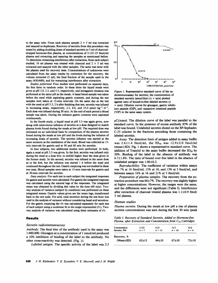
Secretin radioimmunoassayAntibody. The final titer of the antibody used in the assay was1:400,000. Glucagon at a concentration of 1 nmol/ml produceda 10% inhibition of binding of the label to the antibody. Noother cross-reactivity was detected. (Fig. 1).
Labeled antigen. The specific activity of the label was 2.5
B/F1.0-
0.5-
0
GLUCAGON
SECRETIN
1 10 lo2 i03 104 lo 106
CONCENTRATION,pmol/liter
Figure 1. Representative standard curve of the ra-dioimmunoassay for secretin; the concentration ofstandard secretin (pmol/liter) (x = axis) plottedagainst ratio of bound-to-free labeled secretin (y= axis). Dilution curves for glucagon, gastric inhibi-tory peptide (GIP), and vasoactive intestinal peptide(VIP) in the same assay system.
,gCi/nmol. The dilution curve of the label was parallel to thestandard curve. In the presence of excess antibody 85% of thelabel was bound. Unlabeled secretin eluted on the SP-SephadexC-25 column in the fractions preceding those containing thelabeled secretin.
Assay. The detection limit of antigen added to assay bufferwas 2.6±1.4 fmol/ml, the ID50 was 12.9±2.0 fmol/ml(mean±SD). Fig. 1 shows a representative standard curve. Theaddition of Trasylol to the assay buffer decreased the ID50 by20%. Binding of the label in the absence of antibody was6 7± 1.8%. The ratio of bound over free label in the absence ofunlabeled antigen was 1.06±0.2.
Reproducibility. The coefficient of variation within assayswas 7% at 16 fmol/ml, 15% at 10, and 13% at 5 fmol/ml, andbetween assays 16% at 16 and 21% at 5 fmol/ml.
Preparation of plasma samples. The recovery from the ex-traction procedure was 66±7%. The recovery was slightly higherat higher concentrations. However, the ranges were the same,and the differences were not significant (Table I). Interferenceafter extraction of charcoal treated plasma was 1.1±0.9 fmol/5 ml plasma.
Human studiesPlasma secretin. During the meals at low pH a rise of plasmasecretin concentrations was seen during the first 30 min (peak
Table I. Recovery of Standard Secretin, Added to Hormone-freePlasma, after Extraction and Concentration from C,8 Cartridges
Concentration 3.125 6.25 12.5 25.0Secretin, PM n = 7 n = 13 n = 10 n = 4
Recovery, %(Mean±SD) 62±13 64±10 67±10 72±10
528 J. H. Kleibeuker, V. E. Eysselein, V. E. Maxwell, and J. H. Walsh
.
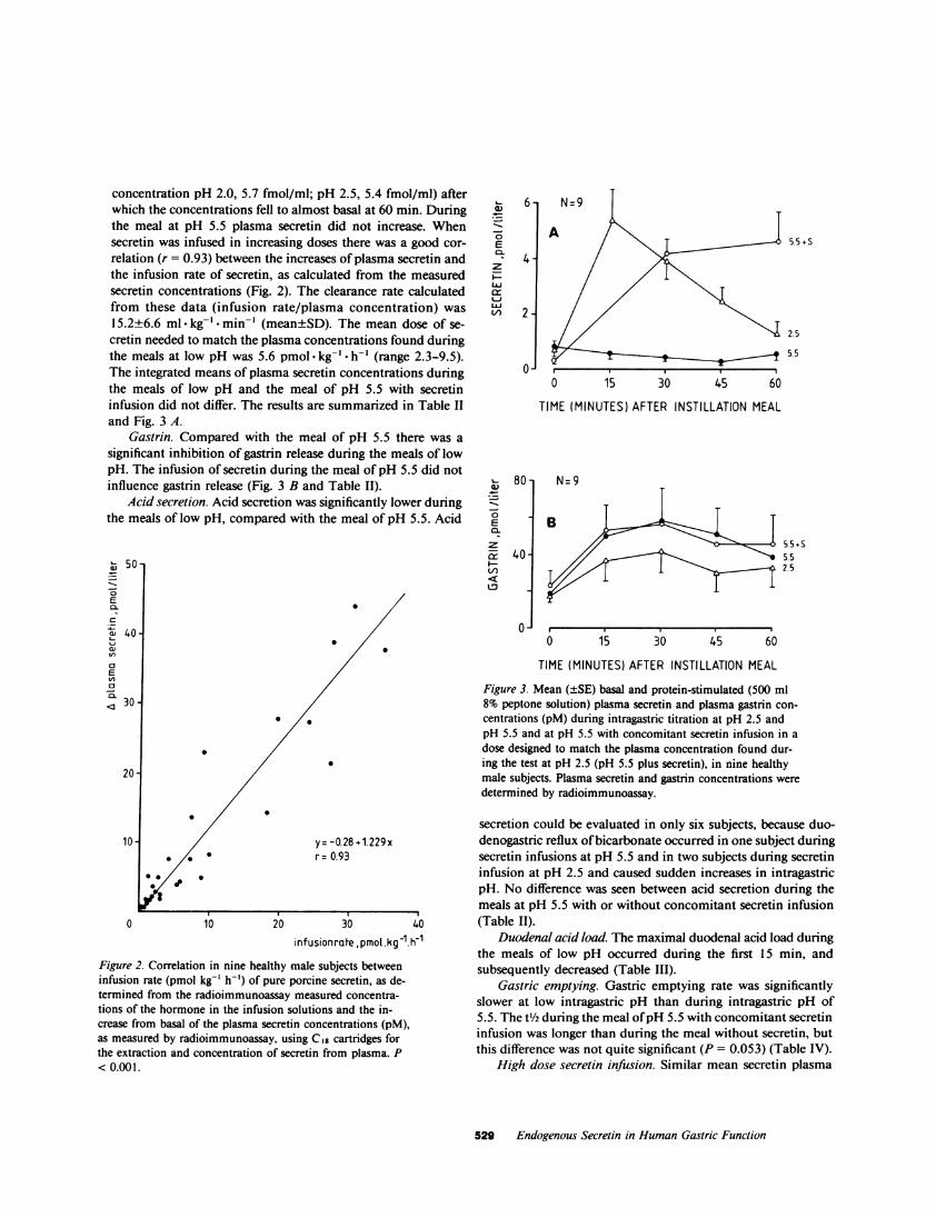
concentration pH 2.0, 5.7 fmol/ml; pH 2.5, 5.4 fmol/ml) afterwhich the concentrations fell to almost basal at 60 min. Duringthe meal at pH 5.5 plasma secretin did not increase. Whensecretin was infused in increasing doses there was a good cor-relation (r = 0.93) between the increases of plasma secretin andthe infusion rate of secretin, as calculated from the measuredsecretin concentrations (Fig. 2). The clearance rate calculatedfrom these data (infusion rate/plasma concentration) was15.2±6.6 ml kg-' -min-' (mean±SD). The mean dose of se-cretin needed to match the plasma concentrations found duringthe meals at low pH was 5.6 pmol -kg-' * h-' (range 2.3-9.5).The integrated means of plasma secretin concentrations duringthe meals of low pH and the meal of pH 5.5 with secretininfusion did not differ. The results are summarized in Table IIand Fig. 3 A.
Gastrin. Compared with the meal of pH 5.5 there was asignificant inhibition of gastrin release during the meals of lowpH. The infusion of secretin during the meal of pH 5.5 did notinfluence gastrin release (Fig. 3 B and Table II).
Acid secretion. Acid secretion was significantly lower duringthe meals of low pH, compared with the meal of pH 5.5. Acid
w 50-
_-6CL
cl
0._
C!< 0
cz30-
a)
0._-6
z
LUL
LU
0.-8E
zixL-L/)
4.-
2-
0-
80 -
40-
0J
0
0
y= -0.28 +1.229xr= 0.93
0 10 20 30 40in fusion rate ,pmol.kg-1.h1
Figure 2. Correlation in nine healthy male subjects betweeninfusion rate (pmol kg-' h-') of pure porcine secretin, as de-termined from the radioimmunoassay measured concentra-tions of the hormone in the infusion solutions and the in-crease from basal of the plasma secretin concentrations (pM),as measured by radioimmunoassay, using Cig cartridges forthe extraction and concentration of secretin from plasma. P< 0.001.
5.5+S
2.5
5.5
0 15 30 45 60
TIME (MINUTES) AFTER INSTILLATION MEAL
N=9
5.5 + S5.52.5
0 15. I I 60 15 30 45 60
TIME (MINUTES) AFTER INSTILLATION MEAL
Figure 3. Mean (±SE) basal and protein-stimulated (500 ml8% peptone solution) plasma secretin and plasma gastrin con-centrations (pM) during intragastric titration at pH 2.5 andpH 5.5 and at pH 5.5 with concomitant secretin infusion in adose designed to match the plasma concentration found dur-ing the test at pH 2.5 (pH 5.5 plus secretin), in nine healthymale subjects. Plasma secretin and gastrin concentrations weredetermined by radioimmunoassay.
secretion could be evaluated in only six subjects, because duo-denogastric reflux of bicarbonate occurred in one subject duringsecretin infusions at pH 5.5 and in two subjects during secretininfusion at pH 2.5 and caused sudden increases in intragastricpH. No difference was seen between acid secretion during themeals at pH 5.5 with or without concomitant secretin infusion(Table II).
Duodenal acid load. The maximal duodenal acid load duringthe meals of low pH occurred during the first 15 min, andsubsequently decreased (Table III).
Gastric emptying. Gastric emptying rate was significantlyslower at low intragastric pH than during intragastric pH of5.5. The t1/2 during the meal of pH 5.5 with concomitant secretininfusion was longer than during the meal without secretin, butthis difference was not quite significant (P = 0.053) (Table IV).
High dose secretin infusion. Similar mean secretin plasma
529 Endogenous Secretin in Human Gastric Function
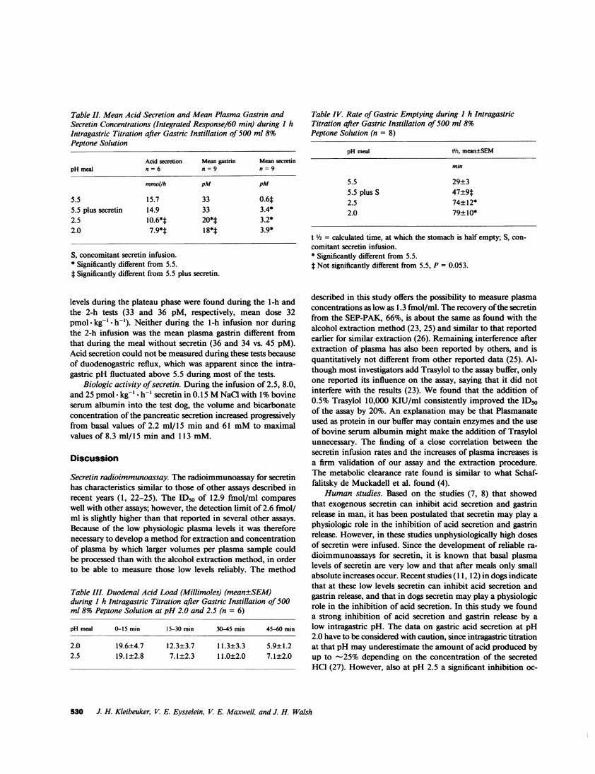
Table II. Mean Acid Secretion and Mean Plasma Gastrin andSecretin Concentrations (Integrated Response/60 min) during I hIntragastric Titration after Gastric Instillation of 500 ml 8%Peptone Solution
Acid secretion Mean gastrin Mean secretinpH meal n = 6 n = 9 n = 9
mmol/h pM pM
5.5 15.7 33 0.6t5.5 plus secretin 14.9 33 3.4*2.5 10.6*t 20*t 3.2*2.0 7.9*t 18*t 3.9*
S, concomitant secretin infusion.* Significantly different from 5.5.t Significantly different from 5.5 plus secretin.
levels during the plateau phase were found during the 1-h andthe 2-h tests (33 and 36 pM, respectively, mean dose 32pmol* kg' - h-'). Neither during the 1-h infusion nor duringthe 2-h infusion was the mean plasma gastrin different fromthat during the meal without secretin (36 and 34 vs. 45 pM).Acid secretion could not be measured during these tests becauseof duodenogastric reflux, which was apparent since the intra-gastric pH fluctuated above 5.5 during most of the tests.
Biologic activity of secretin. During the infusion of 2.5, 8.0,and 25 pmol - kg-' * h-' secretin in 0.15 MNaCl with 1%bovineserum albumin into the test dog, the volume and bicarbonateconcentration of the pancreatic secretion increased progressivelyfrom basal values of 2.2 ml/15 min and 61 mMto maximalvalues of 8.3 ml/15 min and 113 mM.
Discussion
Secretin radioimmunoassay. The radioimmunoassay for secretinhas characteristics similar to those of other assays described inrecent years (1, 22-25). The ID50 of 12.9 fmol/ml compareswell with other assays; however, the detection limit of 2.6 fmol/ml is slightly higher than that reported in several other assays.Because of the low physiologic plasma levels it was thereforenecessary to develop a method for extraction and concentrationof plasma by which larger volumes per plasma sample couldbe processed than with the alcohol extraction method, in orderto be able to measure those low levels reliably. The method
Table III. Duodenal Acid Load (Millimoles) (mean±SEM)during I h Intragastric Titration after Gastric Instillation of 500ml 8% Peptone Solution at pH 2.0 and 2.5 (n = 6)
pH meal 0-15 min 15-30 min 30-45 min 45-0 min
2.0 19.6±4.7 12.3±3.7 11.3±3.3 5.9±1.22.5 19.1±2.8 7.1±2.3 11.0±2.0 7.1±2.0
Table IV. Rate of Gastric Emptying during I h IntragastricTitration after Gastric Instillation of 500 ml 8%Peptone Solution (n = 8)
pH meal t'h, mean±SEM
min
5.5 29±35.5 plus S 47±9$2.5 74±12*2.0 79±10*
t 1/2 = calculated time, at which the stomach is half empty; S. con-comitant secretin infusion.* Significantly different from 5.5.$ Not significantly different from 5.5, P = 0.053.
described in this study offers the possibility to measure plasmaconcentrations as low as 1.3 fmol/ml. The recovery of the secretinfrom the SEP-PAK, 66%, is about the same as found with thealcohol extraction method (23, 25) and similar to that reportedearlier for similar extraction (26). Remaining interference afterextraction of plasma has also been reported by others, and isquantitatively not different from other reported data (25). Al-though most investigators add Trasylol to the assay buffer, onlyone reported its influence on the assay, saying that it did notinterfere with the results (23). We found that the addition of0.5% Trasylol 10,000 KIU/ml consistently improved the ID50of the assay by 20%. An explanation may be that Plasmanateused as protein in our buffer may contain enzymes and the useof bovine serum albumin might make the addition of Trasylolunnecessary. The finding of a close correlation between thesecretin infusion rates and the increases of plasma increases isa firm validation of our assay and the extraction procedure.The metabolic clearance rate found is similar to what Schaf-falitsky de Muckadell et al. found (4).
Human studies. Based on the studies (7, 8) that showedthat exogenous secretin can inhibit acid secretion and gastrinrelease in man, it has been postulated that secretin may play aphysiologic role in the inhibition of acid secretion and gastrinrelease. However, in these studies unphysiologically high dosesof secretin were infused. Since the development of reliable ra-dioimmunoassays for secretin, it is known that basal plasmalevels of secretin are very low and that after meals only smallabsolute increases occur. Recent studies (1 1, 12) in dogs indicatethat at these low levels secretin can inhibit acid secretion andgastrin release, and that in dogs secretin may play a physiologicrole in the inhibition of acid secretion. In this study we founda strong inhibition of acid secretion and gastrin release by alow intragastric pH. The data on gastric acid secretion at pH2.0 have to be considered with caution, since intragastric titrationat that pH may underestimate the amount of acid produced byup to -25% depending on the concentration of the secretedHCO(27). However, also at pH 2.5 a significant inhibition oc-
530 J. H. Kleibeuker, V. E. Eysselein, V. E. Maxwell, and J. H. Walsh
curred. In contrast, exogenous secretin producing plasma levelssimilar to those found during meals of low pH did not inhibitgastrin release and acid secretion during a meal of pH 5.5.Although the peak levels of secretin during the meals of lowpH were slightly higher than the plateau levels during the secretininfusion (5.5 vs. 4.6 fmol/ml), it is unlikely that this explainsthe lack of inhibitory effect by secretin, because the mean levelswere similar in these studies. Several facts may contribute tothe differences found between humans and dogs. Postprandialplasma levels of secretin, as measured by the same radioim-munoassay, are higher in dogs than in men (5, 24). Further,dogs seem to be more sensitive to the acid-inhibiting effect ofsecretin than men (7). A possible explanation for these differencescould be a structural difference between human and porcineand/or canine secretin, that could produce falsely low radioim-munoassay values in men and/or a lower biologic activity.
The doses of secretin infused during the meals of pH 5.5produce a marked increase of pancreatic secretion, and aretherefore thought to produce physiologic plasma concentrations.It might thus be expected that the secretin given in this studycould inhibit the gastrin release if it plays a physiologic role inthis inhibition. Our results are in agreement with an earlierreport in which it was shown that secretin was not responsiblefor the inhibition of pentagastrin-stimulated acid secretion byintraduodenal acidification (28-30), nor was it apparently thecause of inhibition of histamine-stimulated secretion (3 1).
Apparently factors other than secretin are the mediators ofthe acid-induced inhibition of gastrin release, which we foundto be responsible for inhibition of acid secretion by low intra-gastric pH. Whether the mechanism of this inhibition is throughintragastric pathways or whether mechanisms through receptorsin the duodenal or intestinal wall are involved is an unsolvedquestion. The inhibition by intraduodenal acidification is in-dependent of gastrin (28), and thus does not seem to play agreat role in the inhibition by low intragastric pH, which isprimarily an inhibition of gastrin release.
Further studies are necessary to determine the mechanismsinvolved in the acid-induced inhibition of acid secretion. Hor-monal, paracrine, and neural mechanisms and substances suchas somatostatin, vasoactive intestinal peptide, and prostaglandinsmay be involved.
The finding that exogenous secretin at doses five- to sixfoldhigher than those found during the meals at low pH does notsignificantly inhibit the gastrin release is not in disagreementwith other studies, although a real difference might have becomeapparent if larger numbers of subjects had been studied. Daltonet al. (8) found that secretin in doses up to 0.5 CU- kg.- -' * h-equivalent to 41 pmol * kg-' * h-' did not inhibit gastrin releasein response to a steak meal, but these authors did not addprotein to the infusion solution so it may well be that they gavelower doses than they had intended. Londong et al. (9) foundthat peptone-stimulated gastrin release was inhibited by 0.5CU. kg-' * h-' secretin, but this effect first became apparent dur-ing the second half hour of the first hour after the start of thestudy, and the integrated mean over the first hour was probably
not different whether they gave secretin or not. Whether thesehigh levels of secretin inhibit gastrin release and/or acid secretionis probably not important for an understanding of physiologicmechanisms, since these levels are never seen under pure phys-iologic conditions.
A low intragastric pH clearly inhibits gastric emptying. Al-though we did not find a significant inhibition by secretin, se-cretin may still play a role in the inhibition of gastric emptyingas the findings of Valenzuela and Defilippi (13) suggest. Westudied only eight subjects and the borderline significance (P= 0.053) may have been due to this small number.
Weconclude that secretin does not play a role in the acid-induced inhibition of peptone stimulated acid secretion and isunlikely to have a physiologic role in the inhibition of acidsecretion in man. The role that secretin may play in the acid-induced inhibition of gastric emptying seems to be a minor one.
Acknowledgments
Dr. Janet Elashoff and Dr. Terry Reedy are gratefully acknowledgedfor their help in statistical analysis.
This work was supported by National Institutes of Health grantsAM 17294 and AM 17328. Dr. J. H. Kleibeuker was a recipient of aFullbright grant (home institution, Department of Medicine, UniversityHospital, Groningen, The Netherlands). Dr. V. E. Eysselein was supportedby the Deutsche Forschungs Gemeinschaft.
References
1. Greenberg, G. R., R. F. McCloy, J. H. Baron, M. G. Bryant, andS. R. Bloom. 1982. Gastric acid regulates the release of plasma secretinin man. Eur. J. Clin. Invest. 12:361-372.
2. Pelletier, M. J., J. A. P. Chayvialle, and Y. Minaure. 1978. Unevenand transient secretin release after a liquid test meal. Gastroenterology.75:1124-1132.
3. Fahrenkrug, J., 0. B. Schaffalitsky de Muckadell, and S. J. Rune.1978. pH threshold for release of secretin in normal subjects and inpatients with duodenal ulcer and patients with chronic pancreatitis.Scand. J. Gastroenterol. 13:177-186.
4. Schaffalitsky de Muckadell, 0. B., J. Fahrenkrug, S. W. Boolsen,and H. Worming. 1978. Pancreatic response and plasma secretin con-centration during infusion of low dose secretin in man. Scand. J. Gas-troenterol. 13:305-311.
5. Chey, W. Y., M. S. Kim, K. Y. Lee, and T. M. Chang. 1979.Effect of rabbit antisecretin serum on postprandial pancreatic secretionin dogs. Gastroenterology. 77:1266-1275.
6. Waldum, H. L., N. Walde, and P. G. Burhol. 1981. The effectof secretin on gastric H' and pepsin secretion and on urinary electrolyteelevation in man. Scand. J. Gastroenterol. 16:999-1004.
7. Brooks, A. M., and M. I. Grossman. 1970. Effect of secretin andcholecystokinin on pentagastrin-stimulated gastric secretion in man.Gastroenterology. 59(1): 114-119.
8. Dalton, M. D., A. M. Eisenstein, J. H. Walsh, and J. S. Fordtran.1976. Effect of secretin on gastric function in normal subjects and inpatients with duodenal ulcer. Gastroenterology. 71:24-29.
9. Londong, W., V. Londong, L. E. Hanssen, and A. Schwanner.1981. Gastric effects and side effects of synthetic secretin in man. Regul.Pept. 2:231-244.
531 Endogenous Secretin in Human Gastric Function
10. Jansen, J. B. M. J., and C. B. M. W. Lamers. 1981. Calcitoninand secretin inhibit bombesin-stimulated serum gastrin and gastric acidsecretion in man. Regul. Pept. 1:415-421.
11. Chey, W. Y., M. S. Kim, K. Y. Lee, and T. M. Chang. 1981.Secretin is an enterogastrone in the dog. Am. J. Physiol. 240:G239-G244.
12. Kim, Y. M., K. Y. Lee, and W. Y. Chey. 1981. Role of secretinon postprandial gastrin release in the dog: A further study. Surgery (St.Louis). 90:504-508.
13. Valenzuela, J. E., and C. Defilippi. 1981. Inhibition of gastricemptying in humans by secretin, the octapeptide of cholecystokinin andintraduodenal fat. Gastroenterology. 81:898-902.
14. Eysselein, V. E., J. H. Kleibeuker, V. Maxwell, T. Reedy, andJ. H. Walsh. 1983. Inhibition of gastric acid secretion at low intragastricpH in man: relation to plasma gastrin. Gastroenterology. 84:1147a.(Abstr.)
15. Hassan, M. A., and M. Hobsley. 1970. Positioning of subjectand of nasogastric tube during a gastric secretion study. Br. Med. J.1:458-460.
16. Lam, S. K., J. I. Isenberg, M. I. Grossman, W. H. Lane, andJ. H. Walsh. 1980. Gastric acid secretion is abnormally sensitive toendogenous gastrin released after peptone test meals in duodenal ulcerpatients. J. Clin. Invest. 65:555-562.
17. George, J. D. 1968. Newclinical method for measuring the rateof gastric emptying: the double-sampling test meal. Gut. 9:237-242.
18. Gross, R. A., J. I. Isenberg, D. Hogan, and I. M. Samloff. 1978.Effect of fat on meal-stimulated duodenal acid load, duodenal pepsinload, and serum gastrin in duodenal ulcer and normal subjects. Gas-troenterology. 75:357-362.
19. Rosenquist, G. L., and J. H. Walsh. 1980. Radioimmunoassayof gastrin. In Gastrointestinal Hormones. G. B. Jerzy Glass, editor.Raven Press, New York, 769-795.
20. Rodbard, D., et al. 1975. Radioimmunoassay Data Processing:Listings and Documentation, Third ed. The Logistic Method and QualityControl (PB246 or 224). U. S. Department of Commerce, NationalTechnical Information Service, Springfield, VA. Vol. 2.
21. Elashoff, J. D., T. R. Reedy, and J. H. Meyer. 1982. Analysisof gastric emptying data. Gastroenterology. 83:1306-1312.
22. Burhol, P. G., and H. L. Waldun. 1975. Radioimmunoassay ofsecretin in acidified plasma. Acta Hepatogastroenterol. 25:474-481.
23. Hanssen, L. E., and P. Torjesen. 1977. Radioimmunoassay ofsecretin in human plasma. Scand. J. Gastroenterol. 12:481-488.
24. Rominger, J. M., W. Y. Chey, and T. M. Chang. 1981. Plasmasecretin concentrations and gastric pH in healthy subjects and patientswith digestive diseases. Dig. Dis. Sci. 26:591-597.
25. Schaffalitsky de Muckadell, 0. B., and J. Fahrenkrug. 1977.Radioimmunoassay of secretin in plasma. Scand. J. Clin. Lab. Invest.37:155-162.
26. Yang, R.-K., Li, H.-R., Eng, J., Greenstein, R., Straus, E., andYalow, R. S. 1983. Secretin responses to feeding and acid load. J. Lab.Clin. Med. 102:17-23.
27. Spenney, J. G. 1979. Physical chemical and technical limitationsof intragastric titration. Gastroenterology. 76:1025-1036.
28. Ward, A. S., and S. R. Bloom. 1974. The role of secretin in theinhibition of gastric secretion by intraduodenal acid. Gut. 15:889-897.
29. Wormsley, K. G. 1970. Response to duodenal acidification inman. II. Effects on the gastric secretory response to pentagastrin. Scand.J. Gastroenterol. 5:207-215.
30. Berstad, A., and H. Petersen. 1970. Dose-response relationshipof the effect of secretin on acid and pepsin secretion in man. Scand.J. Gastroenterol. 5:647-654.
31. Johnston, D., and H. L. Duthie. 1966. Inhibition of histamine-stimulated gastric secretion by acid in the duodenum in man. Gut.7:58-68.
532 J. H. Kleibeuker, V. E. Eysselein, V. E. Maxwell, and J. H. Walsh