Embed Size (px)
Citation preview
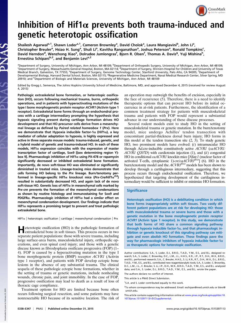
Inhibition of Hif1α prevents both trauma-induced andgenetic heterotopic ossificationShailesh Agarwala,1, Shawn Lodera,1, Cameron Brownleya, David Choloka, Laura Mangiavinib, John Lia,Christopher Breulera, Hsiao H. Sunga, Shuli Lia, Kavitha Ranganathana, Joshua Petersona, Ronald Tompkinsc,David Herndond, Wenzhong Xiaoe, Dolrudee Jumlongrasf, Bjorn R. Olsenf, Thomas A. Davisg, Yuji Mishinah,Ernestina Schipanib,2, and Benjamin Levia,2
aDepartment of Surgery, University of Michigan, Ann Arbor, MI 48109; bDepartment of Orthopedic Surgery, University of Michigan, Ann Arbor, MI 48109;cDepartment of Surgery, Massachusetts General Hospital, Boston, MA 02114; dDepartment of Surgery, Shriners Hospital for Children and University of TexasMedical Branch, Galveston, TX 77555; eDepartment of Surgery, Genome Technology Center, Stanford University, Palo Alto, CA 94305; fDepartment ofDevelopmental Biology, Harvard Dental School, Boston, MA 02115; gRegenerative Medicine Department, Naval Medical Research Center, Silver Spring, MD20910; and hDepartment of Biologic and Materials Sciences, University of Michigan, Ann Arbor, MI 48109
Edited by Gregg L. Semenza, The Johns Hopkins University School of Medicine, Baltimore, MD, and approved December 4, 2015 (received for review August4, 2015)
Pathologic extraskeletal bone formation, or heterotopic ossifica-tion (HO), occurs following mechanical trauma, burns, orthopedicoperations, and in patients with hyperactivating mutations of thetype I bone morphogenetic protein receptor ACVR1 (Activin type 1receptor). Extraskeletal bone forms through an endochondral pro-cess with a cartilage intermediary prompting the hypothesis thathypoxic signaling present during cartilage formation drives HOdevelopment and that HO precursor cells derive from a mesenchy-mal lineage as defined by Paired related homeobox 1 (Prx). Herewe demonstrate that Hypoxia inducible factor-1α (Hif1α), a keymediator of cellular adaptation to hypoxia, is highly expressed andactive in three separate mouse models: trauma-induced, genetic, anda hybrid model of genetic and trauma-induced HO. In each of thesemodels, Hif1α expression coincides with the expression of mastertranscription factor of cartilage, Sox9 [(sex determining region Y)-box 9]. Pharmacologic inhibition of Hif1α using PX-478 or rapamycinsignificantly decreased or inhibited extraskeletal bone formation.Importantly, de novo soft-tissue HO was eliminated or significantlydiminished in treated mice. Lineage-tracing mice demonstrate thatcells forming HO belong to the Prx lineage. Burn/tenotomy per-formed in lineage-specific Hif1α knockout mice (Prx-Cre/Hif1αfl:fl)resulted in substantially decreased HO, and again lack of de novosoft-tissue HO. Genetic loss of Hif1α in mesenchymal cells marked byPrx-cre prevents the formation of the mesenchymal condensationsas shown by routine histology and immunostaining for Sox9 andPDGFRα. Pharmacologic inhibition of Hif1α had a similar effect onmesenchymal condensation development. Our findings indicate thatHif1α represents a promising target to prevent and treat pathologicextraskeletal bone.
HIF1α | heterotopic ossification | cartilage | mesenchymal condensation | Prx
Heterotopic ossification (HO) is the pathologic formation ofextraskeletal bone in soft tissues. This process occurs in two
separate patient populations: those with severe trauma, includinglarge surface-area burns, musculoskeletal injury, orthopedic op-erations, and even spinal cord injury; and those with a geneticdisease known as fibrodysplasia ossificans progressiva (FOP) (1–4). FOP is caused by a hyperactivating mutation in the type Ibone morphogenetic protein (BMP) receptor ACVR1 (Activintype 1 receptor), and patients with FOP develop ectopic bonelesions in the absence of any substantial trauma. The clinicalsequela of these pathologic ectopic bone formations, whether inthe setting of trauma or genetic mutations, include nonhealingwounds, chronic pain, and joint immobility. In the case of FOP,progressive ossification may lead to death as a result of loss ofthoracic cage compliance.Treatment options for HO are limited because bone often
recurs following surgical resection, and some patients may havenonresectable HO because of its sensitive location. The risk of
an operation may outweigh the benefits of excision, especially inthe face of recurrence (5). Therefore, there is a need to identifytherapeutic options that can prevent HO before its initial oc-currence in at-risk patients. Furthermore, the identification of acommon treatment strategy for patients with musculoskeletaltrauma and patients with FOP would represent a substantialadvance in our understanding of these disease processes.Several rodent models exist to study HO in the setting of
musculoskeletal trauma or genetic mutation. In the burn/tenotomymodel, mice undergo Achilles’ tendon transection withconcomitant partial-thickness dorsal burn injury; HO forms inthis model at the tendon transection site (2, 3). To study geneticHO, two prominent models have evolved: (i) intramuscular HOthrough Ad.cre-inducible constitutively active ACVR1 (caACVR1:ACVR1 Q207D) with cardiotoxin injection (1), and (ii) congenitalHO in conditional caACVR1 knockin mice [Nfatc1 (nuclear factor ofactivated T-cells, cytoplasmic 1)-cre/caACVR1fl/wt] (6). HO in theburn/tenotomy model and the ACVR1fl/wt models has been shown todevelop through a cartilaginous intermediary, suggesting that thisprocess occurs through endochondral ossification. Therefore, wehypothesized that targeting development of the cartilaginous in-termediary would be sufficient to inhibit or minimize HO formation.
Significance
Heterotopic ossification (HO) is a debilitating condition in whichbone forms inappropriately within soft tissues. Two vastly dif-ferent patient populations are at risk for developing HO: thosewith musculoskeletal trauma or severe burns and those with agenetic mutation in the bone morphogenetic protein receptorACVR1 (Activin type 1 receptor). In this study, we demonstratethat both forms of HO share a common signaling pathwaythrough hypoxia inducible factor-1α, and that pharmacologic in-hibition or genetic knockout of this signaling pathway can miti-gate and even abolish HO formation. These findings pave theway for pharmacologic inhibitors of hypoxia inducible factor-1αas therapeutic options for heterotopic ossification.
Author contributions: S.A., S. Loder, D.J., B.R.O., T.A.D., Y.M., E.S., and B.L. designed re-search; S.A., S. Loder, C. Brownley, D.C., L.M., J.L., H.H.S., K.R., J.P., R.T., D.H., W.X., B.R.O.,and B.L. performed research; S.A., C. Breuler, H.H.S., S. Li, K.R., R.T., D.H., W.X., D.J., B.R.O.,T.A.D., Y.M., E.S., and B.L. contributed new reagents/analytic tools; S.A., S. Loder, C. Brownley,D.C., L.M., J.L., C. Breuler, S. Li, J.P., D.H., W.X., B.R.O., T.A.D., Y.M., E.S., and B.L. analyzeddata; and S.A., S. Loder, D.J., B.R.O., T.A.D., Y.M., E.S., and B.L. wrote the paper.
The authors declare no conflict of interest.
This article is a PNAS Direct Submission.1S.A. and S. Loder contributed equally to this work.2To whom correspondence may be addressed. Email: [email protected] or [email protected].
This article contains supporting information online at www.pnas.org/lookup/suppl/doi:10.1073/pnas.1515397113/-/DCSupplemental.
E338–E347 | PNAS | Published online December 31, 2015 www.pnas.org/cgi/doi/10.1073/pnas.1515397113
Dow
nloa
ded
by g
uest
on
Feb
ruar
y 15
, 202
0
Hypoxia inducible factor-1α (Hif1α) is one particular signalingmediator that has been shown to be critical for normal chondro-genesis (7–10). Conditional Hif1α knockout mice have demonstratedthat Hif1α is critical for chondrocyte survival and differentiation.Given the critical role of Hif1α in normal cartilage development andthat HO forms through a cartilaginous intermediary, we hypothe-sized that targeting Hif1α through drug treatment or conditionalHif1α knockout would inhibit HO formation.
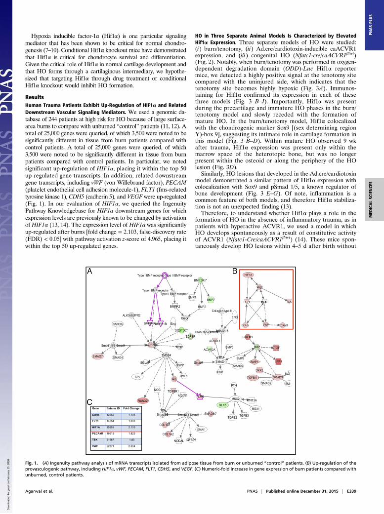
ResultsHuman Trauma Patients Exhibit Up-Regulation of HIF1α and RelatedDownstream Vascular Signaling Mediators. We used a genomic da-tabase of 244 patients at high risk for HO because of large surface-area burns to compare with unburned “control” patients (11, 12). Atotal of 25,000 genes were queried, of which 3,500 were noted to besignificantly different in tissue from burn patients compared withcontrol patients. A total of 25,000 genes were queried, of which3,500 were noted to be significantly different in tissue from burnpatients compared with control patients. In particular, we notedsignificant up-regulation of HIF1α, placing it within the top 50up-regulated gene transcripts. In addition, related downstreamgene transcripts, including vWF (von Willebrand factor), PECAM(platelet endothelial cell adhesion molecule-1), FLT1 (fms-relatedtyrosine kinase 1), CDH5 (cadherin 5), and VEGF were up-regulated(Fig. 1). In our evaluation of HIF1α, we queried the IngenuityPathway Knowledgebase for HIF1α downstream genes for whichexpression levels are previously known to be changed by activationof HIF1α (13, 14). The expression level of HIF1α was significantlyup-regulated after burns [fold change = 2.103, false-discovery rate(FDR) < 0.05] with pathway activation z-score of 4.965, placing itwithin the top 50 up-regulated genes.
HO in Three Separate Animal Models Is Characterized by ElevatedHif1α Expression. Three separate models of HO were studied:(i) burn/tenotomy, (ii) Ad.cre/cardiotoxin-inducible caACVR1expression, and (iii) congenital HO (Nfatc1-cre/caACVR1fl/wt)(Fig. 2). Notably, when burn/tenotomy was performed in oxygen-dependent degradation domain (ODD)-Luc Hif1α reportermice, we detected a highly positive signal at the tenotomy sitecompared with the uninjured side, which indicates that thetenotomy site becomes highly hypoxic (Fig. 3A). Immunos-taining for Hif1α confirmed its expression in each of thesethree models (Fig. 3 B–J). Importantly, Hif1α was presentduring the precartilage and immature HO phases in the burn/tenotomy model and slowly receded with the formation ofmature HO. In the burn/tenotomy model, Hif1α colocalizedwith the chondrogenic marker Sox9 [(sex determining regionY)-box 9], suggesting its intimate role in cartilage formation inthis model (Fig. 3 B–D). Within mature HO observed 9 wkafter trauma, Hif1α expression was present only within themarrow space of the heterotopic bone, but was no longerpresent within the osteoid or along the periphery of the HOlesion (Fig. 3D).Similarly, HO lesions that developed in the Ad.cre/cardiotoxin
model demonstrated a similar pattern of Hif1α expression withcolocalization with Sox9 and pSmad 1/5, a known regulator ofbone development (Fig. 3 E–G). Of note, inflammation is acommon feature of both models, and therefore Hif1α stabiliza-tion is not an unexpected finding (13).Therefore, to understand whether Hif1α plays a role in the
formation of HO in the absence of inflammatory trauma, as inpatients with hyperactive ACVR1, we used a model in whichHO develops spontaneously as a result of constitutive activityof ACVR1 (Nfatc1-Cre/caACVR1fl:wt) (14). These mice spon-taneously develop HO lesions within 4–5 d after birth without
Fig. 1. (A) Ingenuity pathway analysis of mRNA transcripts isolated from adipose tissue from burn or unburned “control” patients. (B) Up-regulation of theprovasculogenic pathway, including HIF1α, vWF, PECAM, FLT1, CDH5, and VEGF. (C) Numeric-fold increase in gene expression of burn patients compared withunburned, control patients.
Agarwal et al. PNAS | Published online December 31, 2015 | E339
MED
ICALSC
IENCE
SPN
ASPL
US
Dow
nloa
ded
by g
uest
on
Feb
ruar
y 15
, 202
0
concomitant trauma or Ad.Cre or cardiotoxin injections. Lesionsare generally localized to the joints, including ankles, knees, elbows,and digits (14). Immunostaining confirmed robust Hif1α expressionwithin immature HO also in this model, which indicates that Hif1α
plays a role in HO formation in the setting of hyperactive BMPreceptor signaling despite absence of inflammatory trauma (Fig. 3H–J). Again, robust colocalization of Hif1α with Sox9 andpSmad 1/5 was noted.
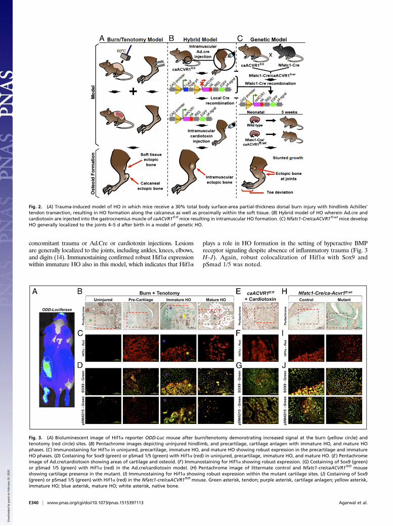
Fig. 2. (A) Trauma-induced model of HO in which mice receive a 30% total body surface-area partial-thickness dorsal burn injury with hindlimb Achilles’tendon transection, resulting in HO formation along the calcaneus as well as proximally within the soft tissue. (B) Hybrid model of HO wherein Ad.cre andcardiotoxin are injected into the gastrocnemius muscle of caACVR1fl:fl mice resulting in intramuscular HO formation. (C) Nfatc1-Cre/caACVR1fl:wt mice developHO generally localized to the joints 4–5 d after birth in a model of genetic HO.
Fig. 3. (A) Bioluminescent image of Hif1α reporter ODD-Luc mouse after burn/tenotomy demonstrating increased signal at the burn (yellow circle) andtenotomy (red circle) sites. (B) Pentachrome images depicting uninjured hindlimb, and precartilage, cartilage anlagen with immature HO, and mature HOphases. (C) Immunostaining for Hif1α in uninjured, precartilage, immature HO, and mature HO showing robust expression in the precartilage and immatureHO phases. (D) Costaining for Sox9 (green) or pSmad 1/5 (green) with Hif1α (red) in uninjured, precartilage, immature HO, and mature HO. (E) Pentachromeimage of Ad.cre/cardiotoxin showing areas of cartilage and osteoid. (F) Immunostaining for Hif1α showing robust expression. (G) Costaining of Sox9 (green)or pSmad 1/5 (green) with Hif1α (red) in the Ad.cre/cardiotoxin model. (H) Pentachrome image of littermate control and Nfatc1-cre/caACVR1fl/fl mouseshowing cartilage presence in the mutant. (I) Immunostaining for Hif1α showing robust expression within the mutant cartilage sites. (J) Costaining of Sox9(green) or pSmad 1/5 (green) with Hif1α (red) in the Nfatc1-cre/caACVR1fl/fl mouse. Green asterisk, tendon; purple asterisk, cartilage anlagen; yellow asterisk,immature HO; blue asterisk, mature HO; white asterisk, native bone.
E340 | www.pnas.org/cgi/doi/10.1073/pnas.1515397113 Agarwal et al.
Dow
nloa
ded
by g
uest
on
Feb
ruar
y 15
, 202
0
Taken together, these data show that Hif1α expression ap-pears to be a common denominator in trauma-induced and ge-netic models of HO, and precedes cartilage formation andcartilage ossification, thereby validating it as a therapeutic target.
Pharmacologic Inhibition of Hif1α Limits HO After Burn/Tenotomy.Next, we tested the hypothesis that Hif1α inhibition can preventHO. For this purpose, we used the drug PX-478, which has beenshown to inhibit Hif1α transcription and translation (15). In vitrotreatment of cells derived from the tenotomy site 3 weeks afterinjury (3WLST) and cultured in hypoxia showed diminishedlevels of the Hif1α transcript and of the chondrogenic genetranscripts Sox9 and Acan (aggrecan) upon treatment with PX-478 (Fig. 4A). Additionally, PX-478 and rapamycin, a previouslydescribed Hif1α inhibitor (16), both significantly diminishedHif1α produced by mesenchymal cells isolated from tendon,confirming again that these drugs affect Hif1α levels in cells localto the future HO site (Fig. S1). We next tested whether treat-ment with PX-478 decreases Hif1α expression and cartilageformation in vivo, and consequently inhibits overall developmentof HO. Mice received burn/tenotomy and were subsequentlytreated with PX-478; histologic evaluation after 3 wk confirmed asubstantial decrease in the cartilage anlagen, which is typicallypresent after 3 wk (Fig. 4B). Furthermore, we found diminishedHif1α expression 3 wk after injury (Fig. 4C). Consistent withthese data, expression of Sox9 was considerably diminished inthe PX-478–treated group (Fig. 4C). Moreover, burn/tenotomymice treated with PX-478 demonstrated a significant reductionin total HO volume at 5 wk (4.3 mm3 vs. 1.5 mm3, P < 0.05) and 9wk (5.8 mm3 vs. 2.3 mm3, P < 0.05) after injury (Fig. 4 D and E).Finally, PX-478 treatment completely inhibited “soft tissue”HO—extraskeletal bone, which forms within the proximal
transected tendon and distal gastrocnemius but away from thecalcaneus—after 9 wk, as shown by binary analysis (yes/no: χ2 = 9.5,P < 0.01) and quantitative comparison (0.90 mm3 vs. 0.00 mm3,P = 0.05) (Fig. 4E). This result is notable, as soft tissue HOlikely forms de novo without the influence of adjacent carti-lage, bone, or periosteum normally located in close proximity toextraskeletal bone at the calcaneus. Taken together, these findingssuggest that Hif1α is a permissive factor for chondrogenesis and itsinhibition can prevent transition of nonosteochondro progenitorlineage cells into cells forming cartilage and ultimately extraskeletalbone. Notably, we found no adverse effects of PX-478 on woundhealing of the burn or at the hindlimb tenotomy sites (Fig. S2). Totest a second Hif1α inhibitor, mice were treated with rapamycin(16), resulting in significantly diminished de novo HO formation(1.60 mm3 vs. 0.81 mm3, P < 0.05) (Fig. 4 F and G) (16).
Pharmacologic Inhibition of Hif1α Limits HO Caused by ACVR1Constitutive Activity. We next confirmed these findings in mod-els of constitutive ACVR1 activity caused by expression of thecaACVR1 (ACVR1 Q207D) mutation. caACVR1fl/fl mice injectedwith cardiotoxin and Ad.cre develop robust HO and this modelhas been used to study inhibitors of ACVR1 signaling. caACVR1fl/fl
mice treated with PX-478 demonstrated near elimination ofcartilage or bone based on pentachrome staining (Fig. 5A) afterAd.cre/cardiotoxin induction. Similarly, there was elimination ofHif1α and Sox9 based on immunostaining (Fig. 5B). Finally,microCT analysis confirmed the complete absence of HO in thePX-478 treated group based on binary analysis (yes/no; χ2 =13.6, P < 0.001) and quantitative comparison (18.1 mm3 vs.0.01 mm3, P = 0.01) (Fig. 5 C and D). These findings werestriking because of the substantially improved efficacy overother BMP inhibitors in the literature. Pentachrome staining
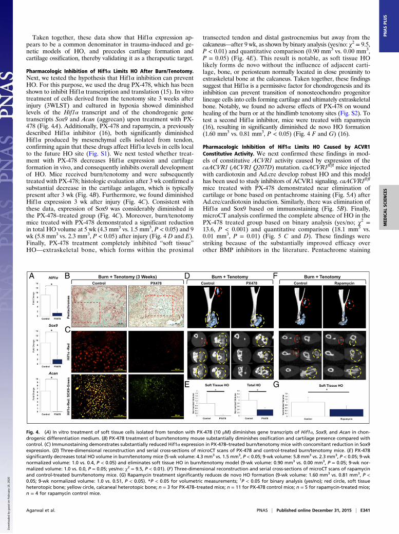
Fig. 4. (A) In vitro treatment of soft tissue cells isolated from tendon with PX-478 (10 μM) diminishes gene transcripts of Hif1α, Sox9, and Acan in chon-drogenic differentiation medium. (B) PX-478 treatment of burn/tenotomy mouse substantially diminishes ossification and cartilage presence compared withcontrol. (C) Immunostaining demonstrates substantially reduced Hif1α expression in PX-478–treated burn/tenotomy mice with concomitant reduction in Sox9expression. (D) Three-dimensional reconstruction and serial cross-sections of microCT scans of PX-478 and control-treated burn/tenotomy mice. (E) PX-478significantly decreases total HO volume in burn/tenotomy mice (5-wk volume: 4.3 mm3 vs. 1.5 mm3, P < 0.05; 9-wk volume: 5.8 mm3 vs. 2.3 mm3, P < 0.05; 9-wknormalized volume: 1.0 vs. 0.4, P < 0.05) and eliminates soft tissue HO in burn/tenotomy model (9-wk volume: 0.90 mm3 vs. 0.00 mm3, P = 0.05; 9-wk nor-malized volume: 1.0 vs. 0.0, P = 0.05; yes/no: χ2 = 9.5, P < 0.01). (F) Three-dimensional reconstruction and serial cross-sections of microCT scans of rapamycinand control-treated burn/tenotomy mice. (G) Rapamycin treatment significantly reduces de novo HO formation (9-wk volume: 1.60 mm3 vs. 0.81 mm3, P <0.05; 9-wk normalized volume: 1.0 vs. 0.51, P < 0.05). *P < 0.05 for volumetric measurements; †P < 0.05 for binary analysis (yes/no); red circle, soft tissueheterotopic bone; yellow circle, calcaneal heterotopic bone; n = 3 for PX-478–treated mice; n = 11 for PX-478 control mice; n = 5 for rapamycin-treated mice;n = 4 for rapamycin control mice.
Agarwal et al. PNAS | Published online December 31, 2015 | E341
MED
ICALSC
IENCE
SPN
ASPL
US
Dow
nloa
ded
by g
uest
on
Feb
ruar
y 15
, 202
0
confirmed absence of cartilage and bone (Fig. 5C) andimmunostaining further confirmed absence of Hif1α andSox9 expression (Fig. 5D). Again, these findings were repli-cated using rapamycin which showed complete absence of HOin treated mice (17.5 mm3 vs. 0.0 mm3, P < 0.001; yes/no; χ2 =14.3, P < 0.001) (Fig. 5 E and F).Finally, PX-478 was administered to mice with congenital HO
(Nfatc1-cre/caACVR1fl/fl) every other day starting from birth for2 wk. Treated mice had significantly less ectopic bone at theankle joints compared with mutant mice treated with vehicle(6.8 mm3 vs. 2.2 mm3, P < 0.01) (Fig. 5 G and H).
Genetic Loss of in Mesenchymal Progenitors Prevents Formation ofHeterotopic Ossifications. To strengthen our findings, we next useda conditional Hif1α knockout mouse because global Hif1αknockout is embryonic lethal. We first established that hetero-topic ossification following burn/tenotomy consists of cells fromthe paired related homeobox (Prx)-lineage using a series of lineagetracing experiments with Prx-cre/ROSA26mTmG mice (Fig. 6). Impor-tantly, we found that HO which formed within the soft tissue andmore proximally within the soft tissue both exhibited nearly100% presence of Prx-cre cells. In fact, we noted that the onlynon–Prx-cre cells present were those forming the marrow spaceof the mature HO.We therefore used a mouse model of conditional Hif1α knock-
out in Prx-cre cells (Prx-cre/Hif1αfl/fl). These mice have been shownto exhibit defective normal cartilage development (7). However,the impact on pathologic heterotopic ossification has not been
demonstrated; therefore, burn/tenotomy was performed in Prx-cre/Hif1αfl/fl mice. Mutant mice developed minimal HO only aroundthe calcaneus, and even these lesions were substantially smallerthan in controls (5.02 mm3 vs. 0.18 mm3, P < 0.01) (Fig. 7 A and B).
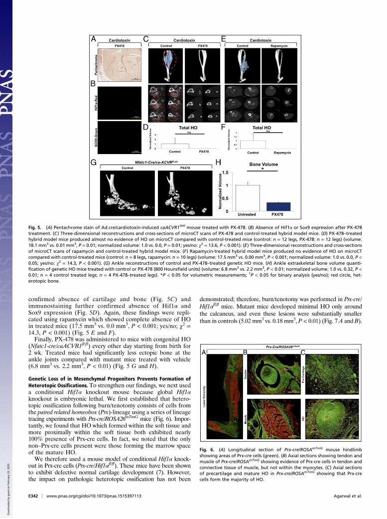
Fig. 5. (A) Pentachrome stain of Ad.cre/cardiotoxin-induced caACVR1fl/fl mouse treated with PX-478. (B) Absence of Hif1α or Sox9 expression after PX-478treatment. (C) Three-dimensional reconstructions and cross-sections of microCT scans of PX-478 and control-treated hybrid model mice. (D) PX-478–treatedhybrid model mice produced almost no evidence of HO on microCT compared with control-treated mice (control: n = 12 legs, PX-478: n = 12 legs) (volume:18.1 mm3 vs. 0.01 mm3, P = 0.01; normalized volume: 1.0 vs. 0.0, P = 0.01; yes/no: χ2 = 13.6, P < 0.001). (E) Three-dimensional reconstructions and cross-sectionsof microCT scans of rapamycin and control-treated hybrid model mice. (F) Rapamycin-treated hybrid model mice produced no evidence of HO on microCTcompared with control-treated mice (control: n = 8 legs, rapamycin: n = 10 legs) (volume: 17.5 mm3 vs. 0.00 mm3, P < 0.001; normalized volume: 1.0 vs. 0.0, P <0.05; yes/no: χ2 = 14.3, P < 0.001). (G) Ankle reconstructions of control and PX-478–treated genetic HO mice. (H) Ankle extraskeletal bone volume quanti-fication of genetic HO mice treated with control or PX-478 (800 Hounsfield units) (volume: 6.8 mm3 vs. 2.2 mm3, P < 0.01; normalized volume: 1.0 vs. 0.32, P <0.01; n = 4 control treated legs; n = 4 PX-478–treated legs). *P < 0.05 for volumetric measurements; †P < 0.05 for binary analysis (yes/no); red circle, het-erotopic bone.
Fig. 6. (A) Longitudinal section of Prx-cre/ROSAmTmG mouse hindlimbshowing areas of Prx-cre cells (green). (B) Axial sections showing tendon andmuscle of Prx-cre/ROSAmTmG showing evidence of Prx-cre cells in tendon andconnective tissue of muscle, but not within the myocytes. (C) Axial sectionsof precartilage and mature HO in Prx-cre/ROSAmTmG showing that Prx-crecells form the majority of HO.
E342 | www.pnas.org/cgi/doi/10.1073/pnas.1515397113 Agarwal et al.
Dow
nloa
ded
by g
uest
on
Feb
ruar
y 15
, 202
0
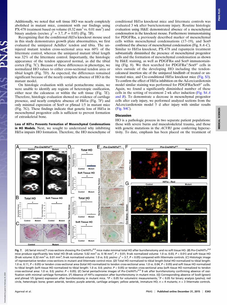
Additionally, we noted that soft tissue HO was nearly completelyabolished in mutant mice, consistent with our findings usingPX-478 treatment based on volume (0.32 mm3 vs. 0.01 mm3) andbinary analysis (yes/no; χ2 = 3.7, P = 0.05) (Fig. 7B).Recognizing that the conditional Hif1α knockout mouse used
in this study demonstrates growth plate abnormalities, we firstevaluated the uninjured Achilles’ tendon and tibia. The un-injured mutant tendon cross-sectional area was 60% of thelittermate control, whereas the uninjured mutant tibial lengthwas 32% of the littermate control. Importantly, the histologicappearance of the tendon appeared normal, as did the tibialcortex (Fig. 7C). Because of these differences in phenotype, wenormalized HO values to either cross-sectional tendon area ortibial length (Fig. 7D). As expected, the differences remainedsignificant because of the nearly complete absence of HO in themutant model.On histologic evaluation with serial pentachrome stains, we
were unable to identify any regions of heterotopic ossification,either near the calcaneus or within the soft tissue (Fig. 7E).Therefore, histologic evaluation showed no evidence of cartilagepresence, and nearly complete absence of Hif1α (Fig. 7F) andonly minimal expression of Sox9 or pSmad 1/5 in mutant mice(Fig. 7G). These findings indicate that genetic loss of Hif1α inmesenchymal progenitor cells is sufficient to prevent formationof extraskeletal bone.
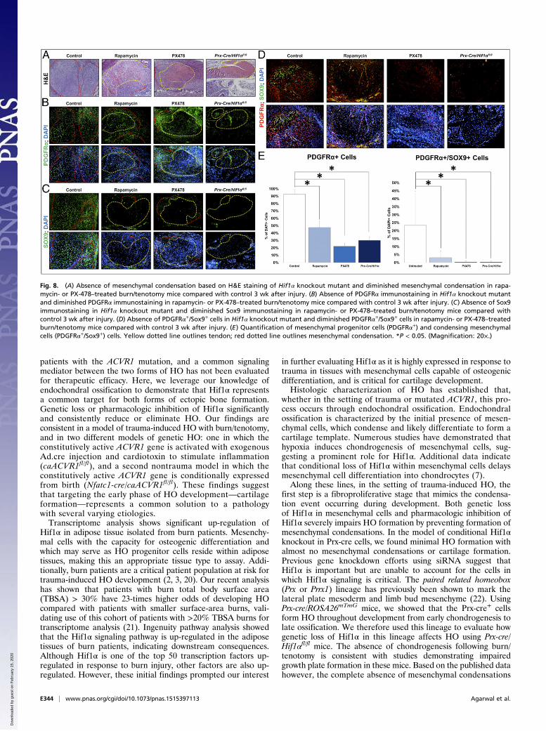
Loss of Hif1α Prevents Formation of Mesenchymal Condensationsin HO Models. Next, we sought to understand why inhibitingHif1α impairs HO formation. Therefore, the HO mesenchyme of
conditional Hif1α knockout mice and littermate controls wasevaluated 3 wk after burn/tenotomy injury. Routine histologicevaluation using H&E demonstrated absence of a mesenchymalcondensation in the knockout mouse. Furthermore immunostainingfor PDGFRα, a previously described marker of mesenchymalcells within mesenchymal condensations (17–19), and Sox9confirmed the absence of mesenchymal condensation (Fig. 8 A–C).Similar to Hif1α knockout, PX-478 and rapamycin treatmentsubstantially diminished the presence of mesenchymal progenitorcells and the formation of mesenchymal condensation as shownby H&E staining, as well as PDGFRα and Sox9 immunostain-ing (Fig. 8). We then searched for PDGFRα+/Sox9+ cells insites outside of the developing HO including the tendon-calcaneal insertion site of the uninjured hindlimb of treated or un-treated mice, and Cre-conditional Hif1α knockout mice (Fig. S3).To confirm the effect of Hif1α inhibition on the Ad.cre/cardiotoxinmodel similar staining was performed for PDGFRα/Sox9+ cells.Again, we found a significantly diminished number of thesecells in the setting of treatment 2 wk after induction (Fig. S4 Aand B). To demonstrate a decrease in mesenchymal progenitorcells after early injury, we performed analyzed sections from theAd.cre/cardiotoxin model 5 d after injury with similar results(Fig. S4C).
DiscussionHO is a pathologic process in two separate patient populations:those with severe burns and musculoskeletal trauma, and thosewith genetic mutations in the ACVR1 gene conferring hyperac-tivity. To date, emphasis has been placed on the treatment of
Fig. 7. (A) Serial microCT cross-sections showing Prx-Cre/Hif1αfl:fl mice make minimal total HO after burn/tenotomy and no soft tissue HO. (B) Prx-Cre/Hif1αfl:fl
mice produce significantly less total HO (9-wk volume: 5.02 mm3 vs. 0.18 mm3, P < 0.01; 9-wk normalized volume: 1.0 vs. 0.03, P < 0.01) and soft tissue HO(9-wk volume: 0.32 mm3 vs. 0.01 mm3; 9-wk normalized volume: 1.0 vs. 0.0; yes/no: χ2 = 3.7, P < 0.05) compared with littermate controls. (C) Histologic imageof representative tendon cross-sections in mutant and littermate control mice. (D) Total HO normalized to tibial length (total HO normalized to tibial length:1.0 vs. 0.11, P < 0.05) or tendon cross-sectional area (total HO normalized to tendon cross-sectional area: 1.0 vs. 0.04, P < 0.05) and soft tissue HO normalizedto tibial length (soft tissue HO normalized to tibial length: 1.0 vs. 0.0; yes/no: P < 0.05) or tendon cross-sectional area (soft tissue HO normalized to tendoncross-sectional area: 1.0 vs. 0.0; yes/no: P < 0.05). (E) Serial pentachrome images of Prx-Cre/Hif1αfl:fl 9 wk after burn/tenotomy confirming absence of ossi-fication with minimal cartilage formation. (F) Absence of Hif1α expression after burn/tenotomy in mutant mice. (G) Corresponding absence of Sox9 (green)and pSmad 1/5 (green) expression after burn/tenotomy in mutant mice. *P < 0.05 for volumetric measurements; †P < 0.05 for binary analysis (yes/no); redcircle, heterotopic bone; green asterisk, tendon; purple asterisk, cartilage anlagen; yellow asterisk, immature HO; n = 4 mutants; n = 3 littermate controls.
Agarwal et al. PNAS | Published online December 31, 2015 | E343
MED
ICALSC
IENCE
SPN
ASPL
US
Dow
nloa
ded
by g
uest
on
Feb
ruar
y 15
, 202
0
patients with the ACVR1 mutation, and a common signalingmediator between the two forms of HO has not been evaluatedfor therapeutic efficacy. Here, we leverage our knowledge ofendochondral ossification to demonstrate that Hif1α representsa common target for both forms of ectopic bone formation.Genetic loss or pharmacologic inhibition of Hif1α significantlyand consistently reduce or eliminate HO. Our findings areconsistent in a model of trauma-induced HO with burn/tenotomy,and in two different models of genetic HO: one in which theconstitutively active ACVR1 gene is activated with exogenousAd.cre injection and cardiotoxin to stimulate inflammation(caACVR1fl/fl), and a second nontrauma model in which theconstitutively active ACVR1 gene is conditionally expressedfrom birth (Nfatc1-cre/caACVR1fl/fl). These findings suggestthat targeting the early phase of HO development—cartilageformation—represents a common solution to a pathologywith several varying etiologies.Transcriptome analysis shows significant up-regulation of
Hif1α in adipose tissue isolated from burn patients. Mesenchy-mal cells with the capacity for osteogenic differentiation andwhich may serve as HO progenitor cells reside within adiposetissues, making this an appropriate tissue type to assay. Addi-tionally, burn patients are a critical patient population at risk fortrauma-induced HO development (2, 3, 20). Our recent analysishas shown that patients with burn total body surface area(TBSA) > 30% have 23-times higher odds of developing HOcompared with patients with smaller surface-area burns, vali-dating use of this cohort of patients with >20% TBSA burns fortranscriptome analysis (21). Ingenuity pathway analysis showedthat the Hif1α signaling pathway is up-regulated in the adiposetissues of burn patients, indicating downstream consequences.Although Hif1α is one of the top 50 transcription factors up-regulated in response to burn injury, other factors are also up-regulated. However, these initial findings prompted our interest
in further evaluating Hif1α as it is highly expressed in response totrauma in tissues with mesenchymal cells capable of osteogenicdifferentiation, and is critical for cartilage development.Histologic characterization of HO has established that,
whether in the setting of trauma or mutated ACVR1, this pro-cess occurs through endochondral ossification. Endochondralossification is characterized by the initial presence of mesen-chymal cells, which condense and likely differentiate to form acartilage template. Numerous studies have demonstrated thathypoxia induces chondrogenesis of mesenchymal cells, sug-gesting a prominent role for Hif1α. Additional data indicatethat conditional loss of Hif1α within mesenchymal cells delaysmesenchymal cell differentiation into chondrocytes (7).Along these lines, in the setting of trauma-induced HO, the
first step is a fibroproliferative stage that mimics the condensa-tion event occurring during development. Both genetic lossof Hif1α in mesenchymal cells and pharmacologic inhibition ofHif1α severely impairs HO formation by preventing formation ofmesenchymal condensations. In the model of conditional Hif1αknockout in Prx-cre cells, we found minimal HO formation withalmost no mesenchymal condensations or cartilage formation.Previous gene knockdown efforts using siRNA suggest thatHif1α is important but are unable to account for the cells inwhich Hif1α signaling is critical. The paired related homeobox(Prx or Prrx1) lineage has previously been shown to mark thelateral plate mesoderm and limb bud mesenchyme (22). UsingPrx-cre/ROSA26mTmG mice, we showed that the Prx-cre+ cellsform HO throughout development from early chondrogenesis tolate ossification. We therefore used this lineage to evaluate howgenetic loss of Hif1α in this lineage affects HO using Prx-cre/Hif1αfl/fl mice. The absence of chondrogenesis following burn/tenotomy is consistent with studies demonstrating impairedgrowth plate formation in these mice. Based on the published datahowever, the complete absence of mesenchymal condensations
Fig. 8. (A) Absence of mesenchymal condensation based on H&E staining of Hif1α knockout mutant and diminished mesenchymal condensation in rapa-mycin- or PX-478–treated burn/tenotomy mice compared with control 3 wk after injury. (B) Absence of PDGFRα immunostaining in Hif1α knockout mutantand diminished PDGFRα immunostaining in rapamycin- or PX-478–treated burn/tenotomy mice compared with control 3 wk after injury. (C) Absence of Sox9immunostaining in Hif1α knockout mutant and diminished Sox9 immunostaining in rapamycin- or PX-478–treated burn/tenotomy mice compared withcontrol 3 wk after injury. (D) Absence of PDGFRα+/Sox9+ cells in Hif1α knockout mutant and diminished PDGFRα+/Sox9+ cells in rapamycin- or PX-478–treatedburn/tenotomy mice compared with control 3 wk after injury. (E) Quantification of mesenchymal progenitor cells (PDGFRα+) and condensing mesenchymalcells (PDGFRα+/Sox9+) cells. Yellow dotted line outlines tendon; red dotted line outlines mesenchymal condensation. *P < 0.05. (Magnification: 20×.)
E344 | www.pnas.org/cgi/doi/10.1073/pnas.1515397113 Agarwal et al.
Dow
nloa
ded
by g
uest
on
Feb
ruar
y 15
, 202
0
at the tendon transection site was unexpected but striking (7–9).Additionally, gene knockout reduced the number of mesenchy-mal progenitor cells, as determined by expression of PDGFRα.Treatment or gene knockout did not alter the presence ofcondensing mesenchymal cells in uninjured sites (e.g., tendon-calcaneal insertion or enthesis), as these cells are not present inthe absence of injury. To confirm the absence of the mesenchymalcondensations, we performed routine histologic evaluation withH&E in addition to immunostaining for PDGFRα (17–19) andSox9 (7). Both PDGFRα and Sox9 have been previously describedas markers of the developing mesenchyme, whereas H&E canalso been used to identify the mesenchymal condensation (23).In this study, we used two different drugs, PX-478 and rapa-
mycin, to inhibit Hif1α (15, 16, 24–29). PX-478 has been shownto decrease Hif1α both in vitro and in vivo by decreasing Hif1αmRNA levels and blocking Hif1α mRNA translation (27, 30, 31).Constitutive VEGF signaling abrogates the effect of PX-478 ondownstream angiogenic signaling, confirming that its effect isupstream of VEGF. Furthermore, the effect of PX-478 is limitedto hypoxia, as angiogenic signaling is not altered in the presenceof PX-478 under normoxic conditions (28). PX-478 does notappear to alter retinoic acid signaling, a pathway previouslyshown to affect HO formation (32). Separately, we used rapa-mycin, which inhibits Hif1α through the mammalian target ofrapamycin (mTOR) (16, 33). When we tested these drugs in vitroon mesenchymal cells isolated from the tendon and cultured inhypoxia, we observed a significant decrease in Hif1α. Althoughboth PX-478 and rapamycin may have off-target effects, theirshared effect on Hif1α along with results from our conditionalHif1α knockout mouse indicate that pharmacologic inhibition ofHif1α is a viable therapeutic strategy to prevent HO. Similar tothe effect of genetic loss, pharmacologic inhibition of Hif1αsignificantly diminished or eliminated de novo heterotopic bone,and diminished the number of mesenchymal progenitor cells andmesenchymal condensations.Strikingly, in the setting of ACVR1 mutation, pharmacologic
Hif1α inhibition with PX-478 or rapamycin again prevents HOformation. This finding suggests that constitutive ACVR1 ac-tivity alone is not sufficient to induce HO, and is consistent withour clinical knowledge that patients with fibrodysplasia ossificansprogressiva who have a hyperactivating mutation in ACVR1(ACVR1 R206H) develop ectopic bone lesions following minortrauma (4). Similar to the burn/tenotomy model, we found thatmesenchymal cells marked by coexpression of PDGFRα andSox9 were present in the developing lesion of untreated mice,but eliminated in the setting of therapeutic Hif1α inhibition. Theeffect of PX-478 on heterotopic bone in the Nfatc1-cre model isconsistent with the role of Hif1α in cartilage maintenance, aspreviously reported (9, 12). To our knowledge, for the first time acommon target has been demonstrated between trauma-inducedand genetic HO that is consistent with the developmental role ofHif1α in endochondral ossification. These findings also suggestthat pharmacologic agents with Hif1α inhibitory potential, suchas PX-478 or rapamycin, may serve as therapeutic options evenfor HO caused by hyperactive ACVR1 signaling. Previously,siRNA directed against Hif1α has been shown to decrease HOformation in a burn/tenotomy model (34). Our study developsthese preliminary findings by demonstrating that availablepharmacologic agents deserve attention for inhibitory potentialin patients. siRNA currently lacks therapeutic efficacy in patientswith heterotopic ossification, prompting the need for therapeuticoptions such as PX-478 or rapamycin. It is nearly impossible todetermine the exact anatomic location where HO will form,making it difficult to precisely deliver local treatments. Thischallenge associated with local delivery can be obviated withsystemic delivery of pharmacologic options. However, we ac-knowledge that further studies must be performed to determinethe optimal dosing and timing of administration of these drugs.
Our findings suggest a new paradigm for treatment of heterotopicossifications that targets Hif1α. We found that pharmacotherapywith Hif1α inhibitors, such as rapamycin or PX-478, can potentlydiminish extraskeletal bone formation in different models of HO.This effect appears to be related to diminution or absence ofmesenchymal condensations which precede HO formation.
MethodsEthics Statement. All animal experiments described were approved by theUniversity Committee on Use and Care of Animals at the University ofMichigan, Ann Arbor (Protocols: #05909, 05182, and 05716). This study wascarried out in strict accordance with the recommendations in the Guide forthe Care and Use of Laboratory Animals of the National Institutes of Health(35). All animal procedures were carried out in accordance with the guide-lines provided in the Guide for the Use and Care of Laboratory Animals:Eighth Edition from the Institute for Laboratory Animal Research (35). In-stitutional review board approval was obtained through the University ofMichigan (HUM051190), University of Texas Medical Branch, University ofFlorida, and Massachusetts General Hospital.
Patient Enrollment and Sampling for Gene-Expression Profiling. Writtenconsent was obtained for all human studies. Institutional review board ap-proval was obtained through the University of Texas Medical Branch. Patientenrollment and sample collection for patients have been described pre-viously (36). Between 2000 and 2009, 244 burn patients were enrolled at oneof four burn centers if admission occurred within 96 h postinjury, at least20% of the TBSA was affected, and at least one excision and grafting pro-cedure was required. Additionally, 35 healthy control subjects (16–55 y) wererecruited between 2004 and 2007. In both the burn patients and the controlpatients, adipose tissue was collected and analyzed for RNA transcript levels.Using a fine scissor or scalpel, 80 mg of adipose tissue was obtained andimmediately placed on an iced Petri dish and cut into a 2- to 5-mm cube. Thesample was placed in a cryogenic tube containing 2 mL RNAlater to stabilizethe tissue according to standard operating procedure B001.03, and the tis-sue was processed to total cellular RNA using a commercial RNA purificationkit (RNeasy, Qiagen) according to standard operating procedure G026.01.Biotinylated cRNA was generated from 4 μg of total cellular RNA, hybridizedonto HU133 Plus 2.0 GeneChips, stained, and washed according to themanufacturer’s recommendations. A total of 25,000 genes were queried, ofwhich 3,500 were significantly changed with an FDR < 0.001 and definedfold-change ≥ 1.5.
Analysis of Time-Course Gene-Expression Data. Specimens were immediatelystabilized using RNAlater (Ambion). Total cellular RNA was extracted fromthe remaining specimens with good quality using a commercial RNA puri-fication kit (RNeasy, Qiagen). Biotinylated cRNA was generated from 1 μg oftotal cellular RNA using the 3′ IVT Express Kit and protocol of Affymetrix,and hybridized onto an HU133 Plus 2.0 GeneChip (Affymetrix). EDGE (Ex-traction of Differential Gene Expression) was used to estimate the signifi-cance of expression changes for each gene by 1,000 random permutations.Significant genes were selected by FDR < 0.001 and fold-change ≥ 1.5. Thesegenes were further analyzed using ingenuity pathway analysis (37).
Animals. Mice included for extraskeletal bone evaluation were wild-typeC57BL/6 (Charles River Laboratory), Cdh5-Cre/tdTomatofl/wt, Prx-Cre/Hif1αfl/fl,Prx-Cre/ROSA26mTmG, caAcvr1fl/fl, Nfatc1-Cre/caAcvr1fl/wt, or littermate con-trols. All breeding was performed at the University of Michigan in facilitiesmanaged by the Unit for Laboratory Animal Medicine at the University ofMichigan. Tail genomic DNA was used for genotyping. Mice used for bio-luminescent imaging were homozygous for the ODD-luc transgene. In thesemice, the C-terminal portion of the hip1α ODD is fused to the firefly lucif-erase (luc) gene. Hypoxia causes stabilization of the fusion protein therebyincreasing fluorescence upon luciferin administration.
Extraskeletal Bone Models. All mice received presurgical analgesia consistingof 0.1 mg/kg buprenorphine, followed by anesthesia with inhaled isoflurane,and close postoperative monitoring with analgesic administration.
Burn/tenotomy mice received a 30% TBSA partial-thickness burn on theshaved dorsum followed by left hindlimb Achilles’ tendon transection. Thedorsum was burned using a metal block heated to 60 °C in a water bath andapplied to the dorsum for 18 s continuously. The tenotomy site was closedwith a single 5-0 vicryl stitch placed through the skin only.
caAcvr1fl:fl mice received hindlimb cardiotoxin and Ad.cre injection at post-natal day 24. Mice were then killed after 22 d (PX-478) or 15 d (rapamycin).
Agarwal et al. PNAS | Published online December 31, 2015 | E345
MED
ICALSC
IENCE
SPN
ASPL
US
Dow
nloa
ded
by g
uest
on
Feb
ruar
y 15
, 202
0
Separate controls were used for each drug treatment to account for dif-ferences in the day of killing.
Nfatc1-Cre/caAcvr1fl:wt mice were generated by crossing Nfatc1-Cre+ micewith caAcvr1fl:wt mice. Resulting mutants developed extraskeletal bone bypostnatal day 4–5.
Drug Treatment. Burn/tenotomy or hybrid HOmice were administered PX-478(100 mg/kg) or rapamycin (5 mg/kg) in PBS solution via intraperitoneal in-jection.Mice received injections every other day for the duration of the study.Nfatc1-Cre/caACVR1fl:wt mice were administered PX-478 (100 mg/kg) everyother day for a total of 2 wk.
Isolation and Culture of Mesenchymal Stem Cells. Mouse mesenchymal stemcells were harvested from the tendon transection site originating from thecalcaneus to the confluence of the fibula and tibia in wild-typemice. All tissuewas mechanically minced and digested with collagenase A and dispase, andsubsequently plated. To test drug treatment on Hif1α expression, cells werecultured in a hypoxia chamber with 0.5% oxygen. Cell treatment with PX-478 (10 μM) or rapamycin (5 μM) was initiated 24 h before hypoxia treat-ment and redosed in hypoxia for 24 h. Protein was harvested and analyzedusing Western blot for Hif1α and α-tubulin. To test effect of PX-478 treat-ment on chondrogenesis, cells isolated from the tendon were cultured inchondrogenic differentiation medium (PT-3925 and PT-4121; Lonza). All invitro experiments were performed in biologic and technical triplicate.
Histology and Immunofluorescence. Histologic evaluation was performed atindicated time points in burn/tenotomy, Ad.cre/cardiotoxin, or Nfatc1-Cre/ca-Acvr1fl:wt mutants. Hind limbs were fixed in formalin overnight at 4 °C andsubsequently decalcified in 19% (mass/vol) EDTA solution for 3–5 wk at 4 °Cuntil X-ray verification of decalcification. Hind limbs were paraffin- or cryo-embedded, and 5- to 7-μm sections were cut and mounted on Superfrostplus slides (Fisher) and stored at room temperature. H&E and Movat’s pen-tachrome staining were performed of the ankle region. Immunostainingstaining of extraskeletal ectopic bone was performed on rehydrated waxsections with the following primary antibodies: mouse anti-mouse anti-Hif1α(Santa Cruz, Cat No. 53546), goat anti-mouse anti-Cdh5 (Santa Cruz, Cat No.6458), goat anti-mouse anti-pSmad 1/5 (Santa Cruz, Cat No. 12353), goatanti-mouse anti-CD31 (Santa Cruz, Cat No. 1506), rabbit anti-mouse anti-Sox9 (Santa Cruz, Cat No. 20095), or anti-mouse PDGFRα. Appropriate dilu-tions were determined before achieving final images. The appropriatefluorescent secondary antibody was applied and visualized using fluorescentmicroscopy. Secondary antibodies consisted of anti-rabbit or anti-goatAlexafluor-488 (green) or -594 (red). All mouse sections were taken 3 wkafter burn/tenotomy. All counts were performed by blinded observer with15 high-power fields for each sample.
Fluorescent and Bioluminescent Imaging. All fluorescent and bioluminescentimaging was acquired using a PerkinElmer IVIS Spectrum system. Wild-typeC57BL/6 mice were used for fluorescent imaging to assess vascular perfusion.Mice were administered Angiosense 750 EX via tail vein injection. Fluorescentimaging was acquired 24 h after injection at 770-nm wavelength. ODD-lucwere used for all bioluminescent imaging. Mice received luciferin in-traperitoneal injection ten minutes before imaging.
Quantitative PCR. Tissue was harvested from the tenotomy site of burn/tenotomy mice, or from the corresponding contralateral, control hindlimb atindicated time points. RNA was collected from tissue using RNeasy Mini Kit(Qiagen) according to the manufacturer’s specifications. Reverse transcrip-tion was performed with 1 μg RNA using Taqman Reverse TranscriptionReagents (Applied Biosystems). Quantitative real-time PCR was carried out
using the Applied Biosystems Prism 7900HT Sequence Detection System andSybr Green PCR Master Mix (Applied Biosystems). Specific primers for thesegenes were chosen based on their PrimerBank sequence (Table S1).
MicroCT and Nano-CT Analysis. MicroCT scans (Siemens Inveon using 80-kVp,80-mA, and1,100-msexposure)wereused toquantify extraskeletalbonegrowthin burn/tenotomy, Ad.cre/cardiotoxin, or mutant Nfatc1-cre/caAcvr1fl:wt mice.Burn/tenotomy mice received scans at 5 and 9 wk after tenotomy. Ad.cre/car-diotoxin mice received microCT scans at day 22 after induction with Ad.cre andcardiotoxin injection. Nfatc1-cre/caAcvr1fl:wt mice and littermate controls re-ceived microCT scansat day 13 after birth. Images were reconstructed and HOvolume quantified using a calibrated imaging protocol as previously describedwith the MicroView microCT viewer (Parallax Innovations).
Microscopy. All fluorescently stained images were taken using an OlympusBX-51 upright light microscope equipped with standard DAPI, 488 nm, andTRITC cubes attached to an Olympus DP-70 high-resolution digital camera.Each site was imaged in all channels and overlaid in DPViewer before ex-amination in Adobe Photoshop.
Statistical Analysis. A power analysis was first performed to determine howmanymicewere needed for our PX-478 treatment groups. For power analysis,the primary outcome of interest is differences in HO volume with treatment.To confirm a 50% decrease in HO volume with power of 0.8, assuming an SDof 1.5 mm3 and mean HO volume of 7.5 mm3 in untreated mice, we requiredthree mice per group. Means and SDs were calculated from numerical data,as presented in the text, figures, and figure legends. In figures, bar graphsrepresent means, whereas error bars represent one SD. Statistical analysiswas performed using an appropriate analysis of variance when more thantwo groups were compared, followed by a post hoc Student’s t test (with aBonferroni correction) to directly compare two groups. Inequality of SDs wasexcluded by using the Levene’s test. Outliers were excluded using theGrubb’s test for outliers. P values are included in the figure legends.
ACKNOWLEDGMENTS. We thank the Department of Radiology at TheUniversity of Michigan for the use of The Center for Molecular Imagingand the Tumor Imaging Core which are supported in part by NIH Grant P30CA046592. This work was supported in part by a Coller Society ResearchFellowship (to S.A.); National Institutes of Health (NIH) Loan RepaymentProgram (S.A.); the Plastic Surgery Foundation (S.A.); NIH F32 Fellowship(to S.A. and K.R.); the Howard Hughes Medical Institute Medical FellowsProgram (S. Loder); NIH Grant R01 DE020843 (to Y.M.); Department ofDefense Grant W81XWH-11-2-0073 (to Y.M.); NIH Grant U54GM062119(to R.T.); NIH Grants R01 AR036820 and P01 AR048564 (to B.R.O.); andNIH National Institute of Arthritis and Musculoskeletal and Skin DiseasesGrant R01AR065403-02 (to E.S.). B.L. received funding from NIH/NationalInstitute of General Medical Sciences Grant K08GM109105-0, Plastic SurgeryFoundation National Endowment Award, the Association for AcademicSurgery Roslyn Award, American Association for the Surgery of TraumaResearch & Education Foundation Scholarship, DOD: W81XWH-14-DMRDP-CRMRP-NMSIRA and American Association of Plastic Surgery ResearchFellowship. Some of the authors are employees of the United StatesGovernment. This work was prepared as part of their official duties. Title17 U.S.C. §105 provides that “Copyright protection under this title is notavailable for any work of the United States Government.” Title 17 U.S.C§101 defined a US Government work as a work prepared by a militaryservice member or employees of the United States Government as part ofthat person’s official duties. The opinions or assertions contained in this paperare the private views of the authors and are not to be construed as reflectingthe views, policy or positions of the Department of the Navy, Department ofDefense nor the United States Government. This work was partially supportedby DOD work units W81XWH-14-2-0010 and 602115HP.3720.001.A1014.
1. Yu PB, et al. (2008) BMP type I receptor inhibition reduces heterotopic [corrected]
ossification. Nat Med 14(12):1363–1369.2. Peterson JR, et al. (2014) Treatment of heterotopic ossification through remote ATP
hydrolysis. Sci Transl Med 6(255):255ra132.3. Peterson JR, et al. (2014) Burn injury enhances bone formation in heterotopic ossifi-
cation model. Ann Surg 259(5):993–998.4. Shore EM, et al. (2006) A recurrent mutation in the BMP type I receptor ACVR1 causes
inherited and sporadic fibrodysplasia ossificans progressiva. Nat Genet 38(5):525–527.5. Pavey GJ, et al. (2015) What risk factors predict recurrence of heterotopic ossifica-
tion after excision in combat-related amputations? Clin Orthop Relat Res 473(9):
2814–2824.6. Asai S, et al. (2014) Tendon progenitor cells in injured tendons have strong chon-
drogenic potential: the CD105-negative subpopulation induces chondrogenic de-
generation. Stem Cells 32(12):3266–3277.
7. Provot S, et al. (2007) Hif-1alpha regulates differentiation of limb bud mesenchyme
and joint development. J Cell Biol 177(3):451–464.8. Schipani E (2006) Hypoxia and HIF-1alpha in chondrogenesis.AnnN YAcad Sci 1068:66–73.9. Schipani E, et al. (2001) Hypoxia in cartilage: HIF-1alpha is essential for chondrocyte
growth arrest and survival. Genes Dev 15(21):2865–2876.10. Wang Y, et al. (2007) The hypoxia-inducible factor alpha pathway couples angio-
genesis to osteogenesis during skeletal development. J Clin Invest 117(6):1616–1626.11. Rajicic N, et al.; Inflammation and the Host Response to Injury Large Scale Collabo-
rative Research Program (2010) Identification and interpretation of longitudinal gene
expression changes in trauma. PLoS One 5(12):e14380.12. Desai KH, et al.; Inflammation and the Host Response to Injury Large-Scale Collabo-
rative Research Program (2011) Dissecting inflammatory complications in critically
injured patients by within-patient gene expression changes: A longitudinal clinical
genomics study. PLoS Med 8(9):e1001093.
E346 | www.pnas.org/cgi/doi/10.1073/pnas.1515397113 Agarwal et al.
Dow
nloa
ded
by g
uest
on
Feb
ruar
y 15
, 202
0
13. Albina JE, et al. (2001) HIF-1 expression in healing wounds: HIF-1alpha inductionin primary inflammatory cells by TNF-alpha. Am J Physiol Cell Physiol 281(6):C1971–C1977.
14. Agarwal S, et al. (2015) BMP signaling mediated by constitutively active Activin type 1receptor (ACVR1) results in ectopic bone formation localized to distal extremity joints.Dev Biol 400(2):202–209.
15. Zhao T, et al. (2015) Inhibition of HIF-1α by PX-478 enhances the anti-tumor effect ofgemcitabine by inducing immunogenic cell death in pancreatic ductal adenocarci-noma. Oncotarget 6(4):2250–2262.
16. Zhang H, et al. (2008) Digoxin and other cardiac glycosides inhibit HIF-1alpha syn-thesis and block tumor growth. Proc Natl Acad Sci USA 105(50):19579–19586.
17. Ataliotis P (2000) Platelet-derived growth factor A modulates limb chondrogenesisboth in vivo and in vitro. Mech Dev 94(1-2):13–24.
18. Orr-Urtreger A, Lonai P (1992) Platelet-derived growth factor-A and its receptor areexpressed in separate, but adjacent cell layers of the mouse embryo. Development115(4):1045–1058.
19. Uezumi A, et al. (2014) Identification and characterization of PDGFRα+ mesenchymalprogenitors in human skeletal muscle. Cell Death Dis 5:e1186.
20. Downey J, et al. (2015) Prospective heterotopic ossification progenitors in adult hu-man skeletal muscle. Bone 71:164–170.
21. Levi B, et al. (2015) Risk factors for the development of heterotopic ossification inseriously burned adults: A National Institute on Disability, Independent Living andRehabilitation Research burn model system database analysis. J Trauma Acute CareSurg 79(5):870–876.
22. Lu MF, et al. (1999) Paired-related homeobox genes cooperate in handplate andhindlimb zeugopod morphogenesis. Dev Biol 205(1):145–157.
23. Barna M, Pandolfi PP, Niswander L (2005) Gli3 and Plzf cooperate in proximal limbpatterning at early stages of limb development. Nature 436(7048):277–281.
24. Flannigan KL, et al. (2015) Proresolution effects of hydrogen sulfide during colitis aremediated through hypoxia-inducible factor-1α. FASEB J 29(4):1591–1602.
25. Lee K, Kim HM (2011) A novel approach to cancer therapy using PX-478 as a HIF-1αinhibitor. Arch Pharm Res 34(10):1583–1585.
26. Jacoby JJ, et al. (2010) Treatment with HIF-1alpha antagonist PX-478 inhibits pro-gression and spread of orthotopic human small cell lung cancer and lung adenocar-cinoma in mice. J Thorac Oncol5(7):940–949(2010).
27. Schwartz DL, et al. (2009) The selective hypoxia inducible factor-1 inhibitor PX-478provides in vivo radiosensitization through tumor stromal effects. Mol Cancer Ther8(4):947–958.
28. Koh MY, et al. (2008) Molecular mechanisms for the activity of PX-478, an antitumorinhibitor of the hypoxia-inducible factor-1alpha. Mol Cancer Ther 7(1):90–100.
29. Wang Y, Lei F, Rong W, Zeng Q, Sun W (2015) Positive feedback between oncogenicKRAS and HIF-1α confers drug resistance in colorectal cancer. Onco Targets Ther 8:1229–1237.
30. Schwartz DL, et al. (2010) Radiosensitization and stromal imaging response correlatesfor the HIF-1 inhibitor PX-478 given with or without chemotherapy in pancreaticcancer. Mol Cancer Ther 9(7):2057–2067.
31. Sun K, Halberg N, Khan M, Magalang UJ, Scherer PE (2013) Selective inhibition ofhypoxia-inducible factor 1α ameliorates adipose tissue dysfunction. Mol Cell Biol33(5):904–917.
32. Bilbija D, et al. (2012) Retinoic acid signalling is activated in the postischemic heartand may influence remodelling. PLoS One 7(9):e44740.
33. Land SC, Tee AR (2007) Hypoxia-inducible factor 1alpha is regulated by the mam-malian target of rapamycin (mTOR) via an mTOR signaling motif. J Biol Chem 282(28):20534–20543.
34. Lin L, et al. (2011) Synergistic inhibition of endochondral bone formation by si-lencing Hif1α and Runx2 in trauma-induced heterotopic ossification. Mol Ther 19(8):1426–1432.
35. National Research Council (2011) Guide for the Use and Care of Laboratory Animals,Eighth Ed. (National Academies, Washington, DC).
36. Cobb JP, et al.; Inflammation and Host Response to Injury Large-Scale CollaborativeResearch Program (2005) Application of genome-wide expression analysis to humanhealth and disease. Proc Natl Acad Sci USA 102(13):4801–4806.
37. Calvano SE, et al.; Inflamm and Host Response to Injury Large Scale Collab. Res.Program (2005) A network-based analysis of systemic inflammation in humans.Nature 437(7061):1032–1037.
Agarwal et al. PNAS | Published online December 31, 2015 | E347
MED
ICALSC
IENCE
SPN
ASPL
US
Dow
nloa
ded
by g
uest
on
Feb
ruar
y 15
, 202
0