Embed Size (px)
Citation preview

ARTICLE
Endogenous stimulus-powered antibiotic releasefrom nanoreactors for a combination therapy ofbacterial infectionsYang Wu1,4, Zhiyong Song 2,4, Huajuan Wang3 & Heyou Han1,2,3*
The use of an endogenous stimulus instead of external trigger has an advantage for targeted
and controlled release in drug delivery. Here, we report on cascade nanoreactors for bacterial
toxin-triggered antibiotic release by wrapping calcium peroxide (CaO2) and antibiotic in a
eutectic mixture of two fatty acids and a liposome coating. When encountering pathogenic
bacteria in vivo these nanoreactors capture the toxins, without compromising their structural
integrity, and the toxins form pores. Water enters the nanoreactors through the pores to
react with CaO2 and produce hydrogen peroxide which decomposes to oxygen and drives
antibiotic release. The bound toxins reduce the toxicity and also stimulate the body’s immune
response. This works to improve the therapeutic effect in bacterially infected mice. This
strategy provides a Domino Effect approach for treating infections caused by bacteria that
secrete pore-forming toxins.
https://doi.org/10.1038/s41467-019-12233-2 OPEN
1 State Key Laboratory of Agricultural Microbiology, College of Life Science and Technology, Huazhong Agricultural University, No. 1 Shizishan Street,Hongshan District, Wuhan, Hubei 430070, China. 2 State Key Laboratory of Agricultural Microbiology, College of Science, Huazhong Agricultural University,No. 1 Shizishan Street, Hongshan District, Wuhan, Hubei 430070, China. 3 State Key Laboratory of Agricultural Microbiology, College of Food Science andTechnology, Huazhong Agricultural University, No. 1 Shizishan Street, Hongshan District, Wuhan, Hubei 430070, China. 4These authors contributed equally:Yang Wu, Zhiyong Song. *email: [email protected]
NATURE COMMUNICATIONS | (2019) 10:4464 | https://doi.org/10.1038/s41467-019-12233-2 | www.nature.com/naturecommunications 1
1234
5678
90():,;

Multi-drug resistant bacterial infections have become oneof the most pressing public health threats in theworld1,2. Long-term and excessive use of antibiotic
treatment will result in stronger resistance to bacteria and sideeffects on normal tissues, and several intensive efforts have beenmade in the area of advanced functional micro- and nanoma-terials to avoid the side effects of current and developing thera-pies3–5. In this case, controlled drug release systems have beendeveloped for the purpose of maintaining a therapeuticallyeffective drug concentration in systemic circulation for a longerperiod of time, as well as reducing side effects by using an activesubstance at the right time and place, overwhelm drug resistancemechanisms with high, sustained local drug concentrations6,7. Inthis process, the concept of a nanoreactor was introduced for thedesign of a stimuli-responsive drug delivery and release nano-system8–11. The potential applications of nanoreactors are notonly involved in chemical synthesis, but also in many cross-cutting fields such as biomedicine12–14. In particular, the in vivouse of micro-/nanoreactors has attracted the attention of moreand more researchers for therapy and diagnosis of variousdiseases15,16. For construction of nanoreactors, the substrate andproduct should be exchanged between the inner and outerregions, that is, appropriate permeability is required for the wallof nanocompartments17. Moreover, the encapsulation of a widevariety of catalytic materials is another essential challenge.Despite the development of several nanoreactor systems, pro-blems still remain in the encapsulation process and permeation ofthe substrate and products18,19.
Pathogenic bacteria possess a range of virulence factors thatenable them to colonize, invade, and replicate in immune com-petent hosts, and bacterial toxins are one of the most sophisti-cated virulence factors20. These effectors can target and disruptcellular membranes, or act intracellularly and be highly specific totheir target cells21. Alpha-toxin, also named α-toxin, is one of themajor cytotoxic agents elaborated by Staphylococcus aureus (S.aureus) and the first bacterial exotoxin identified as a pore for-mer22. These toxins disrupt cells by forming pores in cellularmembranes and altering their permeability23. The pore size is~ 2.5 nm, which promotes uncontrolled permeation of water,ions, and small molecules as well as rapid discharge of importantmolecules, such as ATP, dissipation of the membrane potentialand ionic gradients, and irreversible osmotic swelling leading tocell lysis22,24. This strategy can be used for targeted treatment ofbacterial infections to avoid ligand off-target problems and anydamage to normal organizations.
In recent years, a new type of functional material, phase changematerial (PCM), has been found to be able to quickly respond totemperature and transform into a transparent liquid phase for acontrollable release of drugs25,26.
Thus motivated, here, we report an endogenous stimulus-powered targeted delivery and controllable drug release con-cept for the treatment of bacterial infections in combinationwith PCMs and toxin pore-formation activity (Fig. 1a). In thissystem, lecithin (Lec) and DSPE-PEG3400 are used to coatthe eutectic mixture of two fatty acids as a gate material infabricating toxin-responsive nanoreactors for drug release.Calcium peroxide (CaO2) and rifampicin (RFP) are added intothe eutectic mixture to form liposome-based nanoreactors.Once encountering pathogenic bacteria in vivo, the nanor-eactors are pierced by the toxins secreted by the bacteria toform pores, and through the pores, water molecules enter thenanoreactors to react with CaO2 and produce hydrogen per-oxide (H2O2). Meanwhile, partial H2O2 decomposes to oxygen(O2) to power the release of antibiotics, and the nanoreactorssimultaneously stimulate the body’s immune response aftercapturing bacterial toxins, significantly reduce the toxicity of
toxin and thus improve the therapy effect of bacterial infectedmice.
Results and discussionDesign and characterization of liposome-based nanoreactors.Our strategy for the rational design of an endogenous stimulus-driven liposome-based nanoreactor takes the advantage of thetoxin that is secreted by the bacteria to form pores and in situ gasgeneration that drives the release of antimicrobial agents (Fig. 1b).As a natural saturated fatty acid, lauric acid (LA) and stearic acid(SA) have good chemical stability, biocompatibility, anddegradability, thus are often used as a carrier for drug release26,27.It is similar to the previous report that the eutectic mixture for-mulated from LA (m.p.= 45.7–46.2 °C) and SA (m.p.=71.8–72.3 °C) at a weight ratio of 4:1 exhibits a melting point at35.2−38.3 °C28, a temperature close to that of human bodies (36.2−37.2 °C) (Fig. 1c and Supplementary Table 1), then the Lec andDSPE-PEG3400 were used to coat the eutectic mixture and forma toxin-reactive nanoreactor for drug release, which was mixed ata mass ratio of 3:1 to prevent hemolysis and also maintain theability to adsorb toxin (Supplementary Fig. 1). The nanoreactorswere characterized using the transmission electron microscopy(TEM) and the scanning electron microscopy (SEM). As shownin Fig. 1d and Supplementary Fig. 2, the image confirms theformation of a spherical structure with a relatively uniform size inthe range of 150–200 nm. Based on the characteristic absorptionspectra shown in Fig. 1e and ICP-MS (Supplementary Table 2),both RFP and CaO2 were successfully loaded. When RFP-CaO2@PCM@Lec (nanoreactors) are dissolved in ethanol, theabsorption peak of RFP can be detected at 473 nm, but whendispersed in deionized (DI) water, the absorption peak cannot bedetected, and then absorption peak can be recovered by addingtoxin. Ca2+ was successfully detected by ICP-MS. The drugloading contents were further determined to be 5.4 ± 0.9% and17.2 ± 1.2% for RFP and CaO2, respectively (± SD, n= 3). Fig-ure 1f shows the spectrum of LA-SA, with the peaks at 2917 cm−1
and 2848 cm−1 attributed to the stretching vibration of –CH3 and–CH2 group, respectively. The absorption peak at 1700 cm−1 isassigned to the C=O stretching vibration. The peak at 721 cm−1
corresponds to the out-of-plane bending vibration of the C–Hgroup29,30. Meanwhile, the TEM elemental mappings showed theuniform distribution of C, N, O, P, and Ca elements, which alsodemonstrates the presence of CaO2 and RFP in the nanoreactors,such as the Ca and N atoms shown in Fig. 1g.
Stimulus-triggered drug release from the nanoreactors. Theability of the nanoreactors to capture toxin was tested by mixingthe toxin with different concentrations of nanoreactors, and100 μg of the nanoreactors was found to be able to capture 4 μgof toxin (Fig. 2a and Supplementary Fig. 3). The immunoglodstaining experiment showed that the nanoreactors without toxintreatment did not display any specific binding, whereas toxin-treated nanoreactor surface could combine very distinct goldnanoparticles. These results indicate that nanoreactors can effi-ciently capture toxins without affecting their structural integrity(Fig. 2b). The pore formation of nanoreactors was evaluated bySEM and fluorescence assays. SEM results indicated the forma-tion of pores on the nanoreactors (Fig. 2c). The stability ofliposomes was evaluated by using 8-aminonaphthalene-1,3,6-tri-sulfonic acid disodium salt (ANTS) and p-xylene-bis-pyridiniumbromide (DPX) as a pair of fluorophore/quencher21,31. Figure 2dshows the fluorescence emission signals of ANTS/DPX-loadednanoreactors in the presence of methicillin-resistant Staphylo-coccus aureus (MRSA) (secretes toxins), Bacillus subtilis (B. sub-tilis) (does not secrete toxins and is harmless to humans, plants
ARTICLE NATURE COMMUNICATIONS | https://doi.org/10.1038/s41467-019-12233-2
2 NATURE COMMUNICATIONS | (2019) 10:4464 | https://doi.org/10.1038/s41467-019-12233-2 | www.nature.com/naturecommunications

and animals) and phosphate-buffered saline (PBS) buffer. It canbe seen that a negligible signal was detected from ANTS when thenanoreactors were in contact with PBS buffer and B. subtilis;however, a significant signal increase occurred in the presence ofpore-forming toxin secreted by MRSA bacterium. This resultsuggests that once the toxins insert into the membrane and formtransmembrane pores, drug payloads can be released from thenanoreactors through these pores.
To confirm the above proposed mechanism, we preparednanoreactors and evaluated the RFP drug release effect byincubating the as-prepared nanoreactors with the toxin at adifferent temperature (28, 30, 33, 35, 37 °C) for a different periodof time (30, 60, 90, 120, 150 min). As shown in Fig. 2e, at atemperature below the eutectic point (35.2–38.3 °C), the nanor-eactors exist in the solid state, thus preventing the leakage ofpayloads through diffusion. However, when the local temperatureis above the eutectic point, the nanoreactors will melt, leading to arapid release of payloads32. In our previous study33, ORCAprogram34 is employed to calculate the structure of RFP at thelevel of 6-311G(d, p), and the calculated data (SupplementaryFig. 4) showed that RFP has a diameter of 17.96 Å. The previousexperimental and theoretical work indicates most atomic long-ranged interactions are >5 Å35, thus RFP appears overly large topass through even the largest pore. The CaO2 encapsulated in thePCM nanoparticles reacts with the water through these pores,leading to the production of calcium hydroxide [Ca(OH)2](Fig. 2f) and hydrogen peroxide (H2O2) (Fig. 2g), with H2O2
spontaneously decomposed to form oxygen (O2) (Fig. 2h). We
detected the content of H2O2 in the solution using the HydrogenPeroxide Assay Kit. As shown in Fig. 2g and SupplementaryFig. 5, when the nanoreactors was incubated with the toxin at37 °C, the yield of H2O2 in solution gradually increased within the120 min time point, and the concentration of H2O2 reached amaximum of 2.09 mmol L−1, accounting for 79.15% of thetheoretical production. With the extension of time, the concen-tration of H2O2 was greatly reduced, probably due to the slowdecomposition of H2O2 at 37 °C. However, when the nanor-eactors was incubated with DI water at 37 °C, the maximumconcentration of H2O2 was only 0.32 mmol L−1 at the 60 mintime point, which is only 12.10% of the theoretical production.These results further confirm that the toxin induces the formationof pores and then the water enters these pores to produce H2O2.As a medical reagent, H2O2 is widely used in wound disinfectionto avoid bacterial infection and it is spontaneously decomposed toform oxygen36. As shown in Fig. 2h, a large amount of O2 couldbe produced from the incubation of the nanoreactors with toxin,but not the DI water treatment, suggesting that the pore-formingtoxin mainly contributes to the formation of pores and the watermolecules can penetrate the pores and react with the CaO2 togenerate O2. The creation of gas causes the volume of thenanoreactors to expand, and observable changes were found inthe sizes of nanoreactors (Fig. 2i).
In order to confirm that the gas generation could drive thedrug release, we prepared RFP@PCM@Lec in the absence ofCaO2 as a control. Supplementary Fig. 6 and Fig. 2j show therelease profiles of RFP after incubation of RFP@PCM@Lec and
20 30 40 50 60 70 80–10
–8
–6
–4
–2
0
Hea
t flo
w (
mW
mg–1
)
Temperature (°C)
LA
SA71.8–72.3 °C45.7–46.2 °C
200 nm
400 450 500 550 600
0.00
0.02
0.04
0.06
0.08
Abs
orba
nce
(a.u
.)
Wavelength (nm)
RFPRFP-CaO2@PCM@Lec+ethanolRFP-CaO2@PCM@Lec+toxinRFP-CaO2@PCM@Lec+DI
RFP-CaO2@PCM@Lec RFP@PCM@Lec
CaO2@PCM@Lec PCM@Lec RFP
2917 2848 1700 721
4000 3500 3000 2500 2000 1500 1000 500
Wavelength (cm–1)
100 nmHADDF C N
O P CaPESE-PEG 3400 Lecithin Rifampin CaO2 Melted PCM
37 °C
a c
e f
g
d
b
37 °C
Solid PCM
MRSA bacteria (inactive) MRSA bacteria (active)
Toxin Rifampin
O2
H2O
H2O2 CaO2
Fig. 1 Design and characterization of liposome-based nanoreactors. a The scheme of endogenous stimulus-powered antibiotic release from RFP-CaO2@PCM@Lec nanoreactors for bacterial infection combination therapy. b The solid PCM was dissolved in melted PCM at 37 °C. c Differential scanningcalorimetry (DSC) curves of LA and SA. d A typical TEM image of the RFP-CaO2@PCM@Lec nanoreactors. e UV absorption spectra of RFP under differentconditions. Free RFP (black), RFP in ethanol (red), in toxin (blue) and in DI water (green). f FI-IR absorption spectra of different materials. g Mapping ofRFP-CaO2@PCM@Lec nanoreactors. Source data are provided as a Source Data file
NATURE COMMUNICATIONS | https://doi.org/10.1038/s41467-019-12233-2 ARTICLE
NATURE COMMUNICATIONS | (2019) 10:4464 | https://doi.org/10.1038/s41467-019-12233-2 | www.nature.com/naturecommunications 3

nanoreactors with DI water and toxin at 37 °C for a series ofdifferent time points (30, 60, 90, 120, 150 min). The resultsshowed that the addition of CaO2 greatly promoted the release ofRFP, and the release of the drug was obviously improved with theprolongation of treatment time. Furthermore, we have evaluatedthe correlation between gas generation and drug release, and asshown in Fig. 2j, there was a significant positive correlationbetween gas production and drug release. The aforementionedresults showed that the addition of CaO2 could promote therelease of antibiotics in a similar way to that of carbon dioxide(CO2) power, i.e., the gas bubbles permeate the membrane andform a transient pore, through which to enable drug release37–39.Meanwhile, the Ca2+ produced by the CaO2 increases theconcentration of ions inside the liposome, resulting in largervoids on the liposome and accelerating the escape of antibiotics inthe manner of calcium phosphate as a drug delivery vehicle, sothat drugs can successfully escape from lysosomes40.
In vitro target antibacterial activity of nanoreactors. To provethat the nanoreactors can be targeted to combat against patho-genic bacteria, we selected MRSA as a model and B. subtilis as acontrol. As shown in Fig. 3a–c, the nanoreactors exhibit
antibacterial activity in a concentration-dependent manner,PCM@Lec has no obvious antibacterial activity against MRSAeven at a high concentration (100 μg mL−1), and 100 μg mL−1 ofnanoreactors displays a low antibacterial effect against B. subtilis(22.64%), but a significantly high antibacterial ability againstMRSA (98.19%). However, when 100 μg mL−1 nanoreactors andtoxins are incubated together with B. subtilis, the inhibition ratehas reached 96.71%, with the toxin treatment alone showing noeffect on the bacterial growth (Supplementary Fig. 7). We alsoevaluated the antibacterial effect of 100 μg mL−1 nanoreactorsagainst MRSA. As shown in Fig. 3d, nanoreactors almost com-pletely inhibited bacterial growth, but RFP@PCM@Lec,CaO2@PCM@Lec, and PCM@Lec showed varying degrees ofincomplete antibacterial effects, respectively. In vitro antibacterialactivity (Fig. 3e, f) tests also showed that nanoreactors haveefficient antibacterial ability (3.02 Log), and similar results wereobserved in live/dead staining (Fig. 3g). Furthermore, we eval-uated the antibacterial efficiency of RFP and CaO2 at differentconcentrations, and the pure RFP and H2O2 (SupplementaryFig. 8) were shown to have limited antibacterial activity. Overall,nanoreactors (96.71%) show higher antibacterial activity thanRFP@PCM@Lec (30.87%) and CaO2@PCM@Lec (40.85%).
100 nm 100 nm
400 nm 100 nm
a b c
d f
h
Treated Untreated
e
k
4
0 20 40
Quality (μg)
PBS
Added MRSAAdded B.subtilis
RFP-CaO2@PCM@Lec+ toxin
RFP-CaO2@PCM@Lec+ DI
RFP-CaO2@PCM@Lec+ toxin RFP-CaO2@PCM@LecPDI = 0.190 result quality:goodRFP-CaO2@PCM@Lec+toxinPDI = 0.220 result quality:good
RFP-CaO2@PCM@Lec+toxin
RFP-CaO2@PCM@Lec+toxin RFP-CaO2@PCM@Lec+toxin
RFP-CaO2@PCM@Lec+DICaO2@PCM@Lec+toxinCaO2@PCM@Lec+DI
RFP-CaO2@PCM@Lec+DIRFP@PCM@Lec+toxinRFP@PCM@Lec+DI
RFP-CaO2@PCM@Lec+ DI
60 80 100
100 nm 100 nm
400 nm 100 nm
100%
80%
Ads
orpt
ion
effic
ienc
y (%
)
60%
40%
20%
0%
Ads
orpt
ion
toxi
n qu
ality
(μg
)
3
2
1
0
400
300
200
Flu
ores
cenc
e in
tens
ity (
a.u.
)O
2 pr
oduc
tion
(mg
L–1)
100
450 500
1.2
0.9
0.6
0.3
0.0
0 30 60 90 120
Time (min)
150
20%
15%
10%
5%
0%
Size (nm)102 103 104
Num
ber
(%)
550Wavelength (nm)
600
100%28 °C
30 °C
33 °C
35 °C
37 °C
80%
60%
40%
20%
Rel
ease
of R
FP
at
diffe
rnen
t tem
pera
ture
s
0 30 60
Time (min)
90 120 150 20
100%
80%
60%
Rel
ease
of R
FP
(%
)
40%
20%
0%
30 60 90 120
100%
80%
60%
40%
20%Yie
ld o
f H2O
2 (%
)
0%
100%
80%
60%
40%
20%
Rel
ease
of R
FP
(%
)
0%
0 30
0.0 0.2 0.4 0.6 0.8 1.0O2 concentration (mg/L)
60 90Time (min)
120 150
y = 1.14x + 0.0025y = 0.155x + 0.618
R2 = 0.98
Time (min)
150 180
Inte
nsity
(a.
u.)
30 40 502θ (deg)
60
Free CaO2
0 h
1 h
2 h
CaO2
Ca(OH)2
0%0
ji
g
Fig. 2 Endogenous stimulus-triggered drug release from the nanoreactors. a The RFP-CaO2@PCM@Lec nanoreactors-captured toxin assay. b Transmissionelectron microscopy (TEM) images of nano-toxin immunostained with rabbit anti-toxin as the primary antibody and gold-labeled anti-rabbit IgG as thesecondary antibody. c SEM images of toxin forming pore. d The fluorescence of ANTS recovered owing to the release of the ANTS under the influence ofMRSA (blue), B. sublitis (red),and PBS buffer (black). e The RFP release from the RFP-CaO2@PCM@Lec nanoreactors and RFP@PCM@Lec incubated withtoxin and DI water at different temperatures (28, 30, 33, 35, 37 °C) and different periods of time (30, 60, 90, 120, 150min). f Characterization of Ca(OH)2formation by XRD. g Ratio of H2O2 production to theoretical yield at different time points (15, 30, 60, 90, 120, 180, 360, 540, 720min). h O2 production at37 °C at different time points (30, 60, 90, 120, 150min). i The size changes after toxin was anchored into RFP-CaO2@PCM@Lec nanoreactors. j The RFPrelease from the RFP-CaO2@PCM@Lec nanoreactors and RFP@PCM@Lec incubated with toxin and DI water at 37 °C for different periods of time (30, 60,90, 120, 150min). k The correlation between gas generation and drug release. Error bars= standard deviation (n= 3). Source data are provided as aSource Data file
ARTICLE NATURE COMMUNICATIONS | https://doi.org/10.1038/s41467-019-12233-2
4 NATURE COMMUNICATIONS | (2019) 10:4464 | https://doi.org/10.1038/s41467-019-12233-2 | www.nature.com/naturecommunications
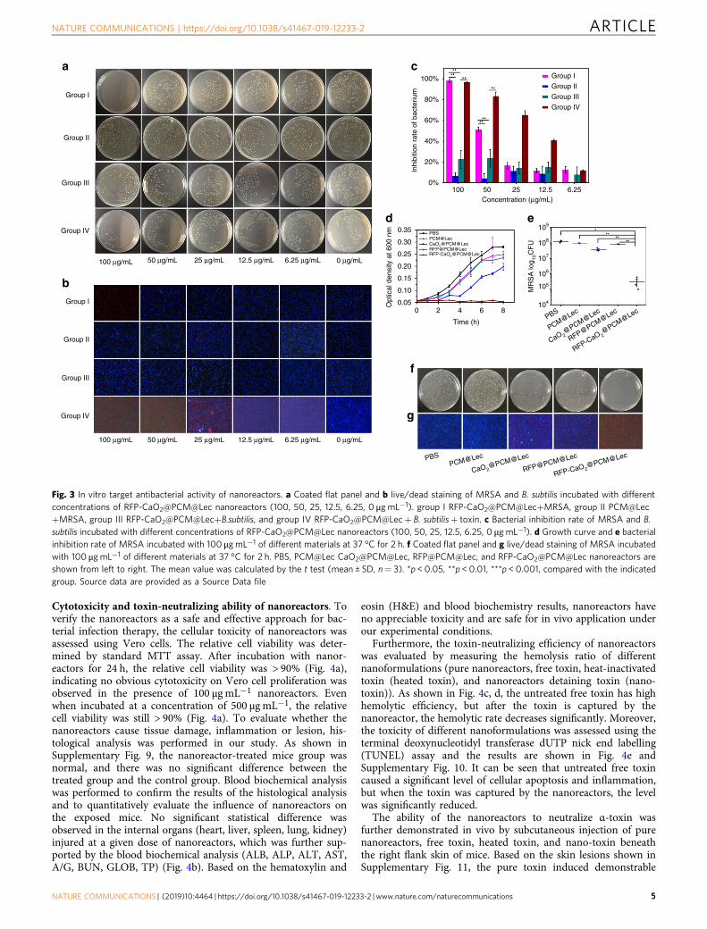
Cytotoxicity and toxin-neutralizing ability of nanoreactors. Toverify the nanoreactors as a safe and effective approach for bac-terial infection therapy, the cellular toxicity of nanoreactors wasassessed using Vero cells. The relative cell viability was deter-mined by standard MTT assay. After incubation with nanor-eactors for 24 h, the relative cell viability was > 90% (Fig. 4a),indicating no obvious cytotoxicity on Vero cell proliferation wasobserved in the presence of 100 μg mL−1 nanoreactors. Evenwhen incubated at a concentration of 500 μg mL−1, the relativecell viability was still > 90% (Fig. 4a). To evaluate whether thenanoreactors cause tissue damage, inflammation or lesion, his-tological analysis was performed in our study. As shown inSupplementary Fig. 9, the nanoreactor-treated mice group wasnormal, and there was no significant difference between thetreated group and the control group. Blood biochemical analysiswas performed to confirm the results of the histological analysisand to quantitatively evaluate the influence of nanoreactors onthe exposed mice. No significant statistical difference wasobserved in the internal organs (heart, liver, spleen, lung, kidney)injured at a given dose of nanoreactors, which was further sup-ported by the blood biochemical analysis (ALB, ALP, ALT, AST,A/G, BUN, GLOB, TP) (Fig. 4b). Based on the hematoxylin and
eosin (H&E) and blood biochemistry results, nanoreactors haveno appreciable toxicity and are safe for in vivo application underour experimental conditions.
Furthermore, the toxin-neutralizing efficiency of nanoreactorswas evaluated by measuring the hemolysis ratio of differentnanoformulations (pure nanoreactors, free toxin, heat-inactivatedtoxin (heated toxin), and nanoreactors detaining toxin (nano-toxin)). As shown in Fig. 4c, d, the untreated free toxin has highhemolytic efficiency, but after the toxin is captured by thenanoreactor, the hemolytic rate decreases significantly. Moreover,the toxicity of different nanoformulations was assessed using theterminal deoxynucleotidyl transferase dUTP nick end labelling(TUNEL) assay and the results are shown in Fig. 4e andSupplementary Fig. 10. It can be seen that untreated free toxincaused a significant level of cellular apoptosis and inflammation,but when the toxin was captured by the nanoreactors, the levelwas significantly reduced.
The ability of the nanoreactors to neutralize α-toxin wasfurther demonstrated in vivo by subcutaneous injection of purenanoreactors, free toxin, heated toxin, and nano-toxin beneaththe right flank skin of mice. Based on the skin lesions shown inSupplementary Fig. 11, the pure toxin induced demonstrable
100 μg/mL 50 μg/mL 25 μg/mL 12.5 μg/mL 6.25 μg/mL 0 μg/mL
100 μg/mL 50 μg/mL 25 μg/mL 12.5 μg/mL 6.25 μg/mL 0 μg/mL
Group I
Group II
Group II
Group III
Group IV
Group I
Group III
Group IV g
RFP-CaO2@PCM@Lec
RFP@PCM@Lec
CaO2@PCM@Lec
PCM@Lec PBS
RFP-CaO 2@PCM@Lec
RFP@PCM@Lec
CaO 2@PCM@Lec
PCM@LecPBS
f
a
b
c
d e
Group I100%
80%
60%
40%
20%
0.35 PBSPCM@Lec
RFP-CaO2@PCM@LecRFP@PCM@LecCaO2@PCM@Lec
Opt
ical
den
sity
at 6
00 n
m
0.30
0.25
0.20
0.15
0.10
0.050 2 4 6 8
Time (h)
109
108
107
106
105
104
Inhi
bitio
n ra
te o
f bac
teriu
m
0%100 50 25 12.5 6.25
Concentration (μg/mL)
MR
SA
log 10
CF
U
Group II
Group III
Group IV
Fig. 3 In vitro target antibacterial activity of nanoreactors. a Coated flat panel and b live/dead staining of MRSA and B. subtilis incubated with differentconcentrations of RFP-CaO2@PCM@Lec nanoreactors (100, 50, 25, 12.5, 6.25, 0 μg mL−1). group I RFP-CaO2@PCM@Lec+MRSA, group II PCM@Lec+MRSA, group III RFP-CaO2@PCM@Lec+B.subtilis, and group IV RFP-CaO2@PCM@Lec+ B. subtilis+ toxin. c Bacterial inhibition rate of MRSA and B.subtilis incubated with different concentrations of RFP-CaO2@PCM@Lec nanoreactors (100, 50, 25, 12.5, 6.25, 0 μg mL−1). d Growth curve and e bacterialinhibition rate of MRSA incubated with 100 μg mL−1 of different materials at 37 °C for 2 h. f Coated flat panel and g live/dead staining of MRSA incubatedwith 100 μgmL−1 of different materials at 37 °C for 2 h. PBS, PCM@Lec CaO2@PCM@Lec, RFP@PCM@Lec, and RFP-CaO2@PCM@Lec nanoreactors areshown from left to right. The mean value was calculated by the t test (mean ± SD, n= 3). *p < 0.05, **p < 0.01, ***p < 0.001, compared with the indicatedgroup. Source data are provided as a Source Data file
NATURE COMMUNICATIONS | https://doi.org/10.1038/s41467-019-12233-2 ARTICLE
NATURE COMMUNICATIONS | (2019) 10:4464 | https://doi.org/10.1038/s41467-019-12233-2 | www.nature.com/naturecommunications 5
edema and inflammation with the extension of time (7 d, 14 d, 21d), and this phenomenon became more and more serious, withobvious suppuration and muscle rot being observed in the skintissue at the toxin injection site after 21 days of treatment.However, the nanoreactor-toxin showed no significant damage tothe skin. Furthermore, the H&E, immunocytochemistry (IHC)and blood routine assays were used to evaluate the toxicity ofdifferent nanoformulations at 21 days post injection. The toxintreatment was shown to induce stronger tissue damage,inflammation or lesion by H&E and IHC analysis (Fig. 4f), incontrast to a similar result between the nanoreactor-toxin and thecontrol, which was further supported by the analysis results ofblood routine (Supplementary Fig. 12). All the above test resultsreveal that the nanoreactors can effectively neutralize toxinswithout causing significant cytotoxicity or physiological toxicity.
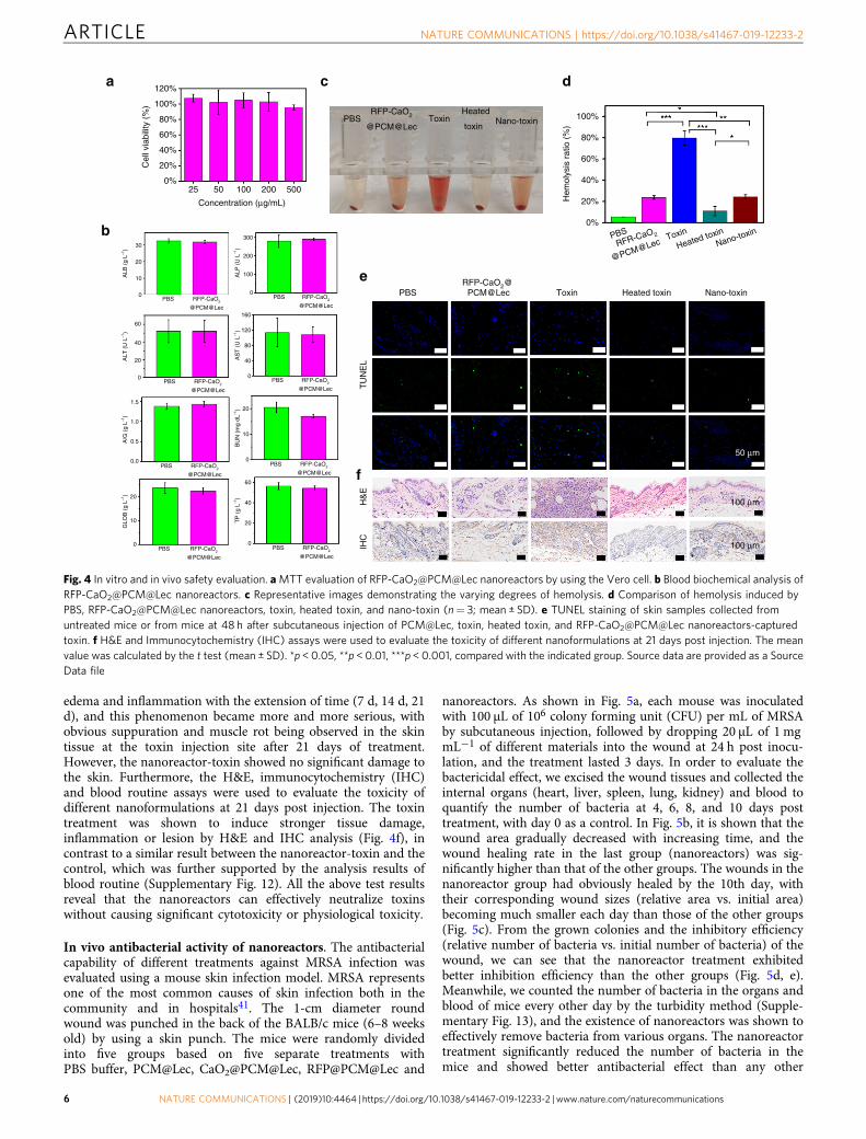
In vivo antibacterial activity of nanoreactors. The antibacterialcapability of different treatments against MRSA infection wasevaluated using a mouse skin infection model. MRSA representsone of the most common causes of skin infection both in thecommunity and in hospitals41. The 1-cm diameter roundwound was punched in the back of the BALB/c mice (6–8 weeksold) by using a skin punch. The mice were randomly dividedinto five groups based on five separate treatments withPBS buffer, PCM@Lec, CaO2@PCM@Lec, RFP@PCM@Lec and
nanoreactors. As shown in Fig. 5a, each mouse was inoculatedwith 100 µL of 106 colony forming unit (CFU) per mL of MRSAby subcutaneous injection, followed by dropping 20 µL of 1 mgmL−1 of different materials into the wound at 24 h post inocu-lation, and the treatment lasted 3 days. In order to evaluate thebactericidal effect, we excised the wound tissues and collected theinternal organs (heart, liver, spleen, lung, kidney) and blood toquantify the number of bacteria at 4, 6, 8, and 10 days posttreatment, with day 0 as a control. In Fig. 5b, it is shown that thewound area gradually decreased with increasing time, and thewound healing rate in the last group (nanoreactors) was sig-nificantly higher than that of the other groups. The wounds in thenanoreactor group had obviously healed by the 10th day, withtheir corresponding wound sizes (relative area vs. initial area)becoming much smaller each day than those of the other groups(Fig. 5c). From the grown colonies and the inhibitory efficiency(relative number of bacteria vs. initial number of bacteria) of thewound, we can see that the nanoreactor treatment exhibitedbetter inhibition efficiency than the other groups (Fig. 5d, e).Meanwhile, we counted the number of bacteria in the organs andblood of mice every other day by the turbidity method (Supple-mentary Fig. 13), and the existence of nanoreactors was shown toeffectively remove bacteria from various organs. The nanoreactortreatment significantly reduced the number of bacteria in themice and showed better antibacterial effect than any other
PBS ToxinHeated
toxinNano-toxin
RFP-CaO2
@PCM@Lec
a
b
PBS
H&
EIH
C
RFP-CaO2@ PCM@Lec
e
f
Toxin Heated toxin Nano-toxin
TU
NE
L
50 μm
100 μm
100 μm
c d
100%
80%
60%
40%
20%
0%
PBS Toxin
Heated toxinNano-toxin
RFR-CaO2
@PCM@Lec
Hem
olys
is r
atio
(%
)
120%
100%
80%
60%
40%
20%
0%25
30
60
160
300
200
100
0
120
80
40
0
1.5
1.0
0.5
0.0
20
60
40
20
0
10
0
20
10
0
40
20
0
20
10ALB
(g
L–1)
ALT
(U
L–1
)A
/G (
g L–1
)G
LOB
(g
L–1)
TP
(g
L–1)
BU
N (
mg
dL–1
)A
ST
(U
L–1
)A
LP (
U L
–1)
0PBS RFP-CaO
2
@PCM@Lec
PBS RFP-CaO2
@PCM@Lec
PBS RFP-CaO2
@PCM@Lec
PBS RFP-CaO2
@PCM@Lec
PBS RFP-CaO2
@PCM@Lec
PBS RFP-CaO2
@PCM@LecPBS RFP-CaO
2
@PCM@Lec
PBS RFP-CaO2
@PCM@Lec
50 100 200 500
Concentration (μg/mL)
Cel
l via
bilit
y (%
)
Fig. 4 In vitro and in vivo safety evaluation. aMTT evaluation of RFP-CaO2@PCM@Lec nanoreactors by using the Vero cell. b Blood biochemical analysis ofRFP-CaO2@PCM@Lec nanoreactors. c Representative images demonstrating the varying degrees of hemolysis. d Comparison of hemolysis induced byPBS, RFP-CaO2@PCM@Lec nanoreactors, toxin, heated toxin, and nano-toxin (n= 3; mean ± SD). e TUNEL staining of skin samples collected fromuntreated mice or from mice at 48 h after subcutaneous injection of PCM@Lec, toxin, heated toxin, and RFP-CaO2@PCM@Lec nanoreactors-capturedtoxin. f H&E and Immunocytochemistry (IHC) assays were used to evaluate the toxicity of different nanoformulations at 21 days post injection. The meanvalue was calculated by the t test (mean ± SD). *p < 0.05, **p < 0.01, ***p < 0.001, compared with the indicated group. Source data are provided as a SourceData file
ARTICLE NATURE COMMUNICATIONS | https://doi.org/10.1038/s41467-019-12233-2
6 NATURE COMMUNICATIONS | (2019) 10:4464 | https://doi.org/10.1038/s41467-019-12233-2 | www.nature.com/naturecommunications

treatment. Furthermore, H&E and Masson staining were per-formed on the tissues of mouse wounds on the first and the 10thday. As shown in Fig. 5f, g, all mouse wounds had a large numberof inflammatory cells on the first day, whereas on the 10th day,only a small number of inflammatory cells were found on thewounds treated with nanoreactors. Masson’s trichrome stainingassay was used to verify the formation of collagen fiber (blue)during wound healing (Fig. 5g). Under the four control treat-ments, unrepaired collagen fibers were observed in the samples,whereas well-established collagen fibers were found in the sam-ples under the nanoreactor treatment, which is consistent withthe aforementioned test results.
Immunity and in vivo toxin neutralization of nanoreactors.Following the in vivo antibacterial assessment, we studied theability of the nanoreactors to elicit potent humoral immunity(Fig. 6a). Germinal centers are the primary sites for the affinity-based maturation of B cells, with the affinity of serum antibodiesincreasing with time after immunization42,43. These high-affinityantibodies of specific isotypes provide excellent protection againsta variety of pathogenic microbial infections. To investigate theperformance of different nanoformulations in immune effect and
in vivo toxin neutralization, draining lymph nodes were collectedat 21 days post immunization to analyze the presence of B cellswith the corresponding phenotype. Flow cytometric analysisshowed that the toxin captured by the nanoreactors significantlyincreased the percentage of germinal center labeled GL-7 B cellsto 24.43%, compared with 10.35% in the control group (P= 0.04)(Fig. 6b and Supplementary Fig. 14 and 15).
To test how the increased response to the nanoformulation isconverted to antigen-specific immunity, we sampled the serumand analyzed the titer by indirect enzyme-linked immunosorbentassay (ELISA) at 21 days post immunization, around the peak ofIgG responses (Fig. 6c). The result is consistent with a previousreport on a nano-toxin formulated with purified toxin44. In thepresent study, the nano-toxin induced significantly higher toxin-specific antibody titers (100-fold, p= 0.04, n= 6) than the heat-treated toxin (60 min treatment). Furthermore, the in vivo toxinneutralization ability of nanoreactors was evaluated by measuringhemolysis ratio (Fig. 6d, e). It can be seen that the nanoreactorshave better toxin-neutralizing ability and can significantly reducethe hemolysis rate. Finally, the protective immunity bestowed bythe nanoreactors was evaluated by subjecting the vaccinated miceto toxin administration at a toxin dose of 120 μg kg−145, whichresulted in 100% mortality within 2 h in the unvaccinated group.
Day 0
RF
P-C
aO2@
PC
M@
Lec
RF
P@
PC
M@
Lec
CaO
2@P
CM
@Le
cP
CM
@Le
cP
BS
RFP@PCM@Lec
RFP-CaO2@PCM@LecCaO2@PCM@LecPCM@Lec
RF
P-C
aO2@
PC
M@
Lec
RF
P@
PC
M@
Lec
CaO
2@P
CM
@Le
cP
BS
PBS
Day 4 Day 6 Day 8 Day 10
Day 1
d
c
a b
100%
PBSPCM@Lec
RFP@PCM@LecRFP-CaO2@PCM@Lec
CaO2@PCM@Lec
PBSPCM@Lec
RFP@PCM@LecRFP-CaO2@PCM@Lec
CaO2@PCM@Lec
PBSPCM@Lec
RFP@PCM@LecRFP-CaO2@PCM@Lec
CaO2@PCM@Lec
80%
60%
40%
20%
0%0 4 6 8 10
100%
Inoculation
Day 0 Day 1 Day 2 Day 3 Day 4 Day 6 Day 8 Day 10
KidneyLungSpleenLiverHeartBloodSkin
100 ppmSubcutaneous injection
106 CFU MRSA
Experimental procedure
3 × Treatment 4 × Assessment
80%
60%
Inhi
bitio
n ra
te o
f bac
teriu
m
40%
20%
0%
0 4 6 8 10
Time (day)After infection (d)
Wou
nd s
ize
(%)
e
f
g
Day 0 Day 4 Day 6 Day 8 Day 10
5 mm
50 μm
50 μm
Day 10
Day 1
Day 10
PC
M@
Lec
Fig. 5 Evaluation of antibacterial activity in vivo. a The study protocol including MRSA inoculation and infection development on the BALB/c mice(6–8 weeks old), followed by the different treatments. b Photographs of wounds infected by MRSA. c Wound size (%) on different days (0, 4, 6, 8, 10).d The grown colonies and e the inhibitory efficiency (relative number of bacteria vs. initial number of bacteria) of wound. f H&E and g Masson images ofthe wounds on the first day and tenth day. Error bars= standard deviation (n= 3), Source data are provided as a Source Data file
NATURE COMMUNICATIONS | https://doi.org/10.1038/s41467-019-12233-2 ARTICLE
NATURE COMMUNICATIONS | (2019) 10:4464 | https://doi.org/10.1038/s41467-019-12233-2 | www.nature.com/naturecommunications 7
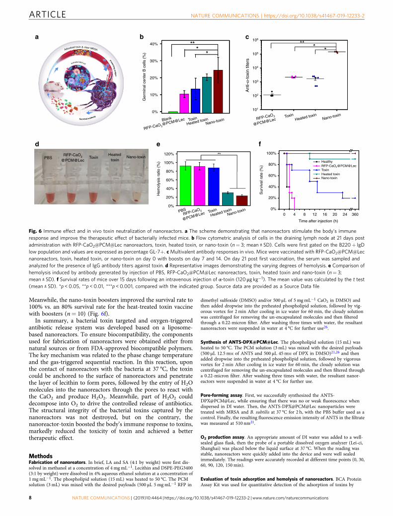
Meanwhile, the nano-toxin boosters improved the survival rate to100% vs. an 80% survival rate for the heat-treated toxin vaccinewith boosters (n= 10) (Fig. 6f).
In summary, a bacterial toxin targeted and oxygen-triggeredantibiotic release system was developed based on a liposome-based nanoreactors. To ensure biocompatibility, the componentsused for fabrication of nanoreactors were obtained either fromnatural sources or from FDA-approved biocompatible polymers.The key mechanism was related to the phase change temperatureand the gas-triggered sequential reaction. In this reaction, uponthe contact of nanoreactors with the bacteria at 37 °C, the toxincould be anchored to the surface of nanoreactors and penetratethe layer of lecithin to form pores, followed by the entry of H2Omolecules into the nanoreactors through the pores to react withthe CaO2 and produce H2O2. Meanwhile, part of H2O2 coulddecompose into O2 to drive the controlled release of antibiotics.The structural integrity of the bacterial toxins captured by thenanoreactors was not destroyed, but on the contrary, thenanoreactor-toxin boosted the body’s immune response to toxins,markedly reduced the toxicity of toxin and achieved a bettertherapeutic effect.
MethodsFabrication of nanoreators. In brief, LA and SA (4:1 by weight) were first dis-solved in methanol at a concentration of 4 mgmL−1. Lecithin and DSPE-PEG3400(3:1 by weight) were dissolved in 4% aqueous ethanol solution at a concentration of1 mgmL−1. The phospholipid solution (15 mL) was heated to 50 °C. The PCMsolution (3 mL) was mixed with the desired payloads (500 μL 5 mgmL−1 RFP in
dimethyl sulfoxide (DMSO) and/or 500 μL of 5 mgmL−1 CaO2 in DMSO) andthen added dropwise into the preheated phospholipid solution, followed by vig-orous vortex for 2 min After cooling in ice water for 60 min, the cloudy solutionwas centrifuged for removing the un-encapsulated molecules and then filteredthrough a 0.22-micron filter. After washing three times with water, the resultantnanoreactors were suspended in water at 4 °C for further use26.
Synthesis of ANTS-DPX@PCM@Lec. The phospholipid solution (15 mL) washeated to 50 °C. The PCM solution (3 mL) was mixed with the desired payloads(500 μL 12.5 mM of ANTS and 500 μL 45mM of DPX in DMSO)21,26 and thenadded dropwise into the preheated phospholipid solution, followed by vigorousvortex for 2 min After cooling in ice water for 60 min, the cloudy solution wascentrifuged for removing the un-encapsulated molecules and then filtered througha 0.22-micron filter. After washing three times with water, the resultant nanor-eactors were suspended in water at 4 °C for further use.
Pore-forming assay. First, we successfully synthesized the ANTS-DPX@PCM@Lec, while ensuring that there was no or weak fluorescence whendispersed in DI water. Then, the ANTS-DPX@PCM@Lec nanoparticles weretreated with MRSA and B. subtilis at 37 °C for 2 h, with the PBS buffer used as acontrol. Finally, the resulting fluorescence emission intensity of ANTS in the filtratewas measured at 510 nm21.
O2 production assay. An appropriate amount of DI water was added to a well-sealed glass flask, then the probe of a portable dissolved oxygen analyzer (Lei-ci,Shanghai) was placed below the liquid surface at 37 °C. When the reading wasstable, nanoreactors were quickly added into the device and were well sealedimmediately. The readings were accurately recorded at different time points (0, 30,60, 90, 120, 150 min).
Evaluation of toxin adsorption and hemolysis of nanoreactors. BCA ProteinAssay Kit was used for quantitative detection of the adsorption of toxins by
40%
a b c
d e f
106
105
104
103
102
101
30%
Ger
min
al c
ente
r B
cel
ls (
%)
20%
120% 100%
80%
60%
40%
20%
0%0 4 8 12 16 20 24 360
Healthy
ToxinHeated toxinNano-toxin
RFP-CaO2@PCM@Lec
Time after injection (h)
Sur
viva
l rat
e (%
)
100%
80%
60%
40%
20%
0%PBS Toxin
Heated toxinNano-toxin
RFP-CaO2
@PCM@Lec
Hem
olys
is r
atio
(%
)
10%
0%
PBS ToxinHeated
toxinNano-toxinRFP-CaO2
@PCM@Lec
Ant
i-α-t
oxin
tite
rs
Blank
Heated toxinNano-toxin Nano-toxin
Heated toxinToxinRFP-CaO2
@PCM@LecToxin
RFP-CaO2@PCM@Lec
Adsorbed toxin & clear MRSA
Activated B&Tcell
Se ecr ted antibody
Presenting antige
n
B cell
T cell
Fig. 6 Immune effect and in vivo toxin neutralization of nanoreactors. a The scheme demonstrating that nanoreactors stimulate the body’s immuneresponse and improve the therapeutic effect of bacterially infected mice. b Flow cytometric analysis of cells in the draining lymph node at 21 days postadministration with RFP-CaO2@PCM@Lec nanoreactors, toxin, heated toxin, or nano-toxin (n= 3; mean ± SD). Cells were first gated on the B220+ IgDlow population and values are expressed as percentage GL-7+. cMultivalent antibody responses in vivo. Mice were vaccinated with RFP-CaO2@PCM@Lecnanoreactors, toxin, heated toxin, or nano-toxin on day 0 with boosts on day 7 and 14. On day 21 post first vaccination, the serum was sampled andanalyzed for the presence of IgG antibody titers against toxin. d Representative images demonstrating the varying degrees of hemolysis. e Comparison ofhemolysis induced by antibody generated by injection of PBS, RFP-CaO2@PCM@Lec nanoreactors, toxin, heated toxin and nano-toxin (n= 3;mean ± SD). f Survival rates of mice over 15 days following an intravenous injection of α-toxin (120 μg kg−1). The mean value was calculated by the t test(mean ± SD). *p < 0.05, **p < 0.01, ***p < 0.001, compared with the indicated group. Source data are provided as a Source Data file
ARTICLE NATURE COMMUNICATIONS | https://doi.org/10.1038/s41467-019-12233-2
8 NATURE COMMUNICATIONS | (2019) 10:4464 | https://doi.org/10.1038/s41467-019-12233-2 | www.nature.com/naturecommunications

materials. In brief, 200 μL of 500 μg mL−1 nanoreactors synthesized in differentmass proportions (Lec: DSPE-PEG= 1:1,3:1,6:1,9:1,12:1, and 1:0) was mixed with10 μL of 400 μg mL−1 toxin to interact with each other at 37 °C for 2 h, using PBSas a control. The mass of the adsorbed toxin was calculated by the absorbance at462 nm according to the detection method of the BCA kit. Under the sameexperimental protocol, the hemolysis rate of the material can also be calculated bythe following formula. In brief, 150 μL of different materials synthesized at differentproportions (Lec: DSPE-PEG= 1:1, 3:1, 6:1, 9:1, 12:1, and 1:0) and 150 μL of 2%RBCs were incubated for 30 min at room temperature. After centrifugation at2000 × g for 5 min, the hemolysis was determined for each sample by measuringthe absorbance of the supernatant at 540 nm using a microplate reader. A 100%lysis control was prepared by treating RBCs with Triton X-100. The hemolysis rateof each group was calculated as follows.
Hemolysis rate ¼ AbsðexperimentÞAbsðX� 100Þ ´ 100% ð1Þ
Bacterial culture. In brief, 200 μL of 108 CFUmL−1 bacteria was incubated withdifferent concentrations of nanoreactors, RFP and CaO2 at 37 °C for 2 h at120 rpm. To evaluate the bacterial mortality, the treated bacteria were diluted anduniformly plated in Luria-Bertani (LB) solid medium, followed by incubation at37 °C for 24 h. Finally, CFU was counted and compared with the control plate.Each treatment was prepared in triplicate and the mean values were compared witheach other.
In vivo safety. In brief, the BALB/c mice (6–8 weeks old) were first shaved toremove the hair on the back. Subsequently, 200 μL of 100 μg mL−1 of nanoreactors(20 μg) was injected subcutaneously, using PBS as a control. At 24 h post injection,the mice were killed, and the internal organs (heart, liver, spleen, lung, kidney)were collected for histological analysis by H&E staining. Meanwhile, the plasmawas collected for biochemical indicator detection (ALB, ALP, ALT, AST, A/G,BUN, GLOB, TP).
Assessment was also performed on the toxicity of nanoreactors (100 μg), toxin(4 μg), heated toxin (4 μg, 70 °C inactivated for 1h), and nano-toxin (4 μg toxinabsorbed by 100 μg RFP-CaO2@PCM@Lec) using PBS as control. In brief, BALB/cmice were first shaved to remove the hair on their back and the above materialswere injected subcutaneously and separately to each group of mice. At 24 h postinjection, the mice were killed, and skin samples at the injection site were collectedfor histological analysis by H&E and TUNEL. TUNEL staining andIpwin32 software were used to count the number of cells with a different colorfluorescence.
After 21 days of immunization, H&E skin staining and IHC were performed onthe dorsal skin of each group to judge the viable toxicity of different treatments. Atthe same time, the blood of the mice was collected, and blood routine tests wereperformed to observe the number of white blood cells and neutrophils (Gran). Allanimal experiments were in compliance with the Huazhong Agriculture University(HZAUMO-2018-036, approved by The Scientific Ethic Committee of HuazhongAgricultural University).
Inhibitory effect of nanoformulations on hemolysis. The ability of nano-formulations to prevent hemolysis was investigated under five different experi-mental groups: PBS, nanoreactors (100 μg), toxin (4 μg), heated toxin (4 μg, 70 °Cinactivated for 1h), and nano-toxin (4 μg toxin absorbed by 100 μg nanoreactors).In brief, 150 μL of different materials and 150 μL of 2% red blood cells (RBCs) wereincubated for 30 min at room temperature, followed by centrifugation at 2000 × gfor 5 min Next, the hemolysis of each group was determined by measuring theabsorbance of the supernatant at 540 nm using a microplate reader. Meanwhile, a100% lysis control was prepared by treating RBC with Triton X-100. Finally, thehemolysis rate of each group was calculated according to formula (1).
Mice injury model. The injury model was established on the BALB/c mice(6–8 weeks old). The 1-cm diameter round wound was punched in the back of themouse using a skin punch, followed by infecting the wound with 100 µL of 1 × 10⁶
CFUmL−1 of MRSA, and the initial infection time was recorded as the zero day.After infection for 24 h, three mice were randomly killed for counting the bacteriaof their wound, blood and internal organs by plate colony counting and turbidi-metry. Furthermore, each wound was stained with H&E and Masson to identifywhether the infection was established or not. The remaining mice were randomlydivided into five groups, each group consisting of 12 mice. After the infection wasestablished, the mice in different groups were treated separately with PBS bufferonly, 20 µL of 1 mgmL−1 PCM@Lec, CaO2@PCM@Lec, RFP@PCM@Lec, andnanoreactors once a day for 3 days. After the wound was photographed, the micewere killed for counting the bacteria of their wound, blood, and internal organs bythe same method at 4, 6, 8, and 10 days post infection. All animal experiments werein compliance with the Huazhong Agriculture University (HZAUMO-2018-036,approved by The Scientific Ethic Committee of Huazhong Agricultural University)
Bacterial statistics. Bacteria at the wound were counted using the plate colonycounting method43. First, bacteria at the wound were collected by a cotton swab
and soaked in LB medium. After the medium was diluted 100 times, 20 microliterswere applied for plate counting. Bacteria in internal organs (heart, liver, spleen,lung, kidney) and blood were counted using the turbidimetric method. First, theinternal organs were crushed, then 20 μL of the polishing solution was taken outand spread in 180 μL of LB medium, using 20 μL of MRSA in logarithmic phase asa control. After incubation for 7 h, the optical density was measured at 600 nm by amicroplate reader.
Germinal center analysis. BALB/c mice were first shaved to remove the hair ontheir back. The materials used included nanoreactors (100 μg), toxin (4 μg), heatedtoxin (4 μg, 70 °C inactivated for 1h), and nano-toxin (4 μg toxin absorbed by100 μg nanoreactors), and PBS as control. These materials were injected sub-cutaneously on day 0, followed by a boost on day 7 and day 14. On day 21 afterimmunization, the lymph nodes were collected and dissociated into single cellsuspensions for flow cytometric analysis. After staining the lymph nodes with APCRat Anti-Mouse IgD (BD Pharmingen, 560868, 1:100), Alexa Fluor 488 Rat Anti-Mouse CD45R (BD Pharmingen, 557669, 1:100), PE Anti-mouse/human GL-7Antigen (T- and B-cell Activation Marker) (BioLegend, 144607, 1:100), PurifiedRat Anti-Mouse CD16/CD32 (Mouse BD Fc Block) (BD Pharmingen, 553142,1:100) data were collected on a flow cytometer and analyzed using Flowjo software.
Anti-α-toxin titer analysis. Mice were subcutaneously administered with nanor-eactors (100 μg), toxin (4 μg), heated toxin (4 μg, 70 °C inactivated for 1h) and nano-toxin (4 μg toxin absorbed by 100 μg nanoreactors) on day 0, followed by a boost onday 7 and day 14. On day 21, the serum of each mouse was collected for measuringtoxin-specific antibody titers by an ELISA. First, a 96-well plate was coated overnightwith 2 µgmL−1 toxin using commercial coating buffer. Next, the wells were blockedwith 1 wt% bovine serum albumin, followed by the addition of serially diluted serumsamples as the primary antibody and horseradish peroxidase-conjugated Goat Anti-Mouse lgG (Sangon Biotech (Shanghai) Co., Ltd. D110087, 1:5000) as the secondaryantibody. The plate was developed with H2O2/TMB-ELISA substrate, and the reactionwas terminated by adding 2% sulfuric acid (H2SO4). Finally, toxin-specific antibodytiters were measured at 450 nm using a Microplate reader.
Toxin-neutralizing ability of nanoreactors. After 21 days of immunization, serawere collected from the different experimental groups of mice. In brief, 20 μL serumwas incubated with 10 μL of 50 μgmL−1 toxin and 20 μL of Hank solution for 30minat room temperature, followed by the addition of 50 μL of 2% RBCs and incubationfor another 30min According to the above experimental method, the hemolysisefficiency after serum toxin neutralization can be calculated by formula 1.
Mouse survival rate. After the end of the 21-day immunization, the mice treatedwith PBS, nanoreactors (100 μg), toxin (4 μg), heated toxin (4 μg, 70 °C inactivatedfor 1 h), and nano-toxin (4 μg toxin absorbed by 100 μg nanoreactors) wereinjected in the tail vein with toxin at the dosage of 120 μg kg−1, and the survivalrate of the decimals was observed for each group within 360 h.
Statistical analysis. All the results were presented as the mean value plus astandard deviation (± SD) from at least three independent experiments. Statisticalanalyses were performed using the t test. Values of *p < 0.05, **p < 0.01, and ***p <0.001 were considered statistically significant.
Data availabilityThe authors declare that data supporting the findings of this study are available withinthe paper and its supplementary information files. Source data are provided as a SourceData file by figshare (https://doi.org/10.6084/m9.figshare.9429287, hyperlink: https://figshare.com/s/ee97bba58d8aee5158c8).
Received: 4 March 2019; Accepted: 28 August 2019;
References1. Willyard, C. The drug-resistant bacteria that pose the greatest health threats.
Nature 543, 15 (2017).2. Li, X. N. et al. Functional gold nanoparticles as potent antimicrobial agents
against multi-drug-resistant bacteria. ACS Nano 28, 10682–10686 (2014).3. Karimi, M. G. et al. Smart micro/nanoparticles in stimulus-responsive drug/
gene delivery systems. Chem. Soc. Rev. 45, 1457–1501 (2016).4. Huh, A. J. & Kwon, Y. J. “Nanoantibiotics”: a new paradigm for treating
infectious diseases using nanomaterials in the antibiotics resistant era. J.Control. Release 156, 128–145 (2011).
5. Mao, C. X. et al. Local photothermal/photodynamic synergistic therapy bydisrupting bacterial membrane to accelerate reactive oxygen species permeationand protein leakage. ACS Appl. Mater. Interfaces 11, 17902–17914 (2019).
NATURE COMMUNICATIONS | https://doi.org/10.1038/s41467-019-12233-2 ARTICLE
NATURE COMMUNICATIONS | (2019) 10:4464 | https://doi.org/10.1038/s41467-019-12233-2 | www.nature.com/naturecommunications 9

6. Xiong, M. H. et al. Bacteria-responsive multifunctional nanogel for targetedantibiotic delivery. Adv. Mater. 24, 6175–6180 (2012).
7. Li, W., Dong, K., Ren, J. & Qu, X. A β-lactamase-imprinted responsivehydrogel for the treatment of antibiotic-resistant bacteria. Angew. Chem. Int.Ed. 55, 8049–8053 (2016).
8. Fan, W. Z., Tong, X., Farnia, F., Yu, B. & Zhao, Y. CO2-responsive polymersingle-chain nanoparticles and self-assembly for gas-tunable nanoreactors.Chem. Mater. 29, 5693–5701 (2017).
9. Zhao, Y. et al. A preloaded amorphous calcium carbonate/doxorubicin@silicananoreactor for pH-responsive delivery of an anticancer drug. Angew. Chem.Int. Ed. 54, 919–922 (2015).
10. Li, J. et al. Therapeutic vesicular nanoreactors with tumor-specific activationand self-destruction for synergistic tumor ablation. Angew. Chem. Int. Ed. 56,14025–14030 (2017).
11. Che, H. L., van Hest & Jan, C. M. Stimuli-responsive polymersomes andnanoreactors. J. Mater. Chem. B 4, 4632–4647 (2016).
12. Zhu, W. et al. Functionalization of hollow nanomaterials for catalyticapplications: nanoreactor construction. Adv. Mater. 31, 1800426 (2018).
13. Zhang, W., Hu, X., Shen, Q. & Xing, D. Mitochondria-specific drug releaseand reactive oxygen species burst induced by polyprodrug nanoreactors canenhance chemotherapy. Nat. Commun. 10, 1704–1717 (2019).
14. Yu, Z., Zhou, P., Pan, W., Li, N. & Tang, B. A biomimetic nanoreactor forsynergistic chemiexcited photodynamic therapy and starvation therapyagainst tumor metastasis. Nat. Commun. 9, 5044–5052 (2018).
15. Larrañaga, A., Lomora, M., Sarasua, J. R., Palivan, C. G. & Pandit, A. Polymercapsules as micro-/nanoreactors for therapeutic applications: current strategies tocontrol membrane permeability. Pro. Mater. Sci. 90, 325–357 (2017).
16. Ke, W. et al. Therapeutic polymersome nanoreactors with tumor-specificactivable cascade reactions for cooperative cancer therapy. ACS Nano 13,2357–2369 (2019).
17. Gaitzsch, J., Appelhans, D., Wang, L., Battaglia, G. & Voit, B. Synthetic bio-nanoreactor:mechanical and chemical control of polymersome membranepermeability. Angew. Chem. Int. Ed. 51, 4448–4451 (2012).
18. Anraku, Y. et al. Systemically injectable enzyme-loaded polyion complexvesicles as in vivo nanoreactors functioning in tumors. Angew. Chem. Int. Ed.55, 560–565 (2016).
19. Tanner, P. et al. Polymeric vesicles: from drug carriers to nanoreactors andartificial organelles. Acc. Chem. Res. 44, 1039–1049 (2011).
20. Penades, J. R., Chen, J., Quiles-Puchalt, N., Carpena, N. & Novick, R. P.Bacteriophage-mediated spread of bacterial virulence genes. Curr. Opin.Microbiol. 23, 171–178 (2015).
21. Pornpattananangkul, D. et al. Bacterial toxin-triggered drug release from goldnanoparticle-stabilized liposomes for the treatment of bacterial infection. J.Am. Chem. Soc. 133, 4132–4139 (2011).
22. Sucharit, B. & Jorgen, T. Alpha-toxin of staphylococcus aureus. Microbiol.Mol. Biol. Rev. 55, 733–751 (1991).
23. Fussle, R. On the mechanism of membrane damage by Staphylococcus aureusalpha- toxin. J. Cell Biol. 91, 83–94 (1981).
24. Bantel, H. et al. Alpha-toxin is a mediator of Staphylococcus aureus-inducedcell death and activates caspases via the intrinsic death pathway independentlyof death receptor signaling. J. Cell Biol. 155, 637–648 (2001).
25. Hyun, D. C., Levinson, N. S., Jeong, U. & Xia, Y. Emerging applications ofphase-change materials (PCMs): teaching an old dog new tricks. Angew.Chem. Int. Ed. 53, 3780–3795 (2014).
26. Zhu, C. et al. A eutectic mixture of natural fatty acids can serve as the gatingmaterial for near-infrared-triggered drug release. Adv. Mater. 29,1703702–1703707 (2017).
27. Motulsky, A. et al. Characterization and biocompatibility of organogels basedon L-alanine for parenteral drug delivery implants. Biomaterials 26,6242–6253 (2005).
28. Ahmet, S. & Kamil, K. Thermal performance of a eutectic mixture of lauricand stearic acids as PCM encapsulated in the annulus of two concentric pipes.Sol. Energy 72, 493–504 (2002).
29. Zhang, N., Yuan, Y. P., Yuan, Y. G., Li, T. Y. & Cao, X. L. Lauric–palmitic–stearicacid/expanded perlite composite as form-stable phase change material:preparation and thermal properties. Energ. Build. 82, 505–511 (2014).
30. Liu, C., Yuan, Y., Zhang, N., Cao, X. L. & Yang, X. J. A novel PCM oflauric–myristic–stearic acid/expanded graphite composite for thermal energystorage. Mater. Lett. 120, 43–46 (2014).
31. Wang, Y., C., C. H., Hu, D., Ulmschneider, M. B. & Ulmschneider, J. P.Spontaneous formation of structurally diverse membrane channel architecturesfrom a single antimicrobial peptide. Nat. Commun. 7, 13535–13543 (2016).
32. Moon, G. D. et al. A new theranostic system based on gold nanocages andphase-change materials with unique features for photoacoustic imaging andcontrolled release. J. Am. Chem. Soc. 133, 4762–4765 (2011).
33. Song, Z. Y. et al. pH-responsive, light-triggered on-demand antibiotic releasefrom functional metal-organic framework for bacterial infection combinationtherapy. Adv. Funct. Mater. 28, 1800011–1800018 (2018).
34. Neese, F. The ORCA program system. Wires Comput. Mol. Sci. 2, 73–78 (2012).35. Dion, M., Rydberg, H., Schroder, E., Langreth, D. C. & Lundqvist, B. I. Van
der waals density functional for general geometries. Phys. Rev. Lett. 92,246401–246404 (2004).
36. Sun, H., Gao, N., Dong, K., Ren, J. & Qu, X. Graphene quantum dots-band-aids used for wound disinfection. ACS Nano 8, 6202–6210 (2014).
37. Chung, M. F. et al. A liposomal system capable of generating CO2 bubbles toinduce transient cavitation, lysosomal rupturing, and cell necrosis. Angew.Chem. Int. Ed. 51, 10089–10093 (2012).
38. Lin, Y. J. et al. Recent advances in CO2 bubble-generating carrier systems forlocalized controlled release. Biomaterials 133, 154–164 (2017).
39. Huang, C. C. et al. An implantable depot that can generate oxygen in situ forovercoming hypoxia-induced resistance to anticancer drugs in chemotherapy.J. Am. Chem. Soc. 138, 5222–5225 (2016).
40. Banerjee, S. S. et al. Calcium phosphate nanocapsule crowned multiwalledcarbon nanotubes for pH triggered intracellular anticancer drug release. J.Mater. Chem. B 3, 3931–3939 (2015).
41. Stryjewski, M. E. & Corey, G. R. Methicillin-resistant staphylococcus aureus:an evolving pathogen. Clin. Infect. Dis. 58, S10–S19 (2014).
42. Ersching, J. et al. Germinal center selection and affinity maturation requiredynamic regulation of mTORC1 kinase. Immunity 46, 1045–1058 (2017).
43. Wei, X. et al. In situ capture of bacterial toxins for antivirulence vaccination.Adv. Mater. 29, 1701644–1701651 (2017).
44. Wang, F. et al. Nanoparticle-based antivirulence vaccine for the managementof methicillin-resistant staphylococcus aureus skin infection. Adv. Funct.Mater. 26, 1628–1635 (2016).
45. Hu, C. M., Fang, R. H., Luk, B. T. & Zhang, L. Nanoparticle-detained toxinsfor safe and effective vaccination. Nat. Nanotechnol. 8, 933–938 (2013).
AcknowledgementsWe are grateful to the financial support by the National Natural Science Foundation ofChina (21807036, 21778020), the Fundamental Research Funds for the Central Uni-versities (2662016QD027), Sci-tech Innovation Foundation of Huazhong AgricultureUniversity (2662017PY042, 2662018PY024), Science and Technology Major Project ofGuangxi (Gui Ke AA18118046). We are also thankful to professor Hanchang Zhu forediting of the language, professor Xiangru Wang for providing us with the bacterialstrain, Fengrui Wu for the help with SEM characterization.
Author contributionsY.W., Z.Y.S. and H.J.W performed the experiments. Y.W. and Z.Y.S. were involved indata analysis. Y.W., Z.Y.S. and H.Y.H. designed experiments, interpreted results, andwrote the manuscript.
Competing interestsThe authors declare no competing interests.
Additional informationSupplementary information is available for this paper at https://doi.org/10.1038/s41467-019-12233-2.
Correspondence and requests for materials should be addressed to H.H.
Peer review information Nature Communications thanks Shuilin Wu and other,anonymous, reviewers for their contributions to the peer review of this work. Peer reviewreports are available.
Reprints and permission information is available at http://www.nature.com/reprints
Publisher’s note Springer Nature remains neutral with regard to jurisdictional claims inpublished maps and institutional affiliations.
Open Access This article is licensed under a Creative Commons Attri-bution 4.0 International License, which permits use, sharing, adaptation,
distribution and reproduction in any medium or format, as long as you give appropriatecredit to the original author(s) and the source, provide a link to the Creative Commonslicense, and indicate if changes were made. The images or other third party material in thisarticle are included in the article’s Creative Commons license, unless indicated otherwise ina credit line to the material. If material is not included in the article’s Creative Commonslicense and your intended use is not permitted by statutory regulation or exceeds thepermitted use, you will need to obtain permission directly from the copyright holder. Toview a copy of this license, visit http://creativecommons.org/licenses/by/4.0/.
© The Author(s) 2019
ARTICLE NATURE COMMUNICATIONS | https://doi.org/10.1038/s41467-019-12233-2
10 NATURE COMMUNICATIONS | (2019) 10:4464 | https://doi.org/10.1038/s41467-019-12233-2 | www.nature.com/naturecommunications