Embed Size (px)
Citation preview
CLINICAL ARTICLEJ Neurosurg Pediatr 20:91–98, 2017
Craniosynostosis is a rare condition that involves premature fusion of one or multiple cranial sutures, resulting in progressive skull deformity as calvarial
growth continues parallel rather than perpendicular to the fused sutures.5 Early techniques for the surgical correction of craniosynostosis involved suturectomies, also known as strip craniectomies, through an open approach.15 Due to the high rate of refusion, however, calvarial vault recon-struction was developed as an alternative strategy, through
which large portions of the skull are removed and remod-eled.21 As expected, such procedures are associated with significant morbidity, including large amounts of blood loss and lengthy hospitalizations.
In the 1990s, David Jimenez and Constance Barone pioneered the use of endoscopic-assisted surgery for per-forming a suturectomy through a minimal incision, fol-lowed by postoperative orthotic therapy.1,12 Multiple case series have established this technique as a viable option
ABBREVIATIONS CI = cephalic index; CMD = cervicomedullary decompression; EBL = estimated blood loss; ETV = endoscopic third ventriculostomy; FOA = frontoorbital advancement; LOS = length of hospital stay; pRBCs = packed red blood cells; TDD = transcranial diameter difference; TI = towering index. SUBMITTED December 23, 2016. ACCEPTED February 27, 2017.INCLUDE WHEN CITING Published online May 5, 2017; DOI: 10.3171/2017.2.PEDS16710.
Endoscopic surgery for patients with syndromic craniosynostosis and the requirement for additional open surgeryDavid S. Hersh, MD,1 Julie E. Hoover-Fong, MD, PhD,2–4 Natalie Beck, MGC, CGC,2–4 Amir H. Dorafshar, MBChB,5 and Edward S. Ahn, MD3,6
1Department of Neurosurgery, University of Maryland School of Medicine; and 2McKusick-Nathans Institute of Genetic Medicine, 3Department of Pediatrics, 4Greenberg Center for Skeletal Dysplasias, 5Department of Plastic and Reconstructive Surgery, and 6Division of Pediatric Neurosurgery, Department of Neurosurgery, Johns Hopkins University School of Medicine, Baltimore, Maryland
OBJECTIVE Recent reports have described early endoscopic suturectomy as a treatment option for patients with syn-dromic craniosynostosis, but such patients often require subsequent calvarial remodeling. The authors describe their experience with this patient population and seek to identify predictors of sufficiency of endoscopic surgery alone.METHODS The medical records of patients with syndromic craniosynostosis who underwent endoscopic repair were retrospectively reviewed. Demographic data, operative details, and follow-up data were collected.RESULTS A total of 6 patients with syndromic craniosynostosis underwent endoscopic surgery followed by helmet therapy during the study period. Of these, 3 patients were male. The involved syndromes included Crouzon, Pfeiffer, Jackson-Weiss, Muenke, Saethre-Chotzen, and craniosynostosis-3 (n = 1 each). The patients underwent endoscopic surgery at a median age of 2.1 months (range 0.9–4.1 months). The median estimated blood loss was 30 ml (range 20–100 ml), with 2 patients requiring a transfusion. The median length of stay in the hospital was 1.5 days (range 1–4 days), and the median follow-up was 29.0 months (range 16.8–81.7 months), with 1 patient (16.7%) requiring an open revision. Three patients (50%) were classified as Whitaker Category I at the last follow-up. The patients for whom additional open surgery was performed or recommended (Whitaker Category IV) were the oldest patients in the cohort, ranging from 2.6 to 4.1 months at the time of surgery.CONCLUSIONS This series demonstrates that endoscopic surgery can be sufficient to treat syndromic craniosyn-ostosis without subsequent open calvarial remodeling over a median follow-up period of at least 2 years. The findings suggest that younger age at the time of endoscopic surgery may be an important factor in determining the sufficiency of this procedure. Even among patients who require subsequent open calvarial remodeling, early endoscopic surgery may allow for growth and development of the brain and skull while delaying the need for open remodeling until the patient is older and can better tolerate the procedure.https://thejns.org/doi/abs/10.3171/2017.2.PEDS16710KEY WORDS craniosynostosis; syndromic; coronal; endoscopic; craniofacial
©AANS, 2017 J Neurosurg Pediatr Volume 20 • July 2017 91
Unauthenticated | Downloaded 12/14/21 08:47 AM UTC

D. S. Hersh et al.
J Neurosurg Pediatr Volume 20 • July 201792
for young infants (generally less than 3 months old) with single-suture, nonsyndromic craniosynostosis. In these cases, endoscopic surgery provides effective treatment while limiting surgical time, blood loss, transfusion rates, and length of hospital stay (LOS).11,14 Recently, endoscopic suturectomy with postoperative helmet therapy has also been expanded to patients with multiple-suture, nonsyn-dromic craniosynostosis. Again, the safety and efficacy of the endoscopic approach were demonstrated.13
Few reports, however, have described the use of en-doscopic suturectomy and postoperative helmet thera-py for patients with syndromic craniosynostosis.8,10, 19,20 Syndromic craniosynostosis accounts for 8%–24% of all patients with craniosynostosis. Multiple syndromes exist, many of which result from 1 or more mutations in 1 of at least 57 currently recognized genes. These mutations may occur de novo or they may be inherited in an auto-somal recessive, dominant, or X-linked fashion with vari-able penetrance and expressivity.3,7,9,16 Patients with syn-dromic craniosynostosis have additional anomalies often involving the digits and limbs, hearing, and midface, and may have cognitive impairment and/or a family history of craniosynostosis. Furthermore, patients with premature fusion of multiple cranial sutures are more likely to have a syndromic diagnosis, and they have a high incidence of intracranial hypertension.9 As a result, they represent a complex subset of patients. Indeed, among the few syn-dromic patients who are reported as having undergone en-doscopic suturectomy and postoperative helmet therapy, almost half have required subsequent calvarial remodel-ing due to either persistent or recurrent deformity.8,19,20 In these cases, one may question the utility of the primary surgery with helmet therapy if subsequent open surgery is ultimately required. We present our experience with this patient population and seek to identify factors that may contribute to the sufficiency of endoscopic surgery alone.
MethodsPatient Population
Following approval by the institutional review board, the medical records of pediatric patients who underwent endoscopic suturectomy and postoperative helmet therapy between January 2008 and May 2016 were retrospectively reviewed. Only patients with molecular confirmation of a syndromic craniosynostosis diagnosis were included. Data were collected including demographic information, estimated blood loss (EBL), transfusion status, complica-tions, LOS, and outcomes.
Preoperative EvaluationAll patients were evaluated by a multidisciplinary team
consisting of a pediatric neurosurgeon, plastic surgeon, and medical geneticist. Patients who were evaluated by the medical genetics team and suspected to have syndromic craniosynostosis were offered genetic testing, with variable patient compliance. Five patients were evaluated between 0.9 and 14.1 weeks of age. One patient was diagnosed with craniosynostosis prenatally and was evaluated in the new-born nursery. Five patients underwent a preoperative head CT to define which sutures were fused and to assess any
intracranial pathology; the remaining patient underwent only skull radiographs. All patients underwent anthro-pometric measurements performed using calipers at the bedside, and 1 or more indices were calculated, including the cephalic index (CI), towering index (TI), and transcra-nial diameter difference (TDD).18 The CI was measured for the patient with sagittal suture involvement and was defined as the eurion-to-eurion diameter divided by the glabella-to-opisthocranion diameter, multiplied by 100.4 The TI was measured for the patients with bilateral coro-nal involvement and was defined as the coronal distance (measured from tragus to tragus) over the cranial vault di-vided by the occipitofrontal circumference, multiplied by 100.18 The TDD was measured for the patient with unilat-eral coronal involvement and was defined as the difference (in millimeters) between the occipital-frontal transcranial diameters on each side of the head (i.e., the left occipital-to-right frontal diameter subtracted from the right occip-ital-to-left frontal diameter).6,17 Given the patients’ young age at evaluation, all families in this series were counseled regarding both endoscopic surgery and open calvarial vault remodeling. All patients who presented at or prior to 3 months of age were offered the endoscopic surgery, and all underwent the procedure accordingly. Only 1 patient with syndromic craniosynostosis—who was not included in this series—underwent initial open surgery during the study period because of older age at evaluation.
Operative ProcedureA similar procedure for endoscopic-assisted cranio-
synostosis surgery has been described previously.2 The patient was brought to the operating room and general endotracheal anesthesia was induced. Appropriate intra-venous and intraarterial access were obtained and a uri-nary catheter was placed. The patient was placed supine with his or her head on a horseshoe headrest. The hair was then clipped and 2 incisions were marked, just behind and along the midpoint of the fused coronal suture on either side. Each incision was approximately 2 cm in length. A single incision was used for the patient with unilateral cor-onal synostosis. The skin was then prepared and draped in sterile fashion.
In each case, the skin was incised with a No. 15 blade scalpel. Electrocautery was used to dissect through the subcutaneous tissue to the bone, and the pericranium was elevated. A craniectomy was started at the site of an exist-ing bony defect, or alternatively after creating a bur hole with a high-speed drill. In 1 case, an ultrasonic bone-cut-ting device was used to perform square-shaped craniec-tomies. A 4-mm, 30° rigid endoscope (Karl Storz) with an attached guard was then inserted to assist in visualiza-tion as the dissection was continued inferiorly along the fused coronal suture. A dissection was performed under the temporalis muscle, and rongeurs were used to continue the craniectomy in the direction of the sphenoid ridge. The craniectomy was then extended along the posterior aspect of the sphenoid ridge toward the temporal fossa until the skull base was reached. The craniectomy was approxi-mately 1 cm in width. Bipolar electrocautery, bone wax, and hemostatic agents were used to obtain hemostasis. The endoscope was then aimed in the opposite direction,
Unauthenticated | Downloaded 12/14/21 08:47 AM UTC

Endoscopic surgery for syndromic craniosynostosis
J Neurosurg Pediatr Volume 20 • July 2017 93
toward the lateral corner of the anterior fontanelle. Mayo scissors or the ultrasonic bone-cutting device were used to create a cut on either side of the fused suture. The bone was then removed in piecemeal fashion with the aid of a rongeur. The wound was then irrigated and Gelfoam strips soaked in thrombin were placed over the craniectomy site. The incision was closed in 2 layers, with 3-0 Vicryl (Ethi-con Inc.) galeal stitches followed by 5-0 Caprosyn (Covi-dien) stitches for the skin. The contralateral suturectomy was performed in identical fashion.
The patient with both sagittal synostosis and bicoronal synostosis underwent a third incision in addition to the bi-coronal incisions described above. The additional incision was planned along the posterior aspect of the anterior fon-tanelle. Again, a No. 15 blade scalpel and electrocautery were used to incise the skin and dissect through the subcu-taneous tissue. Bony defects were identified on either side of the incision, and blunt dissection was used to separate the dura mater from the overlying bone. Rongeurs were used to perform the craniectomy until the defect commu-nicated with the lateral craniectomies performed along the coronal sutures. A large midline keel invaginating into the dura was identified, and was removed using rongeurs. A 3-cm-wide craniectomy was then completed over the midline, and was continued anteriorly until the anterior fontanelle was reached. The incision was then closed as described above.
Postoperative CareAll patients were observed in the intermediate care unit
for 1–2 nights. Blood for postoperative hemoglobin and reticulocyte counts was drawn on the first postoperative morning. Patients were evaluated at the helmet therapy center, underwent surface laser scans 1 week postopera-tively, and were fitted with the helmet within 10 days of surgery. Following discussion with the neurosurgeon, the orthotists created the helmets to address the dominant de-formity in patients with multisuture craniosynostosis. For those with bilateral coronal craniosynostosis, the helmet therapy chiefly addressed towering of the calvaria. Howev-er, in the only patient with additional sagittal craniosynos-tosis, the dominant feature was scaphocephaly; therefore, the bilateral parietal and vertex restrictions were the chief deformities that were addressed. Helmets were worn 23 of 24 hours per day. Patients underwent helmet therapy for 10.5–19.5 months postoperatively, with the exception of the patient with Jackson-Weiss syndrome, who underwent helmet therapy for only 2.43 months due to the develop-ment of hydrocephalus. In his case, there was concern for the hydrocephalus as an intrinsic expanding force, which was a relative contraindication for the restrictive forces of helmet therapy. The CI, TI, and/or TDD were measured at each clinic visit. The duration of helmet therapy was determined by the stability of the anthropometric indices over multiple visits.
The patients were followed in a multidisciplinary cra-niofacial clinic. Additional open calvarial remodeling was recommended if the preoperative deformity remained uncorrected, recurred, or progressed. The need and tim-ing of further surgery was determined by both the plas-tic surgeon and the neurosurgeon. The criteria to proceed
with additional open calvarial remodeling included the following: 1) progression of the original deformity based on craniometric indices, and 2) agreement on the need for additional surgery between both parties. The postopera-tive aesthetic outcomes of the 5 patients who did not un-dergo additional open calvarial remodeling were assessed by one of the authors (A.H.D.), who was not involved in their long-term clinical follow-up. Photographs obtained at the most recent clinic visit were reviewed, and a Whita-ker category23 was assigned: 1) Category I, no need for additional surgery; 2) Category II, soft-tissue and/or minor bone contouring is advisable; 3) Category III, more exten-sive revisions involving major alternative osteotomies or bone graft repositioning are advisable; and 4) Category IV, major surgical revisions are advisable.
ResultsA total of 81 nonsyndromic and syndromic patients
underwent endoscopic suturectomy and postoperative hel-met therapy during the study period. Of these, 6 patients (7.4%) had a disease-causing mutation in a gene implicated in craniosynostosis (Table 1). The demographic, clinical, and surgical features of the patients are summarized in Table 2. Three of the patients were male. The involved syndromes included Crouzon, Pfeiffer, Jackson-Weiss, Muenke, Saethre-Chotzen, and craniosynostosis-3 (n = 1 each). Five patients had involvement of the coronal sutures bilaterally (Fig. 1), whereas 1 patient had unilateral coronal synostosis. Of the 5 patients with bicoronal synostosis, 1 also had involvement of the sagittal suture (Fig. 2), whereas in another the lambdoid sutures were involved bilaterally.
The median age at the time of surgery was 2.1 months (range 0.9–4.1 months), and the median weight at the time of surgery was 4.8 kg (range 3.4–5.2 kg). Preoperative he-moglobin and hematocrit levels were available for 4 of the 6 patients. Among these patients, the median preoperative hemoglobin was 11.0 g/dl (range 9.0–19.0 g/dl), and the median preoperative hematocrit was 32.1% (range 26.5%–54.6%). Postoperative levels were obtained for all patients, with a median postoperative hemoglobin of 8.2 g/dl (range 5.3–11.9 g/dl) and a median postoperative hematocrit of 21.9% (range 17.0%–34.9%).
The median EBL during surgery was 30 ml (range 20–100 ml). The patient with craniosynostosis-3 represented a probable outlier, with an EBL of 100 ml, and because she weighed only 3.4 kg at the time of surgery, she required an intraoperative transfusion of packed red blood cells (pRBCs) and fresh frozen plasma. During this patient’s surgery, a right-sided coronal suturectomy was performed without complications, after which attention was turned to the left side. A small durotomy was encountered, with some bleeding from the dural edges. However, on cauter-ization and shrinkage of the dura, a cortical vein ruptured, resulting in the high EBL. The bleeding was controlled with Gelfoam soaked in thrombin, and the procedure was completed without further complications, but due to the amount of blood loss the patient underwent an intraopera-tive transfusion. In addition, the patient with Jackson-Weiss syndrome underwent a single postoperative transfusion of pRBCs. Although his surgery involved an EBL of only 30
Unauthenticated | Downloaded 12/14/21 08:47 AM UTC
D. S. Hersh et al.
J Neurosurg Pediatr Volume 20 • July 201794
ml, and his hemoglobin level dropped by only 3.7 g/dl, he had the lowest preoperative hemoglobin of the cohort, with a level of 9 g/dl. His immediate postoperative hemoglobin level was 5.3 g/dl, for which he received a transfusion of 70 ml pRBCs and responded appropriately, with a follow-up hemoglobin level of 9.7 g/dl. Other than blood loss re-sulting in a transfusion, the most common complication encountered was durotomy, which occurred in 3 of the 6 patients and was associated with the removal of bony spic-ules that had grown into the dura (Fig. 3). None of the pa-tients with durotomies required reoperation for a CSF leak.
The median LOS was 1.5 days (range 1–4 days). Post-operatively, patients were followed for a median duration of 29.0 months (range 16.8–81.7 months). Preoperative an-thropometric measurements were obtained for 5 patients. Repeat measurements obtained at each interval visit dem-onstrated stable anthropometric indices over time. Among these patients, the one with Jackson-Weiss syndrome un-derwent a planned staged procedure, first undergoing en-doscopic bilateral coronal strip craniectomies, followed by nonendoscopic bilateral lambdoid craniectomies 2 weeks later. He then underwent cervicomedullary decompres-sion (CMD) and endoscopic third ventriculostomy (ETV) 3 months after the index endoscopic surgery. Although the patient initially did well, he died 17 months after the in-dex procedure due to unrelated respiratory complications. Of the remaining 4 patients, 1 also underwent CMD, but none required additional craniofacial procedures during the follow-up period (Fig. 4).
One patient required further correction via open sur-gery. The index endoscopic coronal strip craniectomies had successfully released the frontal bones bilaterally and limited the patient’s calvarial towering, but he contin-ued to have restriction of the orbital bandeau, and there-
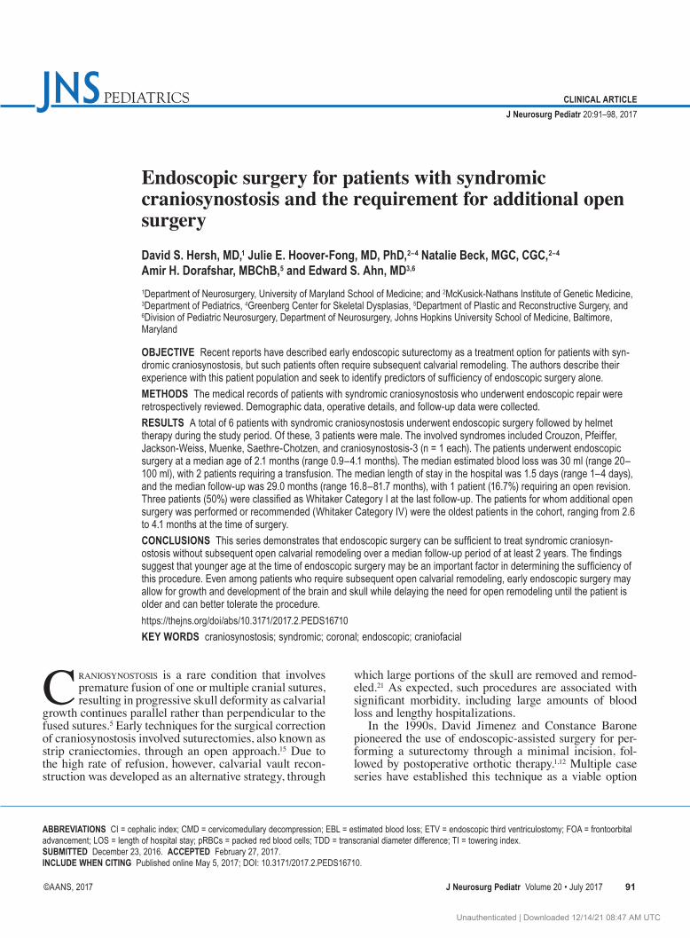
TABLE 1. Features of 6 patients with syndromic craniosynostosis who underwent endoscopic surgery with postoperative helmet therapy
Case No. Sex Syndrome
Gene, Mutation Sutures
Age at Op (mos) Transfusion
LOS (days)
Preop Index
Postop Index*
Whitaker Category
Follow-Up Findings
1 M Saethre-Chotzen
TWIST1, unavailable
Bilat coronal 4.1 No 3 NA 75, TI IV FOA 9 mos after IP; subtotal calvarial vault reconstruction 4.25 yrs after IP
2 M Crouzon FGFR2, p.Cys324Tyr
Bilat coronal 2.6 No 1 70, TI 72, TI IV Exorbitism & midface hypoplasia at 3.5 yrs; will require mono-bloc frontofacial advancement
3 F Muenke FGFR3, p.Pro250Arg
Unilat coro-nal
1.6 No 1 0, TDD 2, TDD I Stable after 3 yrs
4 F Pfeiffer FGFR2, c.940-3T>G
Bilat coronal, sagittal
0.9 No 2 73, CI 81, CI I Stable after 2 yrs
5 M Jackson- Weiss
FGFR2, c.1025G>C
Bilat coronal, bilat lamb-doid
3.1 Postop 4 83, TI 77, TI IV Lambdoid suturectomy 2 wks after IP; CMD & ETV 3 mos after IP; died 17 mos after IP due to unrelated respiratory complications
6 F Craniosyn-ostosis-3
TCF12, c.1491dupT
Bilat coronal 1.0 Intraop 1 74, TI 76, TI I Stable after 1.5 yrs
IP = index procedure; NA = not available.* The anthropometric index measured at the most recent clinic visit. The TDD’s unit of measure is millimeters.
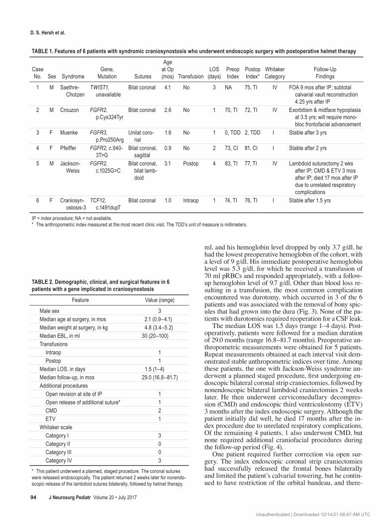
TABLE 2. Demographic, clinical, and surgical features in 6 patients with a gene implicated in craniosynostosis
Feature Value (range)
Male sex 3Median age at surgery, in mos 2.1 (0.9–4.1)Median weight at surgery, in kg 4.8 (3.4–5.2)Median EBL, in ml 30 (20–100)Transfusions Intraop 1 Postop 1Median LOS, in days 1.5 (1–4)Median follow-up, in mos 29.0 (16.8–81.7)Additional procedures Open revision at site of IP 1 Open release of additional suture* 1 CMD 2 ETV 1Whitaker scale Category I 3 Category II 0 Category III 0 Category IV 3
* This patient underwent a planned, staged procedure. The coronal sutures were released endoscopically. The patient returned 2 weeks later for nonendo-scopic release of the lambdoid sutures bilaterally, followed by helmet therapy.
Unauthenticated | Downloaded 12/14/21 08:47 AM UTC

Endoscopic surgery for syndromic craniosynostosis
J Neurosurg Pediatr Volume 20 • July 2017 95
fore underwent open frontoorbital advancement (FOA) 9 months later (Fig. 5). The patient was later noted to have developed supraorbital rim retrusion and had persistent towering 4.3 years after the index endoscopic surgery, and underwent a subtotal calvarial vault reconstruction at that time. Furthermore, the patients with Crouzon syndrome and Jackson-Weiss syndrome were classified as Whitaker Category IV, whereas the patients with Muenke syndrome, Pfeiffer syndrome, and craniosynostosis-3 were classified as Whitaker Category I at their most recent follow-up.
DiscussionEarly endoscopic suturectomy followed by postoper-
ative helmet therapy is becoming increasingly accepted as an option for the treatment of single-suture and, more recently, multiple-suture craniosynostosis. However, ex-perience with this technique in the syndromic craniosyn-ostosis population has been limited, and furthermore has been characterized by a high rate of subsequent calvarial remodeling. We present a series of 6 patients with a mo-lecular confirmation of syndromic craniosynostosis who underwent endoscopic suturectomy and postoperative helmet therapy. We demonstrate that in approximately 50% of cases, endoscopic surgery can be sufficient to treat syndromic craniosynostosis by stabilizing the deformity,
which most commonly involves calvarial towering, with-out subsequent open calvarial remodeling over a median follow-up period of more than 2 years. Furthermore, we observe that younger age at the time of the index proce-dure may be a factor in determining the success of en-doscopic surgery. In the remaining cases, endoscopic sur-gery must be followed by more definitive open calvarial remodeling, but may be useful in delaying open surgery until the patient is older.
Other than an isolated case report,10 prior descriptions of endoscopic surgery for syndromic craniosynostosis have been included within broader analyses of all patients (both syndromic and nonsyndromic) with multiple-suture craniosynostosis (Table 3). In the earliest series, Rivero-Garvía et al.19 described 7 patients 4 months of age or younger with multiple-suture craniosynostosis. Of these 7 patients, 2 were diagnosed with syndromic craniosynos-tosis—one patient had Muenke syndrome and the other had Crouzon syndrome. The patients were 52 days and 62 days old, respectively, at the time of surgery. Neither required a blood transfusion. At follow-up visits, both pa-tients displayed a “satisfactory” result and neither required additional craniofacial procedures. Additionally, neither patient developed ventricular dilation or a Chiari I malfor-mation, and the authors hypothesized that early treatment prevented the development of secondary malformations. However, the patient with Crouzon syndrome underwent only 9 months of follow-up, potentially limiting the ability to determine the effect of early endoscopic surgery on the need for additional surgical intervention.
Recently, in a large series that examined the complica-tions associated with open or endoscopic surgery for cra-niosynostosis, Han et al.8 described 295 nonsyndromic pa-tients and 33 syndromic patients; of the latter, 10 patients were treated endoscopically. These 10 patients were treated at a mean age of 3.8 months. Four of the patients had sin-gle-suture synostosis, 5 had 2-suture synostosis, and 1 had involvement of 3 or more sutures. One patient had an intra-operative durotomy, and none of the patients required an intraoperative or postoperative transfusion. Three patients required a reoperation due to “suboptimal aesthetics,” and an additional patient required reoperation for a CSF leak.
An additional 18 patients with bilateral coronal cra-niosynostosis treated endoscopically, 9 of whom were di-
FIG. 2. Anteroposterior view (A), vertex view (B), and lateral (C) 3D reconstructions of a CT scan demonstrating bilateral coronal and sagittal synostosis associated with Pfeiffer syndrome in a 1-day-old girl. Multifocal thinning and thumbprinting of the calvarium is noted, associated with localized intracranial pressure. Figure is available in color online only.
FIG. 1. Anteroposterior (left) and lateral (right) 3D reconstructions of a CT scan demonstrating bilateral coronal synostosis associated with a mutation in the TCF12 gene in a 1-month-old girl. Figure is available in color online only.
Unauthenticated | Downloaded 12/14/21 08:47 AM UTC

D. S. Hersh et al.
J Neurosurg Pediatr Volume 20 • July 201796
agnosed with syndromic craniosynostosis, were recently reported by Rottgers and colleagues.20 The syndromes that were involved included Saethre-Chotzen (n = 3), Apert (n = 2), Muenke (n = 3), and Pfeiffer (n = 1). Although the authors did not analyze syndromic and nonsyndromic pa-tients separately, the overall mean age at surgery was 2.6 months. Two patients required transfusions, and 1 patient
with Apert syndrome had a durotomy resulting in a pseu-domeningocele and a CSF leak. Only 1 of the 9 patients (11%) with nonsyndromic craniosynostosis required ad-ditional craniofacial surgery (secondary FOA), whereas 5 of the 9 patients (56%) with syndromic craniosynostosis underwent secondary FOA.
An analysis of the CIs and head circumference of the patients over time revealed that the patients who required secondary FOA demonstrated improved head size and shape in the early phase following endoscopic release, but later underwent refusion or fusion of additional sutures, resulting in growth restriction and the need for additional surgery 1–2 years after the index endoscopic procedure. The authors emphasized that although subsequent FOA was often required, early endoscopic surgery at least of-fered the possibility of effective treatment via a single pro-cedure, as opposed to early posterior cranial vault distrac-tion followed by FOA. Additionally, among patients with syndromic craniosynostosis, younger age (i.e., less than 6 months) at the time of cranial vault remodeling is associ-ated with a significantly higher risk of needing a subse-quent major reoperation (odds ratio 4.10, 95% confidence interval 1.31–12.87, p = 0.016).22 At the least, one role of endoscopic surgery in this population could be to delay the initial open reconstruction to reduce the subsequent revision rate. Another possible advantage of early release of the suture is an increased likelihood of correcting sur-rounding abnormalities involving the skull base and su-praorbital ridge.
Despite the high number of syndromic patients report-ed by Han et al.8 and Rottgers et al.20 who required addi-tional, open cranial remodeling (3 of 10 patients and 5 of 9 patients, respectively), the underlying factors that con-tribute to the sufficiency of endoscopic surgery alone re-
FIG. 4. Preoperative (A), 3-month (B), 8-month (C), 1-year (D), 15-month (E), and 2-year (F) postoperative photographs of a female patient with bilateral coronal and sagittal synostosis associated with Pfeiffer syndrome demonstrating improved scapho-cephaly and calvarial towering following endoscopic surgery, without additional open procedures. Corresponding lateral (G–L) and overhead (M–R) views of this patient at the same time points are also shown. Figure is available in color online only.
FIG. 3. Endoscopic view of a bony spicule (asterisk) at a coronal suture fusion, which invaginates into the underlying dura. These sites are asso-ciated with a higher risk of durotomy during removal. Figure is available in color online only.
Unauthenticated | Downloaded 12/14/21 08:47 AM UTC

Endoscopic surgery for syndromic craniosynostosis
J Neurosurg Pediatr Volume 20 • July 2017 97
main unclear. In the current series, only 1 of the 6 patients underwent additional open cranial vault remodeling. This patient was diagnosed with Saethre-Chotzen syndrome, as were 3 of the 9 patients reported by Rottgers et al. One possibility is that the genetic and phenotypic features of individual craniosynostosis syndromes may impact the need for additional surgery. However, the diagnoses of the patients who underwent secondary FOA in that particular series are unknown.
In addition to the patient in the current series who un-derwent additional cranial vault remodeling, the patients with Crouzon syndrome and Jackson-Weiss syndrome were classified as Whitaker Category IV based on their last set of clinical photographs, indicating that open cal-varial remodeling is advisable. In these cases, calvarial re-modeling and monobloc frontofacial advancement will be necessary to correct exorbitism, midface hypoplasia, and supraorbital ridge retraction. Of note, endoscopic surgery appears to have been sufficient for the youngest patients in this series, with ages ranging from 0.9 to 1.6 months when surgery was performed. In contrast, those who required additional cranial vault remodeling tended to be older, with ages ranging from 2.6 to 4.1 months at surgery.
Ultimately, 3 of the 6 patients in our series were clas-sified as Whitaker Category I at the last follow-up. These
outcomes are similar to our previously published experi-ence with calvarial vault remodeling for syndromic cra-niosynostosis. In a series of patients followed over a 13-year period, 47.7% of 71 procedures in 58 patients were considered Whitaker Category I, 14.4% were Category II, 4.6% were Category III, and 32.3% were Category IV.22 Although the numbers in the current endoscopic series are much smaller, our preliminary results indicate that out-comes are similar after endoscopic surgery and that revi-sion rates with major surgery may be higher after open remodeling in this patient group.
Our study has several limitations. In addition to the inherent limitations of a retrospective study, several deci-sions regarding the management of the condition in pa-tients with craniosynostosis are subjective in nature. In particular, decisions regarding indications for revision sur-gery vary from provider to provider and are not necessar-ily generalizable. Although the Whitaker scale was used to measure postoperative aesthetic outcomes, this too is based on a subjective analysis. However, our patients were followed with objective anthropometric indices, which may provide more uniform bases to guide decisions about revision surgery. An additional limitation of our study is the duration of follow-up. However, although our median follow-up was only 29 months, our mean follow-up was
FIG. 5. Lateral photographs of a male patient with bilateral coronal synostosis associated with Saethre-Chotzen syndrome. Photographs were obtained prior to endoscopic surgery (A), 4 months postoperatively (B), and 2 months following an open FOA (C). Despite endoscopic suturectomy and postoperative helmet therapy, the patient was noted to have persistent restriction of the orbital bandeau (B) and an FOA was therefore recommended. Following FOA, the restriction was corrected (C). Figure is available in color online only.
TABLE 3. Literature review of patients with syndromic craniosynostosis treated endoscopically
Authors & Year
No. of Syndromic
Patients
Mean Age at
Op (mos) Syndromes
Mean LOS
(days)No. of
TransfusionsSecondary
Op
Mean Follow-Up in Mos (range)
Jimenez & Barone, 2012 1 2.0 Apert 1 0 0 9Rivero-Garvía et al., 2012 2 1.9 Muenke, Crouzon 3.5 0 0 21 (9–33)Han et al., 2016 10 3.8 Goldenhar, Crouzon, Charcot-Marie-
Tooth, Saethre-Chotzen, DiGeorge, Down, VATER, CDAGS
1.4 0 3 36.8 (2.3–74.9)
Rottgers et al., 2016 9 2.6* Saethre-Chotzen, Apert, Muenke, Pfeiffer 1.2* 2* 5 37 (6–102)*
CDAGS = craniosynostosis, anal anomalies, porokeratosis; VATER = vertebral anomalies, anal atresia, tracheo-esophageal fistula and/or esophageal atresia, renal and radial anomalies, and limb defects.* These values reflect the 18 patients with syndromic and nonsyndromic bilateral coronal craniosynostosis who were presented in the series. The study’s authors did not present individual data for the 9 patients with syndromic craniosynostosis.
Unauthenticated | Downloaded 12/14/21 08:47 AM UTC

D. S. Hersh et al.
J Neurosurg Pediatr Volume 20 • July 201798
36.3 months, which is similar to the mean follow-up of 36.8 months and 37 months reported by Han et al.8 and Rottgers et al.,20 respectively. Furthermore, Rottgers et al. reported that in their series, secondary FOAs were per-formed an average of 15 months after endoscopic surgery, with the latest revision taking place 22.7 months after the endoscopic procedure. Our duration of follow-up should therefore have been sufficient for identifying most patients in need of a delayed calvarial reconstruction. Last, due to the low incidence of syndromic craniosynostosis, there were a limited number of patients in the current cohort. A larger, prospective study is necessary for a statistical anal-ysis and a more comprehensive evaluation of the factors that predict the sufficiency of endoscopic surgery.
ConclusionsPatients with syndromic craniosynostosis represent a
complex subset within the overall craniosynostosis popu-lation. In a subset of patients, endoscopic surgery can be sufficient to treat syndromic craniosynostosis without sub-sequent open calvarial reconstruction, and at the least can delay the need for open surgery. Although historically syn-dromic craniosynostosis has been associated with a high rate of subsequent need for open calvarial vault remodel-ing, our findings suggest that younger age at the time of endoscopic surgery may be an important factor in deter-mining the sufficiency of the endoscopic procedure alone.
References 1. Barone CM, Jimenez DF: Endoscopic craniectomy for
early correction of craniosynostosis. Plast Reconstr Surg 104:1965–1975, 1999
2. Chaichana KL, Jallo GI, Dorafshar AH, Ahn ES: Novel use of an ultrasonic bone-cutting device for endoscopic-assisted cra-niosynostosis surgery. Childs Nerv Syst 29:1163–1168, 2013
3. Chim H, Manjila S, Cohen AR, Gosain AK: Molecular signaling in pathogenesis of craniosynostosis: the role of fibroblast growth factor and transforming growth factor-b. Neurosurg Focus 31(2):E7, 2011
4. Christofides EA, Steinmann ME: A novel anthropometric chart for craniofacial surgery. J Craniofac Surg 21:352–357, 2010
5. Delashaw JB, Persing JA, Broaddus WC, Jane JA: Cranial vault growth in craniosynostosis. J Neurosurg 70:159–165, 1989
6. Glasgow TS, Siddiqi F, Hoff C, Young PC: Deformational plagiocephaly: development of an objective measure and determination of its prevalence in primary care. J Craniofac Surg 18:85–92, 2007
7. Governale LS: Craniosynostosis. Pediatr Neurol 53:394–401, 2015
8. Han RH, Nguyen DC, Bruck BS, Skolnick GB, Yarbrough CK, Naidoo SD, et al: Characterization of complications asso-ciated with open and endoscopic craniosynostosis surgery at a single institution. J Neurosurg Pediatr 17:361–370, 2016
9. Jezela-Stanek A, Krajewska-Walasek M: Genetic causes of syndromic craniosynostoses. Eur J Paediatr Neurol 17:221–224, 2013
10. Jimenez DF, Barone CM: Bilateral endoscopic craniectomies in the treatment of an infant with Apert syndrome. J Neuro-surg Pediatr 10:310–314, 2012
11. Jimenez DF, Barone CM: Early treatment of coronal synos-tosis with endoscopy-assisted craniectomy and postoperative cranial orthosis therapy: 16-year experience. J Neurosurg Pediatr 12:207–219, 2013
12. Jimenez DF, Barone CM: Endoscopic craniectomy for early surgical correction of sagittal craniosynostosis. J Neurosurg 88:77–81, 1998
13. Jimenez DF, Barone CM: Multiple-suture nonsyndromic craniosynostosis: early and effective management using en-doscopic techniques. J Neurosurg Pediatr 5:223–231, 2010
14. Jimenez DF, Barone CM, McGee ME, Cartwright CC, Baker CL: Endoscopy-assisted wide-vertex craniectomy, barrel stave osteotomies, and postoperative helmet molding thera-py in the management of sagittal suture craniosynostosis. J Neurosurg 100 (5 Suppl Pediatrics):407–417, 2004
15. Mehta VA, Bettegowda C, Jallo GI, Ahn ES: The evolution of surgical management for craniosynostosis. Neurosurg Focus 29(6):E5, 2010
16. Miller KA, Twigg SR, McGowan SJ, Phipps JM, Fenwick AL, Johnson D, et al: Diagnostic value of exome and whole genome sequencing in craniosynostosis. J Med Genet [epub ahead of print], 2016
17. Mulliken JB, Vander Woude DL, Hansen M, LaBrie RA, Scott RM: Analysis of posterior plagiocephaly: deformation-al versus synostotic. Plast Reconstr Surg 103:371–380, 1999
18. Pindrik J, Molenda J, Uribe-Cardenas R, Dorafshar AH, Ahn ES: Normative ranges of anthropometric cranial indices and metopic suture closure during infancy. J Neurosurg Pediatr 25:667–673, 2016
19. Rivero-Garvía M, Marquez-Rivas J, Rueda-Torres AB, Olle-ro-Ortiz A: Early endoscopy-assisted treatment of multiple-suture craniosynostosis. Childs Nerv Syst 28:427–431, 2012
20. Rottgers SA, Lohani S, Proctor MR: Outcomes of endoscopic suturectomy with postoperative helmet therapy in bilateral coronal craniosynostosis. J Neurosurg Pediatr 18:281–286, 2016
21. Tessier P: The definitive plastic surgical treatment of the se-vere facial deformities of craniofacial dysostosis. Crouzon’s and Apert’s diseases. Plast Reconstr Surg 48:419–442, 1971
22. Utria AF, Mundinger GS, Bellamy JL, Zhou J, Ghasemzadeh A, Yang R, et al: The importance of timing in optimizing cranial vault remodeling in syndromic craniosynostosis. Plast Reconstr Surg 135:1077–1084, 2015
23. Whitaker LA, Bartlett SP, Schut L, Bruce D: Craniosynostosis: an analysis of the timing, treatment, and complications in 164 consecutive patients. Plast Reconstr Surg 80:195–212, 1987
DisclosuresDr. Dorafshar receives support for non–study-related clinical or research effort that he oversees from DePuy Synthes and from KLS Martin. He is also a patent holder with KLS Martin.
Author ContributionsConception and design: Ahn. Acquisition of data: Ahn, Hersh, Hoover-Fong, Beck. Analysis and interpretation of data: Hersh, Hoover-Fong, Beck, Dorafshar. Drafting the article: Hersh. Criti-cally revising the article: Ahn, Hoover-Fong, Dorafshar. Reviewed submitted version of manuscript: all authors. Approved the final version of the manuscript on behalf of all authors: Ahn. Study supervision: Ahn.
Supplemental InformationPrevious PresentationsThis work was presented in abstract form as a poster at the AANS/CNS Section on Pediatric Neurological Surgery at its 2016 Annual Meeting in Orlando, Florida.
CorrespondenceEdward S. Ahn, Department of Pediatrics, Johns Hopkins University School of Medicine, 600 N Wolfe St., Phipps Ste. 560, Baltimore, MD 21287. email: [email protected].
Unauthenticated | Downloaded 12/14/21 08:47 AM UTC