Embed Size (px)
Citation preview

1
Endothelial dysfunction in femoral artery of the hypertensive rats is nitric oxide
independent
Angelika Puzserova1, Jana Kopincova1,2, Peter Slezak1, Peter Balis1, Iveta Bernatova1
1Institute of Normal and Pathological Physiology, Centre of excellence for examination of
regulatory role of nitric oxide in civilization diseases, Slovak Academy of Sciences,
Sienkiewiczova 1, 813 71 Bratislava, Slovak Republic
2Department of Physiology, Jessenius Faculty of Medicine, Comenius University, Mala Hora
4, 036 01 Martin, Slovak Republic
Correspondence:
Angelika Puzserova, Ph.D.
Institute of Normal and Pathological Physiology
Slovak Academy of Sciences
Sienkiewiczova 1
Bratislava 813 71
Slovak Republic
Tel.: +421-2-32296013
Fax: +421-2-52968516
E-mail: [email protected]
Short title
Endothelial function of hypertensive rats

2
Abstract
This study examined nitric oxide (NO) production, oxidative load and endothelium-dependent
relaxation (NO-dependent and NO-independent) in adult male borderline hypertensive (BHR)
and spontaneously hypertensive (SHR) rats as compared to normotensive Wistar-Kyoto
(WKY) rats.
Systolic blood pressure (BP) was determined by tail-cuff. NO production was determined by
conversion of [3H]-L-arginine. Conjugated dienes (CD) and concentrations of thiobarbituric
acid-reactive substances (TBARS) were measured for assessment of oxidative load. Vascular
function was investigated in rings of the femoral artery (FA) using a wire myograph.
BP of WKY, BHR and SHR was 106±2, 143±3 and 191±3 mmHg, respectively (p<0.01 for
each). Significant left ventricle (LV) hypertrophy and elevated levels of CD and TBARS in
the LV were present in BHR and SHR as compared to WKY. NO production was elevated
significantly in the aorta of BHR and SHR vs. WKY as well as in the LV of SHR vs. WKY.
Acetylcholine (ACh)-induced relaxation of the FA was reduced significantly in both BHR and
SHR vs. WKY. The NO-dependent component of ACh-induced relaxation had increasing
tendency in hypertensive groups and it correlated positively with BP. The NO-independent
component of vasorelaxation was reduced significantly in BHR and SHR vs. WKY and it
correlated negatively with BP.
In conclusion, the results showed that endothelial dysfunction in the experimental model of
borderline hypertensive and hypertensive rats is NO-independent. The results suggest that
borderline hypertension represents a risk of other cardiovascular disorders which is
qualitatively similar to that of fully developed hypertension.
Key words: acetylcholine, borderline hypertension, endothelium, SHR, vascular reactivity

3
Introduction
Chronic hypertension is generally considered to be a risk factor of several cardiovascular
diseases. However, prehypertension is not a disease category (Chobanian et al. 2003) and
relatively little attention has been paid to prevention of cardiovascular risk resulting from
marginal elevation of blood pressure (BP) in adult individuals. Although the vascular
mechanisms involved in prehypertension and hypertension are not well-defined, there are
studies suggesting the involvement of endothelial dysfunction (ED) in conduit and resistance
arteries in association with altered nitric oxide (NO) production and oxidative stress, which
may significantly contribute to the pathogenesis of hypertension by increasing peripheral
vascular resistance.
Observations in spontaneously hypertensive rats (SHR), a widely used experimental model of
human essential hypertension (Yamori and Okamoto 1973, Pintérová et al. 2011, Kuneš et al.
2012), showed considerable differences related to ED (Vapaatalo et al. 2000, Török 2008,
Gündüz et al. 2011). As shown in the literature, ED in SHR depends on several factors
including age, artery size and methodological approaches (Bernatova et al. 2009). Since
hypertension in SHR occurs already at juvenile age (about 5-8 weeks), it is impossible to
examine metabolic and vascular alterations in the prehypertensive period in adult (12+ week-
old) SHR. For this reason, borderline hypertensive rats (BHR) are more suitable for
investigating the early stage of hypertension development in adulthood, which is obvious in
the human population, and needs to be better understood.
Borderline hypertensive rats, an experimental model of human prehypertension and early
stage hypertension, can be produced by mating spontaneously hypertensive dams with

4
normotensive sires (Lawler et al. 1980, Fuchs et al. 1998, Bernatova et al. 2007, Pechánová et
al. 2009). Resting systolic BP of adult offspring in the first-generation is in the range 130-150
mmHg (Sanders and Lawler 1992, Mansi and Drolet 1997, Bernatova et al. 2007), which
allows investigating the vascular function in adult animals with mild elevation of blood
pressure.
In general, ED is characterized by alterations in the production and/or bioavailability of
endothelium-derived relaxing factors (EDRFs) and endothelium-derived constricting factors
(EDCFs) (Török 2008, Vanhoutte 2009). The functional status of the endothelium is usually
tested by the acetylcholine (ACh) test in isolated pre-constricted arteries. ACh-induced
relaxation results from stimulated release of at least three different vasodilating agents from
the endothelial cells - nitric oxide (NO), prostacyclin (PGI2), and endothelium-derived
hyperpolarizing factor(s) (EDHFs) (Fujimoto and Fujimoto 2001, Stankevicius et al. 2003,
Gündüz et al. 2011). NO seems to be the most powerful vasorelaxing factor, however the
extent of NO-dependent relaxation depends on the size of the artery. The greatest NO-
dependency of vasorelaxation was observed in the aorta, while the role of NO is much smaller
in the regulation of endothelial function in the small resistant arteries where EDHFs seem to
be more important (Shimokawa et al. 1996, Török 2008).
In this study we investigated vascular function (contractile responses and vasorelaxation
including its NO-dependent and NO-independent components) in the femoral artery as well as
the oxidative load and NO production in selected tissues of normotensive, borderline
hypertensive and spontaneously hypertensive rats in order to determine the role of NO and the
seriousness of vascular changes in conditions of borderline hypertension in adulthood.

5
Methods
Animals
All rats used in the present study were born in our certified animal facility (Institute of
Normal and Pathological Physiology SAS) in order to maintain the same environmental
background of all animals. Adult, 22-week old males of Wistar-Kyoto (WKY, negative
control), BHR and SHR (positive control) were used in this study (n=10 each). BHRs were
the first filial offspring of SHR dams and normotensive WKY sires. Rats were housed in
groups of 5 rats per cage (35/55/20 cm), each genotype separately, in an air-conditioned room
at constant temperature (22 – 24 ˚C) and humidity (45 - 60%) at a 12:12-h light/dark cycle
(06:00 – 18:00 h lights on) and maintained on a standard pellet diet and tap water ad libitum.
Drinking water consumption was measured in each group from the age of 12 weeks
(beginning of adulthood) to 22 weeks of age (end of experiment). All procedures used were in
accordance with the EU Directive 2010/63/EU and they were approved by the State
Veterinary and Food Administration of the Slovak Republic.
Experimental design
Twenty-two-week-old Wistar-Kyoto, borderline hypertensive and spontaneously hypertensive
rats were killed by decapitation after a brief CO2 anesthesia between 7:30 and 9:30 AM. Wet
mass of the left heart ventricle (LVM) and body mass (BM) were determined for calculation
of the relative left ventricular mass (LVM/BM) to evaluate the degree of cardiac hypertrophy.
Blood pressure and heart rate

6
Systolic blood pressure (BP) and heart rate (HR) were determined non-invasively in
conscious rats by tail-cuff plethysmography (using the Statham Pressure Transducer P23XL,
Hugo Sachs, Germany). One week before experimentation, the rats were handled and
accustomed to the tail-cuff procedure of blood pressure recording in three independent
sessions (Durante et al. 2010). Blood pressure and heart rate were determined between 9:00 –
12:00 h and were calculated as the average value of 4 - 5 successive measurements
(Puzserova et al. 2013).
Assessment of vascular reactivity by wire myograph
Femoral arteries (FA) were carefully dissected, immediately immersed in modified cold
physiological salt solution (PSS) and cleaned of adipose or connective tissue. Arteries were
then cut into segments (1.33±0.04 mm long) and mounted as ring-shaped preparations in the
Mulvany–Halpern style small vessel wire myograph chamber (Dual Wire Myograph System
410A, DMT A/S, Aarhus, Denmark) to determine the vascular reactivity during isometric
conditions as described elsewhere (Mulvany and Halpern 1977). After mounting in
oxygenated (5% CO2, 95% O2 mixture) PSS (composition in mmol/l: NaCl 118.99, KCl 4.69,
NaHCO3 25, MgSO4.7H2O 1.17, KH2PO4 1.18, CaCl2.2H2O 2.5, Na2EDTA 0.03, glucose
5.5), pH 7.4, at 37 ˚C, a standardized computer-assisted normalization procedure was
performed to set the pre-tension of the arteries. The arteries were set to a circumference
equivalent to 90% of that which they would have at a transmural pressure of 100 mmHg (90%
of the normalized inner circumference). The change in wall tension (active wall tension) was
calculated as measured force divided by the double segment length and expressed in mN/mm.

7
Resting wall tension (which arises from the properties of the passive elements in the vascular
wall) was also determined after the normalization procedure (Mulvany and Halpern 1977).
Femoral artery reactivity
Before the start of the normalization procedure, the vessels were allowed to stabilize in PSS
for 30 minutes. The experimental protocol consisted of the following steps: 45 min after
normalization, PSS was changed to KPSS in which NaCl was exchanged for an equimolar
concentration of KCl (in whole KPSS 125 mmol/l for 2 min) followed by wash-out with PSS
to baseline (15 min). After noradrenaline (NA) addition (10 μmol/l, maximal stimulation,
waiting to plateau) and wash-out (PSS, 20 min), pre-constriction was achieved by serotonin
(5-hydroxytryptamine, 1 μmol/l, waiting to plateau). When the contraction of the femoral
artery to serotonin reached a steady state, increasing concentrations of the vasodilator
acetylcholine (ACh, 0.001 to 10 μmol/l) were added in cumulative manner to perform
endothelium-dependent concentration-response curves (Fujimoto and Fujimoto 2001). When
the concentration-relaxation curve was completed, the drugs were washed-out (PSS, 20 min)
and the same experiment was repeated in presence of NG-nitro-L-arginine methyl ester (L-
NAME, nitric oxide synthase inhibitor, 300 μmol/l) after a 25-min pre-incubation period. In
the presence of L-NAME, we could preclude ACh-induced NO release, the curve thus
represented L-NAME-resistant (i.e. NO-independent) component of ACh-induced relaxation.
When concentration-response curve for ACh was completed, drugs were wash-out (PSS, 30
min), and the nitric oxide donor sodium nitroprusside (SNP, 0.001 to 10 μmol/l) was added by
cumulative manner to the pre-constricted arteries (serotonin, 1 μmol/l).

8
After the following wash-out (PSS, 20 min), the femoral artery rings were stimulated with
high concentration of K+ (125 mmol/l) in depolarizing solution to induce maximal contraction
(PSS was changed to KPSS) and then left to achieve plateau. The maximal tension achieved
with depolarizing solution was set as 100% to express the active tension generated by
different vasoconstrictors (Puzserova and Bernatova 2010).
The NO-dependent (i.e. L-NAME-sensitive) component of endothelium-dependent relaxation
was calculated as the difference between ACh-induced relaxation before and after acute L-
NAME pre-treatment (Paulis et al. 2008, Puzserova and Bernatova 2010) and expressed as
area under the curve (AUC, in arbitrary units - a.u.) based on the individual concentration-
response curves (Puzserova and Bernatova 2010). The extent of vasorelaxation was expressed
in relative values as the percentage of the initial contraction induced by serotonin as well as in
absolute values as agonist-induced wall tension decrease (mN/mm) (Webb et al. 1987,
Hansen and Nedergaard 1999, Líšková et al. 2011). Vasoconstrictions were determined as the
maximal tension and they were expressed as active wall tension in mN/mm. Responses to
each drug concentration were always allowed to stabilize before addition of a subsequent dose
of the same drug or of another drug (Asano et al. 1982, Webb et al. 1987).
All chemicals used were purchased from Sigma-Aldrich (Germany), except noradrenaline
hydrogenotartras (Zentiva, Czech Republic). NA was prepared daily. All drugs were
dissolved in distilled water and concentrations were expressed as final concentration in the
myograph chamber.
Oxidative stress markers

9
For assessment of oxidative load, conjugated dienes (CD) and concentrations of thiobarbituric
acid-reactive substances (TBARS) were measured in the left ventricle (LV), as described
previously (Hu et al. 1989), with some modifications.
To determine TBARS, 1 ml of tissue homogenate of the left ventricle (10% w:v, in 1.15%
KCl in 0.01 mol/l phosphate buffer, pH 7.4) was added to 2 ml of 7.5% trichloroacetic acid
and mixed. After centrifugation at 1 000g for 10 min, 1 ml of the supernatant was added to 0.5
ml of 0.7% 2-thiobarbituric acid and incubated in a water bath at 100 ˚C for 10 min. After
cooling, TBARS were measured at 532 nm (GBC UV/VIS 911 A). An extinction coefficient
of 156 000 mol-1.l.cm-1 was used for calculation of the results.
CD were determined from the same tissue homogenates as TBARS. Homogenate (0.4 ml) was
mixed with 1.5 ml chloroform:methanol (1:2) for 1 min, and 0.5 ml of chloroform was added
and mixed for 30 s. Afterwards, 0.5 ml of deionized water was added and mixed for additional
30 s. After centrifugation for 10 min at 1000g, 0.5 ml of the chloroform (lower) layer was
removed and dried under nitrogen. The lipid was dissolved in 2 ml of cyclohexane, and
conjugated dienes were measured at 233 nm using an extinction coefficient of 27 000
mol-1.l.cm-1.
All chemicals used were purchased from Sigma-Aldrich (Germany) and Merck Chemicals
(Germany).
Nitric oxide synthase activity
Total NO synthase (NOS) activity was measured in tissue homogenates of the aorta and left
ventricle (200 mg/ml) by determination of [3H]-L-citrulline formation from [3H]-L-arginine,
as described previously (Púzserová et al. 2006, Puzserova et al. 2013) and expressed as

10
pmol/min/mg of tissue proteins as determined by the Lowry method (Lowry et al. 1951).
Briefly, crude homogenates of the aorta and LV containing 200 mg of wet tissue per 1 ml of
homogenization solution containing 50 mmol/l Tris-HCl, pH 7.4 and 1% Protease Inhibitor
Cocktail (Sigma, Germany) were centrifuged at 10 000 g for 10 min at 4 ˚C. After
centrifugation, 50 µl of supernatant was incubated in the presence of 10 μmol/l [3H]-L-
arginine (MP Biomedicals, USA, 50 Ci/mmol), 5 μg/ml calmodulin, 0.5 mmol/l β-NADPH,
250 µmol/l tetrahydrobiopterin, 4 µmol/l FAD, 4 µmol/l FMN, 1 mmol/l Ca2+, 1 mmol/l Mg2+
in the total volume of 100 µl. After 20-min incubation at 37 ˚C, the reaction was stopped by 1
ml of ice-cold stop solution containing 20 mmol/l HEPES, pH 5.5, 2 mmol/l EDTA, 2 mmol/l
EGTA and 1 mmol/l L-citrulline and applied to 50WX-8 Dowex columns (Na+ form). [3H]-L-
citrulline was eluted by 1 ml of distilled water and determined by liquid scintillation counting
(Perkin Elmer Liquid Scintillation Analyzer, TriCarb 2910TR).
Statistical analysis
Data are presented as group mean values ± SEM of the number (n) of observations. Results
were analyzed by analysis of variance (ANOVA). One-way ANOVA was used to compare
basic biometric and cardiovascular parameters, basal tension and inner diameter, vascular
constrictions, nitric oxide synthase activity, CD and TBARS concentrations. In case of
significant results pairwaise comparison with Bonferroni adjustment was employed.
Concentration response curves were compared using two-way ANOVA, followed by vertical
contrast with Bonferroni adjustment. Homogeneity of variances and normality of distribution
were tested by Levene’s test and by Shapiro-Wilk’s test, respectively. To assess depression
present at high concentration of ACh-cumulative concentration response curves, the maximal

11
response and the response at higher ACh concentration at a particular response curve was
compared with Dunnet’s test (see Fig. 2). Means were considered to differ significantly when
p<0.05.
Results
Basic biometric and cardiovascular parameters, NO production and oxidative stress
markers
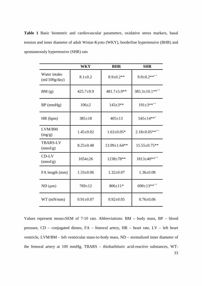
Systolic blood pressure, heart rate, body mass, water intake and LVM/BM ratio of WKY,
BHR and SHR rats are shown in Table 1. Normalized inner diameter was decreased by 10%
in SHR and increased by 5% in BHR compared to WKY (Table 1). There was a significant
positive correlation between normalised inner diameter at 13.3 kPa and BM (r = 0.58, p =
0.006, n = 21). However, there were no significant differences in the resting FA wall tension
of borderline hypertensive and hypertensive rats vs. WKY rats (Table 1). There were also no
significant differences in the FA segment length after mounting among the groups (Table 1).
The NOS activity in the aorta was enhanced by 81% in BHR and by 106% in SHR vs. WKY
(p<0.01; Fig. 1). In the left ventricle, NOS activity was increased only in SHR, as compared
to both BHR and WKY (p<0.01; Fig. 1). Additionally, there were significant increases in
TBARS and CD concentrations in the left ventricle of BHR and SHR (Table 1).
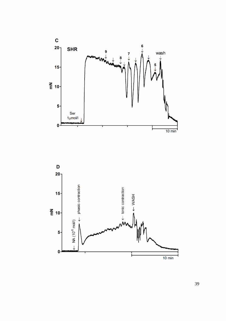
Vascular responsiveness to vasoconstrictors
Both noradrenaline (10 μmol/l) and serotonin (1 μmol/l) induced contractile responses in the
femoral arteries with intact endothelium; the maximum contractions are shown in Table 2.

12
NA-induced responses were biphasic: a transient contraction (early response, phasic
contraction), which occurred within the first 10-15 sec and returned nearly to baseline was
followed by sustained contraction (delayed response, tonic contraction), which reached steady
maximum levels at 5 to 20 min (Fig. 2D). Maximum phasic and tonic contraction induced by
NA was significantly greater in SHR than in WKY, but unchanged in BHR vs. WKY (Table
2). However, only tonic contraction induced by NA determined in relative values (calculated
as percentage of maximal response induced by KPSS) was significantly greater in SHR than
in WKY (Table 2).
In the arteries pre-treated with the NOS inhibitor, the response to serotonin was augmented as
compared to responses before L-NAME administration (Table 2). There were no significant
changes in serotonin responses after L-NAME administration in BHR and SHR rats as
compared to WKY rats. However, the difference between serotonin-induced contraction after
and before L-NAME (to reveal basal, non-stimulated NO synthesis) was significantly
increased in BHR rats compared to WKY and SHR, but comparable between the WKY and
SHR groups (Table 2).
The absolute values for the maximum responses to KPSS were similar among the groups
(Table 2).
Endothelium-dependent and -independent vasorelaxation
ACh (1 nmol/l – 10 μmol/l) and SNP (1 nmol/l – 10 μmol/l) relaxed the femoral artery in a
concentration-dependent manner (Figs. 2, 3A, 3C, 4A, 4C). Since serotonin (1 μmol/l)
induced a reduced response in SHR vessels, resulting in a smaller pre-relaxation active
tension level as compared to WKY (Table 2), the results were quantitatively expressed as

13
mN/mm. ACh in serotonin-contracted femoral arteries could not induce a sustained relaxation
and femoral arteries started to constrict again after a transient relaxation response (Fig. 2).
WKY vessels relaxed significantly more in response to high, yet not to low acetylcholine
concentrations than did BHR and SHR vessels (Fig. 3A, 4A). In BHR and SHR, relaxing
responses produced by ACh at concentrations greater than 0.3 μmol/l were smaller than
maximal relaxations of the given genotype (Fig. 2, 3A, 3B, 4A, 4B). Results revealed a
greater endothelial dysfunction in SHR as compared to BHR (Fig. 3A, 4A). However, there
were small but significant differences in the relative values of ACh-induced relaxations
between BHR and SHR (Fig. 4A). Maximal absolute relaxations induced by ACh, determined
from the individual concentration-response curves, are shown in the Table 2. Relative
maximal relaxations induced by ACh (based on the individual concentration-response curves)
were significantly smaller in SHR (58.47±2.52%, p<0.01) and BHR (60.88±2.75%, p<0.05)
than in WKY (68.88±1.68%). Inhibition of nitric oxide synthesis with L-NAME in WKY and
BHR had no effect on the maximal vasorelaxation, however it reduced significantly in SHR
(Table 2). When the arteries were pre-treated with L-NAME, the femoral arteries from BHR
and SHR rats responded by smaller relaxation to ACh than did the normotensive WKY
arteries (Fig. 3B, 4B). The NO-dependent component of ACh-induced relaxation, calculated
as area under the individual curves, had increasing tendency in WKY, BHR and SHR
(4.15±1.53 a.u., 4.80±1.23 a.u., 5.18±1.04 a.u. calculated from absolute relaxations and
102±17 a.u., 118±17 a.u., 146±16 a.u. calculated from relative relaxations, respectively, n.s.)
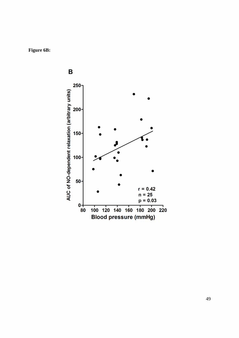
rats. However, significant positive correlation was found between systolic BP and NO-
dependent vasorelaxation (Fig. 6B). The NO-dependent relaxation calculated as the difference
between total relaxation (Fig. 3A/4A) and L-NAME-resistant relaxation (Fig. 3B/4B) are

14
shown in Fig. 5A and Fig. 5B. The NO-independent component of ACh-induced relaxation
was decreased in BHR (12.61±1.47 a.u. as compared to 17.49±1.83 a.u. in WKY, p<0.05) and
SHR (7.32±1.68 a.u. as compared to WKY and BHR, p<0.05). There was also a significant
negative correlation between BP and absolute NO-independent vasorelaxation (r = -0.656, p =
0.0004, n = 25). Moreover, significant negative correlations were found between systolic BP
and relative NO-independent vasorelaxation (Fig. 6A).
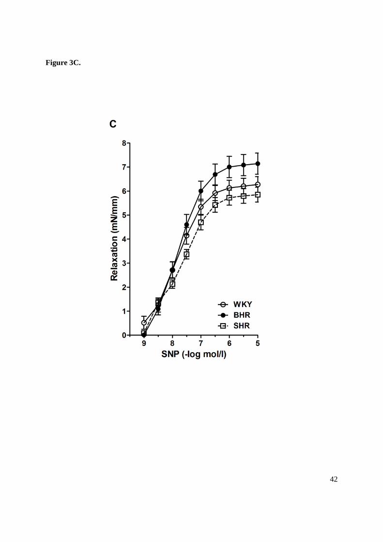
Cumulative addition of NO donor sodium nitroprusside (SNP) produced similar relaxation
responses in the femoral artery from BHR, SHR and WKY rats (Fig. 3C, 4C).
Discussion
The presented study was designed to reveal the severity of vascular alterations in adult
borderline hypertensive rats as compared to normotensive and hypertensive rats. Special
attention was paid to the NO-dependent and independent components of vasorelaxation. The
study revealed attenuated acetylcholine-induced relaxation in the serotonin pre-contracted FA
of both adult BHR and SHR, which was associated with reduction of the NO-independent
component of relaxation. Additionally, there were significant between-strain related
alterations in mechanism of relaxation resulting in negative correlation between BP and NO-
independent component and positive correlation between BP and NO-dependent component
of vasorelaxation. Moreover, biochemical analysis showed elevated NO production as well as
oxidative stress in rats with increased BP.
Elevated systolic blood pressure in both BHR and SHR was accompanied by an increase of
relative left ventricle mass, indicating left ventricular hypertrophy. It is well known that left
ventricular hypertrophy results from the interaction of systemic hemodynamic overload and

15
local non-hemodynamic factors, and thus NO may play a significant role in its development.
Indeed, in the model of NO-deficient hypertension, local reduction of NO production rather
than hemodynamic overload were associated with the degree of LV hypertrophy (Bernátová
et al. 1999). According to our current data, however, LV hypertrophy occurred without
alterations in NO production in the LV of BHR and even in the presence of increased NOS
activity in SHR. It is yet to be noted that the oxidative stress markers were increased in the
LV of both BHR and SHR. Thus oxidative stress may contribute to the development of
hypertrophy induced by multiple mechanisms including G-protein–coupled receptor agonists
and mechanical stretch (Grieve et al. 2006) as well as reduction of NO bioavailability in the
LV (Pechánová et al. 2007, 2009). Similarly, vascular wall remodeling is common feature in
hypertension (Paulis et al. 2007, 2008). We observed reduced internal diameter in the FA in
SHR and increased diameter in BHR without alterations in resting wall tension, which suggest
no changes in the passive elements of the vascular wall (Koprdova et al. 2009). In fact we
observed that internal diameter in the FA correlated positively with BM.
As mentioned above, despite enormous effort, the mechanisms underlying ED in hypertension
are not satisfactorily understood and methodological aspects represent a significant limitation
of our current knowledge. In contrast to our previous findings of unchanged ACh-induced
relaxation in the phenylephrine (Phe) pre-contracted FA of BHR and even improved
relaxation in SHR vs. WKY rats (Púzserová et al. 2007) and vs. Wistar rats (Bernatova et al.
2007), our current study revealed ED in both BHR and SHR as compared to WKY. Since
between-strain differences were detected in the magnitude of pre-contraction induced by
serotonin, the role of differences in pre-relaxation tone cannot be rule out (Li and Bukoski
1993). As Hansen and Nedergaard (1999) have previously demonstrated that the ACh-

16
induced relaxation was inversely related to precontractile tension when expressed as a
percentage, but independent when expressed in absolute values, vasorelaxations in this study
were determined also in absolute values. The results calculated on the basis of absolute
tension were similar to those obtained in percentages (Fig. 3, Fig. 4), however, determination
of relaxation in absolute values revealed a more pronounced ED in SHR vs. BHR, which was
not associated with NO deficiency. The present results also suggest that the responsiveness of
vascular smooth muscles to NO was not impaired since relaxant responses to SNP were
unchanged. Differences in our studies were presumably related to methodological aspects.
Namely, relaxant responses to ACh were determined in serotonin (Fujimoto and Fujimoto
2001) pre-contracted arteries instead of phenylephrine (Bernatova et al. 2007, Púzserová et al.
2007) in order to avoid spontaneous relaxation, which can occur in the Phe pre-contracted FA.
In this context, it has been reported that endothelium-dependent vasorelaxation of SHR
resistance arteries is not impaired under any conditions and appears to be a function of the
agonist used to pre-constrict the vessels (Li and Bukoski 1993).
Moreover, addition of antioxidants, e.g. ascorbic acid (AsA), to solution (to PSS or to solution
for dilution of some drugs) used in vascular in vitro studies may also be of importance
(Bernatova et al. 2009). In addition to above mentioned modifications, our previous
observations of the absence of ED in BHR and SHR were achieved in the presence of AsA
(Bernatova et al. 2007, Púzserová et al. 2007). Similarly to our studies, absence of ED was
shown in the presence of AsA in various arteries of adult SHR (Török and Kristek 2001,
Cacányiová and Kristek 2008, Lísková et al. 2010). However, AsA might artificially reverse
ED by improvement of oxidative status resulting in better NO bioavailability (May 2000,
Bernatova et al. 2009). On the other hand, preserved ACh-induced relaxation was reported

17
also in the adult SHR aorta in absence of antioxidants (Lorkowska et al. 2006). Furthermore
even greater relaxation responses were observed in the adult SHR FA compared to their
normotensive counterparts (Konishi and Su 1983). On the contrary, a reduced ACh-induced
vasorelaxation was reported in the iliac artery in SHR in the presence of AsA (Cacányiová et
al. 2006).
The investigation of vasorelaxation by cumulative addition of ACh revealed also considerable
differences in the course of vascular responses of WKY, BHR and SHR, mainly at higher
ACh concentrations. It is well known that higher concentrations of ACh may elicit production
of endothelium-derived vasoconstrictive cyclooxygenase-generated prostaglandins in adult
SHR arteries, simultaneously with EDRFs (Lüscher and Vanhoutte 1986, Jameson et al. 1993,
Vanhoutte 2009). Previously reported EDCFs include endothelin, cyclooxygenase-derived
prostanoids or free radicals (Li and Bukoski 1993, Paulis et al. 2008, Török 2008, Cacányiová
and Kristek 2008, Vanhoutte et al. 2005, Vanhoutte 2009). The dysbalance between EDRFs
and EDCFs was shown to be progressively increasing not only with advancing age, but also
by hypertension development (Líšková et al. 2011). Similarly, in this study the EDCFs release
was present only in hypertensive animals as determined by stronger depression of relaxation
curves at higher concentration (more than 1 μmol/l in this study) of acetylcholine compared to
normotensive animals. Moreover, the release of EDCFs was not observed in age-matched
WKY rats, similarly as it was found previously (Líšková et al. 2011). In contrast, the EDCFs-
mediated contractions were not seen in the FA of BHR and SHR pre-contracted with
phenylephrine in the presence of AsA (Púzserová et al. 2007), which is in agreement with the
idea that reactive oxygen species, namely superoxide anion, accelerate endothelium-
dependent contractions to ACh in SHR (Jameson et al. 1993, Vanhoutte et al. 2005,

18
Vanhoutte 2009). Furthermore, the EDCFs-mediated contractile responses in the FA of BHR
and SHR were augmented after pre-incubation with L-NAME, suggesting that NO
counterbalances the effect of EDCFs.
An interesting result of this study was the observation of reduced NO-independent (i.e. L-
NAME- resistant) component of ACh-induced relaxation. It is an important finding as many
studies investigating vascular function by ACh test assume that NO deficiency is the major
factor of endothelial dysfunction. As the NG-nitro-L-arginine (an inhibitor of NOS) in the
combination with tetraethylammonium or charybdotoxin (inhibitors of EDHFs-mediated
responses) and indomethacin (cyclooxygenase inhibitor) completely abolished the relaxant
response to ACh in the adult SHR femoral artery (Fujimoto and Fujimoto 2001) we assume
that the NO-independent component is related to endothelium-derived hyperpolarizing factors
and PGI2 dependent vasorelaxation in this artery. This study showed that ED (at least in the
femoral artery) observed in experimental model of genetic prehypertension and hypertension
was related rather to the decreased EDHFs-mediated and/or PGI2-mediated vasorelaxation (Li
et al. 1994, Kähönen et al. 1995, Fujimoto and Fujimoto 2001, Mori et al. 2006) than to lack
of NO. Indeed, there was positive correlation between NO-dependent component of relaxation
and BP, similarly as we observed previously (Púzserová et al. 2007).
Also of interest is our observation that maximal ACh-induced relaxation of SHR vessels was
inhibited by L-NAME, whereas this blocker was without effect on maximal relaxations of
WKY and BHR vessels. Similar observations were reported by Li and Bukoski (1993). This
observation suggests that compensatory EDHFs production may occur in normotensive rats
after acute NOS inhibition (Bauersachs et al. 1996) while this mechanism is impaired in SHR
rats (Púzserová et al. 2010).

19
Regarding vasoconstriction, this study demonstrated that phasic and tonic contractile
responses to noradrenaline were larger in SHR than in WKY. Similar findings were observed
by Asano et al. (1982) in the FA of adult female SHR, which was associated with the decrease
in β-adrenoceptor relaxing responses and accompanied by an enhanced vasoconstriction
induced by NA. Reduced β-adrenoceptor-mediated relaxation was found in the femoral artery
in adult SHR males (Konishi and Su 1983) as well as in young SHR males in the
prehypertensive period (Fujimoto et al. 1987). Thus it is likely that a decrease in β-
adrenoceptor activity would result in potentiation of vasoconstriction by unopposed α-
adrenoceptor stimulation (Asano et al. 1982). Our results also suggest differences in
sensitivity and/or distribution of alpha and beta adrenoceptors between adult BHR and SHR
since NA-induced tonic vasoconstriction of BHR was similar to that in WKY and
significantly lower compared to SHR. Additionally, Líšková et al. (2011) suggested the
enhanced contribution of EDCFs in NA-induced contraction of the femoral arteries in adult
SHR.
Biochemical findings support the idea that the L-arginine/NO system may serve as a
compensatory mechanism activated in response to increased blood pressure in SHR, because
NO is known to counterbalance the effect of sympathetic stimulation on the peripheral as well
as central level (Török 2008). Indeed, several studies, including ours, observed elevated NO
production in SHR (Chen and Hu 1997, Radaelli et al. 1998, Vaziri et al. 2000, Zalba et al.
2001, Hojná et al. 2007, Pechánová et al. 2007, 2009, Púzserová et al. 2007, Török 2008).
Considering the presence of oxidative stress, elevated NO production may be a consequence
of free radical-induced activation of NF-κB, transcriptional factor for NOS (Kopincová et al.
2011). On the other hand, oxidative load may also reduce NO bioavailability (Pechánová et

20
al. 2007), so that NO may be unable to induce greater NO-dependent vasodilatation, despite
increased vascular NO synthesis. Moreover, even though NO-dependent vasorelaxation was
not reduced in hypertensive rats, we cannot exclude relative NO deficiency as compared to
the existing sympathetic overactivity (Kunes et al. 2004, Paulis et al. 2007, Török 2008,
Behuliak et al. 2011) as suggested by increased drinking volume (Kraly et al. 1985) and
elevated HR in SHR as compared to both WKY and BHR.
The observations of elevated CD and TBARS support the idea that oxidative stress may be a
factor contributing to LV hypertrophy as well as ED in genetic hypertension. However,
persistent elevation of NO and superoxide levels can result in generation of peroxynitrite, a
potent oxidant and tissue-damaging agent, resulting in impairment of cellular signalling
(Durackova 2010, Kopincová et al. 2011). It is thus not clear whether NO would remain a
protective compensatory molecule over a prolonged time-course of hypertension. Taking into
account all aforementioned results, the reduced endothelium-dependent relaxations to ACh in
the femoral arteries of BHR and SHR were not due to a decreased release of NO but rather to
other EDRFs and/or to the simultaneous release of EDCFs.
In conclusion, this study showed that LV hypertrophy and ED were present in both adult BHR
and SHR. Moreover, the results revealed that the degree of ED was associated with the level
of BP. LV hypertrophy and ED observed in this study did not result from reduced NO
synthesis. The most important finding of this study was that the NO-independent component
of endothelium-dependent ACh–induced relaxation in serotonin pre-contracted FA correlated
negatively with BP. Thus, the results suggest that both in BHR and SHR rats, ED was not
associated with reduced NO production. Additionally, the results indicate that borderline
hypertension in adulthood represents qualitatively a similar risk of other cardiovascular

21
diseases as does fully developed hypertension. Therefore it is important to study vascular
mechanisms in conditions of moderately increased blood pressure in order to prevent
pressure-induced organ damage, which can further accelerate the development of serious
hypertension.
Acknowledgments
The authors thank to Mrs. Jana Petova for her technical assistance. This study was supported
by the Slovak Grant Agency for Science, grant No. 2/0084/10, Slovak Research and
Development Agency, grant No. APVV-0523-10 and within the project of ‘‘ITMS
26240120020-Establishment of the Centre for the Research on Composite Materials for
Structural, Engineering and Medical Applications-CEKOMAT II”.
Conflict of Interest statement
The authors declare that there are no conflicts of interest.
References
ASANO M, AOKI K, MATSUDA T: Reduced beta adrenoceptor interactions of
norepinephrine enhance contraction in the femoral artery from spontaneously hypertensive
rats. J Pharmacol Exp Ther 223: 207-214, 1982.
BAUERSACHS J, POPP R, HECKER M, SAUER E, FLEMING I, BUSSE R: Nitric oxide
attenuates the release of endothelium-derived hyperpolarizing factor. Circulation 94: 3341-
3347, 1996.

22
BEHULIAK M, PINTÉROVÁ M, KUNEŠ J, ZICHA J: Vasodilator efficiency of endogenous
prostanoids, Ca²⁺-activated K⁺ channels and nitric oxide in rats with spontaneous, salt-
dependent or NO-deficient hypertension. Hypertens Res 34: 968-975, 2011.
BERNATOVA I, CONDE MV, KOPINCOVA J, GONZALEZ MC, PUZSEROVA A,
ARRIBAS SM: Endothelial dysfunction in spontaneously hypertensive rats: focus on
methodological aspects. J Hypertens Suppl 27: S27-31, 2009.
BERNATOVA I, CSIZMADIOVA Z, KOPINCOVA J, PUZSEROVA A: Vascular function
and nitric oxide production in chronic social-stress-exposed rats with various family history of
hypertension. J Physiol Pharmacol 58: 487-501, 2007.
BERNÁTOVÁ I, PECHÁNOVÁ O, KRISTEK F: Mechanism of structural remodelling of
the rat aorta during long-term NG-nitro-L-arginine methyl ester treatment. Jpn J Pharmacol
81: 99-106, 1999.
CACÁNYIOVÁ S, CEBOVÁ M, KUNES J, KRISTEK F. Comparison of vascular function
and structure of iliac artery in spontaneously hypertensive and hereditary
hypertriglyceridemic rats. Physiol Res 55: S73-80, 2006.
CACÁNYIOVÁ S, KRISTEK F: Adaptive vasoactive response to modulatory effects of
endothelin-1 in spontaneously hypertensive rats. Pharmacol Rep 60: 941-949, 2008.
CHEN HI, HU CT: Endogenous nitric oxide on arterial hemodynamics: a comparison
between normotensive and hypertensive rats. Am J Physiol 273: H1816-1823, 1997.
CHOBANIAN AV, BAKRIS GL, BLACK HR, CUSHMAN WC, GREEN LA, IZZO JL JR,
ET AL.: The Seventh Report of the Joint National Committee on Prevention, Detection,
Evaluation, and Treatment of High Blood Pressure: the JNC 7 report. JAMA 289: 2560-2572,
2003.

23
DURACKOVÁ Z: Some current insights into oxidative stress. Physiol Res 59: 459-469,
2010.
DURANTE P, CHÁVEZ M, PÉREZ M, ROMERO F, RIVERA F. Effect of uric acid on
hypertension progression in spontaneously hypertensive rats. Life Sci 86: 957-964, 2010.
FUCHS LC, HOQUE AM, CLARKE NL: Vascular and hemodynamic effects of behavioral
stress in borderline hypertensive and Wistar-Kyoto rats. Am J Physiol 274: R375-382, 1998.
FUJIMOTO S, DOHI Y, AOKI K, ASANO M, MATSUDA T: Diminished beta-
adrenoceptor-mediated relaxation of arteries from spontaneously hypertensive rats before and
during development of hypertension. Eur J Pharmacol 136: 179-187, 1987.
FUJIMOTO S, FUJIMOTO SK: Elcatonin-mediated contractile and relaxant responses in
SHR femoral artery. Acta Pharmacol Sin 22: 595-602, 2001.
GRIEVE DJ, BYRNE JA, SIVA A, LAYLAND J, JOHAR S, CAVE AC, Cave AC, Shah
AM: Involvement of the nicotinamide adenosine dinucleotide phosphate oxidase isoform
Nox2 in cardiac contractile dysfunction occurring in response to pressure overload. J Am Coll
Cardiol 47: 817-826, 2006.
GÜNDÜZ F, KOÇER G, ULKER S, MEISELMAN HJ, BAŞKURT OK, SENTÜRK UK:
Exercise training enhances flow-mediated dilation in spontaneously hypertensive rats. Physiol
Res 60: 589-597, 2011.
HANSEN K, NEDERGAARD OA: Methodologic aspects of acetylcholine-evoked relaxation
of rabbit aorta. J Pharmacol Toxicol Methods 41: 153-159, 1999.
HOJNÁ S, KADLECOVÁ M, DOBESOVÁ Z, VALOUSKOVÁ V, ZICHA J, KUNES J:
The participation of brain NO synthase in blood pressure control of adult spontaneously
hypertensive rats. Mol Cell Biochem 297: 21-29, 2007.
24
HU ML, FRANKEL EN, LEIBOVITZ BE, TAPPEL AL: Effect of dietary lipids and vitamin
E on in vitro lipid peroxidation in rat liver and kidney homogenates. J Nutr 119: 1574-1582,
1989.
JAMESON M, DAI FX, LÜSCHER T, SKOPEC J, DIEDERICH A, DIEDERICH D:
Endothelium-derived contracting factors in resistance arteries of young spontaneously
hypertensive rats before development of overt hypertension. Hypertension 21: 280-288, 1993.
KÄHÖNEN M, MÄKYNEN H, WU X, ARVOLA P, PÖRSTI I: Endothelial function in
spontaneously hypertensive rats: influence of quinapril treatment. Br J Pharmacol 115: 859-
867, 1995.
KONISHI M, SU C: Role of endothelium in dilator responses of spontaneously hypertensive
rat arteries. Hypertension 5: 881-886, 1983.
KOPINCOVÁ J, PÚZSEROVÁ A, BERNÁTOVÁ I: Biochemical aspects of nitric oxide
synthase feedback regulation by nitric oxide. Interdiscip Toxicol 4: 63-68, 2011.
KOPRDOVA R, CEBOVA M, KRISTEK F: Long-term effect of losartan administration on
blood pressure, heart and structure of coronary artery of young spontaneously hypertensive
rats. Physiol Res 58: 327-335, 2009.
KRALY FS, COOGAN LA, SPECHT SM, TRATTNER MS, ZAYFERT C, COHEN A,
GOLDSTEIN JA: Disordered drinking in developing spontaneously hypertensive rats. Am J
Physiol 248: R464-470, 1985.
KUNES J, HOJNÁ S, KADLECOVÁ M, DOBESOVÁ Z, RAUCHOVÁ H, VOKURKOVÁ
M, LOUKOTOVÁ J, PECHÁNOVÁ O, ZICHA J: Altered balance of vasoactive systems in
experimental hypertension: the role of relative NO deficiency. Physiol Res 53: S23-34, 2004.

25
KUNEŠ J, KADLECOVÁ M, VANĚČKOVÁ I, ZICHA J: Critical developmental periods in
the pathogenesis of hypertension. Physiol Res 61 Suppl 1: S9-17, 2012.
LAWLER JE, BARKER GF, HUBBARD JW, SCHAUB RG: Pathophysiological changes
associated with stress-induced hypertension in the borderline hypertensive rat. Clin Sci (Lond)
59: 307-310, 1980.
LI J, BIAN KA, BUKOSKI RD: A non-cyclo-oxygenase, non-nitric oxide relaxing factor is
present in resistance arteries of normotensive but not spontaneously hypertensive rats. Am J
Med Sci 307: 7-14, 1994.
LI J, BUKOSKI RD: Endothelium-dependent relaxation of hypertensive resistance arteries is
not impaired under all conditions. Circ Res 72: 290-296, 1993.
LÍŠKOVÁ S, PETROVÁ M, KAREN P, KUNEŠ J, ZICHA J: Effects of aging and
hypertension on the participation of endothelium-derived constricting factor (EDCF) in
norepinephrine-induced contraction of rat femoral artery. Eur J Pharmacol 667: 265-270,
2011.
LÍSKOVÁ S, PETROVÁ M, KAREN P, KUNES J, ZICHA J: Influence of calcium-
dependent potassium channel blockade and nitric oxide inhibition on norepinephrine-induced
contractions in two forms of genetic hypertension. J Am Soc Hypertens 4: 128-134, 2010.
LORKOWSKA B, BARTUS M, FRANCZYK M, KOSTOGRYS RB, JAWIEN J,
PISULEWSKI PM, CHLOPICKI S: Hypercholesterolemia does not alter endothelial function
in spontaneously hypertensive rats. J Pharmacol Exp Ther 317: 1019-1026, 2006.
LOWRY OH, ROSEBROUGH NJ, FARR AL, RANDALL RJ: Protein measurement with the
Folin phenol reagent. J Biol Chem 193: 265-275, 1951.

26
LÜSCHER TF, VANHOUTTE PM: Endothelium-dependent contractions to acetylcholine in
the aorta of the spontaneously hypertensive rat. Hypertension 8: 344-348, 1986.
MANSI JA, DROLET G: Chronic stress induces sensitization in sympathoadrenal responses
to stress in borderline hypertensive rats. Am J Physiol 272: R813-820, 1997.
MAY JM: How does ascorbic acid prevent endothelial dysfunction? Free Radic Biol Med 28:
1421-1429, 2000.
MORI Y, OHYANAGI M, KOIDA S, UEDA A, ISHIKO K, IWASAKI T: Effects of
endothelium-derived hyperpolarizing factor and nitric oxide on endothelial function in
femoral resistance arteries of spontaneously hypertensive rats. Hypertens Res 29: 187-195,
2006.
MULVANY MJ, HALPERN W: Contractile properties of small arterial resistance vessels in
spontaneously hypertensive and normotensive rats. Circ Res 41: 19-26, 1977.
PAULIS L, LÍSKOVÁ S, PINTÉROVÁ M, DOBESOVÁ Z, KUNES J, ZICHA J:
Nifedipine-sensitive noradrenergic vasoconstriction is enhanced in spontaneously
hypertensive rats: the influence of chronic captopril treatment. Acta Physiol (Oxf) 191: 255-
266, 2007.
PAULIS L, ZICHA J, KUNES J, HOJNA S, BEHULIAK M, CELEC P, KOJSOVA S,
PECHANOVA O, SIMKO F: Regression of L-NAME-induced hypertension: the role of nitric
oxide and endothelium-derived constricting factor. Hypertens Res 31: 793-803, 2008.
PECHÁNOVÁ O, JENDEKOVÁ L, VRANKOVÁ S: Effect of chronic apocynin treatment
on nitric oxide and reactive oxygen species production in borderline and spontaneous
hypertension. Pharmacol Rep 61: 116-122, 2009.

27
PECHÁNOVÁ O, ZICHA J, PAULIS L, ZENEBE W, DOBESOVÁ Z, KOJSOVÁ S,
JENDEKOVÁ L, SLÁDKOVÁ M, DOVINOVÁ I, SIMKO F, KUNES J: The effect of N-
acetylcysteine and melatonin in adult spontaneously hypertensive rats with established
hypertension. Eur J Pharmacol 561: 129-136, 2007.
PINTÉROVÁ M, KUNEŠ J, ZICHA J: Altered neural and vascular mechanisms in
hypertension. Physiol Res 60: 381-402, 2011.
PUZSEROVA A, BERNATOVA I: Chronic social stress increases nitric oxide-dependent
vasorelaxation in normotensive rats. Interdiscip Toxicol 3: 109-117, 2010.
PÚZSEROVÁ A, CSIZMADIOVÁ Z, ANDRIANTSITOHAINA R, BERNÁTOVÁ I:
Vascular effects of red wine polyphenols in chronic stress-exposed Wistar-Kyoto rats. Physiol
Res 55: S39-47, 2006.
PÚZSEROVÁ A, CSIZMADIOVÁ Z, BERNÁTOVÁ I: Effect of blood pressure on L-
NAME-sensitive component of vasorelaxation in adult rats. Physiol Res 56: S77-84, 2007.
PÚZSEROVÁ A, KOPINCOVÁ J, BERNÁTOVÁ I: Evidence for altered feedback
regulation of nitric oxide synthesis in hypertensive rats. Physiol Res 59: 3P, 2010. (Abstract)
PUZSEROVA A, SLEZAK P, BALIS P, BERNATOVA I: Long-term social stress induces
nitric oxide-independent endothelial dysfunction in normotensive rats. Stress 16: 331-339,
2013.
RADAELLI A, MIRCOLI L, MORI I, MANCIA G, FERRARI AU: Nitric oxide dependent
vasodilation in young spontaneously hypertensive rats. Hypertension 32: 735-739, 1998.
SANDERS BJ, LAWLER JE. The borderline hypertensive rat (BHR) as a model for
environmentally-induced hypertension: a review and update. Neurosci Biobehav Rev 16: 207-
217, 1992.

28
SHIMOKAWA H, YASUTAKE H, FUJII K, OWADA MK, NAKAIKE R, FUKUMOTO Y,
TAKAYANAGI T, NAGAO T, EGASHIRA K, FUJISHIMA M, TAKESHITA A: The
importance of the hyperpolarizing mechanism increases as the vessel size decreases in
endothelium-dependent relaxations in rat mesenteric circulation. J Cardiovasc Pharmacol 28:
703-711, 1996.
STANKEVICIUS E, KEVELAITIS E, VAINORIUS E, SIMONSEN U: Role of nitric oxide
and other endothelium-derived factors. Medicina (Kaunas) 39: 333-341, 2003.
TÖRÖK J, KRISTEK F: Functional and morphological pattern of vascular responses in two
models of experimental hypertension. Exp Clin Cardiol 6: 142-148, 2001.
TÖRÖK J: Participation of nitric oxide in different models of experimental hypertension.
Physiol Res 57: 813-825, 2008.
VANHOUTTE PM, FELETOU M, TADDEI S: Endothelium-dependent contractions in
hypertension. Br J Pharmacol 144: 449-458, 2005.
VANHOUTTE PM: Endothelial dysfunction: the first step toward coronary arteriosclerosis.
Circ J 73: 595-601, 2009.
VAPAATALO H, MERVAALA E, NURMINEN ML: Role of endothelium and nitric oxide
in experimental hypertension. Physiol Res 49: 1-10, 2000.
VAZIRI ND, NI Z, OVEISI F, TRNAVSKY-HOBBS DL: Effect of antioxidant therapy on
blood pressure and NO synthase expression in hypertensive rats. Hypertension 36: 957-964,
2000.
WEBB RC, VANDER AJ, HENRY JP: Increased vasodilator responses to acetylcholine in
psychosocial hypertensive mice. Hypertension 9: 268-276, 1987.

29
YAMORI Y, OKAMOTO K: Spontaneous hypertension in rats versus essential hypertension
in man. Singapore Med J 14: 393-394, 1973.
ZALBA G, BEAUMONT FJ, SAN JOSÉ G, FORTUÑO A, FORTUÑO MA, DÍEZ J: Is the
balance between nitric oxide and superoxide altered in spontaneously hypertensive rats with
endothelial dysfunction? Nephrol Dial Transplant 16: 2-5, 2001.

30
Figure legends:
Figure 1. Nitric oxide synthase activity of adult Wistar-Kyoto (WKY), borderline
hypertensive (BHR) and spontaneously hypertensive (SHR) rats. Abbreviations: LV – left
heart ventricle. Values represent mean±SEM of 6-8 rats. Symbols have the following
meanings: **p<0.01, compared to WKY rats; ++p<0.01, compared to BHR rats.
Figure 2. Representative traces showing the typical relaxation response of serotonin (Ser)
pre-contracted femoral artery on acetylcholine (ACh, 1 nmol/l – 10 μmol/l) in Wistar-Kyoto
(WKY, A), borderline hypertensive (BHR, B) and spontaneously hypertensive (SHR, C) rats.
Rings were exposed to increasing concentrations of ACh in –log mol/l units (see charts). The
time axis intervals represent 10 minutes. Charts of BHR and SHR femoral artery depicting
impaired relaxation responses at higher ACh concentrations as compared to maximal
relaxation at lower-concentrations of ACh, indicating release of counterbalancing
vasocontractile factors in hypertensive animals. Fig. D shows the typical contraction response
to noradrenaline (NA, 10 μmol/l) in WKY rats.
Figure 3.
Vascular responses (absolute changes) to acetylcholine (ACh) in isolated femoral arteries of
Wistar-Kyoto (WKY), borderline hypertensive (BHR) and spontaneously hypertensive (SHR)
rats. In serotonin-contracted rings with endothelium of WKY, BHR and SHR, ACh causes
relaxations which at higher concentrations are blunted in the arteries of the hypertensive
strain. Endothelium-dependent relaxations before (A) and after (B) incubation with the nitric

31
oxide (NO) synthase inhibitor NG-nitro-L-arginine methyl ester (L-NAME, i.e. NO-
independent component of ACh-induced relaxation); Sodium nitroprusside (SNP) – induced
endothelium-independent relaxation (C). Values represent mean±SEM of 7-10 rats.
Abbreviations: **p<0.01, *p<0.05, compared to respective value in WKY rats; ++p<0.01,
+p<0.05, compared to respective value in BHR rats; xxp<0.01, xp<0.05, compared to
respective value without L-NAME; §§p<0.01, §p<0.05, compared to maximal relaxation at
ACh concentrations 0.3 µmol/l (Fig. 3A) and 1 µmol/l (Fig. 3B).
Figure 4.
Vascular responses (relative changes) to acetylcholine (ACh) in the isolated femoral arteries
of Wistar-Kyoto (WKY), borderline hypertensive (BHR) and spontaneously hypertensive
(SHR) rats. Relaxation responses were expressed as the percentage of relaxation respect to the
pre-contraction induced by serotonin (1 µmol/l). The pre-contraction plateau value reached
after serotonin administration corresponds to 100%. In serotonin-contracted rings with
endothelium of WKY, BHR and SHR, ACh causes relaxations which at higher concentrations
are blunted in the arteries of the hypertensive strain. Endothelium-dependent relaxations
before (A) and after (B) incubation with the nitric oxide (NO) synthase inhibitor NG-nitro-L-
arginine methyl ester (L-NAME, i.e. NO-independent component of ACh-induced
relaxation); Sodium nitroprusside (SNP) – induced endothelium-independent relaxation (C).
Values represent mean±SEM of 7-10 rats. Abbreviations: **p<0.01, *p<0.05, compared to
respective value in WKY rats; ++p<0.01, +p<0.05, compared to respective value in BHR rats;
xxp<0.01, xp<0.05, compared to respective value without L-NAME; §§p<0.01, §p<0.05,

32
compared to maximal relaxation at ACh concentrations 0.3 µmol/l (Fig. 4A) and 1 µmol/l
(Fig. 4B).
Figure 5.
Nitric oxide (NO)-dependent relaxations (relative and absolute changes) in the isolated
femoral arteries of Wistar-Kyoto (WKY), borderline hypertensive (BHR) and spontaneously
hypertensive (SHR) rats. Nitric oxide (NO)-dependent relaxation (in mN/mm) calculated as
the difference between total acetylcholine (ACh)-induced relaxing curves (Fig. 3A) and L-
NAME-resistant relaxation (Fig. 3B) (A). Nitric oxide (NO)-dependent relaxation (in
percentage) calculated as the difference between total acetylcholine (ACh)-induced relaxing
curves (Fig. 4A) and L-NAME-resistant relaxation (Fig. 4B) (B). Relaxation responses are
expressed as the percentage of relaxation to the pre-contraction induced by serotonin (1
µmol/l). Values represent mean±SEM of 7-10 rats. Abbreviations: L-NAME – NG-nitro-L-
arginine methyl ester; **p<0.01, *p<0.05, compared to respective value in WKY rats.
Figure 6.
Vascular responses (relative changes) to acetylcholine (ACh) in the isolated femoral arteries
of Wistar-Kyoto (WKY), borderline hypertensive (BHR) and spontaneously hypertensive
(SHR) rats. Correlation between NO-independent component of vasorelaxation (calculated
from relative relaxations) and blood pressure (A). Correlation between NO-dependent
component of vasorelaxation (calculated from relative relaxations) and blood pressure (B).
Values represent mean±SEM of 7-10 rats. Abbreviations: AUC – area under the curve.
33
Table 1 Basic biometric and cardiovascular parameters, oxidative stress markers, basal
tension and inner diameter of adult Wistar-Kyoto (WKY), borderline hypertensive (BHR) and
spontaneously hypertensive (SHR) rats
WKY BHR SHR
Water intake (ml/100g/day)
8.1±0.2 8.9±0.2** 9.9±0.2**++
BM (g) 425.7±9.9 481.7±5.9** 385.3±10.1**++
BP (mmHg) 106±2 143±3** 191±3**++
HR (bpm) 385±18 405±13 545±14**+
LVM/BM (mg/g)
1.45±0.02 1.63±0.05* 2.18±0.05**++
TBARS-LV (nmol/g)
8.25±0.48 13.99±1.64** 15.55±0.75**
CD-LV (nmol/g)
1054±26 1238±78** 1813±40**++
FA length (mm) 1.33±0.06 1.32±0.07 1.36±0.08
ND (μm) 769±12 806±11* 690±13**++
WT (mN/mm) 0.91±0.07 0.92±0.05 0.76±0.06
Values represent mean±SEM of 7-10 rats. Abbreviations: BM – body mass, BP – blood
pressure, CD – conjugated dienes, FA – femoral artery, HR – heart rate, LV – left heart
ventricle, LVM/BM – left ventricular mass-to-body mass, ND – normalized inner diameter of
the femoral artery at 100 mmHg, TBARS – thiobarbituric acid-reactive substances, WT-

34
resting wall tension of the femoral artery. Symbols have the following meanings: **p<0.01,
*p<0.05, compared to WKY rats; ++p<0.01, +p<0.05, compared to BHR rats.

35
Table 2 Vascular constrictions induced by noradrenaline, high-potassium solution, pre-
relaxation active tension responses induced by serotonin and maximal vasorelaxations based
on the individual concentration-response curves of the femoral artery of Wistar-Kyoto
(WKY), borderline hypertensive (BHR) and spontaneously hypertensive (SHR) rats
Vasoconstriction (mN/mm)
WKY BHR SHR
NA - phasic 1.07±0.17 1.39±0.16 1.68±0.29*
NA - tonic 1.13±0.19 1.20±0.22 2.30±0.22**++
Ser before L-NAME 7.41±0.15 7.07±0.42 6.05±0.50*
Ser after L-NAME 8.94±0.31xx 9.90±0.40xx 7.80±0.47++xx
∆ Ser after and before L-NAME 1.53±0.30 2.83±0.42* 1.76±0.22+
KPSS 9.17±0.90 8.99±0.58 9.78±0.48
Relative vasoconstriction (%)
WKY BHR SHR
NA - phasic 11.72±1.76 15.60±1.59 16.79±2.36
NA - tonic 13.46±3.19 11.41±1.34 23.35±1.68**++
Ser before L-NAME 85.57±8.41 77.04±6.65 62.09±4.69*
Ser after L-NAME 102.04±8.13 112.76±7.28xx 79.82±3.15*++xx
Maximal vasorelaxation (mN/mm)
WKY BHR SHR
ACh before L-NAME 4.80±0.15 3.96±0.12** 2.83±0.19**++
ACh after L-NAME 4.54±0.36 3.68±0.28 1.99±0.40**++xx

36
Values represent mean±SEM of 7-10 rats. Abbreviations: Δ – difference, ACh –
acetylcholine, KPSS – high-potassium physiological salt solution (identical with PSS, except
with NaCl replaced by KCl on an equimolar basis), NA – noradrenaline (10 µmol/l), L-
NAME – pre-treatment with NG-nitro-L-arginine methyl ester (300 μmol/l, 25 min), Ser –
serotonin (1 µmol/l). Symbols have the following meanings: **p<0.01, *p<0.05, compared to
WKY rats; ++p<0.01, +p<0.05, compared to BHR rats; xxp<0.01, xp<0.05, compared to the
respective value without L-NAME. Receptor-independent, maximum contraction response to
KPSS (125 mmol/l K+) is expressed as 100% for calculation of the relative vasoconstrictions.

37
Figure 1.

38
Figure 2.
39

40
Figure 3A.

41
Figure 3B.
42
Figure 3C.

43
Figure 4A.

44
Figure 4B.

45
Figure 4C.

46
Figure 5A:

47
Figure 5B:

48
Figure 6A:
49
Figure 6B:


![Remote Ischemic Preconditioning of the Femoral Artery and ... · ischemic preconditioning procedure on renal pedicle against I/R-induced AKI [2], the species difference between rats](https://img.pdfslide.net/doc/110x75/6005d98ecae0876c03052ae8/remote-ischemic-preconditioning-of-the-femoral-artery-and-ischemic-preconditioning.jpg)