Embed Size (px)
Citation preview
2
Endothelial Dysfunction in HIV
Vani Subbarao1, David Lowe2,3, Reza Aghamohammadzadeh4,5 and Robert J. Wilkinson2,3,6
1Madwaleni Hospital, Elliotdale, Eastern Cape 2Clinical Infectious Disease Research Initiative,
Institute of Infectious Diseases and Molecular Medicine, University of Cape Town 3Wellcome Trust Centre for Research in Clinical Tropical Medicine,
Department of Medicine, Imperial College London 4British Heart Foundation Clinical Research Fellow
5NIHR Manchester Biomedical Research Centre Clinical Research Fellow 6Division of Mycobacterial Research, MRC National Institute for Medical Research, The
Ridgeway, Mill Hill, London 1,2South Africa
3,4,5,6UK
1. Introduction
The UNAIDS global report estimated that the number of people living with HIV/AIDS by the end of 2009 was 33 million. By 2008, the global funding for HIV/AIDS had climbed to $15.6 billion (Kates et al., 2009) and by 2009, WHO estimated that 5.2 million were on ART in low and middle income countries (WHO, 2010). Over 30 years of the AIDS epidemic and since the introduction of highly active antiretroviral therapy (ART) in 1996, the management of HIV-1 infection has gradually moved from treatment of opportunistic infections towards regular monitoring and maintenance of a suppressed viral load. As expected, this has lead to dramatic improvements in morbidity and mortality from HIV-1. Whilst the median life expectancy following diagnosis with HIV-1 prior to the advent of ART was 7 years, it has now reached 35 years in the developed world (Lohse et al., 2007). Consequently there has been a parallel growth in the complications that arise from chronic infection with HIV-1 and its treatments. Atherosclerotic and ischemic cardiovascular disease, which once predominantly afflicted the elderly, is now increasing in prevalence in HIV-1 infected persons. In the pre-ART era, the cardiac manifestations of HIV-1 were mainly HIV cardiomyopathy and pulmonary hypertension. The first documented case reports of acute myocardial infarction in HIV-1 infected patients were described in 1998 (Bozzette et al., 2003). Supporting these findings were autopsy reports which demonstrated that HIV-1 infected patients without traditional cardiac risk factors also had unexpectedly higher rates of atherosclerosis, with endothelial lymphocytic infiltration, compared with controls (Joshi et al., 1987). The current incidence of coronary artery disease in the HIV-1 infected population is at least three-fold higher than the general population (Vittecoq et al., 2003) even in the absence of traditional risk factors, suggesting that HIV-1 is an independent risk factor for vascular disease.
www.intechopen.com
HIV Infection in the Era of Highly Active Antiretroviral Treatment and Some of Its Associated Complications 18
The pathogenesis of endothelial dysfunction in HIV-1 infection is still being studied. However, several mechanisms have been postulated: HIV-induced endothelial cell injury, activation of endothelial cells by pro-inflammatory cytokines and mediators, and toxicity from ART which may itself have direct and indirect actions. This review will first examine endothelial dysfunction in non-HIV infected people and then explore the determinants of this process in HIV-infected patients. We will cover the most recent studies which suggest an interaction between HIV proteins and endothelium, recent developments in the link between the pro-inflammatory cascade and endothelial dysfunction and the effect of ART on both these mechanisms.
2. Background: The endothelium in non-HIV infected patients
From our knowledge of coronary artery disease in non-HIV infected subjects, we know that the earliest hallmark of vascular abnormalities is endothelial dysfunction. The endothelium is part of the barrier between the vessel wall and the circulation. It serves many purposes including regulation of muscle tone, lipid metabolism, thrombogenesis and vessel permeability (Kharbanda R, 2005). The healthy endothelium is not readily permeable, is anti-adhesive and able to relax vascular smooth muscle. This latter ability is governed by an intricate balance between vasodilatory (e.g. nitric oxide and prostacycline) and vasoconstrictive (predominantly endothelin-1, ET-1) substances that are released by the endothelial cells (Kharbanda R, 2005). Under normal conditions, the vascular endothelium is left in a predominantly dilated state; indeed, endothelial dysfunction is defined as impaired nitric oxide synthesis and vascular reactivity. However, the terminology is also used to describe the associated pro-inflammatory and pro-thrombogenic state.
2.1 Vasodilatation and vasoconstriction In healthy endothelium, nitric oxide is produced from the precursor L-arginine via the constitutively expressed enzyme, endothelial nitric oxide synthase (e-NOS), which is activated in response to physical stimuli, such as shear stress (Kharbanda R, 2005). In addition to its vasodilatory action, the anti-thrombotic effects of nitric oxide are two-fold, inhibiting both leukocyte aggregation and platelet activation. Factors such as smoking, dyslipidemia, diabetes, aging and sedentary lifestyle have all been shown to reduce NO synthesis and therefore impair endothelial function. Endothelial cells also produce vasoconstrictive substances, one of the most potent being ET-1. Endothelin-1 acts via 2 receptor subtypes, Endothelin-A and Endothelin-B (ET-A and ET-B), which are expressed in varying quantities. Endogenous levels of ET-1 act via ET-A to induce coronary vasoconstriction but also serve to increase smooth muscle proliferation and induce cytokine production in vitro (Kharbanda R, 2005). Selective antagonism of ET-A receptors has been shown to improve endothelial function (Verhaar et al., 1998). In a healthy vessel, blood flow is laminar and shear stresses of the blood flow are maximal at the vessel wall. Following shear stress, endothelial cells elongate and align to the direction of blood flow (Lowe, 2003). Work by Virchow, von Rotitansky and Ross (Ross et al., 1977) first generated the hypothesis that endothelial damage was characterized by a loss of the normal orientation of endothelial cells in the direction of flow resulting in low-flow and low-shear circulation of blood cells in contact with the vessel wall. As this mechanical change reduces the release of nitric oxide, vasoreactivity is impaired: this explains why the earliest stages of endothelial dysfunction are characterized by a reduced ability of vessels to vasodilate. Subsequently, there is an
www.intechopen.com
Endothelial Dysfunction in HIV 19
accumulation of platelets, fibrin and monocytes over the injured endothelium; these then release substances (such as platelet derived growth factor, PDGF, and tissue growth factor B, TGF-B) which stimulate smooth muscle proliferation and connective tissue production. Recruited macrophages absorb circulating lipids (such as low density lipoprotein and cholesterol) and are converted to foam cells, which perpetuate a cycle of reduced laminar flow, haemostasis and inflammation (Lowe, 2003).
2.2 Haemostasis
Haemostasis, characterized by activation of the coagulation pathway and fibrin formation, is well described in atherosclerosis. The endothelium synthesizes and releases fibronectin, von Willebrand factor (vWF) and thrombospondin in response to any pro-haemorrhagic stimuli (Kharbanda R, 2005). vWF acts as a ‘glue’ linking platelets to the endothelial matrix. There is a subsequent production of fibrin, which then crosslink the mesh creating a haemostatic seal. Complete vascular occlusion by the matrix is usually prevented by endothelial synthesis and activation of specific anti-thrombotic compounds such as protein C, anti-thrombin and tPA (tissue plasminogen activator) which mediate endogenous fibrinolysis. Pro-coagulant activity is also modulated by nitric oxide which inhibits platelet aggregation and cell-cell adhesion activity. Haemostasis and thrombosis are central to the progression of atherosclerosis and acute arterial occlusion; several studies have looked at the role of pro-coagulant factors in arterial disease and it is suggested that there may be an imbalance of haemostatic factors in the development of atherosclerosis (Signorelli et al., 2007). High plasma levels of fibrinogen have been found in patients with peripheral atherosclerosis and are prognostic predictors for the development of myocardial infarction and cardiac arrest in patients with stable intermittent claudication (Thor et al., 2002). In addition, previous studies have shown that patients with established coronary artery disease were more likely to develop ischemia, as indicated by dobutamine stress echo testing, if they had a hypercoaguable state, which comprised increased levels of fibrinogen and factor VIII (De Lorenzo et al., 2003).
2.3 The effect of inflammation in the non HIV infected-endothelium
Because atherosclerosis is typified by the cycle of haemostasis, lipid accumulation and inflammation, it is considered an inflammatory disease. Factors such as smoking and hyperlipidemia are, in effect, chronically stimulating the endothelium which in turn changes the endothelial architecture and creates a permanent state of endothelial inflammation. In recent years the importance of inflammation in the development of endothelial changes has been increasingly recognized. C-reactive protein (CRP), an acute phase protein synthesized by the liver, is a sensitive marker of inflammation. Increased levels of CRP have been demonstrated in patients with type 2 diabetes and are believed to occur in response to chronic intra-arterial inflammation (Tan et al., 2002). Several studies have shown that the CRP level is closely correlated with the extent of endothelial dysfunction and this marker has been found to be increased in patients who have developed atherosclerosis. CRP was shown to be a strong predictor of cardiovascular events in a large prospective study involving 28,000 women (Ridker et al., 2002). The CRP appears not only to be an indicator of inflammatory disease, but can also directly amplify the inflammatory response via activation of the complement cascade, tissue damage and activation of endothelial cells (Signorelli et al., 2007). Indeed systemic inflammation of any cause, including autoimmune diseases such as rheumatoid arthritis and systemic lupus erythematosus, may initially drive
www.intechopen.com
HIV Infection in the Era of Highly Active Antiretroviral Treatment and Some of Its Associated Complications 20
the process of endothelial damage leading to a greater risk of cardiovascular disease (Turesson et al., 2008). It is hypothesized that HIV as a chronic inflammatory disease gives rise to atherosclerosis in a similar way.
2.4 Cytokines and endothelial dysfunction
Cytokines are polypeptide chemical messengers that play critical roles in the inflammatory cascade, following endothelial injury. Cytokines are able to act at low concentrations and over a range of time-scales; they act through paracrine, autocrine or endocrine routes (Signorelli et al., 2007). Cytokines, including Interleukin-1 (IL-1), interleukin-6 (IL-6), tumour necrosis factor alpha, Interferon-gamma (IFN) and monocyte-chemotactic protein-1 (MCP-1) are proinflammatory. IL-6 may be directly responsible for the production of CRP (Signorelli et al., 2007). IL-2, IFN and TNF-α appear to be responsible for induction of adhesion molecules and chemokines in the vascular wall (Signorelli et al., 2007) and there is mounting evidence that IL-6 works in concert with TNF-α and other cytokines to activate endothelial cells and enhance leukocyte adhesion (Mu et al., 2007). The role of cytokines in endothelial dysfunction has mostly been studied in the obese population, those with established atherosclerosis and in diabetic patients. CRP has been shown to induce macrophage colony stimulating factor (M-CSF) release from mononuclear phagocytes, promoting a positive feedback loop with further proliferation of the macrophages which infiltrate the inflammatory plaque (Devaraj et al., 2009). Type 1 helper T cells (Th1) secrete TNF-α and IFN which can stimulate macrophagic internalization of modified lipoproteins leading to foam cell formation. TNF-α achieves this via up-regulation of receptors on the macrophage for uptake of modified lipoproteins (Hsu et al., 2000) and IFN reduces cholesterol efflux (Wang et al., 2002). The activated macrophages continue to release cytokines which increase the inflammatory response and seem to modulate smooth muscle architecture. Increased TNF levels are also believed to directly inhibit nitric oxide mediated coronary vasodilatation (Zhang et al., 2006). Clinical findings support the in vitro evidence. Elevated levels of TNF-α have been reported in association with myocardial ischemia and may contribute to irreversible myocardial tissue injury (Zhang et al., 2002). Similarly, a recent meta-analysis demonstrated an odds ratio of 3.34 for myocardial infarction or coronary death per two standard deviation increase in long-term average IL-6 level (Danesh et al., 2008).
2.5 Adhesion molecules
Endothelial inflammation induces over-expression of intercellular and vascular cell adhesion molecules (ICAM-1, VCAM-1), P-selectin and E-selectin, all of which attract monocytes and neutrophils to the area (Goldberg, 2009) and hence contribute to plaque formation. ICAM-1 and VCAM-1 mediate adhesion of inflammatory cells at the vascular endothelium. Monocytes then migrate into the sub-endothelial space of the vascular wall and subsequently differentiate into macrophages (Goldberg, 2009). In vivo, P-selectin is not expressed on normal endothelium; its expression on diseased endothelium can occur in response to a number of insults including an oxidized form of internalized low density lipoprotein (oxLDL) (Johnson-Tidey et al., 1994). In a study using rabbits fed only on an atherogenic diet, P-selectin was expressed after one week and infiltration of macrophages with lipoprotein occurred after two weeks (Sakai et al., 1997). Mice with homozygous knockout for the P-selectin gene showed a reduction in the atherosclerotic lesion size within the endothelium compared to wild-type mice (Collins et al.,
www.intechopen.com
Endothelial Dysfunction in HIV 21
2000). Similarly E-selectin is found in increased concentration on atherosclerotic endothelial cells and appears to be induced by TNF and IL-1 alpha (Galkina et al., 2007; Stocker et al., 2000). Combined deficiency of P-selectin and E-selectin in mice elicited an 80% protective effect in the early stages of atherosclerosis (Dong et al., 1998). Likewise, there are several reports showing increased expression of VCAM-1 on aortic endothelium in response to cholesterol accumulation within the intima (Truskey et al., 1999). Furthermore, treatment of human umbilical vein endothelial cells with TNF-α up-regulated VCAM-1 and ICAM-1 expression in vitro (Ramana et al., 2004), implying that adhesion molecule expression can be cytokine dependent.
3. The effect of HIV-1 infection on endothelial dysfunction
In recent years, reports have shown that HIV-infected patients have a greater risk of developing coronary artery disease compared to HIV-uninfected patients of the same age (Vittecoq et al., 2003). In the absence of anti-retrovirals, chronic inflammation, hypercoagulability, cell adhesion and platelet activation appear to drive the pathogenesis behind endothelial dysfunction in HIV-infected individuals (Francisci et al., 2009).
3.1 Measuring atherosclerosis and endothelial dysfunction in HIV-1 Surrogate measures of atherosclerosis include carotid artery intima-media thickness (C-IMT), which directly correlates with the extent of atherosclerosis; other techniques, such as brachial artery flow mediated dilatation, can also be used and these evaluate endothelial dysfunction.
3.1.1 Carotid intima-media thickness Several studies have used C-IMT as a marker to assess sub-clinical atherosclerosis in HIV-infected patients. C-IMT is a non-invasive technique using high resolution B-mode ultrasonography and is a reliable predictor of myocardial infarction and stroke after adjustment for other risk factors (O’Leary et al., 1999). C-IMT can be measured over time and has therefore been used as a primary endpoint for treatment success in clinical trials with cardioprotective drugs. C-IMT appears to be the most sensitive indicator of subclinical atherosclerosis (Hsue et al., 2010b). In a study evaluating methods for assessment of atherosclerosis in HIV-1 infection, C-IMT was compared with coronary artery calcium, measured by computerized tomography (CAC) (Hsue et al., 2010b). Older age, duration of HIV-1 infection, low nadir CD4 count and hypertension in HIV-1 infected patients were shown to be associated with significantly higher C-IMT compared to controls. In contrast, the CAC was only increased in older HIV-infected patients (Hsue et al., 2010b). Hsue et al demonstrated that HIV-infected patients (whether or not they were on antiretroviral treatment) have a higher baseline mean C-IMT compared to age and sex-matched controls (Hsue et al., 2004). In addition, the rate of progression of C-IMT was several fold higher than in HIV-uninfected subjects. In the same study, nadir CD4 T cell count less than 200 cells/microlitre was implicated as a compounding risk factor for increased C-IMT. Similar results have been replicated elsewhere – a case control study of 77 HIV-infected men in the Netherlands showed that they had a 10.8% greater C-IMT compared with controls (van Vonderen et al., 2009). However, these findings are not universal. In an earlier study comparing well-matched cohorts (for age, sex and cardiovascular risk factors) of HIV-infected patients and non-HIV
www.intechopen.com
HIV Infection in the Era of Highly Active Antiretroviral Treatment and Some of Its Associated Complications 22
infected controls, there was no statistically significant difference in the C-IMT as a static measure of atherosclerosis (Currier et al., 2005). One reason for the discrepancy in results may be the lack of uniform approach to C-IMT measurements. Whereas some studies measure C-IMT at the carotid bifurcation, most examine the common carotid. It is believed that the bifurcation may be more susceptible to inflammation and injury therefore could manifest early atherosclerosis (Hsue et al., 2010a).
3.1.2 Brachial artery flow-mediated dilatation
The hallmark of endothelial dysfunction is impaired endothelial dependent vasodilation. This can be non-invasively measured using a technique called brachial artery flow mediated vasodilation (FMD). The technique provokes the release of nitric oxide resulting in vasodilation following transient forearm ischemia and can be quantified as a measure of vasomotor function. FMD measures endothelial dysfunction in response to shear stresswhereas C-IMT measures structural defects and reflects more long term exposure to atherogenic factors (Ho et al., 2009). Studies have previously shown that HIV-1 infected patients have impaired endothelial function as assessed by FMD when compared to non-infected controls (Solages et al., 2006). The severity of impairment may be related to the level of viral replication.
3.2 Pathogenesis
The molecular mechanisms by which HIV-1 induces endothelial dysfunction have yet to be fully elucidated but several theories have been proposed and are currently being researched (Monsuez et al., 2009): 1. Direct endothelial injury from the HIV-1 virus and the component proteins of HIV-1 2. HIV-induced chronic inflammation 3. HIV-induced dyslipidaemia and metabolic syndrome 4. Direct endothelial injury from antiretroviral therapy 5. ART-induced dyslipidaemia and metabolic syndrome It is likely to be the combination of viraemia, elevated inflammatory markers and adhesion molecules, a pro-atherogenic lipid profile and the effects of ART, which heighten the risk of cardiovascular disease in HIV-infected persons.
3.2.1 The effects of HIV viral load
It is likely that the increased viral load provides a permanent “on-switch” which constantly activates the endothelium: this may be via direct toxic insult, the concomitant inflammatory response in HIV infection, or both. One study demonstrated a 4-fold greater cardiovascular mortality in patients with higher viral loads (defined by at least 5 Log10 copies/ml) which was independent of CD4 count – this study suggested that the viral load was a surrogate marker for endothelial activation and IL-6 release (Marin et al., 2009). A study conducted in Argentina showed that patients with detectable HIV-1 viraemia had significantly higher levels of von Willebrand Factor (vWF) which implies endothelial activation and therefore may predict future cardiovascular risk (de Larranaga et al., 2003). Although some studies have shown no relationship between peak viral load and cardiovascular risk (Friis-Moller et al., 2007), there is now a general consensus on the association between viral load, chronic inflammatory activity and endothelial dysfunction. Moreover, recent results confirm that HIV viraemia is a significant predictor of acute myocardial infarction irrespective of CD4 cell count (Triant et al., 2010).
www.intechopen.com
Endothelial Dysfunction in HIV 23
The strategies for the management of antiretroviral therapy (SMART) longitudinal study demonstrated that patients who were initially assigned to intermittent ART therapy had increased cardiovascular events compared to the constant treatment arm which is believed to be due to ‘rebound viraemia’ after stopping treatment (El-Sadr et al., 2006). Similarly, fluctuations in viral load during ART correlate with adverse changes in flow mediated dilatation (Torriani et al., 2008).
3.2.2 The effect of the component proteins within the HIV virion
One of the genes within the HIV virion, “env”, encodes a single protein called Gp160. When Gp160 is synthesized, carbohydrate molecules are attached to it and the complex is turned into a glycoprotein (Wilson et al., 2008). The glycoprotein migrates to the cell surface envelope where it is cleaved into a trimeric complex comprised of a transmembrane protein (Gp41) and a surface glycoprotein (Gp120) which is embedded in the lipid bilayer. The Gp120 facilitates viral entry through interaction with the CD4 receptor and co-receptors on the receiving cell, which are either CXCR4 or CCR5. Studies have shown that during this interaction there may be some damage to the endothelium, which itself expresses CD4 receptors and co-receptors (Ullrich et al., 2000). Contact between Gp120/Gp160 and the CXCR4 co-receptor initiates the apoptotic cascade in umbilical vein endothelium (Huang et al., 2001). Another study showed that Gp120 significantly increased the expression of human endothelial intercellular adhesion molecules (ICAM-1) at both m-RNA and protein levels, although it did not alter expression of VCAM-1 and E-selectin (Ren et al., 2002). Furthermore, Gp120 has been shown to significantly reduce eNOS expression and endothelium dependent vasorelaxation in porcine and coronary arteries pre-treated with TNF-α; the authors also demonstrated that the combination of Gp120 and TNF-α substantially up-regulated ICAM-1 expression in these arteries (Jiang et al., 2010). In a different study the same authors showed that the HIV viral proteins Tat and Nef could also inhibit eNOS expression in endothelial cells. Tat additionally appears to induce expression of several adhesion molecules on endothelium. These results suggest that several viral proteins potentially contribute to the vascular complications seen in HIV-infected patients (Duffy et al., 2009).
3.2.3 HIV induced inflammatory cascade and adhesion markers
Another mechanism by which HIV-1 may contribute to endothelial dysfunction is via systemic inflammation. We know from non-HIV infected patients that inflammation plays an important role in endothelial dysfunction and atherosclerosis. As we have mentioned previously, raised CRP has been implicated in the pathogenesis of atherosclerosis in HIV uninfected individuals. Similarly, higher levels of CRP have been found in HIV-infected patients compared to controls and this has been shown to predict cardiovascular mortality and morbidity even after accounting for viral load and CD4 count (Hsue et al., 2004). Levels of CRP do appear to reduce following ART initiation, but not back to normal levels – data from the AIDS clinical trial group (ACTG 5095) showed that CRP levels did not normalize after 96 weeks of treatment (Shikuma et al., 2011). The CRP is not the only marker of inflammation in HIV-1 infection; HIV-1 appears to be associated with a generalized inflammatory activation of the vascular wall. Proinflammatory markers and adhesion molecules that are implicated in the pathogenesis of cardiovascular disease in non-HIV individuals are similarly studied in the context of HIV-1. TNF-α, for example, is expressed in large quantities by macrophages in HIV-infected
www.intechopen.com
HIV Infection in the Era of Highly Active Antiretroviral Treatment and Some of Its Associated Complications 24
individuals (Herbein et al., 1994). Studies by the Tanga Aids Working Group in Tanzania showed a significant increase in many proinflammatory cytokines in HIV-1 infected people and these displayed a positive correlation with HIV-1 RNA levels, suggesting that HIV-1 replication itself may cause a pathological cytokine response (Haissman et al., 2009). The plasma levels of IL-6 are also higher in HIV-infected patients and are directly associated with the HIV-1 viral load (de Larranaga et al., 2003). Of note, a study which examined the cardiovascular characteristics of a group of HIV-1 positive “elite controllers” (Deeks et al., 2007) (so called as they can maintain undetectable viral loads in the absence of ART), demonstrated raised CRP levels even in these patients (Hsue et al., 2009a). Elite controllers are likely to exhibit a state of viral replication which is not detected by current assays; this low level of replication may be sufficient to increase T-cell specific responses with subsequent IL-6 and CRP release. Likewise, patients who are clinically well on long term ART may still have a low level of replication which is not detectable but which may be driving an atherogenic response. Not only does the increase in pro-inflammatory cytokines correlate with HIV-1 plasma viral load, but also with pro-thrombotic molecules such as vWF. Platelet activation is increased in HIV-1, resulting in increased thrombogenesis (Aukrust et al., 2000). Several studies have shown increased circulating levels of the endothelial adhesion markers VCAM-1 and ICAM-1 as well as selectins in HIV-infected patients and this may also correlate with disease progression (Galea et al., 1997). Moreover, raised levels of vWF, ICAM-1 and VCAM-1 have been associated with raised D-dimer levels, which are fibrin-degradation products produced when fibrinolysis occurs following coagulation (Wolf et al., 2002). The significance of this association may be that endothelial activation correlates with activation of the coagulation cascade and therefore increased thrombogenic potential. Consistently, these biomarkers closely correlate with HIV-1 plasma viraemia corroborating the interplay between inflammatory biomarkers, HIV-1 viral load and endothelial dysfunction (figure 1.). In keeping with these results, another study which looked specifically at risk factors for increased cardiovascular mortality demonstrated that levels of D-dimer and VCAM-1 in HIV-infected patients positively correlated with cardiovascular risk; the D-dimer was identified as an independent risk factor for cardiovascular disease in addition to the traditional risk factors of hypercholesterolaemia and smoking (Ford et al., 2010). These findings may suggest a role for biomarkers in future risk stratification in HIV-infected patients.
3.2.4 AntiRetroviral Therapy (ART) and endothelial dysfunction
There are currently 6 classes of antiretrovirals that have been approved for use. These are the nucleoside reverse transcriptase inhibitors (NRTIs), the non-nucleoside reverse transcriptase inhibitors (NNRTIs), protease inhibitors (PIs), fusion inhibitors, CCR5-antagonists and integrase inhibitors. Recommended initial regimens usually include combinations of two NRTIs and one NNRTI, or two NRTIs and one PI. Antiretroviral therapy appears to be somewhat of a ‘double edged sword’ in terms of cardiovascular effects. Treatment with ART reduces viral load and the concentration of inflammatory markers that are likely to perpetuate cardiovascular risk. However, this may be offset by direct toxic effects of ART on endothelium and ART-induced metabolic syndrome. It seems likely that the effects of ART on endothelial dysfunction may depend on nadir CD4 count and peak viral load prior to ART, the type of antiretroviral given and other contributing ‘classical’ cardiovascular risk factors.
www.intechopen.com
Endothelial Dysfunction in HIV 25
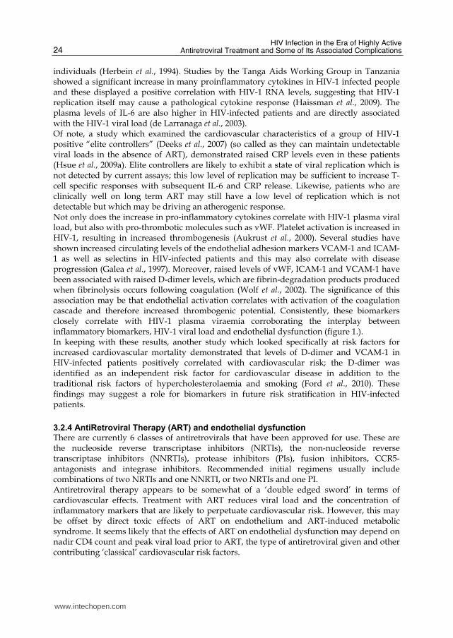
Fig. 1. The key processes involved in plaque formation in anti-retroviral naïve, HIV-1 infected patients. The HIV-1 virus leads to increased levels of adhesion molecules, ICAM-1 and VCAM-1 and increased levels of vWF. Heightened plasma levels of IL-6 contribute to C-reactive protein synthesis from the liver. The adhesion molecules and CRP promote monocyte recruitment to the area. Monocytes migrate to the sub-endothelial tissue and mature into macrophages. Macrophages contribute to foam cell formation and release TNF-α. TNF-α and gp120 inhibit eNOS expression and endothelium dependent vasorelaxtion. The net result is a proliferation of macrophages, foam cell formation and atheromatous plaques with associated haemostasis exacerbated by increased vWF levels.
3.2.4.1 The effects of ART on viral load and cardiovascular disease
It is well established that HIV-1 viraemia and proinflammatory markers are intrinsically linked. Therefore one would presume that by reducing the viral load, ART should also arrest inflammatory processes. The SMART (Lundgren et al., 2008) study showed that interrupted ART was associated with a higher risk of cardiovascular events implying that viral replication and inflammation following treatment cessation is linked with cardiovascular disease. A similar interruption study demonstrated a significant increase in the pro-inflammatory markers IL-6 and D-dimer and an associated increase in cardiovascular mortality in the ART sparing arm compared to those who continued therapy (Kuller, 2008).
3.2.4.2 The effect of ART on inflammatory markers and endothelial dysfunction
Various biological markers of endothelial dysfunction have been shown to increase in HIV-1 infected patients. A longitudinal study comparing biomarkers in HIV-infected patients at ART initiation to two months and then 14 months into treatment demonstrated a normalization or significant reduction in levels of E-selectin, ICAM-1, VCAM-1 and CRP at the two month interval. These changes persisted up to 14 months except for E-selectin which
www.intechopen.com
HIV Infection in the Era of Highly Active Antiretroviral Treatment and Some of Its Associated Complications 26
did not change (Kristoffersen et al., 2009). Similar findings have been reported in other cohort studies, showing significant reductions in both vWF and VCAM-1 levels after six months following initiation of ART, with no demonstrable difference between PI and NNRTI containing regimes (Francisci et al., 2009). In another study the levels of VCAM-1 and vWF correlated positively with viral load in the ART-naïve group; following five months of treatment with a regimen which included either a PI or an NNRTI, there was a significant reduction in levels of VCAM-1 and vWF suggesting a marked reduction in endothelial activation following ART (Wolf et al., 2002). In recent years, the NRTI, abacavir, has been linked with increased cardiovascular risk in certain observational studies (Sabin et al., 2008). The SMART study has also suggested that the use of abacavir is independently associated with a significant increase in plasma levels of CRP and IL-6 (Lundgren et al., 2008), implying that abacavir itself has pro-inflammatory effects compared to other NRTIs. However, these results have not been uniformly replicated in other randomized trials. Indeed the HEAT study, which compared efficacy between abacavir/lamivudine/kaletra and tenofovir/emtricitabine/kaletra regimens, retrospectively showed that there was a decline of CRP, IL-6 and VCAM-1 in both regimens along with viral load reduction. Neither regime was specifically associated with increased cardiovascular events (Smith et al., 2009).
3.2.4.3 The effects of ART on flow mediated dilatation
It seems logical that by reducing systemic inflammation in HIV-1 infected patients, there would be a concurrent improvement in brachial flow mediated dilatation. This concept was demonstrated in a pilot trial which examined the effects of the anti-inflammatory nuclear factor kappaB-inhibitor, salsalate, on HIV-1 infected patients who were not on ART: a significant improvement in FMD was witnessed after 8 weeks of salsalate therapy (Gupta et al., 2008). Since ART also reduces systemic inflammation it might be expected to improve flow mediated dilatation (FMD). Indeed, a study which examined the effects of ART (two NRTIs and one NNRTI, two NRTIs and one PI or one PI and one NNRTI) on FMD, viral load and lipid profile showed that after 24 weeks of treatment there was an increase in brachial artery FMD in all 3 ART regimens. This occurred despite an associated increase in total cholesterol and low density lipoprotein levels in all participants, suggesting that decreased inflammation with ART may have a protective effect on the endothelium (Torriani et al., 2008). However, in another study vascular dilatation in HIV-1 infected patients on ART was significantly impaired compared with ART naïve patients (with no demonstrated difference between PI and NRTI containing regimens) (Andrade et al., 2008). Another study comparing 37 HIV-1 infected patients receiving ART against age and diabetes matched non-HIV-1 infected patients showed that the FMD was equally reduced between the HIV-1 infected patients virally controlled on ART and HIV-1 negative diabetic patients (van Wijk et al., 2006). Again, certain antiretrovirals may be particularly implicated: in HIV-1 infected patients treated with abacavir and achieving virological suppression, a significant reduction in FMD was observed compared to those receiving abacavir-sparing regimes (Hsue et al., 2009b).
3.2.4.4 The effects of ART on vasomotor activity and endothelial cells
Impairment of FMD demonstrates the macro-structural alterations elicited by ART. However, ART also leads to direct microvascular changes. ART can provide a direct insult and subsequent cell death through mitochondrial DNA damage and necrotic pathways, a theory that was demonstrated on human endothelial cells treated with ritonavir in vitro
www.intechopen.com
Endothelial Dysfunction in HIV 27
(Zhong et al., 2002). In vitro cytotoxic effects have also been exhibited by zidovudine (AZT) and indinavir, both damaging intercellular gaps between adjacent endothelial cells (Fiala et al., 2004) thus providing a platform for the inflammatory cascade. There may be a causal relationship between the toxic effects of ART and impaired vasomotor reactivity. Ritonavir has been shown to reduce endothelial NO synthase (eNOS) mRNA and protein levels in cultured human coronary endothelial cells (Fu et al., 2005). Similarly, administration of combination antiretrovirals, which included zidovudine and indinavir in the regimento rats have been shown to increase levels of endothelin-1, a marker of endothelial injury and inducer of vasoconstriction (Jiang et al., 2006). As with other chronic insults, ART-induced endothelial dysfunction may progress to established vascular disease over time. A study by Jiang et al showed that short term treatment (five days) of mice with AZT resulted in a reduction in endothelium-dependent vessel relaxation; however after two weeks of treatment the authors showed a significant increase in injury-induced vascular smooth muscle proliferation and neo-intimal hyperplasia (Jiang et al., 2010). In the same study the authors demonstrated that this increase in neo-intimal hyperplasia correlated with an increase in vascular cell adhesion molecule staining, providing a link between ART and induction of cell adhesion molecules.
4. The metabolic profile of chronic HIV infection and ART and its impact on
cardiovascular risk
Both HIV and antiretrovirals may also induce endothelial damage via modification of ‘classical’ vascular insults, especially blood lipids and glucose. HIV-1 is a known risk factor for hypertriglyceridaemia, elevated low density lipoprotein cholesterol, depressed levels of high density lipoprotein cholesterol and insulin resistance (Oh et al., 2007). In treatment naïve patients, higher HIV-1 RNA levels independently associate with very low density lipoprotein (VLDL) and triglyceride levels. In patients with low CD4 cell counts there is also a higher risk of insulin resistance (El-Sadr et al., 2005). Thus, the metabolic changes that are often attributed to ART may be difficult to interpret because of established abnormalities already present due to infection alone. Nevertheless, it is generally accepted that the PIs and NRTIs are associated with metabolic side effects such as lipodystrophy, and shift the lipid profile to a proatherogenic pattern. The ongoing Data Collection on Adverse Events of Anti-HIV drugs (D:A:D) study showed that the relative risk of a myocardial infarction (MI) associated with cumulative PI use was 1.16 per year of exposure (whereas NNRTIs did not appreciably increase the risk of an MI) (Friis-Moller et al., 2007). However, there appears to be a metabolic difference between various types of PIs. In the CASTLE study, patients treated with lopinavir/ritonavir had significantly raised fasting total cholesterol and triglyceride levels compared to patients given atazanavir/ritonavir (Molina et al., 2010). Other studies have now demonstrated that boosted lopinavir appears to elicit a worse lipid profile compared to other PI-containing regimens (Molina et al., 2010; Mills et al., 2009). The data supporting the role of NRTIs in generating metabolic abnormalities is mainly found in studies which used them in combination with a PI. Again, certain drugs are particularly implicated. The ACTG 5052 study compared efficacy in HIV-infected patients between abacavir/lamivudine and tenofovir/emtricitabine given with efavirenz or ritonavir-boosted atazanavir for 96 weeks. At week 48, fasting lipid levels had significantly increased in the arm receiving abacavir/lamivudine.
www.intechopen.com
HIV Infection in the Era of Highly Active Antiretroviral Treatment and Some of Its Associated Complications 28
The use of NNRTIs may also be associated with adverse lipid effects – recent data from the ACTG 5095 study showed that a regime containing efavirenz significantly increased lipid levels above the baseline values and above those seen in ‘NRTI only’ combinations (Shikuma et al., 2007) 96 weeks after treatment initiation.
5. Assessment and management of patients with increased cardiovascular risk
Given that HIV-1 itself is an independent risk factor for cardiovascular disease, there is an increasing need to apply a cardiovascular risk stratification score in HIV-1 infected patients. A cross-sectional study of HIV-1 infected patients in a Spanish outpatient setting demonstrated that the traditionally used Framingham risk calculation score identified a higher proportion of HIV-1 infected men with a moderate cardiovascular risk compared to other available risk stratification tools (Knobel et al., 2007). However, this tool may not be equally applicable to all populations – for example, in a study which examined the predicted cardiovascular risk in an HIV-1 infected Thai population, the Framingham calculation over estimated the risk of cardiovascular disease compared to other cardiovascular risk equations (Edwards-Jackson et al., 2011). When managing cardiovascular risk in the HIV-1 infected patient, one must advise in the same way as HIV-1 uninfected individuals; for example, addressing lifestyle factors as well as measuring lipid levels, blood pressure and signs of glucose intolerance. However, trials of non-drug therapies and dietary advice alone may not be sufficient to control HIV and ART associated dyslipidaemia. After addressing lifestyle measures it may then be prudent to review the current antiretroviral therapy. Firstly, it may be possible to switch within the class – for example, changing from nelfinavir to atazanavir can reduce the total cholesterol and triglyceride level sufficiently (Calza et al., 2005; Oh et al., 2007). Another strategy, if the patient is on a PI, is to switch them to another class providing there is established viral suppression and a compatible viral resistance profile (Dube et al., 2003). In terms of lipid-lowering pharmacotherapy, the Adult ACTG (Dube et al., 2003) have provided some guidance in approaching HIV-patients with dyslipidemia and raised cardiovascular risk. There are few changes in management compared to the general population and the use of statins (hydroxyl-methyl-glutaryl coenzyme A reductase inhibitors) as a therapy is widely advocated in patients with established isolated hypercholesterolemia (elevated total and LDL-cholesterol and triglyceride level less than 5mmol/l). The advantages of using a statin are two-fold; firstly, statins reduce the levels of cholesterol, which is implicated in endothelial dysfunction and atheroma formation. Secondly, there is increasing evidence that statins also exhibit anti-inflammatory effects (Jain et al., 2005). Recently a double-blinded cross-over trial (8 weeks of high dose 80mg atorvastatin versus placebo in HIV-1 infected ART naïve patients) showed a significant reduction in immune activation with statin therapy, as measured by a fall in activated CD8+ T cells, without any affect on HIV-1 RNA viral load (Ganesan et al., 2011). This further supports the use of statins in HIV-1 infected patients even without established lipid abnormalities. Lowering of oxidized LDL-cholesterol and total LDL cholesterol with 40mg pravastatin has also been shown to improve endothelial dysfunction, as measured by FMD, in patients on a PI-containing ART regime (Hurlimann et al., 2006). However, some caution must be exercised when using statins as there may be significant interactions with PIs. The concentration of pravastatin has been shown to markedly increase
www.intechopen.com
Endothelial Dysfunction in HIV 29
when used with boosted darunavir, although its levels are decreased with all other PIs. Therefore, whilst there is a potential increase in the side effect profile of all statins, pravastatin is usually considered the safest to use with PIs other than darunavir (Seker, 2007). Statins are not the only lipid-lowering drug available; the combination of a statin and a fibrate should be considered (albeit with close monitoring due to the exaggerated side effect profile) when the triglyceride level is above 5mmol/l and may be the best approach to achieve lipid targets in these patients. Ezetimibe is a newer agent that acts by reducing intestinal cholesterol absorption and has been shown to be better tolerated but equally efficacious compared to statins in HIV-infected patients with hypercholesterolaemia (Negredo et al., 2006). Ezetimibe may be used when statins are not tolerated or as an adjunct to other anti-lipid agents in severe lipid disturbance. In non-HIV-1 infected patients, the beneficial effects of aspirin have largely been attributed to its action on thromboxane synthesis and platelet aggregation; however, there is also evidence suggesting that aspirin improves endothelial dysfunction through endothelium dependent vasodilatation (Husain et al., 1998). Current guidelines, as outlined by the U.S Preventative Services Task Force, recommend the use of aspirin in male patients between 45-79 years old when the benefits of a reduction in risk of mycocardial infarction, taking into account overall cardiovascular risk, outweighs the potential risks associated with aspirin therapy (Calonge N, 2009). Spanish researchers applied these standards to their HIV-infected cohort and found that aspirin would be indicated in 30.8% of their male patients (Tornero et al., 2010). Moreover, salsalate, a compound which exists within the same class as aspirin, has been shown to significantly improve flow mediated dilatation in HIV-infected patients after 8 weeks (as discussed above), perhaps suggesting a role for these agents in reducing endothelial dysfunction (Gupta et al., 2008). Whether or not aspirin should be considered as primary prevention in HIV-infected patients is still debatable and certainly it should not be seen as a replacement for timely ART.
6. Conclusions
As the HIV-1 infected population grows, management of patients is increasingly focused on chronic care issues such as cardiovascular comorbidity and metabolic disturbances including lipodystrophy and glucose intolerance. In recent years, there has been increasing recognition that endothelial dysfunction plays a pivotal role in atherosclerosis in HIV-1 infected patients, and that HIV-1 may be as important as other more “traditional” risk factors for accelerated coronary artery disease. The pathology is complex and multifactorial; the HIV-1 virus and its component proteins are likely to perpetuate a cycle of chronic inflammation, haemostasis and endothelial activation. The role of ART is even less well understood, with the benefits of viral suppression being offset by the toxic and metabolic effects of ART itself (figure 2). However, what is certain is that early detection and appropriate management of HIV-1 and its complications is imperative in attempting to reduce the devastating global impact that HIV-1 has had within the last 30 years. Effective viral suppression, establishing coronary risk and modifying behavioral risk factors may provide the best initial approach to endothelial dysfunction. Following this, the option to switch antiretroviral drugs and treat the patient with pharmacotherapeutic agents aiming to optimize lipids, glucose and blood pressure may then be effective.
www.intechopen.com
HIV Infection in the Era of Highly Active Antiretroviral Treatment and Some of Its Associated Complications 30
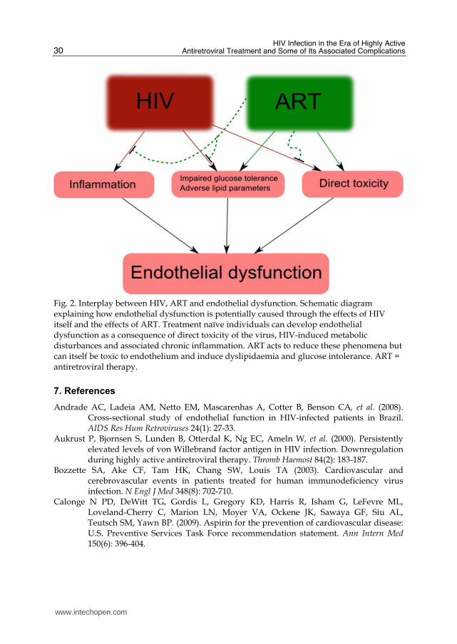
Fig. 2. Interplay between HIV, ART and endothelial dysfunction. Schematic diagram explaining how endothelial dysfunction is potentially caused through the effects of HIV itself and the effects of ART. Treatment naïve individuals can develop endothelial dysfunction as a consequence of direct toxicity of the virus, HIV-induced metabolic disturbances and associated chronic inflammation. ART acts to reduce these phenomena but can itself be toxic to endothelium and induce dyslipidaemia and glucose intolerance. ART = antiretroviral therapy.
7. References
Andrade AC, Ladeia AM, Netto EM, Mascarenhas A, Cotter B, Benson CA, et al. (2008). Cross-sectional study of endothelial function in HIV-infected patients in Brazil. AIDS Res Hum Retroviruses 24(1): 27-33.
Aukrust P, Bjornsen S, Lunden B, Otterdal K, Ng EC, Ameln W, et al. (2000). Persistently elevated levels of von Willebrand factor antigen in HIV infection. Downregulation during highly active antiretroviral therapy. Thromb Haemost 84(2): 183-187.
Bozzette SA, Ake CF, Tam HK, Chang SW, Louis TA (2003). Cardiovascular and cerebrovascular events in patients treated for human immunodeficiency virus infection. N Engl J Med 348(8): 702-710.
Calonge N PD, DeWitt TG, Gordis L, Gregory KD, Harris R, Isham G, LeFevre ML, Loveland-Cherry C, Marion LN, Moyer VA, Ockene JK, Sawaya GF, Siu AL, Teutsch SM, Yawn BP. (2009). Aspirin for the prevention of cardiovascular disease: U.S. Preventive Services Task Force recommendation statement. Ann Intern Med 150(6): 396-404.
www.intechopen.com
Endothelial Dysfunction in HIV 31
Calza L, Manfredi R, Colangeli V, Tampellini L, Sebastiani T, Pocaterra D, et al. (2005). Substitution of nevirapine or efavirenz for protease inhibitor versus lipid-lowering therapy for the management of dyslipidaemia. AIDS 19(10): 1051-1058.
Collins RG, Velji R, Guevara NV, Hicks MJ, Chan L, Beaudet AL (2000). P-Selectin or intercellular adhesion molecule (ICAM)-1 deficiency substantially protects against atherosclerosis in apolipoprotein E-deficient mice. J Exp Med 191(1): 189-194.
Currier JS, Kendall MA, Zackin R, Henry WK, Alston-Smith B, Torriani FJ, et al. (2005). Carotid artery intima-media thickness and HIV infection: traditional risk factors overshadow impact of protease inhibitor exposure. AIDS 19(9): 927-933.
Danesh J, Kaptoge S, Mann AG, Sarwar N, Wood A, Angleman SB, et al. (2008). Long-term interleukin-6 levels and subsequent risk of coronary heart disease: two new prospective studies and a systematic review. PLoS Med 5(4): e78.
de Larranaga GF, Petroni A, Deluchi G, Alonso BS, Benetucci JA (2003). Viral load and disease progression as responsible for endothelial activation and/or injury in human immunodeficiency virus-1-infected patients. Blood Coagul Fibrinolysis 14(1): 15-18.
De Lorenzo F, Xiao H, Scully M, Kadziola Z, Kakkar VV (2003). Pre-thrombotic state and impaired fibrinolytic potential in coronary heart disease patients with left ventricular dysfunction. Blood Coagul Fibrinolysis 14(1): 67-75.
Deeks SG, Walker BD (2007). Human immunodeficiency virus controllers: mechanisms of durable virus control in the absence of antiretroviral therapy. Immunity 27(3): 406-416.
Devaraj S, Yun JM, Duncan-Staley C, Jialal I (2009). C-reactive protein induces M-CSF release and macrophage proliferation. J Leukoc Biol 85(2): 262-267.
Dong ZM, Chapman SM, Brown AA, Frenette PS, Hynes RO, Wagner DD (1998). The combined role of P- and E-selectins in atherosclerosis. J Clin Invest 102(1): 145-152.
Dube MP, Stein JH, Aberg JA, Fichtenbaum CJ, Gerber JG, Tashima KT, et al. (2003). Guidelines for the evaluation and management of dyslipidemia in human immunodeficiency virus (HIV)-infected adults receiving antiretroviral therapy: recommendations of the HIV Medical Association of the Infectious Disease Society of America and the Adult AIDS Clinical Trials Group. Clin Infect Dis 37(5): 613-627.
Duffy P, Wang X, Lin PH, Yao Q, Chen C (2009). HIV Nef protein causes endothelial dysfunction in porcine pulmonary arteries and human pulmonary artery endothelial cells. J Surg Res 156(2): 257-264.
Edwards-Jackson N, Kerr S, Tieu H, Ananworanich J, Hammer S, Ruxrungtham K, et al. (2011). Cardiovascular risk assessment in persons with HIV infection in the developing world: comparing three risk equations in a cohort of HIV-infected Thais. HIV Med. doi: 10.1111/j.1468-1293.2011.00916.x
El-Sadr WM, Lundgren JD, Neaton JD, Gordin F, Abrams D, Arduino RC, et al. (2006). CD4+ count-guided interruption of antiretroviral treatment. N Engl J Med 355(22): 2283-2296.
El-Sadr WM, Mullin CM, Carr A, Gibert C, Rappoport C, Visnegarwala F, et al. (2005). Effects of HIV disease on lipid, glucose and insulin levels: results from a large antiretroviral-naive cohort. HIV Med 6(2): 114-121.
Fiala M, Murphy T, MacDougall J, Yang W, Luque A, Iruela-Arispe L, et al. (2004). HAART drugs induce mitochondrial damage and intercellular gaps and gp120 causes apoptosis. Cardiovasc Toxicol 4(4): 327-337.
www.intechopen.com
HIV Infection in the Era of Highly Active Antiretroviral Treatment and Some of Its Associated Complications 32
Ford ES, Greenwald JH, Richterman AG, Rupert A, Dutcher L, Badralmaa Y, et al. (2010). Traditional risk factors and D-dimer predict incident cardiovascular disease events in chronic HIV infection. AIDS 24(10): 1509-1517.
Francisci D, Giannini S, Baldelli F, Leone M, Belfiori B, Guglielmini G, et al. (2009). HIV type 1 infection, and not short-term HAART, induces endothelial dysfunction. AIDS 23(5): 589-596.
Friis-Moller N, Reiss P, Sabin CA, Weber R, Monforte A, El-Sadr W, et al. (2007). Class of antiretroviral drugs and the risk of myocardial infarction. N Engl J Med 356(17): 1723-1735.
Fu W, Chai H, Yao Q, Chen C (2005). Effects of HIV protease inhibitor ritonavir on vasomotor function and endothelial nitric oxide synthase expression. J Acquir Immune Defic Syndr 39(2): 152-158.
Galea P, Vermot-Desroches C, Le Contel C, Wijdenes J, Chermann JC (1997). Circulating cell adhesion molecules in HIV1-infected patients as indicator markers for AIDS progression. Res Immunol 148(2): 109-117.
Galkina E, Ley K (2007). Vascular adhesion molecules in atherosclerosis. Arterioscler Thromb Vasc Biol 27(11): 2292-2301.
Ganesan A, Crum-Cianflone N, Higgins J, Qin J, Rehm C, Metcalf J, et al. (2011). High dose atorvastatin decreases cellular markers of immune activation without affecting HIV-1 RNA levels: results of a double-blind randomized placebo controlled clinical trial. J Infect Dis 203(6): 756-764.
Goldberg RB (2009). Cytokine and cytokine-like inflammation markers, endothelial dysfunction, and imbalanced coagulation in development of diabetes and its complications. J Clin Endocrinol Metab 94(9): 3171-3182.
Gupta SK, Johnson RM, Saha C, Mather KJ, Greenwald ML, Waltz JS, et al. (2008). Improvement in HIV-related endothelial dysfunction using the anti-inflammatory agent salsalate: a pilot study. AIDS 22(5): 653-655.
Haissman JM, Vestergaard LS, Sembuche S, Erikstrup C, Mmbando B, Mtullu S, et al. (2009). Plasma cytokine levels in Tanzanian HIV-1-infected adults and the effect of antiretroviral treatment. J Acquir Immune Defic Syndr 52(4): 493-497.
Herbein G, Keshav S, Collin M, Montaner LJ, Gordon S (1994). HIV-1 induces tumour necrosis factor and IL-1 gene expression in primary human macrophages independent of productive infection. Clin Exp Immunol 95(3): 442-449.
Ho JE, Hsue PY (2009). Cardiovascular manifestations of HIV infection. Heart 95(14): 1193-1202.
Hsu HY, Twu YC (2000). Tumor necrosis factor-alpha -mediated protein kinases in regulation of scavenger receptor and foam cell formation on macrophage. J Biol Chem 275(52): 41035-41048.
Hsue P, Hunt P, Schnell A, Wu Y, Ho J, Hoh R, et al. (2010a). Rapid Progression of Atherosclerosis at the Carotid Bifurcation Is Linked to Inflammation in HIV-infected Patients. Seventeenth Conference on Retroviruses and Opportunistic Infections (CROI); Abstract 125
Hsue P, Ordovas K, Lee T, Schnell A, Ho JE, Selby V, et al. (2010b). Abstract 10273: Markedly Increased Carotid Intima-Media Thickness in the Absence of Coronary Calcium Among HIV-Infected Individuals. Circulation 122(21-MeetingAbstracts): A10273.
www.intechopen.com
Endothelial Dysfunction in HIV 33
Hsue PY, Hunt PW, Schnell A, Kalapus SC, Hoh R, Ganz P, et al. (2009a). Role of viral replication, antiretroviral therapy, and immunodeficiency in HIV-associated atherosclerosis. AIDS 23(9): 1059-1067.
Hsue PY, Hunt PW, Wu Y, Schnell A, Ho JE, Hatano H, et al. (2009b). Association of abacavir and impaired endothelial function in treated and suppressed HIV-infected patients. AIDS 23(15): 2021-2027.
Hsue PY, Lo JC, Franklin A, Bolger AF, Martin JN, Deeks SG, et al. (2004). Progression of atherosclerosis as assessed by carotid intima-media thickness in patients with HIV infection. Circulation 109(13): 1603-1608.
Huang MB, Khan M, Garcia-Barrio M, Powell M, Bond VC (2001). Apoptotic effects in primary human umbilical vein endothelial cell cultures caused by exposure to virion-associated and cell membrane-associated HIV-1 gp120. J Acquir Immune Defic Syndr 27(3): 213-221.
Hurlimann D, Chenevard R, Ruschitzka F, Flepp M, Enseleit F, Bechir M, et al. (2006). Effects of statins on endothelial function and lipid profile in HIV infected persons receiving protease inhibitor-containing anti-retroviral combination therapy: a randomised double blind crossover trial. Heart 92(1): 110-112.
Husain S, Andrews NP, Mulcahy D, Panza JA, Quyyumi AA (1998). Aspirin improves endothelial dysfunction in atherosclerosis. Circulation 97(8): 716-720.
Jain MK, Ridker PM (2005). Anti-inflammatory effects of statins: clinical evidence and basic mechanisms. Nat Rev Drug Discov 4(12): 977-987.
Jiang B, Hebert VY, Zavecz JH, Dugas TR (2006). Antiretrovirals induce direct endothelial dysfunction in vivo. J Acquir Immune Defic Syndr 42(4): 391-395.
Jiang B, Khandelwal AR, Rogers LK, Hebert VY, Kleinedler JJ, Zavecz JH, et al. (2010). Antiretrovirals induce endothelial dysfunction via an oxidant-dependent pathway and promote neointimal hyperplasia. Toxicol Sci 117(2): 524-536.
Johnson-Tidey RR, McGregor JL, Taylor PR, Poston RN (1994). Increase in the adhesion molecule P-selectin in endothelium overlying atherosclerotic plaques. Coexpression with intercellular adhesion molecule-1. Am J Pathol 144(5): 952-961.
Joshi VV, Pawel B, Connor E, Sharer L, Oleske JM, Morrison S, et al. (1987). Arteriopathy in children with acquired immune deficiency syndrome. Pediatr Pathol 7(3): 261-275.
Kates J, Lief E, Avila C (2009). 'Financing the Response to AIDS in Low- and Middle- Income Countries: International Assistance from the G8, European Commission and Other Donor Governments in 2008'. . UNAIDS. Available from
http://www.kff.org/hivaids/7347.cfm (Access date June 2011) Kharbanda R MR (2005). The Atherosclerosis Time-Line and the Role of the Endothelium.
Current Medicinal Chemistry - Immunology, Endocrine & Metabolic Agents 5: 47-52. Knobel H, Jerico C, Montero M, Sorli ML, Velat M, Guelar A, et al. (2007). Global
cardiovascular risk in patients with HIV infection: concordance and differences in estimates according to three risk equations (Framingham, SCORE, and PROCAM). AIDS Patient Care STDS 21(7): 452-457.
Kristoffersen US, Kofoed K, Kronborg G, Giger AK, Kjaer A, Lebech AM (2009). Reduction in circulating markers of endothelial dysfunction in HIV-infected patients during antiretroviral therapy. HIV Med 10(2): 79-87.
Kuller L (2008). Elevated Levels of Interleukin-6 and D-dimer Are Associated with an Increased Risk of Death in Patients with HIV. Fifteenth Conference on Retroviruses and Opportunistic Infections (CROI); Abstract 139.
www.intechopen.com
HIV Infection in the Era of Highly Active Antiretroviral Treatment and Some of Its Associated Complications 34
Kumar PN, Rodriguez-French A, Thompson MA, Tashima KT, Averitt D, Wannamaker PG, et al. (2006). A prospective, 96-week study of the impact of Trizivir, Combivir/nelfinavir, and lamivudine/stavudine/nelfinavir on lipids, metabolic parameters and efficacy in antiretroviral-naive patients: effect of sex and ethnicity. HIV Med 7(2): 85-98.
Lohse N, Hansen AB, Pedersen G, Kronborg G, Gerstoft J, Sorensen HT, et al. (2007). Survival of persons with and without HIV infection in Denmark, 1995-2005. Ann Intern Med 146(2): 87-95.
Lowe GD (2003). Virchow's triad revisited: abnormal flow. Pathophysiol Haemost Thromb 33(5-6): 455-457.
Lundgren J, Neuhaus J, Babiker A, Cooper D, Duprez D, El-Sadr W, et al. (2008). Use of nucleoside reverse transcriptase inhibitors and risk of myocardial infarction in HIV-infected patients. AIDS 22(14): F17-24.
Marin B, Thiebaut R, Bucher HC, Rondeau V, Costagliola D, Dorrucci M, et al. (2009). Non-AIDS-defining deaths and immunodeficiency in the era of combination antiretroviral therapy. AIDS 23(13): 1743-1753.
Molina JM, Andrade-Villanueva J, Echevarria J, Chetchotisakd P, Corral J, David N, et al. (2010). Once-daily atazanavir/ritonavir compared with twice-daily lopinavir/ritonavir, each in combination with tenofovir and emtricitabine, for management of antiretroviral-naive HIV-1-infected patients: 96-week efficacy and safety results of the CASTLE study. J Acquir Immune Defic Syndr 53(3): 323-332.
Mills AM, Nelson M, Jayaweera D, et al. (2009). Once-daily darunavir/ritonavir vs. lopinavir/ritonavir in treatment-naïve, HIV-1-infected patients: 96-week analysis. AIDS 23(13): 1679–1688.
Monsuez JJ, Charniot JC, Escaut L, Teicher E, Wyplosz B, Couzigou C, et al. (2009). HIV-associated vascular diseases: structural and functional changes, clinical implications. Int J Cardiol 133(3): 293-306.
Mu H, Chai H, Lin PH, Yao Q, Chen C (2007). Current update on HIV-associated vascular disease and endothelial dysfunction. World J Surg 31(4): 632-643.
Negredo E, Molto J, Puig J, Cinquegrana D, Bonjoch A, Perez-Alvarez N, et al. (2006). Ezetimibe, a promising lipid-lowering agent for the treatment of dyslipidaemia in HIV-infected patients with poor response to statins. AIDS 20(17): 2159-2164.
Oh J, Hegele RA (2007). HIV-associated dyslipidaemia: pathogenesis and treatment. Lancet Infect Dis 7(12): 787-796.
O’Leary DH, Polak JF, Kronmal RA, et al. (1999). Carotid-artery intima media thickness as a risk factor for myocardial infarction and stroke in older adults. For the Cardiovascular Health Study Collaborative Research Group. N Engl J Med 340(1):14–22.
Ramana KV, Bhatnagar A, Srivastava SK (2004). Inhibition of aldose reductase attenuates TNF-alpha-induced expression of adhesion molecules in endothelial cells. FASEB J 18(11): 1209-1218.
Ren Z, Yao Q, Chen C (2002). HIV-1 envelope glycoprotein 120 increases intercellular adhesion molecule-1 expression by human endothelial cells. Lab Invest 82(3): 245-255.
Ridker PM, Rifai N, Rose L, Buring JE, Cook NR (2002). Comparison of C-reactive protein and low-density lipoprotein cholesterol levels in the prediction of first cardiovascular events. N Engl J Med 347(20): 1557-1565.
www.intechopen.com
Endothelial Dysfunction in HIV 35
Ross R, Glomset J, Harker L (1977). Response to injury and atherogenesis. Am J Pathol 86(3): 675-684.
Sabin CA, Worm SW, Weber R, Reiss P, El-Sadr W, Dabis F, et al. (2008). Use of nucleoside reverse transcriptase inhibitors and risk of myocardial infarction in HIV-infected patients enrolled in the D:A:D study: a multi-cohort collaboration. Lancet 371(9622): 1417-1426.
Sakai A, Kume N, Nishi E, Tanoue K, Miyasaka M, Kita T (1997). P-selectin and vascular cell adhesion molecule-1 are focally expressed in aortas of hypercholesterolemic rabbits before intimal accumulation of macrophages and T lymphocytes. Arterioscler Thromb Vasc Biol 17(2): 310-316.
Sax PE, Tierney C, Collier AC, Fischl MA, Mollan K, Peeples L, et al. (2009). Abacavir-lamivudine versus tenofovir-emtricitabine for initial HIV-1 therapy. N Engl J Med 361(23): 2230-2240.
Seker V (2007). Pharmacokinetic drug-drug interaction between the new HIV-protease inhibitor darunavir (TMC114) and the lipid lowering agent pravastatin. 8th International work-shop on pharmacology of HIV therapy; Abstract 54.
Shikuma CM, Ribaudo HJ, Zheng Y, Gulick RM, Meyer WA, Tashima KT, et al. (2011). Change in High-Sensitivity C-Reactive Protein Levels Following Initiation of Efavirenz-Based Antiretroviral Regimens in HIV-Infected Individuals. AIDS Res Hum Retroviruses 27(5): 461-468.
Shikuma CM, Yang Y, Glesby MJ, Meyer WA, 3rd, Tashima KT, Ribaudo HJ, et al. (2007). Metabolic effects of protease inhibitor-sparing antiretroviral regimens given as initial treatment of HIV-1 Infection (AIDS Clinical Trials Group Study A5095). J Acquir Immune Defic Syndr 44(5): 540-550.
Signorelli SS, Mazzarino MC, Spandidos DA, Malaponte G (2007). Proinflammatory circulating molecules in peripheral arterial disease. Int J Mol Med 20(3): 279-286.
Smith KY, Patel P, Fine D, Bellos N, Sloan L, Lackey P, et al. (2009). Randomized, double-blind, placebo-matched, multicenter trial of abacavir/lamivudine or tenofovir/emtricitabine with lopinavir/ritonavir for initial HIV treatment. AIDS 23(12): 1547-1556.
Solages A, Vita JA, Thornton DJ, Murray J, Heeren T, Craven DE, et al. (2006). Endothelial function in HIV-infected persons. Clin Infect Dis 42(9): 1325-1332.
Stocker CJ, Sugars KL, Harari OA, Landis RC, Morley BJ, Haskard DO (2000). TNF-alpha, IL-4, and IFN-gamma regulate differential expression of P- and E-selectin expression by porcine aortic endothelial cells. J Immunol 164(6): 3309-3315.
Tan KC, Chow WS, Tam SC, Ai VH, Lam CH, Lam KS (2002). Atorvastatin lowers C-reactive protein and improves endothelium-dependent vasodilation in type 2 diabetes mellitus. J Clin Endocrinol Metab 87(2): 563-568.
Thor M, Yu A, Swedenborg J (2002). Markers of inflammation and hypercoagulability in diabetic and nondiabetic patients with lower extremity ischemia. Thromb Res 105(5): 379-383.
Tornero C, Ventura A, Mafe M (2010). Aspirin Is Indicated for Primary Prevention of Cardiovascular Events in HIV-Infected Patients. J Acquir Immune Defic Syndr 54(5): 560
Torriani FJ, Komarow L, Parker RA, Cotter BR, Currier JS, Dube MP, et al. (2008). Endothelial function in human immunodeficiency virus-infected antiretroviral-
www.intechopen.com
HIV Infection in the Era of Highly Active Antiretroviral Treatment and Some of Its Associated Complications 36
naive subjects before and after starting potent antiretroviral therapy: The ACTG (AIDS Clinical Trials Group) Study 5152s. J Am Coll Cardiol 52(7): 569-576.
Triant VA, Regan S, Lee H, Sax PE, Meigs JB, Grinspoon SK (2010). Association of Immunologic and Virologic Factors With Myocardial Infarction Rates in a US Healthcare System. J Acquir Immune Defic Syndr 55(5):615-619
Truskey GA, Herrmann RA, Kait J, Barber KM (1999). Focal increases in vascular cell adhesion molecule-1 and intimal macrophages at atherosclerosis-susceptible sites in the rabbit aorta after short-term cholesterol feeding. Arterioscler Thromb Vasc Biol 19(2): 393-401.
Turesson C, Jacobsson LT, Matteson EL (2008). Cardiovascular co-morbidity in rheumatic diseases. Vasc Health Risk Manag 4(3): 605-614.
Ullrich CK, Groopman JE, Ganju RK (2000). HIV-1 gp120- and gp160-induced apoptosis in cultured endothelial cells is mediated by caspases. Blood 96(4): 1438-1442.
van Vonderen MG, Smulders YM, Stehouwer CD, Danner SA, Gundy CM, Vos F, et al. (2009). Carotid intima-media thickness and arterial stiffness in HIV-infected patients: the role of HIV, antiretroviral therapy, and lipodystrophy. J Acquir Immune Defic Syndr 50(2): 153-161.
van Wijk JP, de Koning EJ, Cabezas MC, Joven J, op't Roodt J, Rabelink TJ, et al. (2006). Functional and structural markers of atherosclerosis in human immunodeficiency virus-infected patients. J Am Coll Cardiol 47(6): 1117-1123.
Verhaar MC, Strachan FE, Newby DE, Cruden NL, Koomans HA, Rabelink TJ, et al. (1998). Endothelin-A receptor antagonist-mediated vasodilatation is attenuated by inhibition of nitric oxide synthesis and by endothelin-B receptor blockade. Circulation 97(8): 752-756.
Vittecoq D, Escaut L, Chironi G, Teicher E, Monsuez JJ, Andrejak M, et al. (2003). Coronary heart disease in HIV-infected patients in the highly active antiretroviral treatment era. AIDS 17 Suppl 1: S70-76.
Wang XQ, Panousis CG, Alfaro ML, Evans GF, Zuckerman SH (2002). Interferon-gamma-mediated downregulation of cholesterol efflux and ABC1 expression is by the Stat1 pathway. Arterioscler Thromb Vasc Biol 22(5): e5-9.
WHO (2010). World Health Organisation: Towards universal access:scaling up priority HIV/AIDS interventions in the health sector. Progress report Available from
http://www.who.int/hiv/pub/2010progressreport/en (Access date June 2011) Wilson D, Cotton M, Bekker L, Meyers T, Venter F, Maartens G (2008). Oxford Handbook of
HIV Medicine. 2nd edn. Oxford University Press: Cape Town, South Africa. Wolf K, Tsakiris DA, Weber R, Erb P, Battegay M (2002). Antiretroviral therapy reduces
markers of endothelial and coagulation activation in patients infected with human immunodeficiency virus type 1. J Infect Dis 185(4): 456-462.
Zhang C, Hein TW, Wang W, Ren Y, Shipley RD, Kuo L (2006). Activation of JNK and xanthine oxidase by TNF-alpha impairs nitric oxide-mediated dilation of coronary arterioles. J Mol Cell Cardiol 40(2): 247-257.
Zhang DX, Yi FX, Zou AP, Li PL (2002). Role of ceramide in TNF-alpha-induced impairment of endothelium-dependent vasorelaxation in coronary arteries. Am J Physiol Heart Circ Physiol 283(5): H1785-1794.
Zhong DS, Lu XH, Conklin BS, Lin PH, Lumsden AB, Yao Q, et al. (2002). HIV protease inhibitor ritonavir induces cytotoxicity of human endothelial cells. Arterioscler Thromb Vasc Biol 22(10): 1560-1566.
www.intechopen.com
HIV Infection in the Era of Highly Active Antiretroviral Treatmentand Some of Its Associated ComplicationsEdited by Dr. Elaheh Aghdassi
ISBN 978-953-307-701-7Hard cover, 212 pagesPublisher InTechPublished online 14, November, 2011Published in print edition November, 2011
InTech EuropeUniversity Campus STeP Ri Slavka Krautzeka 83/A 51000 Rijeka, Croatia Phone: +385 (51) 770 447 Fax: +385 (51) 686 166www.intechopen.com
InTech ChinaUnit 405, Office Block, Hotel Equatorial Shanghai No.65, Yan An Road (West), Shanghai, 200040, China
Phone: +86-21-62489820 Fax: +86-21-62489821
Human immunodeficiency virus (HIV) infection is a complex illness affecting the immune system. Acquiredimmunodeficiency syndrome (AIDS) is an advanced form of HIV infection in which the patient has developedopportunistic infections or certain types of cancer and/or the CD4+ T cell count has dropped below 200/µL.More than 40 million persons around the world are infected with HIV, with approximately 14,000 new infectionsevery day. The disease causes 3 million deaths worldwide each year, 95% of them in developing countries.Optimal management of human immunodeficiency virus requires strict adherence to highly active antiretroviraltreatment (HAART) regimens, but the complexity of these regimens (e.g., pill burden, food requirements, druginteractions, and severe adverse effects) limits effective treatment. However, more patients with HIV aresurviving longer today because of these drugs. This allows further study of commonly associated adverseeffects. These may affect all body systems and range from serious toxicities to uncomfortable but manageableevents. This book reviews some of HAART-related metabolic and neurological complications.
How to referenceIn order to correctly reference this scholarly work, feel free to copy and paste the following:
Vani Subbarao, David Lowe, Reza Aghamohammadzadeh and Robert J. Wilkinson (2011). EndothelialDysfunction in HIV, HIV Infection in the Era of Highly Active Antiretroviral Treatment and Some of ItsAssociated Complications, Dr. Elaheh Aghdassi (Ed.), ISBN: 978-953-307-701-7, InTech, Available from:http://www.intechopen.com/books/hiv-infection-in-the-era-of-highly-active-antiretroviral-treatment-and-some-of-its-associated-complications/endothelial-dysfunction-in-hiv

© 2011 The Author(s). Licensee IntechOpen. This is an open access articledistributed under the terms of the Creative Commons Attribution 3.0License, which permits unrestricted use, distribution, and reproduction inany medium, provided the original work is properly cited.