Embed Size (px)
Citation preview
Endothelial expression of intercellular adhesion molecule 1and vascular cell adhesion molecule 1 is suppressed bypostbypass plasma containing increased solubleintercellular adhesion molecule 1 and vascular celladhesion molecule 1Michael P. Vallely, MBBSa,b,c
Paul G. Bannon, FRACS, PhDb,c
Clifford F. Hughes, AO, FRACSb,c
Leonard Kritharides, FRACP, PhDa,d
Objective: Endothelial cell dysfunction has been implicated in the inflam-matory response to cardiopulmonary bypass, and the upregulation of endo-thelial cell expression of adhesion molecules might promote leukocyteextravasation in vivo. Soluble endothelial cell adhesion molecules areincreased after bypass. The aim of this study was to investigate therelationship between endothelial cell-surface expression of adhesionmolecules and their concentration in plasma after coronary artery bypassgrafting.
Methods: Ten patients undergoing coronary artery bypass with cardiopulmo-nary bypass had 5 plasma samples taken at defined intervals before, during,and after cardiopulmonary bypass. Plasma was incubated with humanumbilical vein endothelial cell monolayers, and expression of E-selectin,intercellular adhesion molecule 1, and vascular cell adhesion molecule 1on the surface of human umbilical vein endothelial cell monolayers wasmeasured by means of enzyme-linked immunosorbent assay. Plasma solubleadhesion molecules, C-reactive protein, interleukin 8, interleukin 10, trans-forming growth factor �1, and neutrophil counts were determined for eachpatient.
Results: Markers typical of acute inflammation (ie, interleukin 8, neutro-phils, and C-reactive protein) were all increased after bypass. Solubleplasma intercellular and vascular cell adhesion molecule 1 (but not E-selectin)were increased after bypass. However, endothelial cell expression of vas-cular cell adhesion molecule 1 and intercellular adhesion molecule 1 (but notE-selectin) were significantly decreased by exposure to postbypassplasma. Additionally, postbypass plasma inhibited interleukin 1�-stimulatedendothelial cell expression of vascular cell and intercellular adhesionmolecule 1. Interleukin 10 and transforming growth factor �1, both ofwhich are known to inhibit endothelial cell adhesion moleculeexpression, were respectively increased 10-fold and 3-fold (P � .05) afterbypass.
Conclusions: Despite containing increased soluble intercellular and vascular celladhesion molecule 1, postbypass plasma inhibits endothelial cell expression ofintercellular and vascular cell adhesion molecule 1. Upregulated vascular ex-pression of adhesion molecules might not be essential for endothelial activationafter bypass.
From the Clinical Research Group, TheHeart Research Institute,a the Cardiotho-racic Surgical Unit Royal Prince AlfredHospital,b The Baird Institute for Heart andLung Research,c and the Department ofCardiology,d Concord Hospital, Sydney,Australia.
Supported by The Royal Australasian Col-lege of Surgeons’ Foundation and Strath-field Private Hospital.
Address for reprints: Michael Vallely,MBBS, Cardiothoracic Surgical Unit,Royal Prince Alfred Hospital, MissendenRd, Camperdown, New South Wales 2050,Australia (E-mail: [email protected]).
Received for publication Aug 20, 2001; re-visions requested Oct 26, 2001; revisionsreceived Nov 9, 2001; accepted for publi-cation Jan 8, 2002.
J Thorac Cardiovasc Surg 2002;124:758-67
Copyright © 2002 by The American Asso-ciation for Thoracic Surgery
0022-5223/2002 $35.00�0 12/1/123133
doi:10.1067/mtc.2002.123133
Cardiopulmonary Support and Physiology Vallely et al
758 The Journal of Thoracic and Cardiovascular Surgery ● October 2002
CSP
Cardiopulmonary bypass (CPB) is known tocause a systemic inflammatory responsesyndrome, which can contribute to signifi-cant morbidity and mortality.1 The endo-thelial-leukocyte adhesion cascade is cen-tral to the transmigration of activated
leukocytes into the subendothelial space, where they de-granulate, promoting inflammatory injury. Upregulation ofendothelial cell (EC) adhesion molecule expression occursin many inflammatory conditions2-4 and promotes the adhe-sion and transmigration of leukocytes.
CPB has generally been associated with increased levelsof plasma soluble EC adhesion molecules, which have beenattributed to the activation and injury of ECs.1,5-7 However,some studies have shown a decrease in soluble adhesionmolecule levels after CPB.8 Whether soluble adhesion mol-ecules contribute to the causation of the systemic inflam-matory response syndrome after bypass or are merely amarker of other inflammatory processes is unclear. Indeed,they might even have an anti-inflammatory role under somecircumstances.9,10
The relationship between soluble adhesion moleculesand their endothelial expression is not well established.10
This is because adhesion molecules can be expressed onnon-ECs, such as monocytes, tissue macrophages, fibro-blasts, and dendritic cells,11-15 and because it is difficult toevaluate EC adhesion molecule expression in human sub-jects in vivo. The effect of CPB on EC expression ofadhesion molecules might be important for therapeutic tar-geting of the post-CPB inflammatory response and for un-derstanding the mechanism of CPB-induced inflammationwithin the vascular bed.
The aim of this study was to investigate the relationshipbetween EC surface expression of adhesion molecules andtheir concentration in plasma after coronary artery bypassgrafting (CABG). Although soluble intercellular adhesionmolecule 1 (ICAM-1), soluble vascular cell adhesion mol-ecule 1 (VCAM-1), and other inflammatory markers areconfirmed to be increased after CPB, post-CPB plasmadownregulates the expression of ICAM-1 and VCAM-1 onECs. These observations dissociate the regulation of solubleand cell-surface adhesion molecules after CPB.
Material and MethodsPatientsTen adult patients with multivessel coronary artery disease under-going first-time elective CABG with CPB were enrolled in thestudy. The study was undertaken with institutional ethics commit-tee approval, and written informed consent was obtained from eachpatient. Patients taking corticosteroids, nonsteroidal anti-inflam-matory drugs, aspirin, or other immunosuppressing agents wereexcluded, as were patients with diabetes mellitus, renal failure, orother immunocompromising conditions. Patients receiving intra-venous nitrates or heparin were also excluded.
Anesthetic, CPB, and Operative TechniquesAnesthesia induction was performed with 15 to 30 �g/kg fentanyl,0.5 to 1 mg/kg thiopentone, and 0.15 mg/kg pancuronium. Anes-thesia was maintained with a volatile agent (isoflurane) throughoutthe procedure (on and off CPB). No protease inhibitors (aprotinin),antifibrinolytic agents (aminocaproic acid), or corticosteroids wereused in any patients.
Standard systemic heparinization was used (400 IU/kg), and anactivated clotting time of greater than 450 seconds was maintainedduring CPB. CPB was performed with mild-to-moderate hypo-thermia (30°C-32°C). The extracorporeal circuit consisted of aroller pump (Jostra), a membrane oxygenator (Capiox SX18,Terumo), and polyvinyl chloride circuit tubing (Cardio-Re-search) primed with 2500 mL of isotonic Compound SodiumLactate solution (Hartmann’s solution, Baxter) and 10,000 IUof sodium heparin.
Antegrade cold-blood cardioplegic solution (St Thomas Hos-pital solution) was used to arrest the heart in diastole. Myocardialprotection was maintained with intermittent cold-blood cardiople-gic solution (through aortic root and grafts), ice-cold saline topicalcooling, and left ventricular venting (through the aortic root orright superior pulmonary vein).
An in situ, pedicled, left internal thoracic artery graft to the leftanterior descending artery was used in all patients (n � 10). Otherconduits used were aortocoronary long saphenous vein (n � 8),left radial artery (n � 3), and in situ, pedicled, right internalthoracic artery grafts (n � 1).
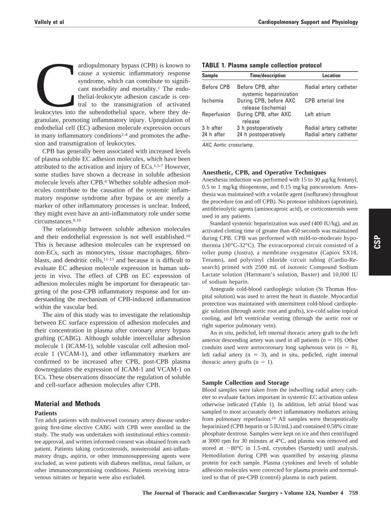
Sample Collection and StorageBlood samples were taken from the indwelling radial artery cath-eter to evaluate factors important in systemic EC activation unlessotherwise indicated (Table 1). In addition, left atrial blood wassampled to most accurately detect inflammatory mediators arisingfrom pulmonary reperfusion.16 All samples were therapeuticallyheparinized (CPB heparin or 5 IU/mL) and contained 0.58% citratephosphate dextrose. Samples were kept on ice and then centrifugedat 3000 rpm for 30 minutes at 4°C, and plasma was removed andstored at �80°C in 1.5-mL cryotubes (Sarstedt) until analysis.Hemodilution during CPB was quantified by assaying plasmaprotein for each sample. Plasma cytokines and levels of solubleadhesion molecules were corrected for plasma protein and normal-ized to that of pre-CPB (control) plasma in each patient.
TABLE 1. Plasma sample collection protocolSample Time/description Location
Before CPB Before CPB, aftersystemic heparinization
Radial artery catheter
Ischemia During CPB, before AXCrelease (ischemia)
CPB arterial line
Reperfusion During CPB, after AXCrelease
Left atrium
3 h after 3 h postoperatively Radial artery catheter24 h after 24 h postoperatively Radial artery catheter
AXC, Aortic crossclamp.
Vallely et al Cardiopulmonary Support and Physiology
The Journal of Thoracic and Cardiovascular Surgery ● Volume 124, Number 4 759
CSP
Human Umbilical Vein Endothelial Cell Isolationand CulturesHuman umbilical vein endothelial cell (HUVEC) cultures wereharvested by using a modified version of the technique describedby Jaffe and colleagues.17 HUVECs were isolated with collagenase(Sigma) and grown to confluence in 175-cm2 Falcon tissue-cultureflasks (Becton Dickinson) in Medium 199 (Biosciences) contain-ing 20% heat-inactivated pooled human serum (CM199), 1% L-glutamine (Biosciences), 0.5% Endothelial Cell Growth Promoter(Starrate Pty Ltd), and penicillin-streptomycin (100 IU/0.1 mg/mL,Sigma). Cells were grown to confluence at 37°C in a 5% CO2
incubator. Cells were subpassaged with 1:250 trypsin/ethylenedi-amine tetraacetic acid (Biosciences) into 96-well tissue-cultureplates (Falcon, Becton Dickinson), at 10,000 cells/well in 100 �Lof media. All glassware was heat treated to ensure an endotoxin-free system, and media were made up in distilled water (Baxter)and then filtered with 0.2-�m Zetapore filters (Cuno) before use.All experiments were conducted with passage 2 HUVECs.
Citrate phosphate dextrose (0.58%) was added to all bloodsamples to overcome cytotoxic fibrin clot formation on theHUVEC cultures. In addition, samples were taken during systemicheparinization or, in the case of postoperative samples, heparinwas added (5 IU/mL). Plasma samples for each patient werenormalized for protein by means of dilution with isotonic crystal-loid (containing no protein) Compound Sodium Lactate solution(Hartmann’s Solution, Baxter) to account for hemodilution duringbypass.
HUVEC Expression of Adhesion MoleculesBy using a modified technique, as described by other investiga-tors,2,4,18,19 HUVECs were exposed for 4 hours to 20% patientplasma and then 20% heat-inactivated pooled human serum. Thisallowed for maximal upregulation of adhesion molecule expres-sion while maintaining normal cell viability. Cell viability wasroutinely assessed by means of lactate dehydrogenase release bymonitoring EC morphology with light microscopy and preserva-tion of cell attachment with quantification of cell protein, aspreviously described.20 Under the conditions described, cell via-bility was preserved (90%-98%, n � 6), and cell protein wasgreater than 95% of control incubations in pooled, heat-inactivatedserum.
After washing, cell-surface adhesion molecules were measuredwith specific enzyme-linked immunosorbent assays (ELISAs) at4°C, as described by McCrohon and colleagues.19 In brief,HUVECs were exposed to 1:1000 purified mouse anti-human
monoclonal primary antibodies to CD54 (ICAM-1), CD62E (E-selectin), and CD106 (VCAM-1; Pharmingen, Becton Dickinson)in Hanks balanced salt solution containing 10% heat-inactivatedpooled human serum, with an irrelevant, nonmammalian mouseIgG1 antibody used as a negative control (DAKO). After washing,cells were incubated with a sheep, anti-mouse horseradish perox-idase secondary antibody (Amersham, 1:500 dilution), and absor-bance was read at 405-nm wavelength (Titertek Multiskan MCC340, Labsystems) after addition of ABTS peroxidase substratesolution (Kirkegard and Perry Laboratories). For each subject, thepatient’s own pre-CPB plasma (sample 1) incubated withHUVECs was used as an internal control. Results were expressedas a percentage of the pre-CPB control (100%) for each subject,with a mean � SD of quadruplicate cultures for each data point.Initial experiments established that control cultures incubated withpooled, heat-inactivated serum and cultures incubated with pre-CPB plasma demonstrated identical expression of adhesion mol-ecules.
Commercially available interleukin 1� (IL-1�; Pharmingen,Becton-Dickinson) was used to investigate whether peri-CABGplasma modulated HUVEC response to IL-1�. Stock solution wasdiluted and incubated with HUVECs at a final concentration of 1ng/mL. Preliminary experiments established that this concentrationof IL-1� generated maximal upregulation of adhesion moleculeexpression under our experimental conditions.
Soluble Adhesion Molecules and Plasma CytokinesPlasma soluble adhesion molecule levels (E-selectin, ICAM-1, andVCAM-1) were measured with commercially available sandwichELISAs (Bender MedSystems). Plasma IL-8, IL-10, and trans-forming growth factor �1 (TGF-�1) levels were measured withcommercially available sandwich ELISAs (Pharmingen, Becton-Dickinson). C-reactive protein (CRP) was measured with an au-tomated rate nephelometry assay (IMMAGE, Beckman).
Statistical AnalysisAll results are expressed as means � SEM (n � 10 patients).Samples were compared with the pre-CPB control. Statisticalanalysis was performed with robust cluster multiple linear regres-sions (STATA 7.0, StataCorp).
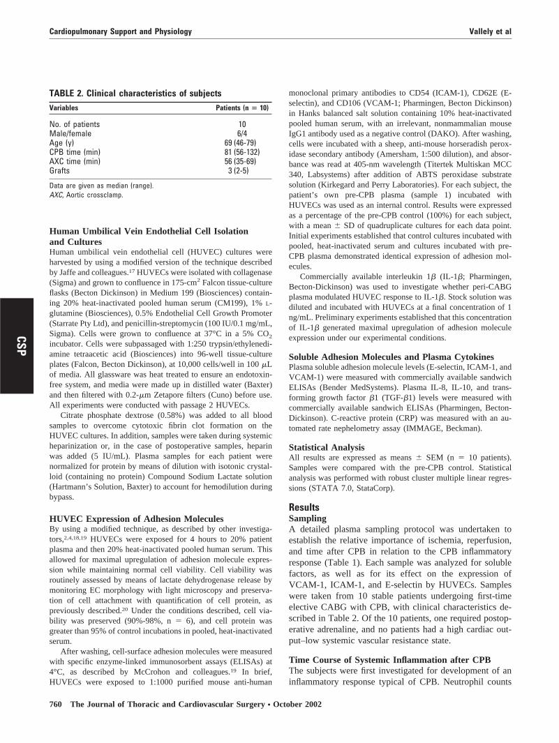
ResultsSamplingA detailed plasma sampling protocol was undertaken toestablish the relative importance of ischemia, reperfusion,and time after CPB in relation to the CPB inflammatoryresponse (Table 1). Each sample was analyzed for solublefactors, as well as for its effect on the expression ofVCAM-1, ICAM-1, and E-selectin by HUVECs. Sampleswere taken from 10 stable patients undergoing first-timeelective CABG with CPB, with clinical characteristics de-scribed in Table 2. Of the 10 patients, one required postop-erative adrenaline, and no patients had a high cardiac out-put–low systemic vascular resistance state.
Time Course of Systemic Inflammation after CPBThe subjects were first investigated for development of aninflammatory response typical of CPB. Neutrophil counts
TABLE 2. Clinical characteristics of subjectsVariables Patients (n � 10)
No. of patients 10Male/female 6/4Age (y) 69 (46-79)CPB time (min) 81 (56-132)AXC time (min) 56 (35-69)Grafts 3 (2-5)
Data are given as median (range).AXC, Aortic crossclamp.
Cardiopulmonary Support and Physiology Vallely et al
760 The Journal of Thoracic and Cardiovascular Surgery ● October 2002
CSP
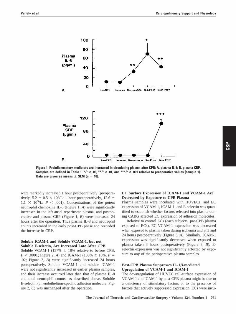
were markedly increased 1 hour postoperatively (preopera-tively, 5.2 � 0.5 � 109/L; 1 hour postoperatively, 12.6 �1.1 � 109/L; P � .001). Concentrations of the potentneutrophil chemokine IL-8 (Figure 1, A) were significantlyincreased in the left atrial reperfusate plasma, and postop-erative and plasma CRP (Figure 1, B) were increased 24hours after the operation. Thus plasma IL-8 and neutrophilcounts increased in the early post-CPB phase and precededthe increase in CRP.
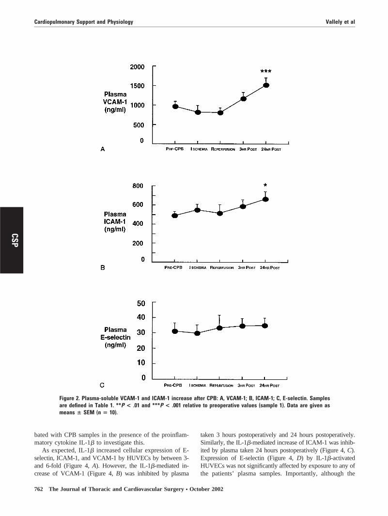
Soluble ICAM-1 and Soluble VCAM-1, but notSoluble E-selectin, Are Increased Late After CPBSoluble VCAM-1 (157% � 18% relative to before CPB,P � .0001; Figure 2, A) and ICAM-1 (135% � 16%, P �.02; Figure 2, B) were significantly increased 24 hourspostoperatively. Soluble VCAM-1 and soluble ICAM-1were not significantly increased in earlier plasma samples,and their increase occurred later than that of plasma IL-8and total neutrophil counts, as described above. SolubleE-selectin (an endothelium-specific adhesion molecule; Fig-ure 2, C) was unchanged after the operation.
EC Surface Expression of ICAM-1 and VCAM-1 AreDecreased by Exposure to CPB PlasmaPlasma samples were incubated with HUVECs, and ECexpression of VCAM-1, ICAM-1, and E-selectin was quan-tified to establish whether factors released into plasma dur-ing CABG affected EC expression of adhesion molecules.
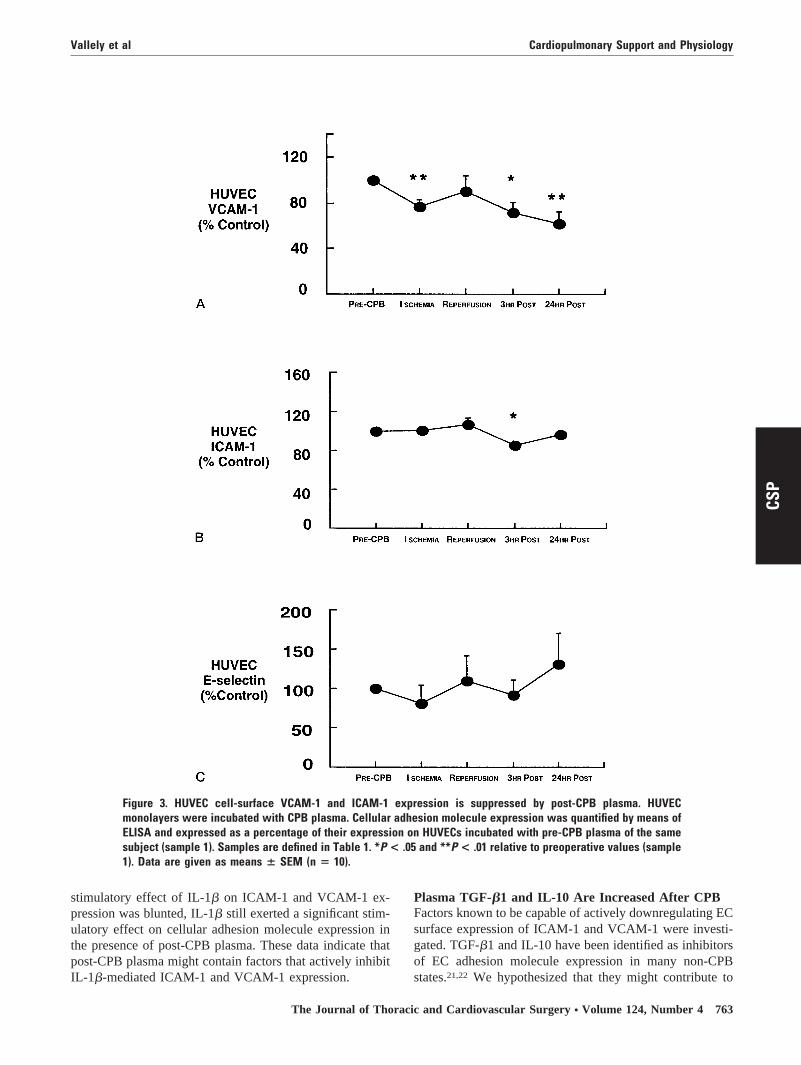
Relative to control ECs (each subjects’ pre-CPB plasmaexposed to ECs), EC VCAM-1 expression was decreasedwhen exposed to plasma taken during ischemia and at 3 and24 hours postoperatively (Figure 3, A). Similarly, ICAM-1expression was significantly decreased when exposed toplasma taken 3 hours postoperatively (Figure 3, B). E-selectin expression was not significantly affected by expo-sure to any of the perioperative plasma samples.
Post-CPB Plasma Suppresses IL-1�-mediatedUpregulation of VCAM-1 and ICAM-1The downregulation of HUVEC cell-surface expression ofVCAM-1 and ICAM-1 by post-CPB plasma might be due toa deficiency of stimulatory factors or to the presence offactors that actively suppressed expression. ECs were incu-
Figure 1. Proinflammatory mediators are increased in circulating plasma after CPB: A, plasma IL-8; B, plasma CRP.Samples are defined in Table 1. *P < .05, **P < .01, and ***P < .001 relative to preoperative values (sample 1).Data are given as means � SEM (n � 10).
Vallely et al Cardiopulmonary Support and Physiology
The Journal of Thoracic and Cardiovascular Surgery ● Volume 124, Number 4 761
CSP
bated with CPB samples in the presence of the proinflam-matory cytokine IL-1� to investigate this.
As expected, IL-1� increased cellular expression of E-selectin, ICAM-1, and VCAM-1 by HUVECs by between 3-and 6-fold (Figure 4, A). However, the IL-1�-mediated in-crease of VCAM-1 (Figure 4, B) was inhibited by plasma
taken 3 hours postoperatively and 24 hours postoperatively.Similarly, the IL-1�-mediated increase of ICAM-1 was inhib-ited by plasma taken 24 hours postoperatively (Figure 4, C).Expression of E-selectin (Figure 4, D) by IL-1�-activatedHUVECs was not significantly affected by exposure to any ofthe patients’ plasma samples. Importantly, although the
Figure 2. Plasma-soluble VCAM-1 and ICAM-1 increase after CPB: A, VCAM-1; B, ICAM-1; C, E-selectin. Samplesare defined in Table 1. **P < .01 and ***P < .001 relative to preoperative values (sample 1). Data are given asmeans � SEM (n � 10).
Cardiopulmonary Support and Physiology Vallely et al
762 The Journal of Thoracic and Cardiovascular Surgery ● October 2002
CSP
stimulatory effect of IL-1� on ICAM-1 and VCAM-1 ex-pression was blunted, IL-1� still exerted a significant stim-ulatory effect on cellular adhesion molecule expression inthe presence of post-CPB plasma. These data indicate thatpost-CPB plasma might contain factors that actively inhibitIL-1�-mediated ICAM-1 and VCAM-1 expression.
Plasma TGF-�1 and IL-10 Are Increased After CPBFactors known to be capable of actively downregulating ECsurface expression of ICAM-1 and VCAM-1 were investi-gated. TGF-�1 and IL-10 have been identified as inhibitorsof EC adhesion molecule expression in many non-CPBstates.21,22 We hypothesized that they might contribute to
Figure 3. HUVEC cell-surface VCAM-1 and ICAM-1 expression is suppressed by post-CPB plasma. HUVECmonolayers were incubated with CPB plasma. Cellular adhesion molecule expression was quantified by means ofELISA and expressed as a percentage of their expression on HUVECs incubated with pre-CPB plasma of the samesubject (sample 1). Samples are defined in Table 1. *P < .05 and **P < .01 relative to preoperative values (sample1). Data are given as means � SEM (n � 10).
Vallely et al Cardiopulmonary Support and Physiology
The Journal of Thoracic and Cardiovascular Surgery ● Volume 124, Number 4 763
CSP

Figure 4. IL-1�-activated HUVEC VCAM-1 and ICAM-1 expression is suppressed by post-CPB plasma: A, upregu-lation of HUVEC expression of VCAM-1, ICAM-1, and E-selectin by IL-1� in the presence of control preoperativeplasma (P < .001 for comparison of control with IL-1� for each adhesion molecule); B-D, effect of post-CPB plasmaon IL-1�-stimulated expression of VCAM-1 (B), ICAM-1 (C), and E-selectin (D). All values are expressed as apercentage expression of IL-1�-exposed pre-CPBB plasma (sample 1). Samples are defined in Table 1. *P < .05,**P < .01, and ***P < .001 relative to preoperative values (sample 1). Data are given as means � SEM (n � 10).
Cardiopulmonary Support and Physiology Vallely et al
764 The Journal of Thoracic and Cardiovascular Surgery ● October 2002
CSP

CPB-mediated suppression and investigated this possibilityirrespective of whether they were increased after CPB.
TGF-�1 at 3 hours was significantly increased 3 hourspostoperatively (2.9-fold relative to before CPB; Figure 5,A). IL-10 was significantly increased during CPB afteraortic crossclamp release during early reperfusion (9.8-fold)and maximally increased 3 hours postoperatively (14-fold;Figure 5, B). The increase of IL-10 was relatively shortlived, whereas the TGF-�1 increase was sustained.
DiscussionInflammation after CABG is understood to involve activa-tion of the endothelium; however, the relative importance ofendothelial expression of cell-surface adhesion molecules inthis process is unclear. This study is the first to relate ECexpression of adhesion molecules to their concentration inplasma after CABG, and suggests that the two are dissoci-ated.
A number of studies have investigated soluble plasmaadhesion molecules after CABG.5-7,23 Although some indi-cate a null effect,8 the majority indicate upregulation ofsoluble adhesion molecules after CPB.5,24 Our data supports
this in relation to ICAM-1 and VCAM-1, particularly insamples taken 24 hours postoperatively. This was not foundfor E-selectin.
The difference between ICAM-1, VCAM-1, and E-se-lectin might indicate that the release of ICAM-1 andVCAM-1 from cells other than endothelium might contrib-ute to plasma soluble adhesion molecules. E-selectin, incontrast to ICAM-1 and VCAM-1, is endothelium specific.Alternatively, the release of E-selectin might be relativelyshort lived, and time points used in our study might havefailed to collect plasma samples containing the highestamounts of E-selectin.
That soluble ICAM-1 and soluble VCAM-1 were in-creased late after surgical intervention does not suggest theiracute release during ischemia or reperfusion during CPB.Other markers of systemic inflammation (polymorphonucle-ar neutrophil count and IL-8) preceded the soluble ICAM-1and soluble VCAM-1 increase. These kinetic considerationssuggest that if EC activation explains the presence of solu-ble ICAM-1 and soluble VCAM-1 after CABG, it mightfollow other inflammatory processes, such as neutrophilactivation. Because all patients underwent operations with
Figure 5. Plasma TGF-�1 and IL-10 are increased after CPB: A, TGF-�1; B, IL-10. Samples 1 to 5 are defined in Table1. *P < .05, **P < .01, and ***P < .001 relative to preoperative values (sample 1). Data are given as means � SEM(n � 10).
Vallely et al Cardiopulmonary Support and Physiology
The Journal of Thoracic and Cardiovascular Surgery ● Volume 124, Number 4 765
CSP
CPB, we cannot delineate the role of the bypass circuit ininfluencing soluble ICAM-1 and VCAM-1. A comparativestudy with patients undergoing surgical intervention withoutCPB would be interesting.
In our study an IL-8 increase preceded postoperativeneutrophilia, and this preceded an increase in CRP. Thissequence is to be expected because IL-8 is a neutrophilchemokine and CRP represents the later effects of systemicinflammation on the liver. The late increase of CRP suggestsongoing inflammation 24 hours postoperatively, which isconsistent with previous literature.25 The more rapid declineof IL-10 postoperatively relative to TGF-�1 is interestingand unexplained. It suggests that different stimuli (eg, reper-fusion injury for IL-10)22 are responsible for upregulation ofthese 2 inflammatory mediators.
The most remarkable finding in the present study was thesuppression of EC ICAM-1 and VCAM-1 expression bypost-CABG plasma. This cannot be attributed to an atypicalpatient population because soluble adhesion molecules andtypical systemic inflammatory markers were increased afterCABG. The postoperative increase in soluble adhesion mol-ecules and the suppression of EC adhesion molecules mightindicate a relationship between the 2 processes. It is possiblethat soluble adhesion molecules are released from the cellsurface as a result of cleavage or shedding26 and that theirincreased plasma concentration represents increased releasefrom the cell surface rather than increased cell expression.Because ICAM-1 and VCAM-1 are constitutively expressedon resting endothelium, and E-selectin expression is in-duced by activation of the endothelium, our data mightsupport cleavage of cell-surface adhesion molecule ratherthan increased EC synthesis and cell-surface expression.Alternatively, the increase in soluble adhesion moleculesand decreased EC adhesion molecule expression might bothbe independent consequences of earlier inflammatory pro-cesses, such as neutrophil activation and cytokine release. Inaddition, future studies might identify whether other EClines, such as microvascular cell lines27 or arterial cells,28
respond differently to HUVECs when exposed to peri-CABG plasma. Similarly, it is possible that other adhesionmolecules, such as P-selectin, might behave differently thanthe adhesion molecules in our study.
The blunting of the IL-1�-mediated expression of ECICAM-1 and VCAM-1 (but not E-selectin) suggests thatthere might be factors released into post-CABG plasma thatinhibit ICAM-1 and VCAM-1 expression. One possiblemechanism by which plasma antagonizes the effect ofIL-1� is the presence of IL-1 receptor antagonist, which canbe present after CPB.29 Second, TGF-�1 and IL-10 bothsuppress adhesion molecule expression30,31 and were bothincreased in our population, which is consistent with apossible role for these cytokines in suppressing EC adhesionmolecule expression by post-CPB plasma.
The findings of this study have important clinical impli-cations. First, they dissociate soluble adhesion moleculeconcentrations from plasma-induced EC expression. Sec-ond, they suggest that prevention of EC E-selectin,ICAM-1, or VCAM-1 upregulation might not be a suitabletherapeutic target for the reduction of inflammation afterCPB. However, inhibition of their expression or activitymight still have a role. Third, they suggest that EC activa-tion after CPB is not an indiscriminate process and thatmediators of irreversible leukocyte adhesion (ICAM-1 andVCAM-1), and not reversible adhesion (E-selectin), aredifferentially modulated after CPB. Future targeting of thepost-CPB systemic inflammatory response will require elu-cidation of mechanisms underlying these observations.
We thank Gail de Lucia, RN, for her invaluable help in con-ducting this study and Jack Chen, PhD, for his statistical analyses.
References
1. Boyle EM Jr, Pohlman TH, Johnson McVerrier ED. Endothelial cellinjury in cardiovascular surgery: the systemic inflammatory response.Ann Thorac Surg. 1997;63:277-84.
2. Butthep P, Bunyaratvej A, Funahara Y, Kitaguchi H, Fucharoen S,Sato S, et al. Possible evidence of endothelial cell activation anddisturbance in thalassemia: an in vitro study. Southeast Asian J TropMed Public Health. 1997;28(suppl 3):141-8A.
3. Drake TA, Cheng J, Chang A, Taylor FB Jr. Expression of tissuefactor, thrombomodulin, and E-selectin in baboons with lethal Esch-erichia coli sepsis [published erratum appears in Am J Pathol. 1993;143:649]. Am J Pathol. 1993;142:1458-70.
4. Heyl W, Handt S, Reister F, Gehlen J, Mittermayer C, Rath W. Therole of soluble adhesion molecules in evaluating endothelial cellactivation in preeclampsia. Am J Obstet Gynecol. 1999;180:68-72.
5. Blume ED, Nelson DP, Gauvreau K, Walsh AZ, Plumb C, Neufeld EJ,et al. Soluble adhesion molecules in infants and children undergoingcardiopulmonary bypass. Circulation. 1997;96:II352-7.
6. Kalawski R, Bugajski P, Smielecki J, Wysocki H, Olszewski R, MoreR, et al. Soluble adhesion molecules in reperfusion during coronarybypass grafting. Eur J Cardiothorac Surg. 1998;14:290-5.
7. Boldt J, Kumle B, Papsdorf M, Hempelmann G. Are circulatingadhesion molecules specifically changed in cardiac surgical patients?Ann Thorac Surg. 1998;65:608-14.
8. Boldt J, Osmer C, Linke LC, Dapper F, Hempelmann G. Circulatingadhesion molecules in pediatric cardiac surgery. Anesth Analg. 1995;81:1129-35.
9. Ross L, Hassman FMolony L. Inhibition of Molt-4-endothelial adher-ence by synthetic peptides from the sequence of ICAM-1. J BiolChem. 1992;267:8537-43.
10. Vallely MP, Bannon PG, Hughes CFL. Endothelial cell adhesionmolecules and cardiopulmonary bypass. Asian Cardiovasc ThoracAnn. 2001;9:353-9.
11. Grigg J, Kukielka GL, Berens KL, Dreyer WJ, Entman ML, SmithCW. Induction of intercellular adhesion molecule-1 by lipopolysac-charide in canine alveolar macrophages. Am J Respir Cell Mol Biol.1994;11:304-11.
12. Maio M, Pinto A, Carbone A, Zagonel V, Gloghini A, Marotta G, etal. Differential expression of CD54/intercellular adhesion molecule-1in myeloid leukemias and in lymphoproliferative disorders. Blood.1990;76:783-90.
13. Vanhee D, Molet S, Gosset P, Tillie-Leblond I, Boitelle A, Wallaert B,et al. Expression of leucocyte-endothelial adhesion molecules is lim-ited to intercellular adhesion molecule-1 (ICAM-1) in the lung ofpneumoconiotic patients: role of tumour necrosis factor-alpha (TNF-alpha). Clin Exp Immunol. 1996;106:541-8.
Cardiopulmonary Support and Physiology Vallely et al
766 The Journal of Thoracic and Cardiovascular Surgery ● October 2002
CSP
14. Freedman AS, Munro JM, Morimoto C, McIntyre BW, Rhynhart K,Lee N, et al. Follicular non-Hodgkin’s lymphoma cell adhesion tonormal germinal centers and neoplastic follicles involves very lateantigen-4 and vascular cell adhesion molecule-1. Blood. 1992;79:206-12.
15. Ryan DH, Nuccie BL, Abboud CN, Winslow JM. Vascular celladhesion molecule-1 and the integrin VLA-4 mediate adhesion ofhuman B cell precursors to cultured bone marrow adherent cells.J Clin Invest. 1991;88:995-1004.
16. Massoudy P, Zahler S, Becker BF, Braun SL, Barankay AMeisner H.Evidence for inflammatory responses of the lungs during coronaryartery bypass grafting with cardiopulmonary bypass. Chest. 2001;119:31-6.
17. Jaffe EA, Nachman RL, Becker CG, Minick CR. Culture of humanendothelial cells derived from umbilical veins. Identification by mor-phologic and immunologic criteria. J Clin Invest. 1973;52:2745-56.
18. Aebert H, Kirchner S, Keyser A, Birnbaum DE, Holler E, AndreesenR, et al. Endothelial apoptosis is induced by serum of patients aftercardiopulmonary bypass. Eur J Cardiothorac Surg. 2000;18:589-93.
19. McCrohon JA, Jessup W, Handelsman DJ, Celermajer DS. Androgenexposure increases human monocyte adhesion to vascular endothe-lium and endothelial cell expression of vascular cell adhesion mole-cule-1. Circulation. 1999;99:2317-22.
20. Rees D, Sloane T, Jessup W, Dean RT, Kritharides L. ApolipoproteinA-I stimulates secretion of apolipoprotein E by foam cell macro-phages. J Biol Chem. 1999;274:27925-33.
21. Opal SM, DePalo VA. Anti-inflammatory cytokines. Chest. 2000;117:1162-72.
22. Yang Z, Zingarelli B, Szabo C. Crucial role of endogenous interleu-kin-10 production in myocardial ischemia/reperfusion injury. Circu-lation. 2000;101:1019-26.
23. Liebold A, Keyl C, Birnbaum DE. The heart produces but the lungsconsume proinflammatory cytokines following cardiopulmonary by-pass. Eur J Cardiothorac Surg. 1999;15:340-5.
24. Boldt J, Osmer C, Schindler E, Linke LC, Stertmann WAHempel-mann G. Circulating adhesion molecules in cardiac operations: influ-ence of high-dose aprotinin. Ann Thorac Surg. 1995;59:100-5.
25. Boralessa H, de Beer FC, Manchie A, Whitwam JG, Pepys MB.C-reactive protein in patients undergoing cardiac surgery. Anaesthe-sia. 1986;41:11-5.
26. Leeuwenberg JF, Smeets EF, Neefjes JJ, Shaffer MA, Cinek T,Jeunhomme TM, et al. E-selectin and intercellular adhesion mole-cule-1 are released by activated human endothelial cells in vitro.Immunology. 1992;77:543-9.
27. Frigerio S, Gelati M, Ciusani E, Corsini E, Dufour A, Massa G, et al.Immunocompetence of human microvascular brain endothelial cells:cytokine regulation of IL-1beta, MCP-1, IL-10, sICAM-1 and sV-CAM-1. J Neurol. 1998;245:727-30.
28. Kalogeris TJ, Kevil CG, Laroux FS, Coe LL, Phifer TJ, Alexander JS.Differential monocyte adhesion and adhesion molecule expression invenous and arterial endothelial cells. Am J Physiol. 1999;276:L9-19.
29. Marie C, Muret J, Fitting C, Payen D, Cavaillon JM. Interleukin-1receptor antagonist production during infectious and noninfectioussystemic inflammatory response syndrome. Crit Care Med. 2000;28:2277-82.
30. Morise Z, Eppihimer M, Granger DN, Anderson DC, Grisham MB.Effects of lipopolysaccharide on endothelial cell adhesion moleculeexpression in interleukin-10 deficient mice. Inflammation. 1999;23:99-110.
31. Gamble JR, Khew-Goodall Y, Vadas MA. Transforming growth fac-tor-beta inhibits E-selectin expression on human endothelial cells.J Immunol. 1993;150:4494-503.
Vallely et al Cardiopulmonary Support and Physiology
The Journal of Thoracic and Cardiovascular Surgery ● Volume 124, Number 4 767
CSP