Embed Size (px)
Citation preview

Engineering a Structure Switching Mechanism into a Steroid-BindingAptamer and Hydrodynamic Analysis of the Ligand BindingMechanismOren Reinstein,† Miguel A. D. Neves,† Makbul Saad,† Sherry N. Boodram,† Stephanie Lombardo,†
Simone A. Beckham,‡,§ Jason Brouwer,§ Gerald F. Audette,† Patrick Groves,⊥ Matthew C. J. Wilce,*,§
and Philip E. Johnson*,†
†Department of Chemistry, York University, Toronto, Ontario, Canada M3J 1P3‡Centre for Cancer Research, Monash Institute of Medical Research, Monash University, Clayton, VIC 3168, Australia§Department of Biochemistry and Molecular Biology, Monash University, Clayton, VIC 3800, Australia⊥Department of Biological Chemistry, Instituto de Tecnologia Quimica e Biologica, Av. da Republica (EAN), 2781-901, Oeiras,Portugal
*S Supporting Information
ABSTRACT: The steroid binding mechanism of a DNAaptamer was studied using isothermal titration calorimetry(ITC), NMR spectroscopy, quasi-elastic light scattering(QELS), and small-angle X-ray spectroscopy (SAXS). Bindingaffinity determination of a series of steroid-binding aptamersderived from a parent cocaine-binding aptamer demonstratesthat substituting a GA base pair with a GC base pair governsthe switch in binding specificity from cocaine to the steroid deoxycholic acid (DCA). Binding of DCA to all aptamers is anenthalpically driven process with an unfavorable binding entropy. We engineered into the steroid-binding aptamer a ligand-induced folding mechanism by shortening the terminal stem by two base pairs. NMR methods were used to demonstrate thatthere is a transition from a state where base pairs are formed in one stem of the free aptamer, to where three stems are formed inthe DCA-bound aptamer. The ability to generate a ligand-induced folding mechanism into a DNA aptamer architecture based onthe three-way junction of the cocaine-binding aptamer opens the door to obtaining a series of aptamers all with ligand-inducedfolding mechanisms but triggered by different ligands. Hydrodynamic data from diffusion NMR spectroscopy, QELS, and SAXSshow that for the aptamer with the full-length terminal stem there is a small amount of structure compaction with DCA binding.For ligand binding by the short terminal stem aptamer, we propose a binding mechanism where secondary structure forms uponDCA binding starting from a free structure where the aptamer exists in a compact form.
Since their initial development, aptamers have gainedwidespread use in biosensor applications.1−3 A large variety
of detection methods such as fluorescence, colorimetric, andelectrochemical signals have been coupled with aptamer−ligandinteractions to indicate the presence of an analyte. In order toproduce the maximum desired signal, and therefore greatestsensitivity, ligand binding has been coupled with the occurrenceof structural changes in the aptamer that results in thegeneration of a signal that is detected.3 A particularly well-developed system for the development of biosensors employingstructural switching is that of the cocaine-binding aptamer.4
Numerous different biosensors have been developed on thebasis of the aptamer−cocaine interaction.5−13 Aside frombiosensors, the cocaine-binding aptamer has been employed insupramolecular and nanotechnology applications that takeadvantage of the structural switching that occurs with ligandbinding.14−16 The common principle among these applicationsis that cocaine binding assembles separate DNA molecules in adesired and controlled manner. Doubtlessly, the widespreaddevelopment and use of these applications are limited by the
legal and regulatory restrictions placed on the use of therequired ligand, cocaine.The cocaine-binding aptamer is composed of three stems
arranged around a three-way junction (Figure 1a).4 Threevariations of the cocaine-binding aptamer have been studied,two of which follow a structural switching or ligand-inducedfolding mechanism. In the first type of cocaine-bindingaptamer, all of the three stems are 4−6 base pairs long (Figure1a). Here, the aptamer has its secondary structure formed inthe free state and binds cocaine with affinity in the lowmicromolar range depending on the exact sequence and bufferconditions.5,17,18 The second type of cocaine-binding aptamerconsists of stem 1 being shortened to three base pairs (Figure1b). In this molecule, little secondary structure is formed in thefree aptamer and ligand binding prompts secondary structure
Received: August 29, 2011Revised: September 25, 2011Published: September 26, 2011
Article
pubs.acs.org/biochemistry
© 2011 American Chemical Society 9368 dx.doi.org/10.1021/bi201361v |Biochemistry 2011, 50, 9368−9376
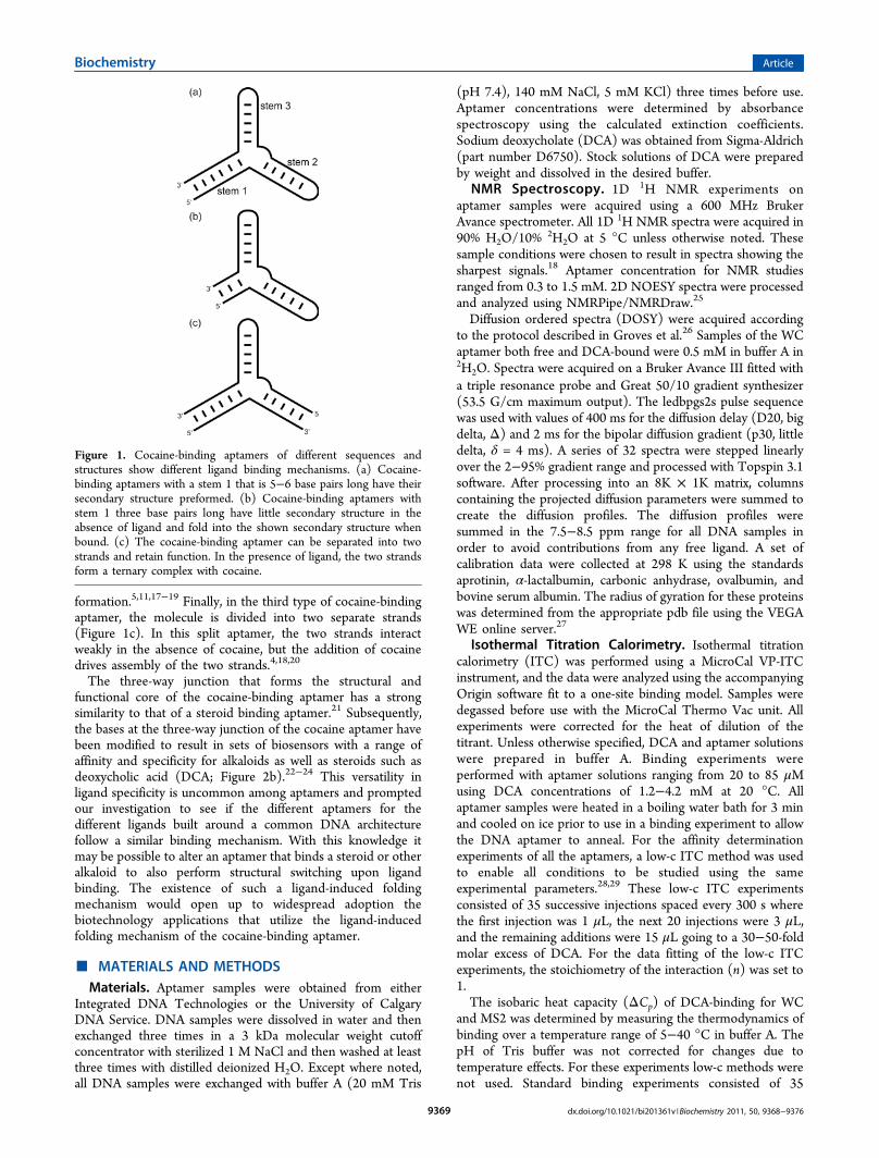
formation.5,11,17−19 Finally, in the third type of cocaine-bindingaptamer, the molecule is divided into two separate strands(Figure 1c). In this split aptamer, the two strands interactweakly in the absence of cocaine, but the addition of cocainedrives assembly of the two strands.4,18,20
The three-way junction that forms the structural andfunctional core of the cocaine-binding aptamer has a strongsimilarity to that of a steroid binding aptamer.21 Subsequently,the bases at the three-way junction of the cocaine aptamer havebeen modified to result in sets of biosensors with a range ofaffinity and specificity for alkaloids as well as steroids such asdeoxycholic acid (DCA; Figure 2b).22−24 This versatility inligand specificity is uncommon among aptamers and promptedour investigation to see if the different aptamers for thedifferent ligands built around a common DNA architecturefollow a similar binding mechanism. With this knowledge itmay be possible to alter an aptamer that binds a steroid or otheralkaloid to also perform structural switching upon ligandbinding. The existence of such a ligand-induced foldingmechanism would open up to widespread adoption thebiotechnology applications that utilize the ligand-inducedfolding mechanism of the cocaine-binding aptamer.
■ MATERIALS AND METHODSMaterials. Aptamer samples were obtained from either
Integrated DNA Technologies or the University of CalgaryDNA Service. DNA samples were dissolved in water and thenexchanged three times in a 3 kDa molecular weight cutoffconcentrator with sterilized 1 M NaCl and then washed at leastthree times with distilled deionized H2O. Except where noted,all DNA samples were exchanged with buffer A (20 mM Tris
(pH 7.4), 140 mM NaCl, 5 mM KCl) three times before use.Aptamer concentrations were determined by absorbancespectroscopy using the calculated extinction coefficients.Sodium deoxycholate (DCA) was obtained from Sigma-Aldrich(part number D6750). Stock solutions of DCA were preparedby weight and dissolved in the desired buffer.NMR Spectroscopy. 1D 1H NMR experiments on
aptamer samples were acquired using a 600 MHz BrukerAvance spectrometer. All 1D 1H NMR spectra were acquired in90% H2O/10%
2H2O at 5 °C unless otherwise noted. Thesesample conditions were chosen to result in spectra showing thesharpest signals.18 Aptamer concentration for NMR studiesranged from 0.3 to 1.5 mM. 2D NOESY spectra were processedand analyzed using NMRPipe/NMRDraw.25
Diffusion ordered spectra (DOSY) were acquired accordingto the protocol described in Groves et al.26 Samples of the WCaptamer both free and DCA-bound were 0.5 mM in buffer A in2H2O. Spectra were acquired on a Bruker Avance III fitted witha triple resonance probe and Great 50/10 gradient synthesizer(53.5 G/cm maximum output). The ledbpgs2s pulse sequencewas used with values of 400 ms for the diffusion delay (D20, bigdelta, Δ) and 2 ms for the bipolar diffusion gradient (p30, littledelta, δ = 4 ms). A series of 32 spectra were stepped linearlyover the 2−95% gradient range and processed with Topspin 3.1software. After processing into an 8K × 1K matrix, columnscontaining the projected diffusion parameters were summed tocreate the diffusion profiles. The diffusion profiles weresummed in the 7.5−8.5 ppm range for all DNA samples inorder to avoid contributions from any free ligand. A set ofcalibration data were collected at 298 K using the standardsaprotinin, α-lactalbumin, carbonic anhydrase, ovalbumin, andbovine serum albumin. The radius of gyration for these proteinswas determined from the appropriate pdb file using the VEGAWE online server.27
Isothermal Titration Calorimetry. Isothermal titrationcalorimetry (ITC) was performed using a MicroCal VP-ITCinstrument, and the data were analyzed using the accompanyingOrigin software fit to a one-site binding model. Samples weredegassed before use with the MicroCal Thermo Vac unit. Allexperiments were corrected for the heat of dilution of thetitrant. Unless otherwise specified, DCA and aptamer solutionswere prepared in buffer A. Binding experiments wereperformed with aptamer solutions ranging from 20 to 85 μMusing DCA concentrations of 1.2−4.2 mM at 20 °C. Allaptamer samples were heated in a boiling water bath for 3 minand cooled on ice prior to use in a binding experiment to allowthe DNA aptamer to anneal. For the affinity determinationexperiments of all the aptamers, a low-c ITC method was usedto enable all conditions to be studied using the sameexperimental parameters.28,29 These low-c ITC experimentsconsisted of 35 successive injections spaced every 300 s wherethe first injection was 1 μL, the next 20 injections were 3 μL,and the remaining additions were 15 μL going to a 30−50-foldmolar excess of DCA. For the data fitting of the low-c ITCexperiments, the stoichiometry of the interaction (n) was set to1.The isobaric heat capacity (ΔCp) of DCA-binding for WC
and MS2 was determined by measuring the thermodynamics ofbinding over a temperature range of 5−40 °C in buffer A. ThepH of Tris buffer was not corrected for changes due totemperature effects. For these experiments low-c methods werenot used. Standard binding experiments consisted of 35
Figure 1. Cocaine-binding aptamers of different sequences andstructures show different ligand binding mechanisms. (a) Cocaine-binding aptamers with a stem 1 that is 5−6 base pairs long have theirsecondary structure preformed. (b) Cocaine-binding aptamers withstem 1 three base pairs long have little secondary structure in theabsence of ligand and fold into the shown secondary structure whenbound. (c) The cocaine-binding aptamer can be separated into twostrands and retain function. In the presence of ligand, the two strandsform a ternary complex with cocaine.
Biochemistry Article
dx.doi.org/10.1021/bi201361v |Biochemistry 2011, 50, 9368−93769369

successive 8 μL injections spaced every 300 s where the firstinjection was 2 μL. Each experiment had a c value of 5.Quasi-Elastic Light Scattering. Free and bound WC
aptamers were analyzed using SEC-QELS and were performedusing an Akta Purifier 10 (GE Healthcare) connected in-line toa Dawn Heleos II and Optilab T-rEX light scattering system(Wyatt Technologies). Analysis of 100 μL of 0.45 mM WC freeand DCA bound samples was performed using the same bufferas used for ITC measurements (20 mM Tris (pH 7.4), 140 mMNaCl, 5 mM KCl). After chromatographic separation on asilica-based SEC column (100 Å pore size; Wyatt Technolo-gies), the column eluate traveled to the QELS flow cell wherelight scattering (658 nm laser light source) by the separatedaptamers or aptamers-DCA samples were monitored by 15angularly separated static light scattering detectors and a quasi-elastic light scattering (QELS) detector at a collection angle of99°. Hydrodynamic radii (Rh) and diffusion coefficients (Dt)were calculated from an autocorrelation function using theaccompanying AstraV software package (Wyatt).Small-Angle X-ray Scattering. Samples of WC and MS2
aptamers both free and DCA-bound were analyzed using SAXS.Measurements were made using the SAXS-WAXS beamline atthe Australian Synchrotron, Melbourne, Australia. Scattering
was obtained over a range of four concentrations; the highestconcentration was 0.45 and 0.51 mM for the WC and MS2aptamers, respectively. All SAXS experiments were carried outwith the aptamer in buffer A. Scattering data were acquired forthe following dilutions: neat sample, 1 in 2, 1 in 4, and 1 in 8.SAXS experiments were performed at room temperature for theWC aptamer and 12 °C for the MS2 aptamer. The samples andmatching buffer solutions were exposed to X-ray as the sampleflowed through the capillary. The 2D scattering images werenormalized for sample transmission and radially averaged. Ineach case, 20 1-s exposures were recorded and averaged.Scattering from the buffer and empty capillaries was subtractedafter scaling scattering intensities to correspond to incidentbeam intensities. Data analysis was performed using the ATSASsuite of software.30 Scattered intensity (I) was plotted against s.Extrapolation of the I(s) profiles to zero angle was used toestimate I(0).
■ RESULTS
Sequence Requirements for Steroid Binding. We usedITC methods to analyze the ability of the WC aptamer and aseries of sequence variants (Figure 2a) to bind DCA (Figure2b). An example of the ITC binding data is shown in Figure 3.
Figure 2. (a) Sequence of the aptamers analyzed in this study. The GC base pair that is important for ligand discrimination between cocaine andDCA binding is shown in red. Dashes between nucleotides indicate Watson−Crick base pairs, and dots indicate non-Watson−Crick base pairs. (b)Chemical structure of deoxycholic acid (DCA).
Biochemistry Article
dx.doi.org/10.1021/bi201361v |Biochemistry 2011, 50, 9368−93769370

The affinity and thermodynamic parameters of DCA bindingfor these constructs are summarized in Table 1. We include thecorresponding affinity data for cocaine binding in Table 1 forcomparison. From this binding data we observe a trend whereconstructs with a cytosine corresponding to position 21 of theWC aptamer, and its equivalent position in other constructs(MN9, MN10, MN11), bind DCA with affinity of 12−19 μM.These same aptamers only weakly bind cocaine. Constructswith an adenine in this position, corresponding to position 21
of WC, are able to bind cocaine but do not bind DCA.Sequence changes outside of the vicinity of the tandem GAmismatch in the aptamer have little impact on ligand binding.The WC, MN9, MN10, and MN11 constructs contain differentcombinations of the GT and GA non-Watson−Crick base pairsobserved in the originally selected cocaine-binding aptamer.4
All these aptamers show similar affinity for DCA and uniformlyweak affinity for cocaine. We analyzed the importance of thepresence of both GA mismatches for DCA binding using theaptamer rWC. In this aptamer, both GA mismatches werechanged to GC base pairs. No binding to either DCA orcocaine was observed for this aptamer. Together, these dataindicate that the determinants for ligand selectivity lie at thetandem GA mismatch at the three-way junction.Effect of Ionic Strength on DCA Binding. At the pH
value these studies were performed (7.4) we expect the DCA tobe negatively charged due to the presence of the carboxylategroup (Figure 2b). The effect of this negative charge on ligandbinding was studied by measuring the affinity and thermody-namics of DCA binding as a function of NaCl concentration.The DCA-binding parameters for the WC aptamer at NaClconcentrations of 0, 140, and 1000 mM are summarized inTable 2. For this aptamer, DCA affinity is tightest at 140 mM
NaCl. At both higher and lower NaCl values affinity is weaker.At all three NaCl concentrations, binding remains enthalpicallydriven with an unfavorable entropy of binding.Engineering of a DCA-Induced Folding Mechanism in
an Aptamer with a Short Stem 1. Cocaine-binding
Figure 3. Sample of ITC data showing the interaction of the (a) WCand (b) MN6 aptamers with DCA. In (a) the WC aptamer binds DCAwith a Kd value of 16 ± 3 μM while in (b) binding is not detectedbetween MN6 and DCA. On top are the raw titration data showingthe heat resulting from each injection of DCA into an aptamersolution. On the bottom are the integrated heats after correcting forthe heat of dilution. Binding experiments were performed at 20 °C in abuffer of 20 mM Tris (pH 7.4), 140 mM NaCl, 5 mM KCl.
Table 1. Thermodynamic Parameters and DissociationConstants of DCA Binding for the Aptamers Presented inThis Studya
DCA cocaineb
aptamer Kd (μM) ΔH (kcal mol−1) −TΔS (kcal mol−1) Kd (μM)
WC 16 ± 3 −7 ± 1 0.7 ± 1.3 204 ± 6MN6 nbd 45.3 ± 0.5MN8 nbd 8.6 ± 0.2MN9 12.2 ± 0.8 −3.7 ± 0.3 2.8 ± 0.1 193 ± 1MN10 18.6 ± 0.1 −8.4 ± 0.8 2.1 ± 0.8 148 ± 1MN11 15.0 ± 0.8 −5.2 ± 0.8 1.2 ± 0.8 123 ± 22rWC nbd ndMS1 nbd ndMS2 25 ± 3 −16 ± 1 10 ± 1 nd
aData acquired in 20 mM Tris (pH 7.4), 140 mM NaCl, 5 mM KCl.Data for WC, MS1, and MS2 were acquired at 15 °C; all others wereacquired at 20 °C. The values reported are averages of 2−4 individualexperiments. The error range reported is one standard deviation. nbdindicates no binding detected. nd indicates that binding for thatcombination was not measured. bThe corresponding data for cocainebinding are included for comparison.18
Table 2. Dissociation Constant and Thermodynamics ofDCA Binding by the WC Aptamer as a Function of NaClConcentrationa
[NaCl] (mM) Kd (μM) ΔH (kcal mol−1) −TΔS (kcal mol−1)
0 68 ± 2 −9.0 ± 0.7 3.5 ± 0.7140 16 ± 3 −7 ± 1 0.7 ± 1.31000 46 ± 5 −8 ± 1 2 ± 1
aData acquired at 15 °C in 20 mM Tris (pH 7.4), 5 mM KCl. Thevalues reported are averages of 2−4 experiments.
Biochemistry Article
dx.doi.org/10.1021/bi201361v |Biochemistry 2011, 50, 9368−93769371

aptamers with a three base pair long stem 1 exhibit a ligand-induced folding mechanism (Figure 1b).17,19 We designed aDCA-binding aptamer that undergoes a similar unfolded tofolded secondary structure transition upon ligand binding. Wefirst tested a modified version of the WC aptamer that has astem 1 that is three base pairs long (MS1; Figure 2a). No DCAbinding was detected by either ITC or NMR methods upon theaddition of the ligand (Table1). 1D 1H NMR spectra of MS1indicate this sequence is unfolded in both the absence andpresence of DCA (Figure 1 of the Supporting Information). Wethen lengthened stem 1 in MS1 by one base pair to make it fourbase pairs long (MS2; Figure 2a). The resulting MS2 aptamerbinds DCA with affinity of 25 ± 3 μM (Table 1).We used NMR spectroscopy to check the extent of
secondary structure formation in the free and bound MS2 asa function of increasing DCA concentration (Figure 4a). In thefree MS2, the imino region of the 1-D 1H NMR shows 4 peaksthat are assigned to the stem 3 nucleotides G27, T28, G29, andG30 (Figure 2 of the Supporting Information). Upon additionof DCA, there is a significant change in the imino region asnumerous additional peaks appear. The appearance of 6−8additional dispersed peaks indicates the formation of additionalsecondary structure elements upon ligand binding. Of particularnote is the presence of the upfield imino at 10.4 ppm, indicatingthe presence of a sheared GA base pair in the aptamer.17
Addition of DCA to WC results in a number of resonanceschanging chemical shift when ligand binding occurs, but thesame number of peaks is observed in the DCA-bound WCaptamer as in the free aptamer.We then analyzed the stability of the DCA-bound MS2
aptamer using NMR spectroscopy. The 1D 1H NMR spectra ofthe imino region of the DCA-bound MS2 recorded as afunction of temperature are shown in Figure 4b. As thetemperature increases for the DCA-bound MS2, two separategroups of transitions are evident. In the first, the group ofdispersed peaks that appear with DCA binding and areindicative of a structure containing three stems disappear by15 °C. The second subset of peaks corresponds to thenucleotides in stem 3, and are those observed in the free MS2.These signals gradually lose intensity and are not visible after35 °C.Change in Heat Capacity with Steroid Binding. In
order to gain further insight into the DCA binding mechanismand compare it with the cocaine binding mechanism, we usedITC methods to measure the change in heat capacity (ΔCp) ofboth WC and MS2 aptamers with DCA binding (Figure 5). Forboth the WC and MS2 aptamers binding DCA, data obtainedat temperatures between 5 and 15 °C were used to determineΔCp. Data acquired at higher temperatures were not used as theeffects of aptamer folding and unfolding complicate the data.For MS2, we know from the temperature-dependent NMRspectra (Figure 4b) that this aptamer−DCA complex has atemperature of thermal denaturation of ∼20 °C. In the plot ofenthalpy versus temperature (Figure 5), the measured enthalpyfor MS2 reflects both folding and binding events. A fit of thesedata for MS2 yields a ΔCp value of −753 ± 200 cal mol−1 K−1
for MS2 binding DCA. For the WC aptamer, the measuredenthalpy between 5 and 15 °C reflects the binding of the foldedfree-aptamer to the DCA ligand. A fit of the enthalpy data atthese temperatures yields a ΔCp° value of −94 ± 75 cal mol−1
K−1.Hydrodynamic Analysis Using Quasi-Elastic Light
Scattering. Initial analysis of the hydrodynamic properties
of free and bound WC was performed using QELS in batchmode. We observed that the DCA-bound WC showed a smallbut consistent decrease in the translational diffusion coefficient(Dt) when compared to the free aptamer. These initial resultswere subsequently confirmed using QELS analysis followingsize exclusion chromatography (SEC-QELS). For the free WCaptamer, SEC-QELS provided a Dt value of (1.12 ± 0.02) ×10−8 cm2 s−1, corresponding to a Rh(z) 28 ± 1 Å. For the DCA-bound WC aptamer SEC-QELS resulted in a Dt value of (1.16± 0.02) × 10−8 cm2 s−1 and an Rh(z) 26 ± 1 Å (Table 3).
Figure 4. (a) Binding of DCA by the MS2 aptamer demonstrated by1D 1H NMR spectra. Shown is the region of the NMR spectrumfocusing on the imino resonances as a function of increasing DCAconcentration. Spectra were acquired in 90% H2O/10%
2H2O at 5 °C.The molar ratios of DCA:aptamer are indicated. (b) Thermal stabilityof the DCA-MS2 complex assayed by 1D 1H NMR spectra. Shown isthe region of the NMR spectrum focusing on the imino resonances asa function of increasing temperature from 5 to 40 °C. Spectra wereacquired in 90% H2O/10%
2H2O.
Biochemistry Article
dx.doi.org/10.1021/bi201361v |Biochemistry 2011, 50, 9368−93769372

Analysis of the Effect of Ligand Binding on DiffusionUsing DOSY Methods. In order to analyze any change inmolecule shape upon DCA binding, DOSY experiments on thefree and ligand-bound WC aptamer were performed. DOSYexperiments were performed at temperatures of 5−25 °C. At alltemperatures, the log Dt value for the WC-DCA complex wasless negative than for the free DCA aptamer (Figure 6; Figure 3of the Supporting Information). At 25 °C six separate DOSYmeasurements were performed with the average log Dt value forthe free WC aptamer being −9.958 ± 0.002. For the DCA-bound WC at the same temperature the average value of log Dtis −9.939 ± 0.005. This observed decrease in log Dt occursdespite the molecular mass of the complex being heavier thanfor the free aptamer by the weight of DCA (391.6 Da). Thisdecrease in log Dt likely reflects a small compaction in structureupon ligand binding.A set of calibration DOSY data was acquired at 25 °C using
protein size standards (Figure 6). We used the published three-dimensional structures of the size standards to calculate theirrespective Rg values in order to get a gauge of the change inmolecular size we observed for the WC aptamer upon DCAbinding. The standards were aprotinin (pdb id: 1GSX; Rg 11.1Å), α-lactalbumin (pdb id: 1A4V; Rg 14.8 Å), carbonicanhydrase (pdb id: 1FLJ; Rg 17.5 Å), and ovalbumin (pdb id:1UHG; Rg 21.9 Å). Bovine serum albumin (66 kDa) was alsoused as a standard, but no structure for BSA was found. From aplot of log Rg vs log Dt (Figure 4 of the SupportingInformation) we estimate the radius of gyration for the freeaptamer to be 17.1 Å and that of the bound aptamer to be 16.3Å (Table 3).Structural Analysis of DCA Binding Using SAXS.
Solution SAXS was conducted on the WC and MS2 aptamersin both the presence and absence of DCA (Figure 7a).
Comparison with water as a standard indicated a molecularmass for all species consistent with no aggregation. The radiusof gyration and the maximum dimension of the aptamers aresmaller for the neat and 1 in 2 dilution data for each sample(Supporting Information Table 1). This finding is consistentwith interparticle interference at these concentrations. The 1 in4 and 1 in 8 diluted samples have consistent Rg and Rmax values,and Guinier plots were linear for sRg < 1.3. The 1 in 4 dilutionsamples were used for all subsequent analysis.The Rg and Rmax are similar for both the bound and free
aptamers with a slight reduction in both Rg and Rmax for theligand-bound MS2 (Table 3). The pair distance distributionfunction, P(r), was calculated using the indirect Fouriertransform method31 (Figure 7b). The P(r) function is similarfor each of the aptamers and is bimodal, having one major peakwith a shoulder at r = ∼33 Å, and with an extended tailconsistent with at least two discrete domains and a slightlyelongated molecule. While three of the P(r) functions areessentially the same, there are noticeable differences in theprofile of the P(r) function for the WC-DCA sample. Themaxima of the curves corresponds to a slightly larger r value,and the shoulder is enhanced indicative of structural changesupon binding DCA. Kratky analysis also suggests that the WC-DCA structure changes upon binding DCA (Figure 7c). Kratkyplots for each of the samples are indicative of a partially foldedor partially flexible molecule; however, the WC-DCA Kratkyplot is suggesting that upon ligand binding WC become moreordered.
■ DISCUSSION
ITC Analysis of Steroid Binding. The sequence require-ment for the change of binding specificity from cocaine to DCAis the single nucleotide replacement of the GA base pairbetween nucleotides 21 and 29, with a GC base pair (Figure 2aand Table 1). The secondary structure of the cocaine-bindingaptamer is comprised of three stems that meet at a three-wayjunction adjacent to a tandem GA mismatch (Figure 1).17 The
Figure 5. Temperature dependence of the enthalpy of DCA bindingfor the WC (blue) and MS2 (red) aptamers derived by ITC. The datavalues are shown while the fit of the data is represented by the solidline. For both aptamers only the low-temperature region where effectsof bound aptamer unfolding do not contribute to the enthalpy wereused in the fit. Binding experiments were performed in 20 mM Tris(pH 7.4), 140 mM NaCl, 5 mM KCl.
Table 3. Summary of the Hydrodynamic Data for the Freeand Bound WC and MS2 Aptamersa
SAXSb DOSY QELS
sample Rg (Å) Rmax (Å) Rg (Å) Rh(z) (Å)
WCFree 17.4 ± 0.1 59.4 17.1 28 ± 1WCbound 17.6 ± 0.1 60.5 16.3 26 ± 1MS2Free 17.6 ± 0.1 61.0MS2bound 16.7 ± 0.1 57.2
aData acquired in 20 mM Tris (pH 7.4), 140 mM NaCl, 5 mM KCl.bFor SAXS-derived Rg values, data from the 1 in 4 dilution ispresented.
Figure 6. Diffusion profiles obtained from DOSY spectra for, from leftto right, (a) aprotinin (6.6 kDa), α-lactalbumin (14.4 kDa), carbonicanhydrase (29 kDa), ovalbumin (44 kDa), and bovine serum albumin(66 kDa). (b) Diffusion profile of free and DCA-bound WC aptamers.The diffusion coefficient of DCA-bound WC aptamer is less negative.This corresponds to a smaller effective molecular weight and smallerRg, upon complex formation with ligand.
Biochemistry Article
dx.doi.org/10.1021/bi201361v |Biochemistry 2011, 50, 9368−93769373

identity of the two GA base pairs is critical to retain high affinitycocaine binding.22,23 As a tandem GA arrangement in a DNAhelix is known to distort the helix,32 it is likely that thedisruption of the structure formed by the tandem GA base pairsresults in loss of cocaine-binding and conveys DCA-bindingability. Verifying this possible change in structure could beaddressed in future studies by employing methods such asSAXS or fluorescence spectroscopy where a 2-aminopurinereplaces an adenine to compare a tandem GA-containingaptamer with a WC-type single GA mismatch aptamer.The WC, MN9, MN10, and MN11 aptamers all bind DCA
within a 2-fold range of affinity (Table 1). This demonstratesthat nucleotide changes away from the three-way junction havelittle effect on DCA affinity. In a similar manner, changesoutside the three-way junction do not affect cocaine bind-ing.17,18 We also tested the necessity of the presence of a GAbase pair for DCA binding through the use of the rWCconstruct (Figure 2a). In this molecule, both GA base pairswere changed to GC base pairs, resulting in elimination of DCAbinding ability. This finding shows that the presence of a GAbase pair is necessary for DCA binding. The sequencerequirements to produce a DCA-binding aptamer presentedhere are consistent with earlier studies that first identified DCA
binding by a modified cocaine-binding aptamer with afluorophore reporter at the three-way junction.22,23
The DCA binding mechanism of the WC aptamer wasanalyzed by measuring the affinity of ligand binding as afunction of NaCl concentration. Both the DNA aptamer andthe DCA ligand are negatively charged at the pH studied, 7.4.The affinity of the aptamer for DCA is reduced at NaClconcentrations of 0 and 1 M compared to 140 mM NaCl. Thisfinding is in contrast to cocaine binding where the affinity istightest at 0 M NaCl and indicates that for the WC aptamerelectrostatic interactions do not play a positive role in DCAbinding by the WC aptamer.Engineering of a Ligand-Induced Binding Mecha-
nism. We have designed a ligand-induced structural switchingbinding mechanism to occur in the steroid-binding aptamerMS2. Stem 1 in MS2 is shortened to contain four base pairs,resulting in an aptamer that has secondary structure formed inonly stem 3 in the ligand-free state. Upon addition of DCA,numerous well-dispersed resonances appear, indicating thefolding of the aptamer into the predicted secondary structure(Figure 4a). The steroid-binding aptamer likely contains thesame secondary structural elements seen previously in thecocaine-binding aptamer, indicating that this DNA frameworkof three stems arranged around a three-way junction appears tobe a versatile architecture for ligand binding, and shortening ofstem 1 enables the engineering of a structural switchingmechanism in a predictable manner.One difference between the MS2 DCA-binding aptamer and
the previously studied MN6 and MN19 cocaine-bindingaptamers17 is that for MN6 and MN19 stem 1 contains threebase pairs while for MS2 four base pairs are needed in stem 1 toproduce an aptamer that is functional. This suggests thatcocaine binding has a greater stabilizing effect than DCAbinding. Temperature-dependent NMR experiments demon-strate that, in comparison to cocaine-bound MN6 and MN19,DCA-bound MS2 is less thermally stable. The last of the well-dispersed imino protons that appear in MS2 due to ligandbinding are visible at 10 °C, compared to 15 °C for MN6 andMN19.17 This lower stability exists despite stem 1 having fourbase pairs in MS2 while MN6 and MN19 have 3 base pairs intheir stem 1. While the disappearance of the imino signals canbe due to either the unfolding of the aptamer or an increase inthe hydrogen exchange rate as the temperature increases, it isclear that DCA binding does not have as much of a stabilizingeffect on the MS2 aptamer as cocaine binding has for MN6 andMN19.One reason why ligand induced folding mechanisms are so
useful in analytical methods is that when the biosensor goesfrom an “off” to an “on” state, the corresponding sensitivity ofthe signal change is maximized.3 It should be straightforward toapply the analytical methods based on structural switching inthe cocaine aptamer to the steroid binding aptamer sequencespresented here. More importantly, new methods that exploitthe structural switching mechanism can now be developedusing a steroid-binding aptamer as opposed to a cocaine-binding aptamer. Unlike cocaine, there are no legal andregulatory restrictions with using DCA.Structural Implications of the Hydrodynamic
Changes upon Steroid Binding. One aim of this study isto understand what tertiary structure changes occurs withligand binding by the WC and MS2 aptamers. We addressedthis by looking at the changes in hydrodynamics of the free andDCA-bound WC and MS2 aptamers. Additionally, we used
Figure 7. (a) SAXS data for WC and MS2 in the presence and absenceof ligand. SAXS intensity profile I(q) as a function of the magnitude ofthe scattering vector q. Error bars indicate the mean, plus or minus onestandard deviation. Line A: WC-DCA; B: free WC; C: MS2-DCAcomplex; D: free MS2. (b) Pair-distance distribution function (P(r))for WC and MS2 SAXS data. WC-DCA: black solid line; free WC:black dashed line; MS2-DCA: gray solid line; free MS2: gray dashedline. (c) Kratky plot for WC and MS2 SAXS data. WC-DCA: blackcircles; free WC: black crosses; MS2-DCA: gray circles; free MS2: graycrosses.
Biochemistry Article
dx.doi.org/10.1021/bi201361v |Biochemistry 2011, 50, 9368−93769374

NMR and calorimetry techniques in order to help provide acomparison with our previous findings with the cocaine-bindingaptamer.17,18
NMR and ITC data indicate a high degree of similaritybetween the DCA binding mechanisms reported here and thecocaine binding mechanisms described previously.17−19 The 1HNMR spectrum shows for MS2 only stem 3 is formed in theabsence of ligand (Figure 4a, Figure 2 of the SupportingInformation), and upon binding DCA, all three stems form(Figure 4a). This behavior is similar to what is seen with shortstem 1 variants of the cocaine-binding aptamer.17 Similarly, theΔCp value of MS2 and WC binding DCA show that the shortstem 1 variant MS2 has a much more negative ΔCp value thanseen for the WC construct. This indicates that more nonpolarsurface area becomes buried upon MS2 binding compared withWC binding which implies that a greater degree of structuralchange occurs for the short stem 1 constructs than for the longstem 1 aptamers.33 An additional similarity between the DCAand cocaine binding mechanisms is that for all aptamersstudied, binding for both ligands is an enthalpically drivenprocess with an unfavorable binding entropy (Table 118).For the WC aptamer, we used three different methods to
obtain hydrodynamic data that consistently showed that nolarge-scale tertiary structural changes occur between the freeand ligand-bound state. For the DOSY and QELS data, therewas evidence of a small increase in the diffusion constant Dt,indicating a small degree of compaction with DCA binding.Using protein size standards, this faster diffusion can betranslated into a slightly smaller Rg in the bound WC aptamercompared to the free aptamer (Table 3). For the SAXS data, weobserved no significant change in Rg with DCA binding by theWC aptamer. This difference between methods may arise fromsmall changes in the roughly flat elongated shape of theaptamer changing the diffusion rate but still giving rise to thesame Rg as measured by SAXS. Despite the lack of difference inRg values between free and DCA-bound WC, analysis of theSAXS data using the P(r) function and the Kratky plot (Figure7b,c) indicates the presence of structural changes in the WCaptamer with DCA binding and indicates that the aptamerbecomes more ordered upon DCA binding. Together, theseresults indicate little change in the overall size of the moleculewith ligand binding but that structural changes do occur in theWC aptamer with DCA binding. Previously, for cocainebinding, we saw little to no secondary structure change withcocaine binding by the MN4 aptamer,17 but we did not have ameans to look at changes in tertiary structure. Given thesimilarity in NMR and ITC trends between the cocaine-bindingMN4 aptamer and DCA-binding WC aptamers, it is likely thatthese hydrodynamic findings would be similar for MN4 bindingcocaine.SAXS methods were also used to look at what structural
changes occur with DCA binding by the MS2 aptamer. Wefound a significant decrease in Rg for the MS2 aptamer withligand binding (Table 3) as may be expected given thesecondary structure formation in this aptamer with DCAbinding. Surprisingly, there is little difference in the Kratky plotbetween free and DCA-bound MS2, indicating that MS2 doesnot become significantly more ordered with ligand binding.This apparent discrepancy with the NMR data that showssecondary structure formation in two stems in MS2 can be bestexplained if we think about what the structure of the unboundstate may look like. If the unbound state exits as an unfolded,random coil, spaghetti-like strand, then secondary structure
formation of two stems should represent a large increase inorder. However, if the unbound form exists in a compact form,one where the base pairs outside of stem 3 are not yet formedbut the strands are already aligned close together, then ligandbinding and base pair formation can occur without a largechange in order as reflected in the Kratky plot (Figure 8).
In summary, we have engineered a ligand-induced foldingmechanism into a steroid binding aptamer. We accomplishedthis by shortening stem 1 in the DCA-binding aptamer in asimilar manner as seen in the cocaine-binding aptamer. Onlysome fine-tuning of the stem 1 length was needed to achieve amechanism where two stems of the three-way junction formwith ligand binding in the MS2 aptamer. Given the propensityshown by the DNA architecture of the cocaine-binding aptamerto be selective for different ligands,22−24 it should be possible touse this structure as a general template to obtain a set ofaptamers that exhibit ligand-induced folding that is specific to awide array of ligands.
■ ASSOCIATED CONTENT*S Supporting InformationFigures showing the 1D 1H NMR spectrum of the MS1construct in the presence and absence of DCA, the assignmentof the free MS2 imino 1H NMR spectrum, a plot of the DOSY-derived log Dt values versus temperature and the log Rg valuesversus log Dt values for the size standards; one table containingthe SAXS-derived Rg and Rmax values for the free and DCA-bound WC and MS2 aptamers at all concentrations studied.This material is available free of charge via the Internet athttp://pubs.acs.org.
■ AUTHOR INFORMATIONCorresponding Author*E-mail [email protected], phone 416-736-2100 x3319, fax416-736-5936 (P.E.J.); e-mail [email protected],phone 613-9902-09244, fax 613-9902-9500 (M.C.J.W.).FundingThis work was supported by funding from the Natural Sciencesand Engineering Research Council of Canada (NSERC); ThePortuguese National NMR Network (REDE/1517/RMN/2005) was supported by Programa Operacional Ciencia eInovacao 2010 and Fundacao para a Ciencia e a Tecnologia.M.C.J.W. is a Senior Research Fellow of the National Healthand Medical Research Council of Australia and is funded by
Figure 8. Proposed mechanism for the structural changes that occurwith ligand binding by the MS2 aptamer. In the free state stem 3 isformed and the molecule exists in a compact prefolded form wherestems 1 and 2 do not exist in a random coil. With ligand binding, basepair formation in stems 1 and 2 take place despite little change inoverall order occurring.
Biochemistry Article
dx.doi.org/10.1021/bi201361v |Biochemistry 2011, 50, 9368−93769375

both the Australian Research Council and the National Healthand Medical Research Council of Australia.
■ ACKNOWLEDGMENTS
We thank Anna Petrov for help with the light scatteringexperiments and members of the Johnson laboratory (YorkUniversity, Toronto) for useful discussions.
■ ABBREVIATIONS
ΔCp, change in heat capacity; DCA, deoxycholic acid; DOSY,diffusion ordered spectroscopy; Dt, translational diffusioncoefficient; ITC, isothermal titration calorimetry; Kd, dissoci-ation constant; NMR, nuclear magnetic resonance; Rg, radius ofgyration; Rh, hydrodynamic radius; SAXS, small-angle X-rayscattering; SEC-QELS, size exclusion chromatography quasi-elastic light scattering.
■ REFERENCES(1) Liu, J., Cao, Z., and Lu, Y. (2009) Functional nucleic acid
sensors. Chem. Rev. 109, 1948−1998.(2) Cho, E. J., Lee, J.-W., and Ellington, A. D. (2009) Applications of
aptamers as sensors. Annu. Rev. Anal. Chem. 2, 241−264.(3) Li, D., Song, S., and Fan, C. (2010) Target-responsive structural
switching for nucleic acid-based sensors. Acc. Chem. Res. 43, 631−641.(4) Stojanovic, M. N., de Prada, P., and Landry, D. W. (2000)
Fluorescent sensors based on aptamer self-assembly. J. Am. Chem. Soc.122, 11547−11548.(5) Stojanovic, M. N., de Prada, P., and Landry, D. W. (2001)
Aptamer-based folding fluorescent sensor for cocaine. J. Am. Chem.Soc. 123, 4928−4931.(6) Stojanovic, M. N., and Landry, D. W. (2002) Aptamer-based
colorimetric probe for cocaine. J. Am. Chem. Soc. 124, 9678−9679.(7) Zhang, J., Wang, L., Pan, D., Song, S., Boey, F. Y. C., Zhang, H.,
and Fan, C. (2008) Visual cocaine detection with gold nanoparticlesand rationally engineered aptamer structures. Small 4, 1196−1200.(8) Madru, B., Chapuis-Hugon, F., Peyrin, E., and Pichon, V. (2009)
Determination of cocaine in human plasma by selective solid-phaseextraction using an aptamer-based sorbent. Anal. Chem. 81, 7081−7086.(9) Baker, B. R., Lai, R. Y., Wood, M. S., Doctor, E. H., Heeger, A. J.,
and Plaxco, K. W. (2006) An electronic, aptamer-based small-moleculesensor for the rapid, label-free detection of cocaine in adulteratedsamples and biological fluids. J. Am. Chem. Soc. 128, 3138−3139.(10) Liu, J., Mazumdar, D., and Lu, Y. (2006) A simple and sensitive
“dipstick” test in serum based on lateral flow separation of aptamer-linked nanostructures. Angew. Chem., Int. Ed. 45, 7955−7959.(11) Li, T., Li, B., and Dong, S. (2007) Adaptive recognition of small
molecules by nucleic acid aptamers through a label-free approach.Chem.Eur. J. 13, 6718−6723.(12) Chen, J., Jiang, J., Gao, X., Liu, G., Shen, G., and Yu, G. (2008)
A new aptameric biosensor for cocaine based on surace-enhancedraman scattering spectroscopy. Chem.Eur. J. 14, 8374−8382.(13) Swensen, J. S., Xiao, Y., Ferguson, B. S., Lubin, A. A., Lai, R. Y.,
Heeger, A. J., Plaxco, K. W., and Soh, H. T. (2009) Continuous, real-time monitoring of cocaine in undiluted blood serum via amicrofluidic, electrochemical aptamer-based sensor. J. Am. Chem. Soc.131, 4262−4266.(14) Freeman, R., Sharon, E., Tel-Vered, R., and Willner, I. (2009)
Supramolecular cocaine-aptamer complexes activate biocatalyticcascades. J. Am. Chem. Soc. 131, 5028−5029.(15) Wang, Z.-G., Wilner, O. I., and Willner, I. (2009) Self-assembly
of aptamer-circular DNA nanostructures for controlled biocatalysis.Nano Lett. 9, 4098−4102.(16) Freeman, R., Sharon, E., Teller, C., and Willner, I. (2010)
Control of Biocatalytic Transformations by Programmed DNAAssemblies. Chem.Eur. J. 16, 3690−3698.
(17) Neves, M. A. D., Reinstein, O., and Johnson, P. E. (2010)Defining a stem length-dependant binding mechanism for the cocaine-binding aptamer. A combined NMR and calorimetry study.Biochemistry 49, 8478−8487.(18) Neves, M. A. D., Reinstein, O., Saad, M., and Johnson, P. E.
(2010) Defining the secondary structural requirements of a cocaine-binding aptamer by a thermodynamic and mutation study. Biophys.Chem. 153, 9−16.(19) Cekan, P., Jonsson, E. O., and Sigurdsson, S. T. (2009) Folding
of the cocaine aptamer studied by EPR and fluorescence spectros-copies using the bifunctional spectroscopic probe C. Nucleic Acids Res.37, 3990−3995.(20) Sharma, A. K., and Heemstra, J. M. (2011) Small-molecule-
dependent split aptamer ligation. J. Am. Chem. Soc. 133, 12426−12429.(21) Kato, T., Yano, K., Ikebukuro, K., and Karube, I. (2000)
Interaction of three-way DNA junctions with steroids. Nucleic AcidsRes. 28, 1963−1968.(22) Stojanovic, M. N., Green, E. G., Semova, S., Nikic, D. B., and
Landry, D. W. (2003) Cross-reactive arrays based on three-wayjunctions. J. Am. Chem. Soc. 125, 6085−6089.(23) Green, E., Olah, M. J., Abramova, T., Williams, L. R., Stefanovic,
D., Worgall, T., and Stojanovic, M. N. (2006) A rational approach tominimal high-resolution cross-reactive arrays. J. Am. Chem. Soc. 128,15278−15282.(24) Pei, R., Shen, A., Olah, M. J., Stefanovic, D., Worgall, T., and
Stojanovic, M. N. (2009) High-resolution cross-reactive array foralkaloids. Chem. Commun., 3193−3195.(25) Delaglio, F., Grzesiek, S., Vuister, G. W., Zhu, G., Pfeifer, J., and
Bax, A. (1995) NMRPipe: A multidimensional spectral processingsystem based on UNIX pipes. J. Biomol. NMR 6, 277−293.(26) Groves, P., Palczewska, M., Molero, M. D., Batta, G., Can ada, F.
J., and Jimenez-Barbero, J. (2004) Protein Molecular weight standardscan compensate systematic errors in Diffusion Ordered Spectroscopy.Anal. Biochem. 331, 395−397.(27) Pedretti, A., Villa, L., and Vistoli, G. (2004) VEGA - An open
platform to develop chemo-bio-informatics applications, using plug-inarchitecture and script programming. J. Comput.-Aided Mol. Des. 18,167−173.(28) Turnbull, W. B., and Daranas, A. H. (2003) On the value of c:
can low affinity systems be studied by isothermal titration calorimetry?J. Am. Chem. Soc. 125, 14859−14866.(29) Tellinghuisen, J. (2008) Isothermal titration calorimetry at very
low c. Anal. Biochem. 373, 395−397.(30) Konarev, P. V., Volkov, V. V., Sokolova, A. V., M.H.J., K., and
Svergun, D. I. (2003) PRIMUS - a Windows-PC based system forsmall-angle scattering data analysis. J. Appl. Crystallogr. 36, 1277−1282.(31) Svergun, D. I. (1991) Mathematical methods in small-angle
scattering data analysis. J. Appl. Crystallogr. 24, 485−492.(32) Gao, Y.-G., Robinson, H., Sanishvili, R., Joachimiak, A., and
Wang, A. H.-L. (1999) Structure and recognition of sheared tandemGA base pairs associated with human centromere DNA sequence atatomic resolution. Biochemistry 38, 16452−16460.(33) Spolar, R. S., and Record, M. Jr. (1994) Coupling of local
folding to site-specific binding of proteins to DNA. Science 263, 777−784.
Biochemistry Article
dx.doi.org/10.1021/bi201361v |Biochemistry 2011, 50, 9368−93769376