Embed Size (px)
Citation preview
Engineering Fracture Mechanics 187 (2018) 74–93
Contents lists available at ScienceDirect
Engineering Fracture Mechanics
journal homepage: www.elsevier .com/locate /engfracmech
Fatigue of double-network hydrogels
https://doi.org/10.1016/j.engfracmech.2017.10.0180013-7944/� 2017 Elsevier Ltd. All rights reserved.
⇑ Corresponding authors.E-mail addresses: [email protected] (T. Lu), [email protected] (Z. Suo).
Wenlei Zhang a, Xiao Liu a, Jikun Wang a, Jingda Tang a, Jian Hu a, Tongqing Lu a,⇑, Zhigang Suo b,⇑a State Key Lab for Strength and Vibration of Mechanical Structures, International Center for Applied Mechanics, Department of Engineering Mechanics,Xi’an Jiaotong University, Xi’an 710049, Chinab School of Engineering and Applied Sciences, Kavli Institute for Bionano Science and Technology, Harvard University, MA 02138, United States
a r t i c l e i n f o a b s t r a c t
Article history:Received 11 September 2017Received in revised form 19 October 2017Accepted 20 October 2017Available online 27 October 2017
Keywords:Double-network hydrogelFatigue fractureThresholdShakedown
The discovery of tough hydrogels of many chemical compositions, and their emergingapplications in medicine, clothing, and engineering, has raised a fundamental question:How do hydrogels behave under many cycles of stretch? This paper initiates the studyof the fatigue behavior of the classic PAMPS/PAAM double network hydrogels discoveredby Gong and her co-workers [25]. We reproduce the hydrogels, and prepare samples oftwo types, with or without a crack cut before the test. When an uncut sample is subjectto cyclic stretches, internal damage accumulates over thousands of cycles until a steadystate is reached. When a cut sample is subject to cyclic stretches, the crack extends cycleby cycle if the amplitude of stretch is above a certain value. A threshold of energy releaserate exists, below which the crack remains stationary as the sample is cycled. We find athreshold around 400 J/m2 for hydrogels containing PAAM networks of a low density ofcrosslinkers, and around 200 J/m2 for hydrogels containing PAAM networks of a high den-sity of crosslinkers. The experimental findings are compared to the Lake-Thomas modeladapted to the double-network hydrogels.
� 2017 Elsevier Ltd. All rights reserved.
1. Introduction
A hydrogel is an aggregate of water and a three-dimensional polymer network (Fig. 1). That is, the hydrogel is a molecularcomposite: polymer-reinforced water. The mesh of the polymer network is on the order of 10 nm, much larger than an indi-vidual water molecule. Consequently, water in the hydrogel retains its molecular properties. In particular, water in thehydrogel is a solvent of small molecules, and transports them. The polymer network gives the hydrogel elasticity. The hydro-gel is compliant under a mechanical force, and recovers its shape after the force is removed. Most tissues of animals andplants are hydrogels. Many synthetic hydrogels are compatible with living tissues chemically, mechanically, and electrically.
The history of synthetic hydrogels is rather short [1]. Initial commercial successes were contact lenses in the 1960s [2]and superabsorbent diapers in the 1980s [3]. Recent decades have seen intense development for medical applications, suchas drug delivery [4] and tissue regeneration [5]. When a hydrogel is immersed in water, the polymer network imbibes orexudes water in response to changes in force, temperature, voltage, light, and concentrations of molecular species. The large,environment-responsive swelling and de-swelling have been used to develop sensors and actuators [6,7]. Hydrogels arestretchable, transparent, ionic conductors, and have enabled many devices, such as transparent loudspeakers [8],muscle-like actuators [8,9], skin-like sensors [10–12], axon-like cables [13], stretchable touchpads [14,15], stretchable
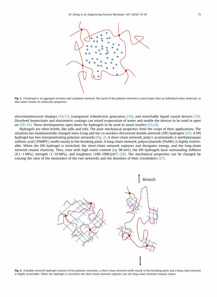
Fig. 1. A hydrogel is an aggregate of water and a polymer network. The mesh of the polymer network is much larger than an individual water molecule, sothat water retains its molecular properties.
W. Zhang et al. / Engineering Fracture Mechanics 187 (2018) 74–93 75
electroluminescent displays [16,17], transparent triboelectric generators [18], and stretchable liquid crystal devices [19].Dissolved humectants and elastomeric coatings can retard evaporation of water and enable the devices to be used in openair [20–23]. These developments open doors for hydrogels to be used in smart textiles [23,24].
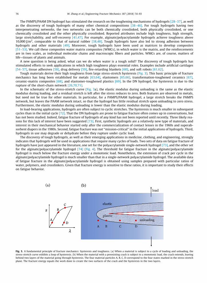
Hydrogels are often brittle, like jello and tofu. The poor mechanical properties limit the scope of their applications. Thesituation has fundamentally changed since Gong and her co-workers discovered double-network (DN) hydrogels [25]. A DNhydrogel has two interpenetrating polymer networks (Fig. 2). A short-chain network, poly(1-acrylanmido-2-methylpropanesulfonic acid) (PAMPS), swells nearly to the breaking point. A long-chain network, polyacrylamide (PAAM), is highly stretch-able. When the DN hydrogel is stretched, the short-chain network ruptures and dissipates energy, and the long-chainnetwork retains elasticity. Thus, even with high water content (ca. 90 wt%), the DN hydrogels have outstanding stiffness(0.1–1 MPa), strength (1–10 MPa), and toughness (100–1000 J/m2) [26]. The mechanical properties can be changed byvarying the ratio of the monomers of the two networks and the densities of their crosslinkers [27].
Fig. 2. A double-network hydrogel consists of two polymer networks: a short-chain network swells nearly to the breaking point, and a long-chain networkis highly stretchable. When the hydrogel is stretched, the short-chain network ruptures, but the long-chain network remains elastic.
76 W. Zhang et al. / Engineering Fracture Mechanics 187 (2018) 74–93
The PAMPS/PAAM DN hydrogel has stimulated the research on the toughening mechanisms of hydrogels [28–37], as wellas the discovery of tough hydrogels of many other chemical compositions [38–46]. For tough hydrogels having twointerpenetrating networks, the two networks can be both chemically crosslinked, both physically crosslinked, or onechemically crosslinked and the other physically crosslinked. Reported attributes include high toughness, high strength,large stretchability, and self-recovery [41,47]. For example, alginate/polyacrylamide hydrogels achieve toughness above10,000 J/m2, comparable to that of natural rubber [38,48]. Tough hydrogels have also led to strong adhesion betweenhydrogels and other materials [49]. Moreover, tough hydrogels have been used as matrices to develop composites[51–53]. We call these composites water matrix composites (WMCs), in which water is the matrix, and the reinforcementsare in two scales, as individual molecular chains and macroscopic fibers and particles. WMCs are, of course, matters oflife—tissues of plants and animals.
A new question is being asked, what can we do when water is a tough solid? The discovery of tough hydrogels hasstimulated efforts to seek applications in which high toughness plays essential roles. Examples include artificial cartilages[54–57], tissue adhesives [58], drug delivery [59], fire-retarding blankets [60], and soft robots [61,62].
Tough materials derive their high toughness from large stress-stretch hysteresis (Fig. 3). This basic principle of fracturemechanics has long been established for metals [63,64], elastomers [65,66], transformation-toughened ceramics [67],ceramic-matrix composites [68], and elastomer-toughened plastics [69]. In the DN hydrogel, the hysteresis is due to therupture of the short-chain network [26,70,71].
In the schematic of the stress-stretch curve (Fig. 3a), the elastic modulus during unloading is the same as the elasticmodulus during loading, and a residual stretch is left after the stress reduces to zero. Both features are observed in metals,but need not be true for other materials. In particular, for a PAMPS/PAAM hydrogel, a large stretch breaks the PAMPSnetwork, but leaves the PAAM network intact, so that the hydrogel has little residual stretch upon unloading to zero stress.Furthermore, the elastic modulus during unloading is lower than the elastic modulus during loading.
In load-bearing applications, hydrogels are often subject to cyclic stretches. The hysteresis is much smaller in subsequentcycles than in the initial cycle [72]. That the DN hydrogels are prone to fatigue fracture often comes up in conversations, buthas not been studied. Indeed, fatigue fracture of hydrogels of any kind has not been reported until recently. Three likely rea-sons for this lack of interest have been suggested [73]. First, synthetic hydrogels are a relatively new type of materials, andinterest in their mechanical behavior started only after the commercialization of contact lenses in the 1960s and superab-sorbent diapers in the 1980s. Second, fatigue fracture was not ‘‘mission-critical’’ in the initial applications of hydrogels. Third,hydrogels in use may degrade or dehydrate before they rupture under cyclic load.
The discovery of tough hydrogels, as well as their emerging applications in medicine, clothing, and engineering, stronglyindicates that hydrogels will be used in applications that require many cycles of loads. Two sets of data on fatigue fracture ofhydrogels have just appeared in the literature, one set for the polyacrylamide single-network hydrogel [73], and the other setfor the alginate/polyacrylamide hydrogel [74] (Fig. 4). The threshold for fatigue fracture in the alginate/polyacrylamidehydrogel is much below the fracture energy under a monotonic load. Nonetheless, the extension of crack per cycle in thealginate/polyacrylamide hydrogel is much smaller than that in a single-network polyacrylamide hydrogel. The available dataof fatigue fracture in the alginate/polyacrylamide hydrogel is obtained using samples prepared with particular ratios ofwater, polymers, and crosslinkers. Given that fracture energy is sensitive to these ratios, it is important to study their effectson fatigue behavior.
Fig. 3. A fundamental principle of fracture mechanics: hysteresis and toughness. (a) When a material is subject to a cycle of loading and unloading, thestress-stretch curve exhibits a loop of hysteresis. (b) When the material with a preexisting crack is subject to a monotonic load, the crack extends, leavingbehind two layers of the material going through hysteresis. The four material particles A, B, C, D correspond to the four states marked in the stress-stretchplane. The fracture energy equals the work done to create the two surfaces of the crack and the hysteresis in the two layers.
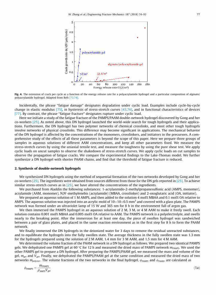
Fig. 4. The extension of crack per cycle as a function of the energy release rate for a polyacrylamide hydrogel and a particular composition of alginate/polyacrylamide hydrogel. Adapted from Refs [73,74].
W. Zhang et al. / Engineering Fracture Mechanics 187 (2018) 74–93 77
Incidentally, the phrase ‘‘fatigue damage” designates degradation under cyclic load. Examples include cycle-by-cyclechange in elastic modulus [75], in hysteresis of stress-stretch curves [43,76], and in functional characteristics of devices[77]. By contrast, the phrase ‘‘fatigue fracture” designates rupture under cyclic load.
Here we initiate a study of the fatigue fracture of the PAMPS/PAAM double-network hydrogel discovered by Gong and herco-workers [25]. As noted above, this DN hydrogel launched the world-wide search for tough hydrogels and their applica-tions. Furthermore, the DN hydrogel has two polymer networks of chemical crosslinks, and most other tough hydrogelsinvolve networks of physical crosslinks. This difference may become significant in applications. The mechanical behaviorof the DN hydrogel is affected by the concentrations of the monomers, crosslinkers, and initiators in the precursors. A com-prehensive study of the effects of all these parameters is beyond the scope of this paper. Here we prepare three groups ofsamples in aqueous solutions of different AAM concentrations, and keep all other parameters fixed. We measure thestress-stretch curves by using the uniaxial tensile test, and measure the toughness by using the pure shear test. We applycyclic loads on uncut samples to observe the shakedown of stress-stretch curves. We apply cyclic loads on cut samples toobserve the propagation of fatigue cracks. We compare the experimental findings to the Lake-Thomas model. We furthersynthesize a DN hydrogel with shorter PAAM chains, and find that the threshold of fatigue fracture is reduced.
2. Synthesis of double-network hydrogels
We synthesized DN hydrogels using the method of sequential formation of the two networks developed by Gong and herco-workers [25]. The ingredients were obtained from sources different from those for the DN gels reported in [25]. To achievesimilar stress-stretch curves as in [25], we have altered the concentrations of the ingredients.
We purchased from Aladdin the following substances: 1-acrylanmido-2-methylpropanesulfonic acid (AMPS, monomer),acrylamide (AAM, monomer), N,N0-methylenebis (acrylamide) (MBAA, crosslinker) and 2-oxoglutaric acid (OA, initiator).
We prepared an aqueous solution of 1 M AMPS, and then added to the solution 4 mol% MBAA and 0.1 mol% OA relative toAMPS. The aqueous solution was injected into an acrylic mold of 10�10�0.5 mm3 and covered with a glass plate. The PAMPSnetwork was formed under an ultraviolet lamp of 15 W and 365 nm for 8 h in the environment full of argon gas.
We then immersed the PAMPS hydrogel in an aqueous solution of 2 M, 3 M, or 4 M AAM to make it freely swell. Eachsolution contains 0.001 mol% MBAA and 0.005 mol% OA relative to AAM. The PAMPS network is a polyelectrolyte, and swellsnearly to the breaking point. After the immersion for at least one day, the piece of swollen hydrogel was sandwichedbetween a pair of glass plates, and placed in the same reaction environment as in the first step for 8 h to form the PAAMnetwork.
We finally immersed the DN hydrogels in the deionized water for 3 days to remove the residual unreacted substances,and to equilibrate the hydrogels into the fully swollen state. The average thickness in the fully swollen state was 1.3 mmfor the hydrogels prepared using the solution of 2 M AAM, 1.4 mm for 3 M AAM, and 1.5 mm for 4 M AAM.
We determined the volume fraction of the PAAM network in a DN hydrogel as follows. We prepared two identical PAMPSgels. We dehydrated one PAMPS gel at 60 �C for 12 h and measured the dried mass of PAMPS network mPAMPS. We used theother PAMPS gel to prepare a DN hydrogel. After preparing the PAMPS/PAAM gel, we measured the mass and volume of thegel, mgel and Vgel. Finally, we dehydrated the PAMPS/PAAM gel at the same condition and measured the dried mass of twonetworks mpolymer . The volume fractions of the two networks in the final hydrogel, /PAMPS and /PAAM , are calculated as
/PAMPS ¼mPAMPS
qAMPSVgelð1aÞ
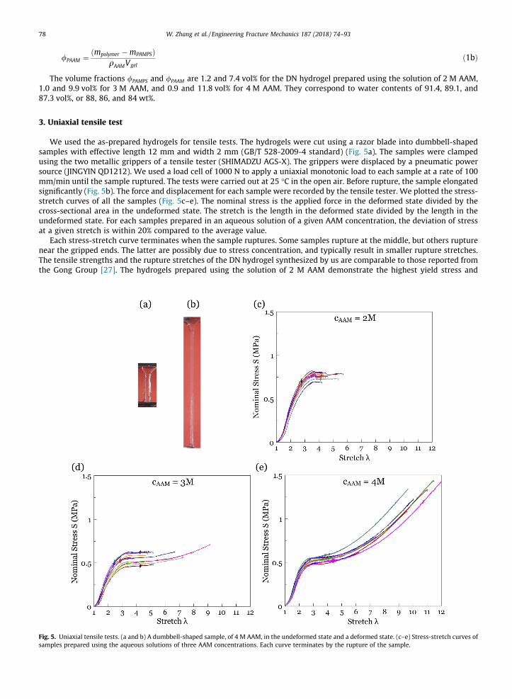
Fig. 5.sample
78 W. Zhang et al. / Engineering Fracture Mechanics 187 (2018) 74–93
/PAAM ¼ ðmpolymer �mPAMPSÞqAAMVgel
ð1bÞ
The volume fractions /PAMPS and /PAAM are 1.2 and 7.4 vol% for the DN hydrogel prepared using the solution of 2 M AAM,1.0 and 9.9 vol% for 3 M AAM, and 0.9 and 11.8 vol% for 4 M AAM. They correspond to water contents of 91.4, 89.1, and87.3 vol%, or 88, 86, and 84 wt%.
3. Uniaxial tensile test
We used the as-prepared hydrogels for tensile tests. The hydrogels were cut using a razor blade into dumbbell-shapedsamples with effective length 12 mm and width 2 mm (GB/T 528-2009-4 standard) (Fig. 5a). The samples were clampedusing the two metallic grippers of a tensile tester (SHIMADZU AGS-X). The grippers were displaced by a pneumatic powersource (JINGYIN QD1212). We used a load cell of 1000 N to apply a uniaxial monotonic load to each sample at a rate of 100mm/min until the sample ruptured. The tests were carried out at 25 �C in the open air. Before rupture, the sample elongatedsignificantly (Fig. 5b). The force and displacement for each sample were recorded by the tensile tester. We plotted the stress-stretch curves of all the samples (Fig. 5c–e). The nominal stress is the applied force in the deformed state divided by thecross-sectional area in the undeformed state. The stretch is the length in the deformed state divided by the length in theundeformed state. For each samples prepared in an aqueous solution of a given AAM concentration, the deviation of stressat a given stretch is within 20% compared to the average value.
Each stress-stretch curve terminates when the sample ruptures. Some samples rupture at the middle, but others rupturenear the gripped ends. The latter are possibly due to stress concentration, and typically result in smaller rupture stretches.The tensile strengths and the rupture stretches of the DN hydrogel synthesized by us are comparable to those reported fromthe Gong Group [27]. The hydrogels prepared using the solution of 2 M AAM demonstrate the highest yield stress and
Uniaxial tensile tests. (a and b) A dumbbell-shaped sample, of 4 M AAM, in the undeformed state and a deformed state. (c–e) Stress-stretch curves ofs prepared using the aqueous solutions of three AAM concentrations. Each curve terminates by the rupture of the sample.
W. Zhang et al. / Engineering Fracture Mechanics 187 (2018) 74–93 79
rupture after yield. The hydrogels prepared using the solution of 3 M AAM yield at a lower stress but a larger stretch in aver-age. The hydrogels prepared using the solution of 4 M AAM show pronounced strain stiffening, with the highest strength andthe rupture stretch.
The shear modulus of each sample is obtained by fitting the beginning portion of its stress-stretch curve to the neo-Hookean model [78]:
Fig. 6.subjecthydrogcrack b
s ¼ lðk� k�2Þ; ð2Þ
where s is the nominal stress, k is the stretch, and l is the shear modulus. The shear modulus for all hydrogels prepared inthis work is roughly 100 kPa (see Appendix A for fitting). As expected, the double-network hydrogels do not follow the neo-Hookean model. Our intention here is to obtain a rough value for the small-strain shear modulus for the materials prepared.This shear modulus will not be used in any of the following calculations.
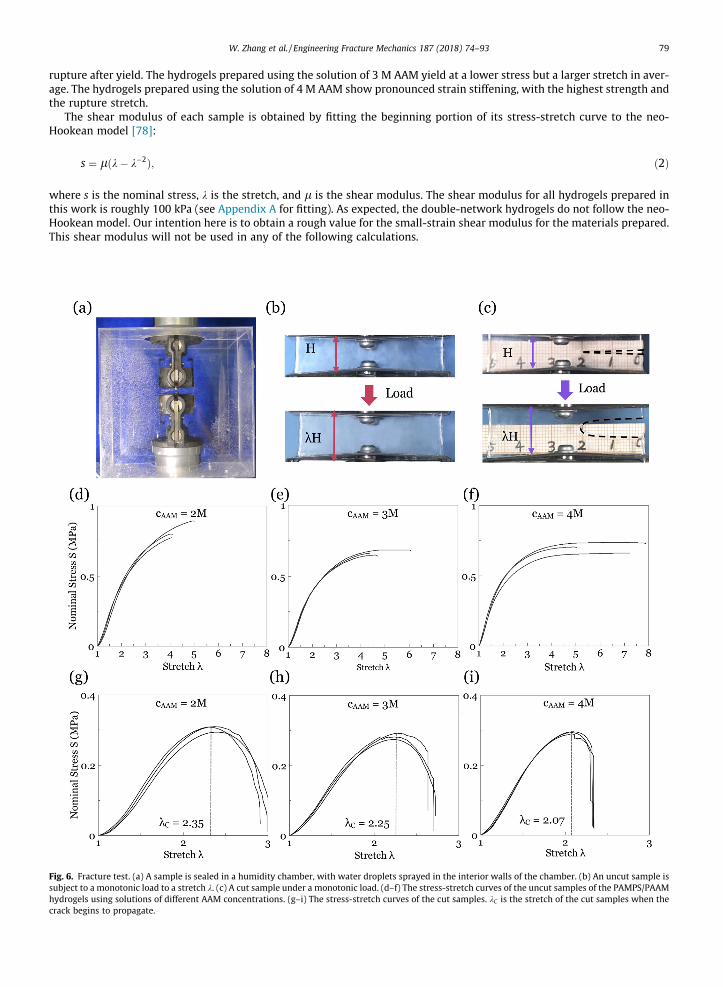
Fracture test. (a) A sample is sealed in a humidity chamber, with water droplets sprayed in the interior walls of the chamber. (b) An uncut sample isto a monotonic load to a stretch k. (c) A cut sample under a monotonic load. (d–f) The stress-stretch curves of the uncut samples of the PAMPS/PAAMels using solutions of different AAM concentrations. (g–i) The stress-stretch curves of the cut samples. kC is the stretch of the cut samples when theegins to propagate.
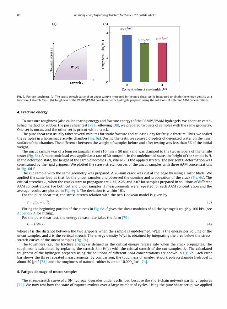
Fig. 7. Facture toughness. (a) The stress-stretch curve of an uncut sample measured in the pure shear test is integrated to obtain the energy density as afunction of stretch, WðkÞ. (b) Toughness of the PAMPS/PAAM double-network hydrogels prepared using the solutions of different AAM concentrations.
80 W. Zhang et al. / Engineering Fracture Mechanics 187 (2018) 74–93
4. Fracture energy
To measure toughness (also called tearing energy and fracture energy) of the PAMPS/PAAM hydrogels, we adopt an estab-lished method for rubber, the pure shear test [79]. Following [38], we prepared two sets of samples with the same geometry.One set is uncut, and the other set is precut with a crack.
The pure shear test usually takes several minutes for static fracture and at least 1 day for fatigue fracture. Thus, we sealedthe samples in a homemade acrylic chamber (Fig. 6a). During the tests, we sprayed droplets of deionized water on the innersurface of the chamber. The difference between the weight of samples before and after testing was less than 5% of the initialweight.
The uncut sample was of a long rectangular sheet (10 mm � 50 mm) and was clamped to the two grippers of the tensiletester (Fig. 6b). A monotonic load was applied at a rate of 30 mm/min. In the undeformed state, the height of the sample is H.In the deformed state, the height of the sample becomes kH, where k is the applied stretch. The horizontal deformation wasconstrained by the rigid grippers. We plotted the stress-stretch curves of the uncut samples with three AAM concentrationsin Fig. 6d-f.
The cut sample with the same geometry was prepared. A 20-mm crack was cut at the edge by using a razor blade. Weapplied the same load as that for the uncut samples and observed the opening and propagation of the crack (Fig. 6c). Thecritical stretches kC when the cracks start to propagate are 2.35, 2.25, and 2.07 for samples prepared in solutions of differentAAM concentrations. For both cut and uncut samples, 3 measurements were repeated for each AAM concentration and theaverage results are plotted in Fig. 6g–i. The deviation is within 10%.
For the pure shear test, the stress-stretch relation with the neo-Hookean model is given by
s ¼ lðk� k�3Þ; ð3Þ
Fitting the beginning portion of the curves in Fig. 6d–f gives the shear modulus of all the hydrogels roughly 100 kPa (seeAppendix A for fitting).For the pure shear test, the energy release rate takes the form [79].
G ¼ HWðkÞ ð4Þ
where H is the distance between the two grippers when the sample is undeformed, WðkÞ is the energy per volume of theuncut samples, and k is the vertical stretch. The energy density WðkÞ is obtained by integrating the area below the stress-stretch curves of the uncut samples (Fig. 7a).The toughness (i.e., the fracture energy) is defined as the critical energy release rate when the crack propagates. Thetoughness is calculated by replacing the stretch k in WðkÞ with the critical stretch of the cut samples, kC . The calculatedtoughness of the hydrogels prepared using the solutions of different AAM concentrations are shown in Fig. 7b. Each errorbar shows the three repeated measurements. By comparison, the toughness of single-network polyacrylamide hydrogel isabout 50 J/m2 [73], and the toughness of natural rubber is about 10,000 J/m2 [74].
5. Fatigue damage of uncut samples
The stress-stretch curve of a DN hydrogel degrades under cyclic load because the short-chain network partially ruptures[72]. We now test how the state of rupture evolves over a large number of cycles. Using the pure shear setup, we applied
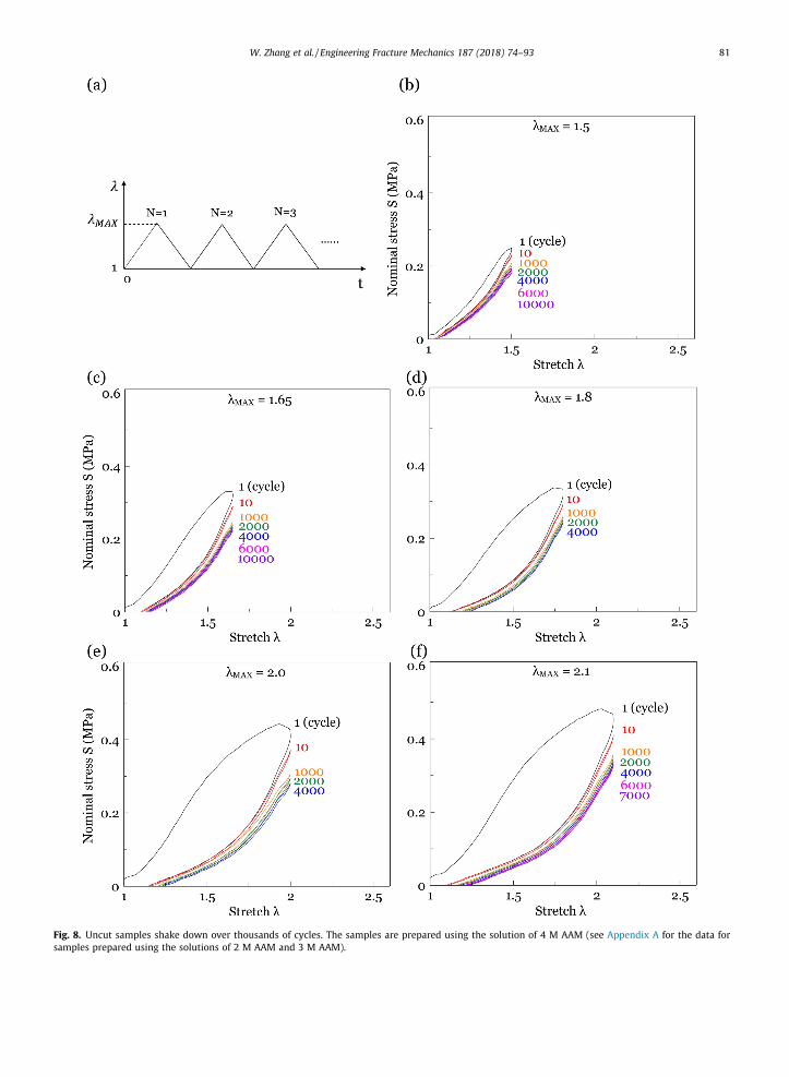
Fig. 8. Uncut samples shake down over thousands of cycles. The samples are prepared using the solution of 4 M AAM (see Appendix A for the data forsamples prepared using the solutions of 2 M AAM and 3 M AAM).
W. Zhang et al. / Engineering Fracture Mechanics 187 (2018) 74–93 81
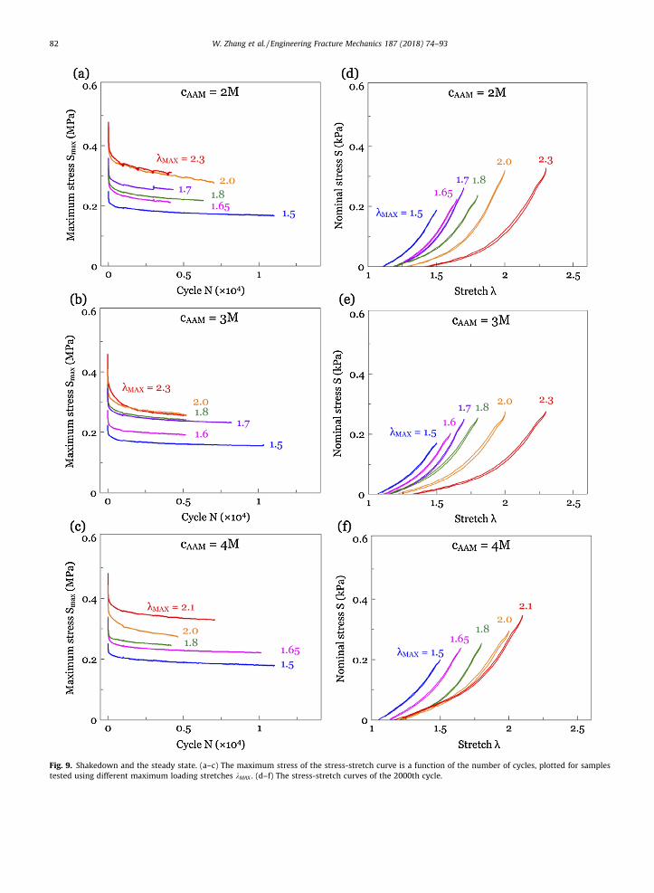
Fig. 9. Shakedown and the steady state. (a–c) The maximum stress of the stress-stretch curve is a function of the number of cycles, plotted for samplestested using different maximum loading stretches kMAX . (d–f) The stress-stretch curves of the 2000th cycle.
82 W. Zhang et al. / Engineering Fracture Mechanics 187 (2018) 74–93
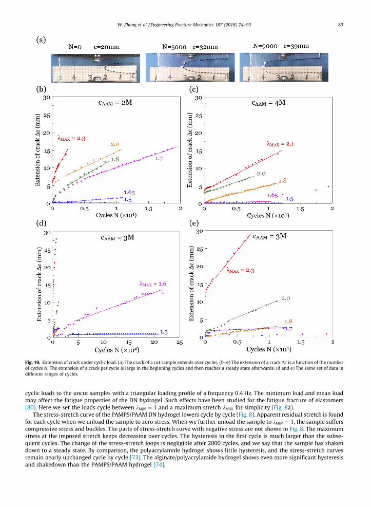
Fig. 10. Extension of crack under cyclic load. (a) The crack of a cut sample extends over cycles. (b–e) The extension of a crack Dc is a function of the numberof cycles N. The extension of a crack per cycle is large in the beginning cycles and then reaches a steady state afterwards. (d and e) The same set of data indifferent ranges of cycles.
W. Zhang et al. / Engineering Fracture Mechanics 187 (2018) 74–93 83
cyclic loads to the uncut samples with a triangular loading profile of a frequency 0.4 Hz. The minimum load and mean loadmay affect the fatigue properties of the DN hydrogel. Such effects have been studied for the fatigue fracture of elastomers[80]. Here we set the loads cycle between kMIN ¼ 1 and a maximum stretch kMAX for simplicity (Fig. 8a).
The stress-stretch curve of the PAMPS/PAAM DN hydrogel lowers cycle by cycle (Fig. 8). Apparent residual stretch is foundfor each cycle when we unload the sample to zero stress. When we further unload the sample to kMIN ¼ 1, the sample sufferscompressive stress and buckles. The parts of stress-stretch curve with negative stress are not shown in Fig. 8. The maximumstress at the imposed stretch keeps decreasing over cycles. The hysteresis in the first cycle is much larger than the subse-quent cycles. The change of the stress-stretch loops is negligible after 2000 cycles, and we say that the sample has shakendown to a steady state. By comparison, the polyacrylamide hydrogel shows little hysteresis, and the stress-stretch curvesremain nearly unchanged cycle by cycle [73]. The alginate/polyacrylamide hydrogel shows even more significant hysteresisand shakedown than the PAMPS/PAAM hydrogel [74].
84 W. Zhang et al. / Engineering Fracture Mechanics 187 (2018) 74–93
The shakedown phenomenon has been observed and studied in elastic-plastic materials, such as metals [83–89]. Theshakedown of hydrogels over a large number cycles was reported for the alginate/polyacrylamide hydrogel [74], and nowfor the PAMPS/PAAM hydrogel. For the PAMPS/PAAM hydrogel, the first polymer network PAMPS is near the breaking point.Under cyclic loads, the PAMPS network partially ruptures into small clusters. The damage of the PAMPS network leads to thehysteresis between the loading and unloading curves. Our data suggests that each subsequent cycle incrementally involvesmore rupture of the PAMPS network, until a steady-state is reached.
The maximum stress drops greatly in the beginning cycles and reaches a steady state after thousands of cycles (Fig. 9a–c).Following [74], we use the stress-stretch curve of the 2000th cycle to represent the deformation behavior of the PAMPS/PAAM hydrogels under cyclic load in the steady state (Fig. 9d–f).
6. Fatigue facture of cut samples
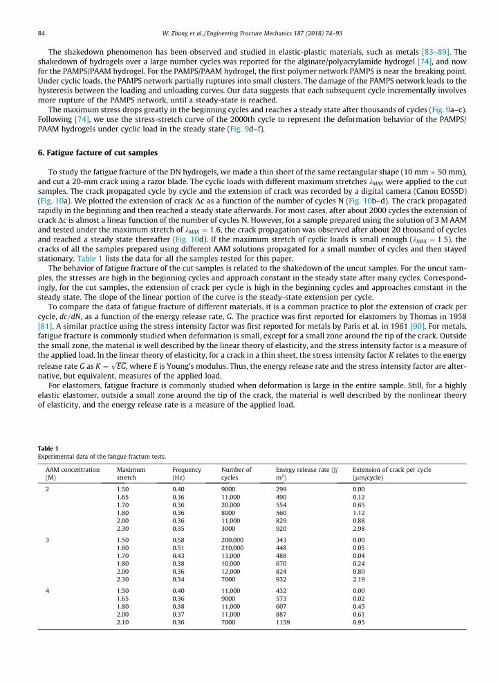
To study the fatigue fracture of the DN hydrogels, we made a thin sheet of the same rectangular shape (10 mm � 50 mm),and cut a 20-mm crack using a razor blade. The cyclic loads with different maximum stretches kMAX were applied to the cutsamples. The crack propagated cycle by cycle and the extension of crack was recorded by a digital camera (Canon EOS5D)(Fig. 10a). We plotted the extension of crack Dc as a function of the number of cycles N (Fig. 10b–d). The crack propagatedrapidly in the beginning and then reached a steady state afterwards. For most cases, after about 2000 cycles the extension ofcrack Dc is almost a linear function of the number of cycles N. However, for a sample prepared using the solution of 3 M AAMand tested under the maximum stretch of kMAX ¼ 1:6, the crack propagation was observed after about 20 thousand of cyclesand reached a steady state thereafter (Fig. 10d). If the maximum stretch of cyclic loads is small enough (kMAX ¼ 1:5), thecracks of all the samples prepared using different AAM solutions propagated for a small number of cycles and then stayedstationary. Table 1 lists the data for all the samples tested for this paper.
The behavior of fatigue fracture of the cut samples is related to the shakedown of the uncut samples. For the uncut sam-ples, the stresses are high in the beginning cycles and approach constant in the steady state after many cycles. Correspond-ingly, for the cut samples, the extension of crack per cycle is high in the beginning cycles and approaches constant in thesteady state. The slope of the linear portion of the curve is the steady-state extension per cycle.
To compare the data of fatigue fracture of different materials, it is a common practice to plot the extension of crack percycle, dc=dN, as a function of the energy release rate, G. The practice was first reported for elastomers by Thomas in 1958[81]. A similar practice using the stress intensity factor was first reported for metals by Paris et al. in 1961 [90]. For metals,fatigue fracture is commonly studied when deformation is small, except for a small zone around the tip of the crack. Outsidethe small zone, the material is well described by the linear theory of elasticity, and the stress intensity factor is a measure ofthe applied load. In the linear theory of elasticity, for a crack in a thin sheet, the stress intensity factor K relates to the energyrelease rate G as K ¼
ffiffiffiffiffiffi
EGp
, where E is Young’s modulus. Thus, the energy release rate and the stress intensity factor are alter-native, but equivalent, measures of the applied load.
For elastomers, fatigue fracture is commonly studied when deformation is large in the entire sample. Still, for a highlyelastic elastomer, outside a small zone around the tip of the crack, the material is well described by the nonlinear theoryof elasticity, and the energy release rate is a measure of the applied load.
Table 1Experimental data of the fatigue fracture tests.
AAM concentration(M)
Maximumstretch
Frequency(Hz)
Number ofcycles
Energy release rate (J/m2)
Extension of crack per cycle(lm/cycle)
2 1.50 0.40 9000 299 0.001.65 0.36 11,000 490 0.121.70 0.36 20,000 554 0.651.80 0.36 8000 560 1.122.00 0.36 11,000 829 0.882.30 0.35 3000 920 2.98
3 1.50 0.58 200,000 343 0.001.60 0.51 210,000 448 0.051.70 0.43 13,000 488 0.041.80 0.38 10,000 670 0.242.00 0.36 12,000 824 0.802.30 0.34 7000 932 2.19
4 1.50 0.40 11,000 432 0.001.65 0.36 9000 573 0.021.80 0.38 11,000 607 0.452.00 0.37 11,000 887 0.612.10 0.36 7000 1159 0.95
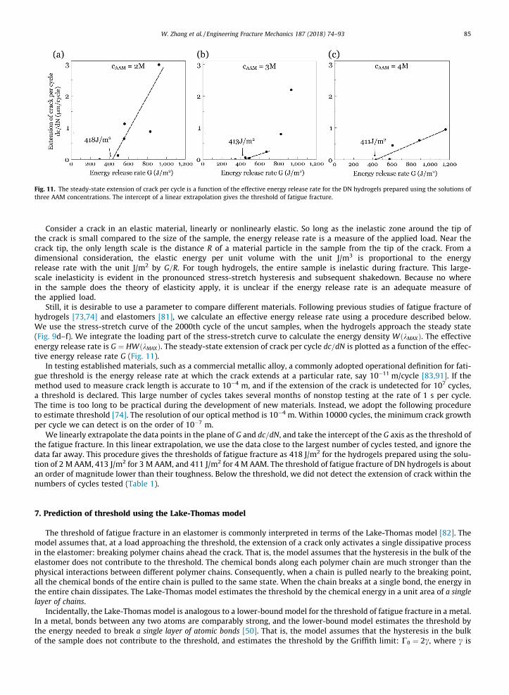
Fig. 11. The steady-state extension of crack per cycle is a function of the effective energy release rate for the DN hydrogels prepared using the solutions ofthree AAM concentrations. The intercept of a linear extrapolation gives the threshold of fatigue fracture.
W. Zhang et al. / Engineering Fracture Mechanics 187 (2018) 74–93 85
Consider a crack in an elastic material, linearly or nonlinearly elastic. So long as the inelastic zone around the tip ofthe crack is small compared to the size of the sample, the energy release rate is a measure of the applied load. Near thecrack tip, the only length scale is the distance R of a material particle in the sample from the tip of the crack. From adimensional consideration, the elastic energy per unit volume with the unit J/m3 is proportional to the energyrelease rate with the unit J/m2 by G=R. For tough hydrogels, the entire sample is inelastic during fracture. This large-scale inelasticity is evident in the pronounced stress-stretch hysteresis and subsequent shakedown. Because no wherein the sample does the theory of elasticity apply, it is unclear if the energy release rate is an adequate measure ofthe applied load.
Still, it is desirable to use a parameter to compare different materials. Following previous studies of fatigue fracture ofhydrogels [73,74] and elastomers [81], we calculate an effective energy release rate using a procedure described below.We use the stress-stretch curve of the 2000th cycle of the uncut samples, when the hydrogels approach the steady state(Fig. 9d–f). We integrate the loading part of the stress-stretch curve to calculate the energy density WðkMAXÞ. The effectiveenergy release rate is G ¼ HWðkMAXÞ. The steady-state extension of crack per cycle dc=dN is plotted as a function of the effec-tive energy release rate G (Fig. 11).
In testing established materials, such as a commercial metallic alloy, a commonly adopted operational definition for fati-gue threshold is the energy release rate at which the crack extends at a particular rate, say 10�11 m/cycle [83,91]. If themethod used to measure crack length is accurate to 10�4 m, and if the extension of the crack is undetected for 107 cycles,a threshold is declared. This large number of cycles takes several months of nonstop testing at the rate of 1 s per cycle.The time is too long to be practical during the development of new materials. Instead, we adopt the following procedureto estimate threshold [74]. The resolution of our optical method is 10�4 m. Within 10000 cycles, the minimum crack growthper cycle we can detect is on the order of 10�7 m.
We linearly extrapolate the data points in the plane of G and dc=dN, and take the intercept of the G axis as the threshold ofthe fatigue fracture. In this linear extrapolation, we use the data close to the largest number of cycles tested, and ignore thedata far away. This procedure gives the thresholds of fatigue fracture as 418 J/m2 for the hydrogels prepared using the solu-tion of 2 M AAM, 413 J/m2 for 3 M AAM, and 411 J/m2 for 4 M AAM. The threshold of fatigue fracture of DN hydrogels is aboutan order of magnitude lower than their toughness. Below the threshold, we did not detect the extension of crack within thenumbers of cycles tested (Table 1).
7. Prediction of threshold using the Lake-Thomas model
The threshold of fatigue fracture in an elastomer is commonly interpreted in terms of the Lake-Thomas model [82]. Themodel assumes that, at a load approaching the threshold, the extension of a crack only activates a single dissipative processin the elastomer: breaking polymer chains ahead the crack. That is, the model assumes that the hysteresis in the bulk of theelastomer does not contribute to the threshold. The chemical bonds along each polymer chain are much stronger than thephysical interactions between different polymer chains. Consequently, when a chain is pulled nearly to the breaking point,all the chemical bonds of the entire chain is pulled to the same state. When the chain breaks at a single bond, the energy inthe entire chain dissipates. The Lake-Thomas model estimates the threshold by the chemical energy in a unit area of a singlelayer of chains.
Incidentally, the Lake-Thomas model is analogous to a lower-bound model for the threshold of fatigue fracture in a metal.In a metal, bonds between any two atoms are comparably strong, and the lower-bound model estimates the threshold bythe energy needed to break a single layer of atomic bonds [50]. That is, the model assumes that the hysteresis in the bulkof the sample does not contribute to the threshold, and estimates the threshold by the Griffith limit: C0 ¼ 2c, where c is
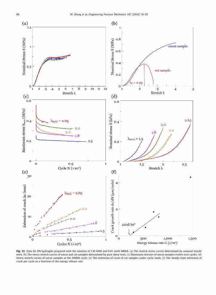
Fig. 12. Data for DN hydrogels prepared with the solution of 2 M AAM and 0.01 mol% MBAA. (a) The stretch-stress curves determined by uniaxial tensiletests. (b) The stress-stretch curves of uncut and cut samples determined by pure shear tests. (c) Maximum stresses of uncut samples evolve over cycles. (d)Stress-stretch curves of uncut samples at the 2000th cycle. (e) The extension of crack of cut samples under cyclic loads. (f) The steady-state extension ofcrack per cycle as a function of the energy release rate.
86 W. Zhang et al. / Engineering Fracture Mechanics 187 (2018) 74–93
W. Zhang et al. / Engineering Fracture Mechanics 187 (2018) 74–93 87
the surface energy. The surface energy of a metal is on the order of 1 J/m2. This model provides a lower bound. The exper-imental values of the threshold for metals are in the range 10–1000 J/m2.
The Lake-Thomas model has been adapted to a single-network PAAM hydrogel [73]. For the PAMPS/PAAM double-network hydrogels, we now hypothesize that the rupture of the PAMPS network does not contribute to the threshold,and estimate the threshold by the chemical energy in one layer of PAAM chains:
C0 ¼ /23PAAM
ffiffiffi
np
bUl ð5Þ
where /PAAM is the volume fraction of the PAAM network in the DN hydrogel, n is the number of monomer units in a PAAMchain, b is the number of bonds per unit volume of the dry polymer, U is CAC bond energy, and l is the length of each mono-mer unit.
We use the values of /PAAM determined by the experiments described above. The number of single bonds per unit volumeof the polymer is estimated by the number of monomers per unit volume of the dry polymer, b ¼ Aq
m ¼ 1:12� 1028m�3, wherem is the molecular weight of acrylamide (71.08 g/mole), q is the density of acrylamide (1.322 g/cm3), and A is the Avogadro
number (6:022� 1023). The length of the monomer is estimated by l ¼ b�13 ¼ 0:446 nm. The energy of a CAC bond is
U ¼ 3:3� 10�19 J [82]. The molar ratio of the crosslinker MBAA to monomer AAM is set to be 0.001 mol%, and we estimatethe number of monomers between two crosslinks by n ¼ 1=0:001 mol% ¼ 105. The threshold of fatigue fracture for PAMPS/PAAM hydrogels prepared using the solution of 2 M AAM is predicted to be 92 J/m2, 112 J/m2 for 3 M AAM, and 125 J/m2 for4 M AAM.
Fig. A1. Fit the beginning part of uniaxial stress-stretch curve by Eq. (2) with neo-Hookean model.
88 W. Zhang et al. / Engineering Fracture Mechanics 187 (2018) 74–93
8. Effect of the length of PAAM chains
The Lake-Thomas model adapted to the DN hydrogel gives a prediction of correct order of magnitude. Here we do notpush for better agreement. Instead, we ascertain if the threshold does change if we change the density of crosslinkers ofthe PAAM network. We prepared another set of samples of DN hydrogels. The formulation of the PAMPS network is the sameas before. We immersed the PAMPS network in a solution of 2 M AAM, with 0.01 mol% MBAA and 0.01 mol% OA relative toAAM. That is, the density of the crosslinkers for the PAAM network is 10 times that used in the above experiments. In thefully swollen DN hydrogel, the volume fractions of PAMPS, PAAM, and water are respectively, 1.1, 7.5, and 91 vol%. Werepeated the same experiments for the samples of this composition (Fig. 12). The fracture energy and the fatigue thresholdof this set of samples are 3066 J/m2 and 220 J/m2. Recall that the fracture energy and the fatigue threshold for the hydrogelsusing the solution of 2 M AAMwith 0.001 mol% MBAA is 3779 J/m2 and 418 J/m2. The Lake-Thomas model predicts that if thecrosslinker density of PAAM increases by ten times, the number of monomers between two crosslinks decreases to one tenth,resulting in a decrease of threshold by a factor of
ffiffiffiffiffiffi
10p
. This prediction is captured in our experiments qualitatively, but notquantitatively.
The number of monomers between two crosslinks is roughly n ¼ 7000 for both PAAM gel and alginate/PAAM gel in Refs.[73,74], and is n ¼ 105 for DN gels prepared in the solution of 10�5 number fraction of MBAA relative to AAM in this work.The corresponding fatigue threshold is 7 J/m2 for pure PAAM gel, 53 J/m2 for alginate/PAAM gel, and 400 J/m2 for DN gel. Wecan see that the comparisons of fatigue threshold from different gel systems do not simply support the assumption that thesacrificial network has no contribution. The detailed reasons are still unknown and are worthy of further investigations.
9. Concluding remarks
This paper is the first study of the fatigue of the classic PAMPS/PAAM double network hydrogels. We apply cyclic stretchto samples with or without cuts. For an uncut sample, damage accumulates over thousands of cycles, until a steady state is
Fig. A2. Fit the beginning part of pure shear stress-stretch curve by Eq. (3) with neo-Hookean model.
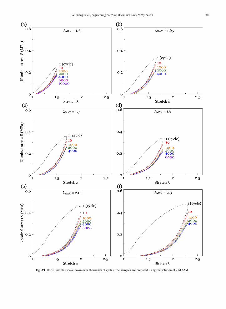
Fig. A3. Uncut samples shake down over thousands of cycles. The samples are prepared using the solution of 2 M AAM.
W. Zhang et al. / Engineering Fracture Mechanics 187 (2018) 74–93 89
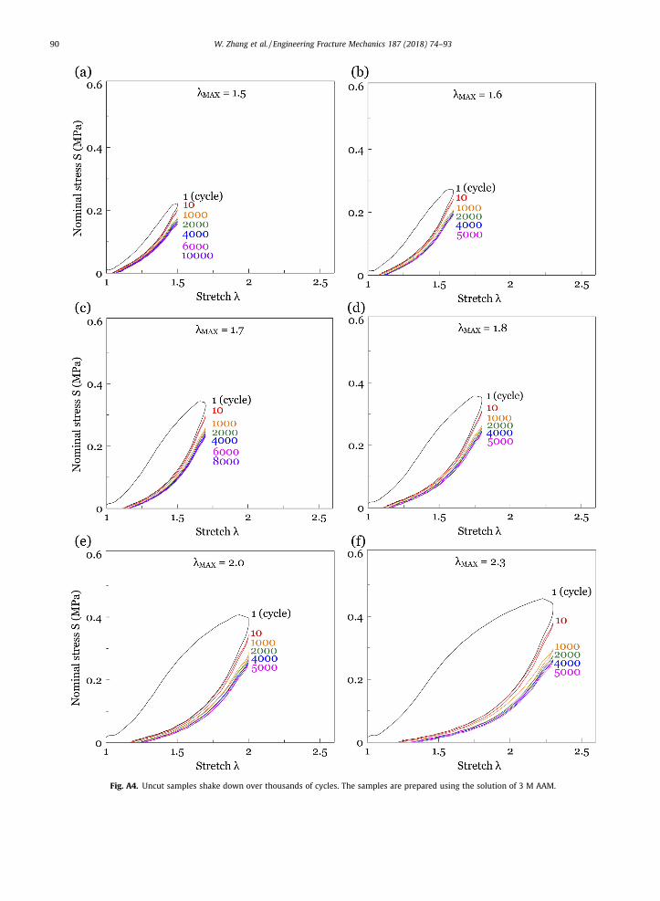
Fig. A4. Uncut samples shake down over thousands of cycles. The samples are prepared using the solution of 3 M AAM.
90 W. Zhang et al. / Engineering Fracture Mechanics 187 (2018) 74–93
W. Zhang et al. / Engineering Fracture Mechanics 187 (2018) 74–93 91
reached. For a cut sample, the crack extends cycle by cycle if the amplitude of the load is above a threshold. We prepare allsamples of the DN hydrogels using PAMPS hydrogels of a fixed composition, but using aqueous solutions of various concen-trations of AAM monomers and MBAA crosslinkers. Within the samples prepared for this work, the effect of the concentra-tion of AAM is modest. The concentration of MBAA has a small effect on fracture energy, but significant effect on thresholdenergy. For the DN hydrogels prepared in the aqueous solution of 2 M AAM and 10�5 number fraction of MBAA relative toAAM, the fracture energy is 3779 J/m2, and the fatigue threshold is 418 J/m2. For the DN hydrogels prepared in the aqueoussolution of 2 M AAM and 10�4 number fraction of MBAA relative to AAM, the fracture energy is 3066 J/m2, and the fatiguethreshold is 220 J/m2.
We adapt the Lake-Thomas model under the hypothesis that the fatigue threshold of a DN hydrogel corresponds to thechemical energy stored in one layer of PAAM chains. This hypothesis predicts the experimentally measured fatigue thresh-olds qualitatively. The available data do not allow a comprehensive comparison of the fatigue-resistance of hydrogels of dif-ferent chemistries, such as the alginate/PAAM (Fig. 4) and PAMPS/PAAM (Fig. 11), given that fatigue resistance depends onthe concentrations of ingredients. It is hoped that researchers worldwide will report the fatigue behavior of their own hydro-gels under development for load-bearing applications, and that an understanding will soon emerge to link the behavior offatigue to the chemistry of hydrogels.
Acknowledgments
ZS acknowledges the support of the NSF MRSEC at Harvard (DMR-1420570), and the visiting appointment at Xian Jiao-tong University. TL acknowledges the support of NSFC (11772249). JH acknowledges the support of NSFC (11702207). JTacknowledges the support of NSFC (11702208) and the Program for Postdoctoral Innovative Talents (No. BX201700192).
Appendix A
See Figs. A1–A4.
References
[1] Buwalda SJ, Boere KWM, Dijkstra PJ, Feijien J, Vermonden T, Hennink WE. Hydrogels in a historical perspective: from simple networks to smartmaterials. J Control Release 2014;190:254–73.
[2] Wichterle O, Lim D. Hydrophilic gels for biological use. Nature 1960;185(4706):117–8.[3] Masuda F. Trends in the development of superabsorbent polymers for diapers; 1994.[4] Li J, Mooney DJ. Designing hydrogels for controlled drug delivery. Nat Rev Mater 2016;1:16071.[5] Zhang YS, Khademhosseini A. Advances in engineering hydrogels. Science 2017;356(6337):eaaf3627.[6] Beebe DJ, Moore JS, Bauer JM, Yu Q, Liu RH, Devadoss C, et al. Functional hydrogel structures for autonomous flow control inside microfluidic channels.
Nature 2000;404(6778):588.[7] Sidorenko A, Krupenkin T, Taylor A, Fratzl P, Aizenberg J. Reversible switching of hydrogel-actuated nanostructures into complex micropatterns.
Science 2007;315(5811):487–90.[8] Keplinger C, Sun J-Y, Foo CC, Rothemund P, Whitesides GM, Suo Z. Stretchable, transparent, ionic conductors. Science 2013;341(6149):984–7.[9] Li T, Li G, Liang Y, Cheng T, Dai J, Yang X, et al. Fast-moving soft electronic fish. Sci Adv 2017;3(4):e1602045.[10] Sun J-Y, Keplinger C, Whitesides GM, Suo Z. Ionic skin. Adv Mater 2014;26(45):7608–14.[11] Robinson SS, O’Brien KW, Zhao H, Peele BN, Larson CM, Mac Murray BC, et al. Integrated soft sensors and elastomeric actuators for tactile machines
with kinesthetic sense. Extreme Mech Lett 2015;5:47–53.[12] Wirthl D, Pichler R, Drack M, Kettlguber G, Moser R, Gerstmayr R, et al. Instant tough bonding of hydrogels for soft machines and electronics. Sci Adv
2017;3(6):e1700053.[13] Yang CH, Chen B, Lu JJ, Yang JH, Zhou J, Chen YM, et al. Ionic cable. Extreme Mech Lett 2015;3:59–65.[14] Kim C-C, Lee H-H, Oh KH, Sun J-Y. Highly stretchable, transparent ionic touch panel. Science 2016;353(6300):682–7.[15] Sarwar MS, Dobashi Y, Preston C, Wyss JKM, Mirabbasi S, Madden JDW. Bend, stretch, and touch: locating a finger on an actively deformed transparent
sensor array. Sci Adv 2017;3(3):e1602200.[16] Yang CH, Chen B, Zhou J, Chen YM, Suo Z. Electroluminescence of giant stretchability. Adv Mater 2016;28(22):4480–4.[17] Larson C, Peele B, Li S, Robinson S, Totaro M, Beccai L, et al. Highly stretchable electroluminescent skin for optical signaling and tactile sensing. Science
2016;351(6277):1071–4.[18] Pu X, Liu M, Chen X, Sun J, Du C, Zhang Y, et al. Ultrastretchable, transparent triboelectric nanogenerator as electronic skin for biomechanical energy
harvesting and tactile sensing. Sci Adv 2017;3(5):e1700015.[19] Yang CH, Zhou S, Shian S, Clarke DR, Suo Z. Organic liquid-crystal devices based on ionic conductors. Mater Horiz 2017. doi: https://doi.org/10.1039/
C7MH00345E.[20] Bai Y, Chen B, Xiang F, Zhou J, Wang H, Suo Z. Transparent hydrogel with enhanced water retention capacity by introducing highly hydratable salt. Appl
Phys Lett 2014;105(15):151903.[21] Yuk H, Zhang T, Parada GA, Liu X, Zhao X. Skin-inspired hydrogel-elastomer hybrids with robust interfaces and functional microstructures. Nat
Commun 2016;7:12028.[22] Parada GA, Yuk H, Liu X, Hsieh AJ, Zhao X. Impermeable robust hydrogels via hybrid lamination. Adv Healthcare Mater 2017:1700520.[23] Le Floch P, Yao X, Liu Q, Wang Z, Nian G, Sun Y, et al. Wearable and washable conductors for active textiles. ACS Appl Mater Inter 2017;9
(30):25542–52.[24] Zhang Q, Wang X, Mu Q, Liu P, Jia S, Chen L, et al. Genipin-cross-linked silk sericin/poly (N-isopropylacrylamide) IPN hydrogels: color reaction between
silk sericin and genipin, pore shape and thermo-responsibility. Mater Chem Phys 2015;166:133–43.[25] Gong JP, Katsuyama Y, Kurokawa T, Osada Y. Double-network hydrogels with extremely high mechanical strength. Adv Mater 2003;15(14):1155–8.[26] Gong JP. Why are double network hydrogels so tough? Soft Matter 2010;6:2583–90.[27] Ahmed S, Nakajima T, Kurokawa T, Haque MA, Gong JP. Brittle-ductile transition of double network hydrogels: mechanical balance of two networks as
the key factor. Polymer 2014;55:914–23.[28] Gong JP. Materials both tough and soft. Science 2014;344(6180):161–2.

92 W. Zhang et al. / Engineering Fracture Mechanics 187 (2018) 74–93
[29] Na Y-H, Kurokaw T, Katsuyama Y, Tsukeshiba H, Gong JP, Osada Y, et al. Structural characteristics of double network gels with extremely highmechanical strength. Macromolecules 2004;37:5370–4.
[30] Nakayama A, Kakugo A, Gong JP, Osada Y, Takai M, Erata T, et al. High mechanical strength double-network hydrogel with bacterial cellulose. Adv FunctMater 2004;14(11):1124–8.
[31] Tsukeshiba H, Huang M, Na YH, Kurokawa T, Kuwabara R, Tanaka Y, et al. Effect of polymer entanglement on the toughening of double networkhydrogels. J Phys Chem B 2005;109(34):16304–9.
[32] Na Y-H, Tanaka Y, Kawauchi Y, Furukawa H, Sumiyoshi T, Gong JP, et al. Necking phenomenon of double-network gels. Macromolecules 2006;39(14):4641–5.
[33] Tirumala VR, Tominaga T, Lee S, Butler PD, Lin EK, Gong JP, et al. Molecular model for toughening in double-network hydrogels. J Phys Chem B2008;112(27):8024–31.
[34] Nakajima T, Furukawa H, Tanaka Y, Kurokawa T, Osada Y, Gong JP. True chemical structure of double network hydrogels. Macromolecules 2009;42(6):2184–9.
[35] Liang S, Wu ZL, Hu J, Kurokawa T, Yu QM, Gong JP. Direct observation on the surface fracture of ultrathin film double-network hydrogels.Macromolecules 2009;44(8):3016–20.
[36] Nakajima A, Kurokawa T, Ahmed S, Wu W-L, Gong JP. Characterization of internal fracture process of double network hydrogels under uniaxialelongation. Soft Matter 2012;9(6):1955–66.
[37] Matsuda T, Nakajima T, Fukuda Y, Hong W, Sakai T, Kurokawa T, et al. Yielding criteria of double network hydrogels. Macromolecules 2012;49(5):1865–72.
[38] Sun J-Y, Zhao X, IIIeperuma WRK, Chauhuri O, Oh KH, Mooney DJ, et al. Highly stretchable and tough hydrogels. Nature 2012;489(7414):133–6.[39] Haraguchi K, Takehisa T. Nanocomposite hydrogels: a unique organic-inorganic network structure with extraordinary mechanical, optical, and
swelling/de-swelling properties. Adv Mater 2002;14(16):1120.[40] Mredha MTI, Kitamura N, Nonoyama T, Wada S, Goto K, Zhang X, et al. Anisotropic tough double network hydrogel from fish collagen and its
spontaneous in vivo bonding to bone. Biomaterials 2017;132:85–95.[41] Zhao X. Multi-scale multi-mechanism design of tough hydrogels: building dissipation into stretchy networks. Soft Matter 2014;10:672–87.[42] Peak CW, Wilker JJ, Schmidt G. A review on tough and sticky hydrogels. Colloid Polym Sci 2013;291(9):2031–47.[43] Haque MA, Kurokawa T, Kamita G, Gong JP. Lamellar bilayers as reversible sacrificial bonds to toughen hydrogel: hysteresis, self-recovery, fatigue
resistance, and crack blunting. Macromolecules 2011;44(22):8916–24.[44] Sun TL, Kurokawa T, Kuroda S, Ihsan AB, Akasaki T, Sato K, et al. Physical hydrogels composed of polyampholytes demonstrate high toughness and
viscoelasticity. Nat Mater 2013;12(10):932–7.[45] Wang W, Zhang Y, Liu W. Bioinspired fabrication of high strength hydrogels from non-covalent interactions. Prog Polym Sci 2017;71:1–25.[46] Zhou X, Guo B, Zhang L, Hu G. Progress in bio-inspired sacrificial bonds in artificial polymeric materials. Chem Soc Rev 2017. doi: https://doi.org/
10.1039/c7cs00276a.[47] Chen Q, Chen H, Zhu L, Zheng J. Engineering of tough double network hydrogels. Macromol Chem Phys 2016;217(9):1022–36.[48] Li J, Illeperuma WRK, Suo Z, Vlassak JJ. Hybrid hydrogels with extremely high stiffness and toughness. ACS Macro Lett 2014;3(6):520–3.[49] Yuk H, Zhang T, Lin S, Parada GA, Zhao X. Tough bonding of hydrogels to diverse nonporous surfaces. Nat Mater 2016;15(2):190.[50] Fleck NA, Muller GM, Ashby MF, Hutchinson JW. Strain gradient plasticity: theory and experiment. Acta Metall Mater 1994;42(2):475–87.[51] Liao I, Moutos FT, Estes BT, Zhao X, Guilak F. Composite three-dimensional woven scaffolds with interpenetrating network hydrogels to create
functional synthetic articular cartilage. Adv Funct Mater 2013;23(47):5833–9.[52] Illeperuma WRK, Rothemund P, Suo Z, Vlassak JJ. Fire-resistant hydrogel-fabric laminates: a simple concept that may save lives. ACS Appl Mater Inter
2016;8(3):2071–7.[53] Huang Y, King DR, Sun TL, Nonoyama T, Kurokawa T, Nakajima T, et al. Energy-dissipative matrices enable synergistic toughening in fiber reinforced
soft composites. Adv Funct Mater 2017;27(9):e1605350.[54] Fukui T, Kitamura N, Kurokawa T, Yokota M, Kondo E, Gong JP, et al. Intra-articular administration of hyaluronic acid increases the volume of the
hyaline cartilage regenerated in a large osteochondral defect by implantation of a double-network gel. J Mater Sci: Mater Med 2014;25(4):1173–82.[55] Arakaki K, Kitamura N, Fujiki H, Kurokawa T, Iwamoto M, Ueno M, et al. Artificial cartilage made from a novel double-network hydrogel: in vivo effects
on the normal cartilage and ex vivo evaluation of the friction property. J Biomed Mater Res A 2010;93(3):1160–8.[56] Azuma C, Yasuda K, Tanabe Y, Taniguro H, Kanaya F, Nakayama A, et al. Biodegradation of high-toughness double network hydrogels as potential
materials for artificial cartilage. J Biomed Mater Res A 2007;81(2):373–80.[57] Yasuda K, Kitamura N, Gong JP, Arakaki K, Kwon HJ, Onodera S, et al. A novel double-network hydrogel induces spontaneous articular cartilage
regeneration in vivo in a Large osteochondral defect. Macromol Biosci 2009;9(4):307–16.[58] Li J, Celiz AD, Yang J, Yang Q, Wamala I, Whyte W, et al. Tough adhesives for diverse wet surfaces. Science 2017;357(6349):378–81.[59] Liu J, Pang Y, Zhang S, Cleveland C, Yin X, Booth L, et al. Triggerable tough hydrogels for gastric resident dosage forms. Nat Commun 2017;8:124.[60] Illeperuma WRK, Sun J, Suo Z, Vlassaka JJ. Fiber-reinforced tough hydrogels. Extreme Mech Lett 2014;1:90–6.[61] Yuk H, Lin S, Ma C, Takaffoli M, Fang NX, Zhao X. Hydraulic hydrogel actuators and robots optically and sonically camouflaged in water. Nat Commun
2017;8:14230.[62] Zheng WJ, An N, Yang JH, Zhou J, Chen YM. Tough al-alginate/poly (n-isopropylacrylamide) hydrogel with tunable lcst for soft robotics. ACS Appl Mater
Inter 2015;7(3):1758–64.[63] Irwin GR. Fracture dynamics. Fract. Metals 1948;152.[64] Orowan E. Fracture and strength of solids. Rep Prog Phys 1949;12(1):185.[65] Andrews EH. Rupture propagation in hysteresial materials: stress at a notch. J Mech Phys Solids 1963;11(4):231–42.[66] Ducrot E, Chen Y, Bulters M, Sijbesma RP, Creto C. Toughening elastomers with sacrificial bonds and watching them break. Science 2014;344
(6180):186–9.[67] McMeeking RM, Evans AG. Mechanics of transformation-toughening in brittle materials. J Am Ceram Soc 1982;65(5):242–6.[68] Evans AG. Perspective on the development of high-toughness ceramics. J Am Ceram Soc 1990;73(2):187–206.[69] Du J, Thouless MD, Yee AF. Development of a process zone in rubber-modified epoxy polymers. Int J Fracture 1998;92(3):271–86.[70] Brown HR. A model of the fracture of double network gels. Macromolecules 2007;40(10):3815–8.[71] Tanaka Y. A local damage model for anomalous high toughness of double-network gels. Europhys Lett 2007;78(5):56005.[72] Webber RE, Creton C, Brown HR, Gong JP. Large strain hysteresis and mullins effect of tough double-network hydrogels. Macromolecules 2007;40
(8):2919–27.[73] Tang J, Li J, Vlassak JJ, Suo Z. Fatigue fracture of hydrogels. Extreme Mech Lett 2017;10:24–31.[74] Bai RB, Yang Q, Tang J, Morelle XP, Vlassak J, Suo Z. Fatigue fracture of tough hydrogels. Extreme Mech Lett 2017;15:91–6.[75] Joshia A, Fussellb G, Thomasa J, Hsuanb A, Lowmanb A, Kardunac A, et al. Functional compressive mechanics of a PVA/PVP nucleus pulposus
replacement. Biomaterials 2006;27(2):176–84.[76] Bai T, Zhang P, Han Y, Liu Y, Liu W, Zhao X, et al. Construction of an ultrahigh strength hydrogel with excellent fatigue resistance based on strong
dipole–dipole interaction. Soft Matter 2011;7(6):2825–31.[77] Yu Q, Bauer JM, Moore JS. Responsive biomimetic hydrogel valve for microfluidics. Appl Phys Lett 2001;78(17):2589–91.[78] Treloar LRG, Montgomery DJ. The physics of rubber elasticity. Phys Today 1959;12:32.[79] Rivlin RS, Thomas AG. Rupture of rubber. I. Characteristic energy for tearing. J Polym Sci 1953;10(3):291–318.[80] Mars WV, Fatemi A. Factors that affect the fatigue life of rubber: a literature survey. Rubber Chem Technol 2004;77(3):391–412.

W. Zhang et al. / Engineering Fracture Mechanics 187 (2018) 74–93 93
[81] Thomas AG. Rupture of rubber. V. Cut growth in natural rubber vulcanizates. J Polym Sci 1958;31(123):467–80.[82] Lake GJ, Thomas AG. The strength of highly elastic materials. Proc R Soc Lond Ser A: Math Phys Sci 1967;300(1460):108–19.[83] Suresh S. Fatigue of materials. Cambridge University Press; 1998.[84] Münster S, Jawerth LM, Leslie BA, Weitz JI, Fabry B, Weitz DA. Strain history dependence of the nonlinear stress response of fibrin and collagen
networks. P Natl Acad Sci 2013;110(30):12197–202.[85] Belytschko T. Plane stress shakedown analysis by finite elements. Int J Mech Sci 1972;14(9):619–25.[86] Mackenzie D, Boyle JT, Hamilton R. The elastic compensation method for limit and shakedown analysis: a review. J Strain Anal Eng 2000;35(3):171–88.[87] Ceradini G. Dynamic shakedown in elastic-plastic bodies. J Eng Mech Div 1980;106(3):481–99.[88] Sawczuk A. Shakedown analysis of elastic-plastic structures. Nucl Eng Des 1974;28(1):121–36.[89] Chinh PD. On shakedown theory for elastic–plastic materials and extensions. J Mech Phys Solids 2008;56(5):1905–15.[90] Paris PC, Gomez MP, Anderson WE. A rational analytic theory of fatigue. Trend Eng 1961;13(1):9–14.[91] Ritchie RO. Near-threshold fatigue-crack propagation in steels. Int Met Rev 1979;24(1):205–30.


![Deformation and Fracture Mechanics of Engineering Material [RichardW.hertzberg]](https://img.pdfslide.net/doc/110x75/5695d03c1a28ab9b029195f8/deformation-and-fracture-mechanics-of-engineering-material-richardwhertzberg.jpg)