Embed Size (px)
DESCRIPTION
-
Citation preview
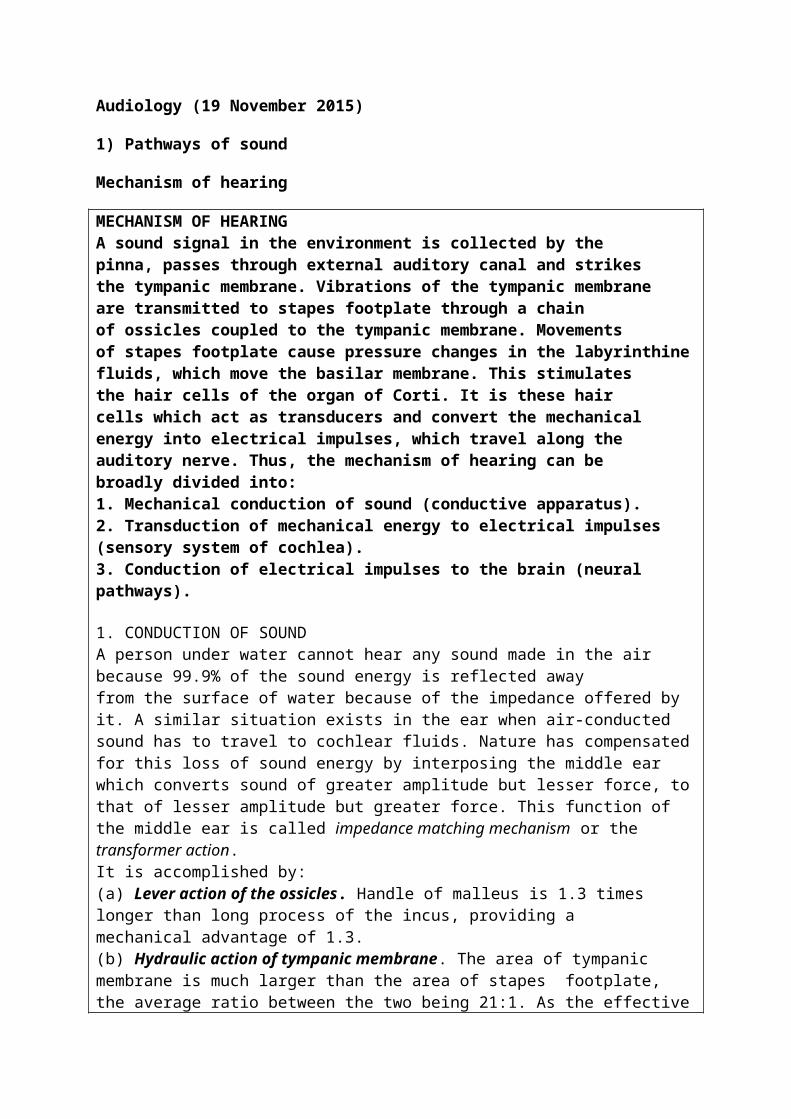
Audiology (19 November 2015)
1) Pathways of sound
Mechanism of hearing
MECHANISM OF HEARINGA sound signal in the environment is collected by thepinna, passes through external auditory canal and strikesthe tympanic membrane. Vibrations of the tympanic membraneare transmitted to stapes footplate through a chainof ossicles coupled to the tympanic membrane. Movementsof stapes footplate cause pressure changes in the labyrinthinefluids, which move the basilar membrane. This stimulatesthe hair cells of the organ of Corti. It is these haircells which act as transducers and convert the mechanicalenergy into electrical impulses, which travel along theauditory nerve. Thus, the mechanism of hearing can bebroadly divided into:1. Mechanical conduction of sound (conductive apparatus).2. Transduction of mechanical energy to electrical impulses(sensory system of cochlea).3. Conduction of electrical impulses to the brain (neuralpathways).
1. CONDUCTION OF SOUNDA person under water cannot hear any sound made in the air because 99.9% of the sound energy is reflected awayfrom the surface of water because of the impedance offered by it. A similar situation exists in the ear when air-conducted sound has to travel to cochlear fluids. Nature has compensated for this loss of sound energy by interposing the middle ear which converts sound of greater amplitude but lesser force, to that of lesser amplitude but greater force. This function of the middle ear is called impedance matching mechanism or the transformer action.It is accomplished by:(a) Lever action of the ossicles. Handle of malleus is 1.3 times longer than long process of the incus, providing amechanical advantage of 1.3.(b) Hydraulic action of tympanic membrane. The area of tympanic membrane is much larger than the area of stapes footplate, the average ratio between the two being 21:1. As the effective vibratory area of tympanic membrane is only two-thirds, the effective areal ratio is reduced to 14:1, and this is the mechanical advantage provided by the tympanic membrane
2. TRANSDUCTION OF MECHANICAL ENERGY TOELECTRICAL IMPULSESMovements of the stapes footplate, transmitted to thecochlear fluids, move the basilar membrane and set up shearing force between the tectorial membrane and the hair

cells. The distortion of hair cells gives rise to cochlear microphonics,which trigger the nerve impulse.A sound wave, depending on its frequency, reaches maximumamplitude on a particular place on the basilar membraneand stimulates that segment (travelling wave theory ofvon Bekesy). Higher frequencies are represented in the basalturn of the cochlea and the progressively lower ones towardsthe apex
3. NEURAL PATHWAYSHair cells get innervation from the bipolar cells of spiralganglion. Central axons of these cells collect to form thecochlear nerve which goes to the ventral and dorsal cochlearnuclei. From there, both crossed and uncrossed fibres travelto the superior olivary nucleus, lateral lemniscus, inferiorcolliculus, medial geniculate body and finally reach theauditory cortex of the temporal lobe.
2) Hearing test
a) What is subjective test
b) What is objective test
Subjective Test Objective TestPure tone audiogramSpeech audiometry
Impedance audiometryEvoked response audiometry (ERA)

Birth - 6 months -- evoked response audiometry
6 months to about 18 months - Distraction test
2 years ++ -- conditioning / cooperation
3) Tympanometry test (impedance test)
- Why do tympanometry
- Explain tympanometry result : type A , type AS , type AD type B , type C

Type A - Normal tympanogram.Type As - Compliance is lower at or near ambient air pressure. Seen in fixation of ossicles, e.g. otosclerosis or malleus fixation.Type Ad - High compliance at or near ambient pressure. Seen in ossicular discontinuity or thin and lax tympanic membrane.Type B - A flat or dome-shaped graph. No change in compliance with pressure changes. Seen in middle ear fluid or thick tympanic membrane.Type C - Maximum compliance occurs with negative pressure in excess of 100 mm H2O. Seen in retracted tympanic membrane and may show some fluid in middle ear.
(a) Tympanometry. It is based on a simple principle, i.e.when a sound strikes tympanic membrane, some of thesound energy is absorbed while the rest is reflected. A stiffertympanic membrane would reflect more of sound energythan a compliant one. By changing the pressures in a sealedexternal auditory canal and then measuring the reflectedsound energy, it is possible to find the compliance or stiffnessof the tympano-ossicular system and thus find thehealthy or diseased status of the middle ear.Essentially, the equipment consists of a probe which snugly

fits into the external auditory canal and has three channels:(i) to deliver a tone of 220 Hz, (ii) to pick up the reflectedsound through a microphone and (iii) to bring about changesin air pressure in the ear canal from positive to normal andthen negative (Figure 4.5). By charting the compliance oftympano-ossicular system against various pressure changes,different types of graphs called tympanograms are obtainedwhich are diagnostic of certain middle ear pathologies.
Testing function of eustachian tube. Tympanometry has also beenused to find function of eustachian tube in cases of intact orperforated tympanic membrane. A negative or a positive pressure(−200 or +200 mm H2O) is created in the middle ear andthe person is asked to swallow five times in 20 s. The ability toequilibrate the pressure indicates normal tubal function. Thetest can also be used to find the patency of the grommet placedin the tympanic membrane in cases of serous otitis media.
4) Type of hearing loss
- Conductive hearing loss
- Sensorineural hearing loss
5) Degree of hearing impairments

6) High risk of hearing impairments
7) Management cases of hearing loss
8) What is aural habilitation & rehabilitation?
- What is early detection & intervention?
- Cochlear implant?