Embed Size (px)
Citation preview
Morpho-Molecular Characterization and Virulence Determination ofEntomopathogenic Fungal Isolates Native to Indian SubcontinentSapna Mishra, Peeyush Kumar and Anushree Malik*
Applied Microbiology Laboratory, Centre for Rural Developmentand Technology, Indian Institute of Technology, Delhi, NewDelhi-110 016, India*Corresponding author: Anushree Malik, Applied Microbiology Laboratory, Centre for Rural Developmentand Technology, IndianInstitute of Technology, Delhi, New Delhi-110 016, India, Tel: +91-11-26591158; Fax: +91-11-26591121, E-mail:[email protected]
Received date: August 13, 2015, Accepted date: November 13, 2015, Published date: November 24, 2015
Copyright: © 2015 Malik A, et al. This is an open-access article distributed under the terms of the Creative Commons AttributionLicense, which permits unrestricted use, distribution, and reproduction in any medium, provided the original author and sourceare credited.
Abstract
Background: The house fly, Musca domestica L. is amajor pest of medical and veterinary significancewhich due to their ubiquitous lifestyle and broad foodpreference achieves a very high density in favorableclimatic conditions of Indian subcontinent. This has ledto several instances of severe health condition amonghuman and animal population, calling attentiontowards M. domestica control through existing andinnovative methods. Use of biological control agent,such as entomopathogenic fungi (ENPF) for M.domestica control has shown potential, and need to befurther explored for the newer isolates suited toparticular environment for greater efficacy. Thepresent study attempted the isolation andidentification of native ENPF strains suitable for M.domestica control.
Method and findings: Five strains of ENPF wereisolated from soil samples, which on preliminaryidentification of morphological, microscopic and sporeattributes were determined to be isolates of Beauveriaspecies. The isolates showed wide variation inpathogenicity against M. domestica, with 72.7%-100%and 36.7%-72.3% mortality against adults and larvaelife stage, respectively. Isolate ‘1’ depicting maximuminsecticidal activity against M. domestica was selectedfor molecular identification using 18s-rRNA, and foundto be Beauveria bassia HQ917687. The molecularanalysis using random amplified polymorphic DNA(RAPD) showed isolate ‘2’ to be most similar to isolate‘3’, while isolate ‘1’ and ‘5’ showed maximum variationbetween them. RAPD result was in conformity withvarious other properties of isolates; such as biomass,spore yield, hydrophobicity and insecticidal activity.
Conclusion: The variations in activity obtained throughpathogenicity assessment of fungal isolates in thisstudy reflected the strain diversity of Beauveriaisolates. The results also signified the importance ofstrain selection for effective control measures as wellas for further consideration of commercial aspects.
Keywords: Entomopathogenic fungi; Isolation;Characterization; Pathogenicity; Musca domestica
IntroductionEntomopathogenic fungi (ENPF) due to their broad host
range, diminution of potential environmental hazards andtheoretical impossibility of resistance development in targetinsects, are advantageous for pest management strategies [1].Also, due to their multiple modes of action for insect kill andability of adapt to pertaining environmental conditions, ENPFshave emerged as bio pesticide of considerable potential. Theliterature is replete with studies employing potential of ENPFs(e.g., Entomophthora muscae, Beauveria bassiana andMetarhizium anisopliae etc.) for insect control [2-5]. Most ofthese studies reports use of ENPFs for control of agriculturalpests [2,3]. However, some other studies depict ENPF activityagainst pests of vector significance, such as mosquitoes andhouse fly [4,5].
The house fly, Musca domestica L. (Diptera: Muscidae) is asignificant vector of more than 100 medical and veterinarypathogens which due to their ubiquitous lifestyle and broadfood preference achieves a very high density in favourableclimatic conditions of Indian subcontinent, leading toenvironment and health menace [1]. The vector potential ofM. domestica has been proved for the propagation ofprotozoan (amoebic dysentery), bacterial (shigellosis,salmonellosis, cholera), helminthic (round worms, hookworms,pinworms and tapeworms), viral (Turkey Corona virus,
Research Article
iMedPub Journalshttp://www.imedpub.com/
Medical Mycology: Open AccessVol.1 No.1:3
2015
© Copyright iMedPub | This article is available from: http://mycology.imedpub.com/ 1
Reticuloendotheliosis virus, H5N1 influenza virus) andrickettsial infections, among others [4]. The study by Barro etal. [6], revealed M. domestica to be predominant fly associatedwith street-food vendors in Ouagadougou which carriedisolates of Coliform, Salmonella, Shigella, Staphylococci andStreptococci on both their legs and proboscis, while Barin et al.[7], demonstrated presence of Newcastle disease virus in thesignificant number of adult muscid flies collected from 20poultry farms near Tehran, Iran. Furthermore, recent reportsabout carriage of antibiotic-resistant pathogens by M.domestica collected from different fast-food restaurants innorthern Kansas (USA) [8] and in hospitals of several countriesof Sub-Saharan Africa [9] are quite alarming and calls forurgent M. domestica control measures.
The ENPFs are significant biological control agents againstM. domestica due to their low mammalian toxicity and naturalprevalence among flies population. However, compared toliterature reports on mosquito control, the studies for M.domestica control is significantly few and far between. Theliterature studies describing M. domestica control using ENPFshave mostly used E. muscae, B. bassiana and M. anisopliae.Most of these studies pertain to control of adult fly [10-12];while few of these reports have explored control of M.domestica larval stage [12,13]. These studies indicatedabsolute mortality of M. domestica in 5-15 days. However, asubstantial part of these studies has been conducted intemperate region, where the control agents have tried tomimic the prevalent environmental conditions. The researchworks carried out in the temperate region with local fungalstrains have little significance in tropical/sub-tropicalconditions of Indian subcontinent. The reason for this partlylays in lack of adaption of fungal strains towards the varyingtemperature, humidity and soil conditions of tropical/sub-tropical region. Furthermore, pathogenicity of ENPFs is straindependent and is influenced by geographical conditions [14].Kryukov et al. [14], reported high variability in virulence of B.bassiana cultures isolated in a small territory or even at thesame site, due to formation of the fungal races adapted to killlocal insect populations or specific hosts. Adaptability of ENPFstrains to a particular environment may also increase itsinfectivity by affecting their genetic polymorphism [15].Considering the above facts, the investigation of native ENPFisolates adaptable to local environment and hence moreefficient for the control of pest population of the region isdesirable, to ensure the production of biopesticidescomparable to conventionally used chemical insecticides interms of cost and activity. Thus, the present study hasundertaken the screening of suitable native isolates of ENPFsfor the use as biopesticidal agents against M. domestica.
Methods
Collection and analysis of soil sampleSoil samples were collected from different fruit orchards in
northern India during september-november, 2009. A total of 8fruit orchards were sampled. Soil samples were taken using acylindrical soil corer with an inner diameter of 5 cm. From each
orchard, 10 randomly selected soil cores (diameter-5 cm,depth-5 cm) were removed and placed as subsamples in aplastic bag. Collected soil samples were analysed for carbonand nitrogen content through CHN analyzer (VARIO EL III). ThepH of the soil solutions was analysed by a digital type pHmeter (HI 2212 pH meter, Hannah instruments, USA) bydissolving 1 g of soil in 10 ml of distilled water.
Isolation of fungal strainsIsolation of ENPF from soil was done by Galleria bait
method. 100 g of air dried and re-moistured soil samples wereplaced in cylindrical plastic bottle (90 mm × 140 mm) alongwith 10 pre heat treated larvae of Galleria mellonella(Lepidoptera: Pyralidae). The heat treatment preventswebbing by G. mellonella larvae [16]. The bottles were sealedwith perforated lids and incubated at 22°C ± 1°C and 65%relative humidity (RH) in a growth chamber for 16-18 days.During the incubation for first week, containers were invertedto make the vigorous bait insects penetrate maximum amountof soil [16]. Containers were examined regularly for the deadlarvae, which were collected and surface-sterilized with 1%sodium hypochlorite solution for 3 min, followed by gentlerinsing (2 times) with distilled water. Dead larvae were placedon Petri plate containing selective medium [1% peptone, 2%glucose and 1.2% agar along with 0.5 ml of 0.6 g/mlStreptomycin, 0.5 ml of 0.05 g/ml Tetracycline, 0.5 ml of 0.1g/ml Dodine and 2.5 ml of 0.05 g/5 ml Cyclohexamide] forisolation of ENPFs. The plates were incubated at 27°C and100% RH in a growth chamber and observed for the presenceof fungi. The fungi were sub-cultured to obtain pure culturesand maintained on potato dextrose agar (PDA) slants.
Growth pattern, spore production, biomassand spore hydrophobicity
Fungal isolates obtained through isolation from soil sampleswere morphologically characterized using culturing on artificialmedia; potato dextrose agar (PDA) on Petri plates and potatodextrose broth (PDB) in culture flasks. Both the type of mediawere inoculated with 1 ml of conidial suspension (1 × 10⁶
conidia/ml) of different fungal isolates and incubated at 28°Cfor 8 days under stationary (PDA) or as shake cultures at 180rpm (PDB). The growth of each fungal strain was studied interms of colony morphology (color and growth pattern), andradial growth of isolates. Sporulation was recorded by excising20 mm2 colony area from fungal growth on PDA medium usinga cork borer, followed by mixing with 1 ml of distilled water todislodge spores [17]. The spores present in the sample werecounted by Automatic Cell Counter (Cellometer® Vision HSL,Nexcelom Bioscience). Biomass production was assessed fromfungal growth on PDB media. After the incubation period, thedry cell weight of the biomass was estimated by filtering outthe contents of flasks through predried and preweighedWhatman no. 1 filter paper, and drying it at 54°C to a constantweight.
The spore hydrophobicity of fungal isolates was determinedby the method described by Shan et al. [18] For each isolate,
Medical Mycology: Open Access
Vol.1 No.1:3
2015
2 This article is available from:http://mycology.imedpub.com/
spores were suspended in PM buffer (per liter: K2HPO4, 6.97 g;KH2PO4, 2.99 g; MgSO4.7H2O, 0.2 g) containing 0.02% (v/v)Tween 80 at pH of 7.12. The spore suspension wasstandardized to 2 × 10⁶ spores/ml, followed by addition of0.1% liquid paraffin. The mixture was vortexed for 2 min toseparate organic phase from the aqueous phase. The spores inthe aqueous phase were counted using an Automatic CellCounter. The hydrophobicity index (HI) of the tested spores offungus isolates was determined using following formulae:HI = 1− CC0
where C0 and C is the spore concentration of the aqueousphase before and after the addition of liquid paraffin,respectively. The assay was repeated thrice for each isolate.
Microscopic observation of fungal isolatesMicroscopic observation of fungal isolates was done using
phase contrast microscope (Leica Microscope; Magnification:40X) and scanning electron microscope (SEM). For SEManalysis, 10 μl inoculum of fungal growth in liquid culture wasprimarily fixed in 2.5% glutaraldehyde in 0.1M phosphatebuffer for 1-2 h. Thereafter, it was washed with distilled waterfor 10-20 min, followed by secondary fixation with 1%-4%osmium tetroxide (OsO4) in distilled water for 1-2 h. Secondaryfixation was followed by washing with distilled water (10-20min) and then serial dehydration with 45%, 60%, 75%, 90%,and 100% ethanol for 10 min each. After dehydration, sampleswere subjected to critical point drying (CPD) in hexamethylenedisilazane (HMDS). All the processes were performed at roomtemperature. Samples were mounted on silver stub for SEMobservation and gold-covered by cathodic spraying (Polarongold). Observations were made on a scanning electronicmicroscope (ZEISS EVO 50) under the following analyticalcondition: EHT ¼ 20.00 kV, WD ¼ 9.5 mm, Signal A ¼ SE1.
Insecticidal activity of fungal isolates againstM. domestica
Insecticidal activity of fungal isolates was determined in aPetri plate bioassay against M. domestica adult and larvaeaccording to the method described earlier by Mishra et al. [1]Fungal dose used for the assays was 1 ml of conidialsuspension in 0.1% Tween 80 at 1 × 10⁸ spores/ml. Control wassprayed with 1 ml of sterile distilled water containing 0.1%Tween 80. Treated plates were incubated at 28 ± 5°C, 65% RHand 14:10 photoperiod. For each treatment, four replicationswere incorporated, while whole set up was repeated thrice.For assay against M. domestica adults, lab reared flies (10 inno., 2-3 days old) were placed on a filter paper (Whatman no.1) in Petri plate (diameter-150 mm, area=17662.5 mm2,BOROSIL®) containing diet consisting of mixture of groundnutoil cake and wheat bran in 1:3 ratios. Dead adult flies werecounted daily during five days of monitoring. Fungal infectionon dead flies was confirmed by incubation of surface sterilizeddead cadaver on PDA media, followed by incubation at 28 ±5°C, 65% RH, for allowing growth of fungus on cadaver. Forlarval assay, a batch of 20 lab reared larvae (2nd in star) of M.
domestica was taken on Petri plates (diameter-90 mm) alongwith larval diet as described previously [1]. The mortality of M.domestica larvae was adjudged by larval wasting andimmobility, monitored daily for five days [1]. Mortality datawere corrected for control mortality using Abbott’s [19]formula and normalized using arcsine transformation beforebeing subjected to a two-way factorial analysis of varianceusing Stat Plus [20]. The means were separated using LSD anddifferences between them were considered significant atP<0.05.
The fungal isolates obtained in the present study werecompared for their insecticidal activity with twelve differentisolates of ENPF procured from microbial type culturecollection (MTCC) and national bureau of agriculturallyimportant insects (NBAII), India (Table 1). Selection of isolatesfor procurement was made on the basis of literature reportsfor their insecticidal activity against various insect/pests. Theprocured isolates were investigated for their insecticidalactivity against M. domestica adults and larvae according tothe method described above.
Molecular characterization of fungal isolatesthrough RAPD-PCR amplification
Variation among different fungal isolates was furtherascertained using molecular amplification method by randomamplified polymorphic DNA (RAPD). For this, DNA was isolatedfrom fungal isolate using freeze drying of fungal mycelia, whichwas further extracted by CTAB method [17]. Quantity of DNAwas estimated by spectrophotometer while its quality analysiswas done by 0.8% agarose gel electrophoresis. For RAPDanalysis, twenty RAPD primers (obtained from BangaloreGenei™) were screened, of which twelve gave amplification,however only six primers gave scorable and reproducibleresults. Polymer chain reaction (PCR) amplification reaction forRAPD analysis was carried out in the final volume of 25 μl [1 ×assay buffer (2.5 μl), 10 mM dNTPs (1.25 μl), 3U Taq DNAPolymerase (1 μl), 2.5 mM MgCl2 (1.25 μl), 25 ng DNAtemplate (5 μl), 20 μM primer (2 μl)]. Amplifications wereexecuted using an initial step of 15 s at 94°C, followed by 45cycles (94°C at 15 s, 36°C at 30 s, 72°C at 1 min), and a 7 minfinal extension at 72°C. RAPD data obtained was clusteranalysed to examine the genotypic relationship among theisolates. Amplification products were scored as either present(1) or absent (0). Numerical analysis was done using NTSYS PCversion 2.0. A data matrix was prepared to determinesimilarities between each pair of genotype. Similarities werecalculated using simple matching coefficient and clusteringwas achieved by unweighted pair group method usingarithmetic averages (UPGMA).
18s-rRNA identification of selected isolateBased on the insecticidal activity data, the isolate with
maximum pathogenic potential was selected for molecularidentification through 18s-rRNA analysis. The processaccompanied DNA isolation followed by amplification reaction.PCR amplification was performed with 3 primers; primers P1
Medical Mycology: Open Access
Vol.1 No.1:3
2015
© Copyright iMedPub 3
(5′-AAGCTTCGACATGGTCTG-3′), ITS1 (5′-TCCGTAGGTGAACCTGCGG-3′) and I21 (5′-CGATCCTTTAGTCCCTCGAC-3′), chosen on the basis of literaturereport for molecular characterization of Beauveria strains[21,22]. PCR amplification reaction was carried out in the finalvolume of 25 μl [1 × assay buffer (2.5 μl), 10 mM dNTPs (1.25μl), 3U Taq DNA Polymerase (1 μl), 2.5 mM MgCl2 (1.25 μl), 25ng DNA template (5 μl), 20 μM primer (2 μl)]. The enzymaticamplification was performed in a thermo cycler programmedfor 1 cycle for 4 min at 94°C, 35 cycles: 45 s at 94°C, 45 s at57°C and 45 s at 72°C (extension of the primer). After 35 cyclesan extra extension step was performed for 4 min at 72°C.Amplified products were separated by submerged gelelectrophoresis on 1.5% agarose gel in 1 × TAE buffer. The gelwas visualized under UV Transilluminator attached with Kodak
camera and the picture was taken with gel documentationsystem (Ultra.lum). PCR product obtained after amplificationand purification in 12 μl of 20% PEG-NaCl (Polyethylene glycol-NaCl) solution was subjected to sequencing reactions based ondideoxynucleotide chain termination method [23]. A 2 μl PCRproduct and 3 pmol of the sequencing primer were used in a20 μl sequencing reaction and subjected to 25 cycles in aPerkin Elmer thermal cycler 9700. Each cycle consisted of 95°Cfor 10 min, 50°C for 5 min and 60°C for 4 min. DNA sequencingwas carried out on ABI 1500 automated sequencer at the DNAsequencing facility of Ocimum Biosolution (P) Ltd. Thesequences obtained were in small fragments and hence it wasaligned properly by overlapping the sequences. The nucleotidesequence was analyzed with the GenBank database usingBLAST program [24].
Table 1: Entomopathogenic fungal isolates procured from Indian Culture Collection Center.
S. No. Fungus isolate Accession No. Strain Host Source
1 Beauveria bassiana MTCC 4562 E90 Fly Mandla, Chattisgarh, India
2 Beauveria bassiana MTCC 4580 E74 Inst Jabalpur, Madhya Pradesh, India
3 Beauveria bassiana MTCC 4514 E123 Hyblaea puera Hosangabad, Madhya Pradesh, India
4 Metarihiziumanisopliae MTCC 3210 E/98/1 Cricket Jorhat, Assam, India
5 Paecilomycesfumosoroseus MTCC 4099 E24 Grub Kalpi, Madhya Pradesh, India
6 Paecilomycesfarinosus MTCC 4114 E20 Plusia (Inst) Dantewara, Chattisgarh, India
7 Beauveria bassiana PDBC-Bb-5a - Hypothenemus hampei(Coffee berry borer) Coorg, Karnataka
8 Verticillium lecanii PDBC-Vl-1 - Aphids of Vegetablecrops Bangalore, Karnataka
9 Metarhiziumanisopliiae PDBC-Ma-4 -
Grubs of Plocaederusferrugineus (Cashew
Stem Borer)Dakshina Kannada, Karnataka
10 Nomuraea riley PDBC-Nr-44 -Larvae of Pieris
brassicae (CabbageButterfly)
Jorhat, Assam
11 Nomuraea riley PDBC-Nr-1 - Larvae of Spodopteralitura from Cotton Fields Kurnool, Andhra Pradesh
12 Paeciliomycesfumosoroseus PDBC-P-1 -
Grubs of Brahminaspecies from Potato
fields (Potato root grub)Solan, Himachal Pradesh
Results
Collection of soil sample and Isolation offungus isolates
Soil samples were collected from different fruit orchardsand nurseries located in Northern India during September-November, 2009. Characterization of collected soil samples forselected parameters is represented in (Table 2). A total of 5ENPF strains (later identified as strains of “Beauveriabassiana”) were isolated. The soil samples harboring ENPF
showed a narrow pH range (7.2-8.5), with inclination towardsalkalinity (pH>8).
Growth pattern, spore production, biomassand spore hydrophobicity
Growth of fungal isolates in different media providedinformation about their colony forming properties, growthpattern and vigorousity. Growth of fungal isolates on PDBmedium was rapid till 3.5 days, followed by decline in growthrate which was quite visible in case of isolate ‘1’. Mycelium ofdifferent fungal isolates was white with fluffy to powderyappearance. The color of colony/or mats was observed to be
Medical Mycology: Open Access
Vol.1 No.1:3
2015
4 This article is available from:http://mycology.imedpub.com/
white, which became yellowish white or pale pinkish (isolate‘3’) with increase in time.
The spore production among the isolates showed significantvariation (p<0.05); with yield for different fungal isolatesranging from 8.3 × 10⁵-2.5 × 10⁸ spores/cm2 (Table 3). The
maximum spore production was observed in isolate ‘1’, whileisolate ‘5’ had lowest spore yield. After an incubation period of8 days, radial growth of fungal isolates varied between 4.2-7.7cm (Table 3).
Table 2: Description of isolation sites for entomopathogenic fungal isolates.
S. No. Field type C (%) N (%) pH Entomopathogenic fungi obtained
1 Mango Orchard 1.996 0.253 8.5 Isolate 1
2 Coriander Nursery 1.188 0.183 8.2 None
3 Mango Orchard 2 1.173 0.192 7.6 Isolate 5
4 Horticultural Research Centre 0.877 0.166 7.2 Isolate 2
5 High density Mango Orchard 0.865 0.15 8.4 Isolate 3
6 Rose Nursery 0.788 0.171 8.3 Isolate 4
7 Aloe Vera Nursery 0.785 0.153 8.2 None
8 Wheat Nursery 0.589 0.153 5.7 None
The maximum mean radial growth (7.7 cm) was observed inisolate ‘1’, followed by isolate ‘4’ (7.3 cm). Isolate ‘5’ showedleast radial growth at 4.2 cm. Radial growth of the fungalisolates represents the frequency of their hyphal branchingand substrate affinity. The biomass production with differentisolates varied between 1.35-5.66 g/L, with maximum yieldfrom isolate ‘1’ (Table 3). The biomass and spore yield fordifferent fungal isolates is indicative of their pathogenicity.Hydrophobicity index of fungal spores varied significantly(F=850.3; df=4; p<0.001) amongst different fungal isolatesranging from 0.54-0.82 (Table 3). The spores of isolate ‘4’ werefound to be most hydrophobic with hydrophobicity index of0.82, followed by that of isolate ‘1’ (0.79).
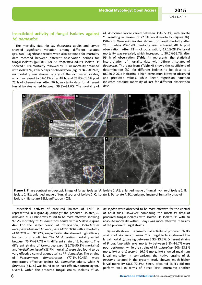
Microscopic observation of fungal isolatesMicroscopic observation of fungal isolates by phase contrast
microscopy is represented in (Figure 1). Mycelial strand of thefungal isolates were observed to be cylindrical, hyaline andseptate. Isolate ‘1’ revealed oval to round spores, whereas
spores of isolate ‘2’ were oval and relatively larger in size.Isolate ‘3’ had flask shaped spores. The shape of spores forisolate ‘4’ and isolate ‘5’ was round and globose to sub-globose, respectively. The spore shape of fungal isolates gavean indication that these isolates belongs to Genus “Beauveria”[25]. The sporogenous cells formed tightly clustered groups,appearing as fluffy balls within the aerial hyphal mass.
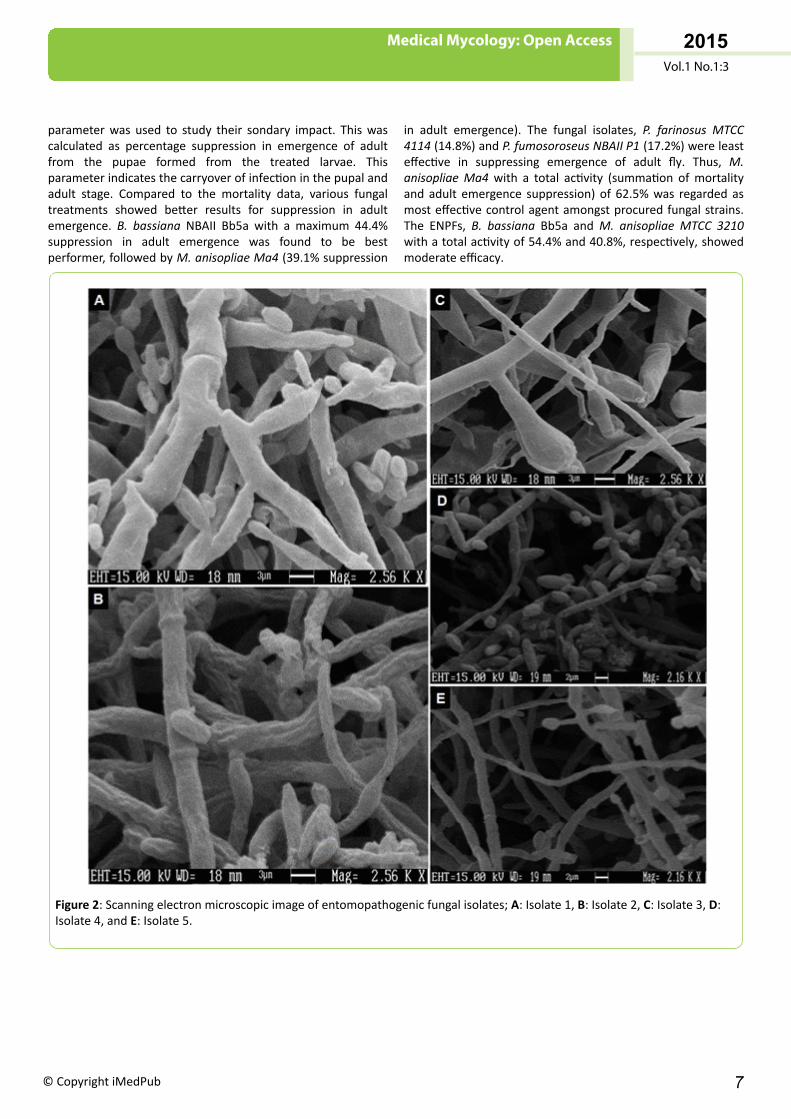
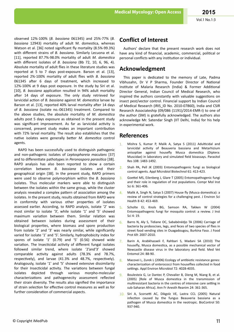
The SEM image for different fungal isolates revealed narrowand septate hyphae (Figure 2). The vesicles generally formedglobose type of structure, with size varying between 1.5 μm-5μm for different fungal isolates. Majority of spores of fungalisolates showed globose to oval shape, while theirarrangement was basifugal. These features provided furtherevidence that the fungal isolates belongs to Genus“Beauveria”. Further characterization of fungal isolates formorphological identification was carried out by indian typeculture collection (ITCC), Indian Agricultural Research Institute(New Delhi), which confirmed the fungal isolates to be strainsof Beauveria bassiana.
Table 3: Mean radial growth, Spore production, Biomass and Hydrophobicity index of different fungal isolates.
Fungus isolates Mean radialgrowth (cm)
Spore production(conidia/cm2) Biomass (g/L, mean ± S.D.) Hydrophobicity index (mean ± S.D.)
Isolate 1 7.7 2.5 × 10⁸ 5.66 ± 0.191 0.795 ± 0.016
Isolate 2 4.8 3.8 × 10⁷ 5.23 ± 0.163 0.542 ± 0.009
Isolate 3 5.4 7.2 × 10⁶ 6.53 ± 0.237 0.589 ± 0.011
Isolate 4 7.3 2.7 × 10⁶ 3.04 ± 0.091 0.822 ± 0.021
Isolate 5 4.2 8.3 × 10⁵ 1.35 ± 0.086 0.558 ± 0.013
Medical Mycology: Open Access
Vol.1 No.1:3
2015
© Copyright iMedPub 5
Insecticidal activity of fungal isolates againstM. domestica
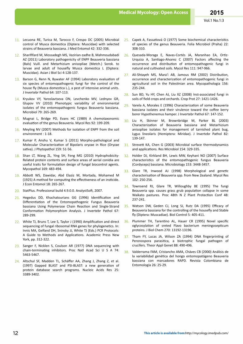
The mortality data for M. domestica adults and larvaeshowed significant variation among different isolates(p<0.001). Significant results were also obtained for mortalitydata recorded between different observation periods forfungal isolates (p<0.01). For M. domestica adults, isolate ‘1’showed 100% mortality, followed by 82.3% mortality obtainedwith isolate ‘4’, after 5 days of observation (Figure 3a). At 24 h,no mortality was shown by any of the Beauveria isolates,which increased to 0%-11% after 48 h, and 21.8%-61.6% post72 h of observation. After 96 h, mortality data for differentfungal isolates varied between 59.8%-82.6%. The mortality of
M. domestica larvae varied between 36%-72.3%, with Isolate‘1’ resulting in maximum 72.3% larval mortality (Figure 3b).Different Beauveria isolates showed no larval mortality after24 h, while 0%-6.4% mortality was achieved 48 h postobservation. After 72 h of observation, 17.1%-28.2% larvalmortality was revealed, which increased to 30.0%-59.7% after96 h of observation (Table 4) represents the statisticalinterpretation of mortality data with different isolates ofBeauveria. The data from (Table 4) shows the coefficient ofdetermination (R2) for different isolates to be close to 1(0.920-0.961) indicating a high correlation between observedand predicted values, while linear regression equationindicates absolute mortality of inst for different observationdays.
Figure 1: Phase contrast microscopic image of fungal isolates; A: Isolate 1; A1: enlarged image of fungal hyphae of isolate 1, B:Isolate 2, B1: enlarged image of fungal spores of isolate 2, C: Isolate 3, D: Isolate 4, D1: enlarged image of fungal hyphae ofisolate 4, E: Isolate 5 [Magnification 40X].
Insecticidal activity of procured isolates of ENPF isrepresented in (Figure 4). Amongst the procured isolates, B.bassiana NBAII Bb5a was found to be most effective showing97.7% mortality of M. domestica adults within 5 days (Figure4a). For the same period of observation, Metarhiziumanisopliae Ma4 and M. anisopliae MTCC 3210 with a mortalityof 94.72% and 92.72%, respectively, also showed high efficacyfor control of adult flies. The M. domestica mortality variedbetween 73.7%-97.7% with different strain of B. bassiana. Thedifferent strains of Nomuraea riley (86.7%-90.1% mortality)and Verticillium lecanii (88.7% mortality) were also found to bevery effective control agent against M. domestica. The strainsof Paecilomyces fumosoroseus (77.1%-80.4%) weremoderately effective against M. domestica adults, while P.farinosus (64.1%) was found to be least effective control agent.Overall, within the procured fungal strains, isolates of M.
anisopliae were observed to be most effective for the controlof adult flies. However, comparing the mortality data ofprocured fungal isolates with Isolate ‘1’, Isolate ‘1’ with anabsolute mortality within 5 days was more effective than anyof the procured fungal strains.
Figure 4b shows the Insecticidal activity of procured ENPFsagainst M. domestica larvae. The fungal isolates showed lowlarval mortality, varying between 3.3%-23.3%. Different strainsof B. bassiana with larval mortality between 3.3%-16.7% werepoor performer, while the strains of M. anisopliae (20%-23.3%mortality) and V. lecanii (16.7% mortality) showed maximumlarval mortality. In comparison, the native strains of B.bassiana isolated in the present study showed much higherlarval mortality (36%-72.3%). Since, procured ENPFs did notperform well in terms of direct larval mortality; another
Medical Mycology: Open Access
Vol.1 No.1:3
2015
6 This article is available from:http://mycology.imedpub.com/
parameter was used to study their sondary impact. This wascalculated as percentage suppression in emergence of adultfrom the pupae formed from the treated larvae. Thisparameter indicates the carryover of infection in the pupal andadult stage. Compared to the mortality data, various fungaltreatments showed better results for suppression in adultemergence. B. bassiana NBAII Bb5a with a maximum 44.4%suppression in adult emergence was found to be bestperformer, followed by M. anisopliae Ma4 (39.1% suppression
in adult emergence). The fungal isolates, P. farinosus MTCC4114 (14.8%) and P. fumosoroseus NBAII P1 (17.2%) were leasteffective in suppressing emergence of adult fly. Thus, M.anisopliae Ma4 with a total activity (summation of mortalityand adult emergence suppression) of 62.5% was regarded asmost effective control agent amongst procured fungal strains.The ENPFs, B. bassiana Bb5a and M. anisopliae MTCC 3210with a total activity of 54.4% and 40.8%, respectively, showedmoderate efficacy.
Figure 2: Scanning electron microscopic image of entomopathogenic fungal isolates; A: Isolate 1, B: Isolate 2, C: Isolate 3, D:Isolate 4, and E: Isolate 5.
Medical Mycology: Open Access
Vol.1 No.1:3
2015
© Copyright iMedPub 7
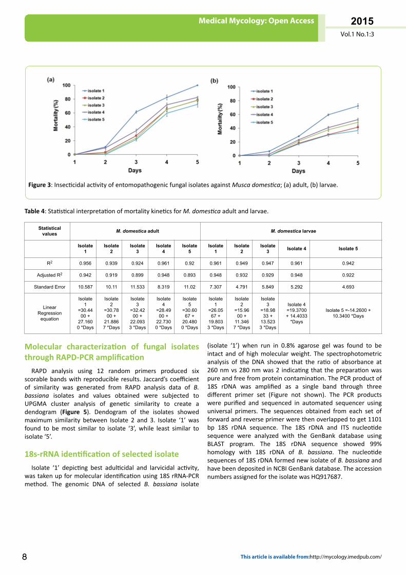
Figure 3: Insecticidal activity of entomopathogenic fungal isolates against Musca domestica; (a) adult, (b) larvae.
Table 4: Statistical interpretation of mortality kinetics for M. domestica adult and larvae.
Statisticalvalues M. domestica adult M. domestica larvae
Isolate1
Isolate2
Isolate3
Isolate4
Isolate5
Isolate1
Isolate2
Isolate3 Isolate 4 Isolate 5
R2 0.956 0.939 0.924 0.961 0.92 0.961 0.949 0.947 0.961 0.942
Adjusted R2 0.942 0.919 0.899 0.948 0.893 0.948 0.932 0.929 0.948 0.922
Standard Error 10.587 10.11 11.533 8.319 11.02 7.307 4.791 5.849 5.292 4.693
LinearRegression
equation
Isolate1
=30.4400 +
27.1600 *Days
Isolate2
=30.7800 +
21.8867 *Days
Isolate3
=32.4200 +
22.0933 *Days
Isolate4
=28.4900 +
22.7300 *Days
Isolate5
=30.6067 +
20.4800 *Days
Isolate1
=26.0567 +
19.8033 *Days
Isolate2
=15.9600 +
11.3467 *Days
Isolate3
=18.9833 +
13.5233 *Days
Isolate 4=19.3700+ 14.4033
*Days
Isolate 5 =-14.2600 +10.3400 *Days
Molecular characterization of fungal isolatesthrough RAPD-PCR amplification
RAPD analysis using 12 random primers produced sixscorable bands with reproducible results. Jaccard’s coefficientof similarity was generated from RAPD analysis data of B.bassiana isolates and values obtained were subjected toUPGMA cluster analysis of genetic similarity to create adendogram (Figure 5). Dendogram of the isolates showedmaximum similarity between Isolate 2 and 3. Isolate ‘1’ wasfound to be most similar to isolate ‘3’, while least similar toisolate ‘5’.
18s-rRNA identification of selected isolateIsolate ‘1’ depicting best adulticidal and larvicidal activity,
was taken up for molecular identification using 18S rRNA-PCRmethod. The genomic DNA of selected B. bassiana isolate
(isolate ‘1’) when run in 0.8% agarose gel was found to beintact and of high molecular weight. The spectrophotometricanalysis of the DNA showed that the ratio of absorbance at260 nm vs 280 nm was 2 indicating that the preparation waspure and free from protein contamination. The PCR product of18S rDNA was amplified as a single band through threedifferent primer set (Figure not shown). The PCR productswere purified and sequenced in automated sequencer usinguniversal primers. The sequences obtained from each set offorward and reverse primer were then overlapped to get 1101bp 18S rDNA sequence. The 18S rDNA and ITS nucleotidesequence were analyzed with the GenBank database usingBLAST program. The 18S rDNA sequence showed 99%homology with 18S rDNA of B. bassiana. The nucleotidesequences of 18S rDNA formed new isolate of B. bassiana andhave been deposited in NCBI GenBank database. The accessionnumbers assigned for the isolate was HQ917687.
Medical Mycology: Open Access
Vol.1 No.1:3
2015
8 This article is available from:http://mycology.imedpub.com/
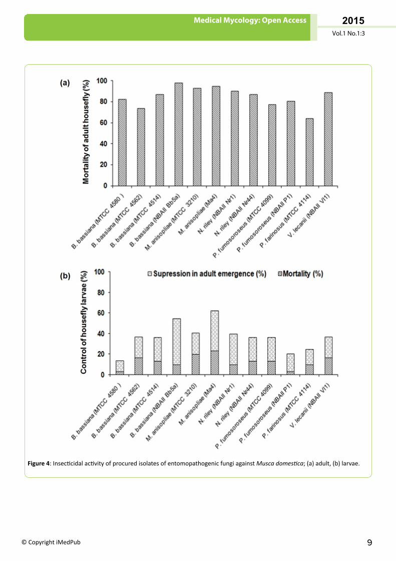
Figure 4: Insecticidal activity of procured isolates of entomopathogenic fungi against Musca domestica; (a) adult, (b) larvae.
Medical Mycology: Open Access
Vol.1 No.1:3
2015
© Copyright iMedPub 9
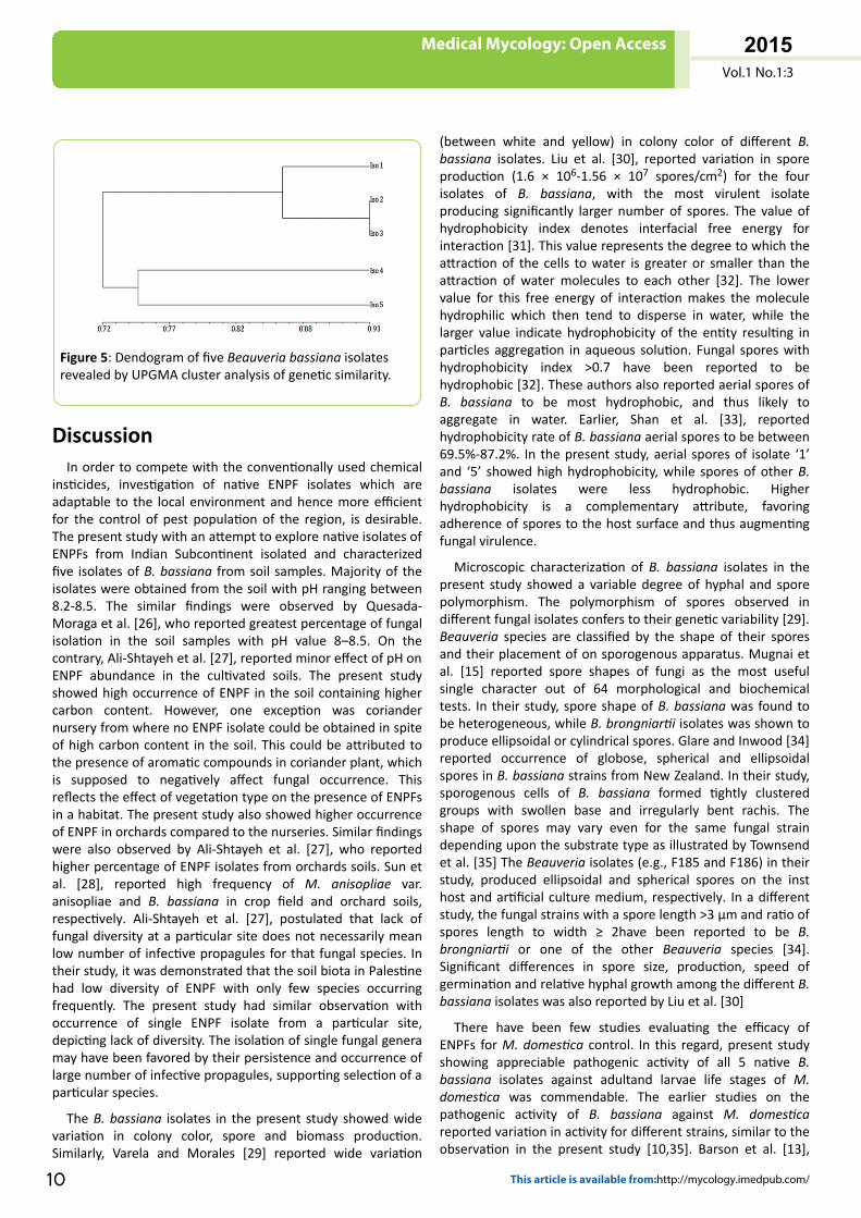
Figure 5: Dendogram of five Beauveria bassiana isolatesrevealed by UPGMA cluster analysis of genetic similarity.
DiscussionIn order to compete with the conventionally used chemical
insticides, investigation of native ENPF isolates which areadaptable to the local environment and hence more efficientfor the control of pest population of the region, is desirable.The present study with an attempt to explore native isolates ofENPFs from Indian Subcontinent isolated and characterizedfive isolates of B. bassiana from soil samples. Majority of theisolates were obtained from the soil with pH ranging between8.2-8.5. The similar findings were observed by Quesada-Moraga et al. [26], who reported greatest percentage of fungalisolation in the soil samples with pH value 8–8.5. On thecontrary, Ali-Shtayeh et al. [27], reported minor effect of pH onENPF abundance in the cultivated soils. The present studyshowed high occurrence of ENPF in the soil containing highercarbon content. However, one exception was coriandernursery from where no ENPF isolate could be obtained in spiteof high carbon content in the soil. This could be attributed tothe presence of aromatic compounds in coriander plant, whichis supposed to negatively affect fungal occurrence. Thisreflects the effect of vegetation type on the presence of ENPFsin a habitat. The present study also showed higher occurrenceof ENPF in orchards compared to the nurseries. Similar findingswere also observed by Ali-Shtayeh et al. [27], who reportedhigher percentage of ENPF isolates from orchards soils. Sun etal. [28], reported high frequency of M. anisopliae var.anisopliae and B. bassiana in crop field and orchard soils,respectively. Ali-Shtayeh et al. [27], postulated that lack offungal diversity at a particular site does not necessarily meanlow number of infective propagules for that fungal species. Intheir study, it was demonstrated that the soil biota in Palestinehad low diversity of ENPF with only few species occurringfrequently. The present study had similar observation withoccurrence of single ENPF isolate from a particular site,depicting lack of diversity. The isolation of single fungal generamay have been favored by their persistence and occurrence oflarge number of infective propagules, supporting selection of aparticular species.
The B. bassiana isolates in the present study showed widevariation in colony color, spore and biomass production.Similarly, Varela and Morales [29] reported wide variation
(between white and yellow) in colony color of different B.bassiana isolates. Liu et al. [30], reported variation in sporeproduction (1.6 × 10⁶-1.56 × 10⁷ spores/cm2) for the fourisolates of B. bassiana, with the most virulent isolateproducing significantly larger number of spores. The value ofhydrophobicity index denotes interfacial free energy forinteraction [31]. This value represents the degree to which theattraction of the cells to water is greater or smaller than theattraction of water molecules to each other [32]. The lowervalue for this free energy of interaction makes the moleculehydrophilic which then tend to disperse in water, while thelarger value indicate hydrophobicity of the entity resulting inparticles aggregation in aqueous solution. Fungal spores withhydrophobicity index >0.7 have been reported to behydrophobic [32]. These authors also reported aerial spores ofB. bassiana to be most hydrophobic, and thus likely toaggregate in water. Earlier, Shan et al. [33], reportedhydrophobicity rate of B. bassiana aerial spores to be between69.5%-87.2%. In the present study, aerial spores of isolate ‘1’and ‘5’ showed high hydrophobicity, while spores of other B.bassiana isolates were less hydrophobic. Higherhydrophobicity is a complementary attribute, favoringadherence of spores to the host surface and thus augmentingfungal virulence.
Microscopic characterization of B. bassiana isolates in thepresent study showed a variable degree of hyphal and sporepolymorphism. The polymorphism of spores observed indifferent fungal isolates confers to their genetic variability [29].Beauveria species are classified by the shape of their sporesand their placement of on sporogenous apparatus. Mugnai etal. [15] reported spore shapes of fungi as the most usefulsingle character out of 64 morphological and biochemicaltests. In their study, spore shape of B. bassiana was found tobe heterogeneous, while B. brongniartii isolates was shown toproduce ellipsoidal or cylindrical spores. Glare and Inwood [34]reported occurrence of globose, spherical and ellipsoidalspores in B. bassiana strains from New Zealand. In their study,sporogenous cells of B. bassiana formed tightly clusteredgroups with swollen base and irregularly bent rachis. Theshape of spores may vary even for the same fungal straindepending upon the substrate type as illustrated by Townsendet al. [35] The Beauveria isolates (e.g., F185 and F186) in theirstudy, produced ellipsoidal and spherical spores on the insthost and artificial culture medium, respectively. In a differentstudy, the fungal strains with a spore length >3 μm and ratio ofspores length to width ≥ 2have been reported to be B.brongniartii or one of the other Beauveria species [34].Significant differences in spore size, production, speed ofgermination and relative hyphal growth among the different B.bassiana isolates was also reported by Liu et al. [30]
There have been few studies evaluating the efficacy ofENPFs for M. domestica control. In this regard, present studyshowing appreciable pathogenic activity of all 5 native B.bassiana isolates against adultand larvae life stages of M.domestica was commendable. The earlier studies on thepathogenic activity of B. bassiana against M. domesticareported variation in activity for different strains, similar to theobservation in the present study [10,35]. Barson et al. [13],
Medical Mycology: Open Access
Vol.1 No.1:3
2015
10 This article is available from:http://mycology.imedpub.com/
observed 12%-100% (B. bassiana 061345) and 25%-77% (B.bassiana 12943) mortality of adult M. domestica, whereasWatson et al. [36] noted significant fly mortality (8.5%-99.3%)with different strains of B. bassiana. Similarly Lecuona et al.[11], reported 87.7%-98.0% mortality of adult M. domesticawith different isolates of B. bassiana (Bb 72, 10, 6, 96, 4).Absolute mortality of adult flies in these literature studies wasreported at 5 to 7 days post-exposure. Barson et al. [13],reported 2%-100% mortality of adult flies with B. bassiana061345 after 6 days of treatment, which increased to12%-100% at 9 days post exposure. In the study by Siri et al.[10], B. bassiana application resulted in 94% adult mortalityafter 14 days of exposure. The only study retrieved forlarvicidal action of B. bassiana against M. domestica larvae byBarson et al. [13], reported 40% larval mortality after 14 daysof B. bassiana (isolate no.-061345) treatment. Compared tothe above studies, the absolute mortality of M. domesticaadults post 5 days exposure as obtained in the present studywas significant improvement. As far as larvicidal activity isconcerned, present study makes an important contributionwith 72% larval mortality. The result also establishes that thenative isolates were generally better M. domestica controlagents.
RAPD has been successfully used to distinguish pathogenicand non-pathogenic isolates of Leptosphareia maculans [37]and to differentiate pathotypes in Peronospora parasitica [38].RAPD analysis has also been reported to show a certaincorrelation between B. bassiana isolates and theirgeographical origin [38]. In the present study, RAPD primerswere used to observe polymorphism within the B. bassianaisolates. Thus molecular markers were able to distinguishbetween the isolates within the same group, while the clusteranalysis revealed a complex pattern of association among theisolates. In the present study, results obtained from RAPD werein conformity with various other properties of isolatesassessed earlier. According, to RAPD analysis, isolate ‘2’ wasmost similar to isolate ‘3’, while isolate ‘1’ and ‘5’ showedmaximum variation between them. Similar relation wasobtained between isolates during assessment of theirbiological properties, where biomass and spore productionfrom isolate ‘2’ and ‘3’ was nearly similar, while significantlyvaried for isolate ‘1’ and ‘5’. Similarly, hydrophobicity index forspores of isolate ‘1’ (0.79) and ‘5’ (0.56) showed widevariation. The Insecticidal activity of different fungal isolatesfollowed similar trend, where isolate ‘2’and‘3’ showedcomparable activity against adults (78.3% and 78.7%,respectively), and larvae (41.3% and 48.7%, respectively).Analogously, isolate ‘1’ and ‘5’ showed extensive dissimilarityfor their Insecticidal activity. The variations between fungalisolates depicted through various morpho-molecularcharacterizations and pathogenecity assessment reflectedtheir strain diversity. The results also signified the importanceof strain selection for effective control measures as well as forfurther consideration of commercial aspects.
Conflict of InterestAuthors’ declare that the present research work does not
have any kind of financial, academic, commercial, political orpersonal conflicts with any institution or individual.
AcknowledgmentThis paper is dedicated to the memory of Late, Padma
Vibhusahn, Dr V P Sharma, Founder Director of NationalInstitute of Malaria Research (India) & Former AdditionalDirector General, Indian Council of Medical Research, whoinspired the authors constantly with valuable suggestions oninsect pest/vector control. Financial support by Indian Councilof Medical Research (IRIS_ID No. 2010-07860), India and CSIRResearch Associateship (09/086 (1191)/2014-EMR-I) to one ofthe author (SM) is gratefully acknowledged. The authors alsoacknowledge Mr. Satendar Singh (IIT Delhi, India) for his helpin experimental work.
References1. Mishra S, Kumar P, Malik A, Satya S (2011) Adulticidal and
larvicidal activity of Beauveria bassiana and Metarhiziumanisopliae against housefly Musca domestica (Diptera:Muscidae) in laboratory and simulated field bioassays. ParasitolRes 108: 1483-1492.
2. Shah PA, Pell JK (2003) Entomopathogenic fungi as biologicalcontrol agents. Appl Microbiol Biotechnol 61: 413-423.
3. Goettel MS, Eilenberg J, Glare T (2005) Entomopathogenic fungiand their role in regulation of inst populations. Compr Mol InstSci 6: 361-406.
4. Malik A, Singh N, Satya S (2007) House fly (Musca domestica): areview of control strategies for a challenging pest. J Environ SciHealth B 42: 453-469.
5. Scholte EJ, Knols BG, Samson RA, Takken W (2004)Entomopathogenic fungi for mosquito control: a review. J InstSci 4: 19.
6. Barro N, Aly S, Tidiane OC, Sababénédjo TA (2006) Carriage ofbacteria by proboscises, legs, and feces of two species of flies instreet food vending sites in Ouagadougou, Burkina Faso. J FoodProt 69: 2007-2010.
7. Barin A, Arabkhazaeli F, Rahbari S, Madani SA (2010) Thehousefly, Musca domestica, as a possible mechanical vector ofNewcastle disease virus in the laboratory and field. Med VetEntomol 24: 88-90.
8. Macovei L, Zurek L (2006) Ecology of antibiotic resistance genes:characterization of enterococci from houseflies collected in foodsettings. Appl Environ Microbiol 72: 4028-4035.
9. Boulesteix G, Le Dantec P, Chevalier B, Dieng M, Niang B, et al.(2005) [Role of Musca domestica in the transmission ofmultiresistant bacteria in the centres of intensive care setting insub-Saharan Africa]. Ann Fr Anesth Reanim 24: 361-365.
10. Siri A, Scorsetti AC, Dikgolz VE, Lastra CCL (2005) Naturalinfection caused by the fungus Beauveria bassiana as apathogen of Musca domestica in the neotropic. BioControl 50:937-940.
Medical Mycology: Open Access
Vol.1 No.1:3
2015
© Copyright iMedPub 11
11. Lecuona RE, Turica M, Tarocco F, Crespo DC (2005) Microbialcontrol of Musca domestica (Diptera: Muscidae) with selectedstrains of Beauveria bassiana. J Med Entomol 42: 332-336.
12. Sharififard M, Mossadegh MS, Vazirian-zadhe B, MahmoudabadiAZ (2011) Laboratory pathogenicity of ENPF Beauveria bassiana(Bals) Vuill. and Metarhizium anisopliae (Metch.) Sorok. tolarvae and adult of housefly Musca domestica L (Diptera:Muscidae). Asian J Biol Sci 4:128-137.
13. Barson G, Renn N, Bywater AF (1994) Laboratory evaluation ofsix species of entomopathogenic fungi for the control of thehouse fly (Musca domestica L.), a pest of intensive animal units.J Invertebr Pathol 64: 107-113.
14. Kryukov VY, Yaroslavtseva ON, Levchenko MV, Lednyov GR,Glupov VV (2010) Phenotypic variability of environmentalisolates of the entomopathogenic fungus Beauveria bassiana.Microbiol 79: 265-269.
15. Mugnai L, Bridge PD, Evans HC (1989) A chemotaxonomicevaluation of the genus Beauveria. Mycol Res 92: 199-209.
16. Meyling NV (2007) Methods for isolation of ENPF from the soilenvironment : 1-18.
17. Kumar P, Anshu V, Kumar S (2011) Morpho-pathological andMolecular Characterization of Bipolaris oryzae in Rice (Oryzaesativa). J Phytopathol 159: 51-56.
18. Shan LT, Wang ZL, Ying SH, Feng MG (2010) Hydrophobicity-Related protein contents and surface areas of aerial conidia areuseful traits for formulation design of fungal biocontrol agents.Mycopathol 169: 483-494.
19. Abbott WS, Dawidar, Abd Elaziz M, Mortada, Mohamed M(1925) A method for computing the effectiveness of an insticide.J Econ Entomol 18: 265-267.
20. StatPlus. Professional build 4.9.0.0. AnalystSoft, 2007.
21. Hegedus DD, Khachatourians GG (1996) Identification andDifferentiation of the Entomopathogenic Fungus Beauveriabassiana Using Polymerase Chain Reaction and Single-StrandConformation Polymorphism Analysis. J Invertebr Pathol 67:289-299.
22. White TJ, Bruns T, Lee S, Taylor J (1990) Amplification and directsequencing of fungal ribosomal RNA genes for phylogenetics. In:Innis MA, Gelfand DH, Sninsky JJ, White TJ (Eds.) PCR Protocols:A Guide to Methods and Applications. Academic Press NewYork, pp. 312-322.
23. Sanger F, Nicklen S, Coulson AR (1977) DNA sequencing withchain-terminating inhibitors. Proc Natl Acad Sci U S A 74:5463-5467.
24. Altschul SF, Madden TL, Schäffer AA, Zhang J, Zhang Z, et al.(1997) Gapped BLAST and PSI-BLAST: a new generation ofprotein database search programs. Nucleic Acids Res 25:3389-3402.
25. Capek A, Fassatiová O (1977) Some biochemical characteristicsof species of the genus Beauveria. Folia Microbiol (Praha) 22:308-310.
26. Quesada-Moraga E, Navas-Cortés JA, Maranhao EA, Ortiz-Urquiza A, Santiago-Alvarez C (2007) Factors affecting theoccurrence and distribution of entomopathogenic fungi innatural and cultivated soils. Mycol Res 111: 947-966.
27. Ali-Shtayeh MS, Mara'i AB, Jamous RM (2002) Distribution,occurrence and characterization of entomopathogenic fungi inagricultural soil in the Palestinian area. Mycopathologia 156:235-244.
28. Sun BD, Yu HY, Chen AJ, Liu XZ (2008) Inst-associated fungi insoils of field crops and orchards. Crop Prot 27: 1421-1426.
29. Varela A, Morales E (1996) Characterization of some Beauveriabassiana isolates and their virulence toward the coffee berryborer Hypothenemus hamper. J Invertebr Pathol 67: 147-152.
30. Liu H, Skinner M, Brownbridge M, Parker BL (2003)Characterization of Beauveria bassiana and Metarhiziumanisopliae isolates for management of tarnished plant bug,Lygus lineolaris (Hemiptera: Miridae). J Invertebr Pathol 82:139-147.
31. Strevett KA, Chen G (2003) Microbial surface thermodynamicsand applications. Res Microbiol 154: 329-335.
32. Holder DJ, Kirkland BH, Lewis MW, Keyhani NO (2007) Surfacecharacteristics of the entomopathogenic fungus Beauveria(Cordyceps) bassiana. Microbiology 153: 3448-3457.
33. Glare TR, Inwood AJ (1998) Morphological and geneticcharacterisation of Beauveria spp. from New Zealand. Mycol Res102: 250-256.
34. Townsend RJ, Glare TR, Willoughby BE (1995) The fungiBeauveria spp. causes grass grub population collapse in someWaikato pastures. Proc 48th N Z Plant Protection Conf 48:237-241.
35. Watson DW, Geden CJ, Long SJ, Rutz DA (1995) Efficacy ofBeauveria bassiana for the controlling of the housefly and Stablefly (Diptera: Muscadiae). Biol Control 5: 405-411.
36. Plummer TH, Tarentino AL, Hauer CR (1995) Novel specificoglycosylation of sreted Flavo bacterium meningosepticumproteins. J Biol Chem 270: 13192-13196.
37. Tham FY, Lucas JA, Wilson ZA (1994) DNA fingerprinting ofPeronospora parasitica, a biotrophic fungal pathogen ofcrucifers. Theor Appl Genet 88: 490-496.
38. Valderrama FAM, Cristancho AMA, Cháves CB (2000) Análisis dela variabilidad genética del hongo entomopatógeno Beauveriabassiana con marcadores RAPD. Revista Colombiana deEntomología 26: 25-29.
Medical Mycology: Open Access
Vol.1 No.1:3
2015
12 This article is available from:http://mycology.imedpub.com/


![ARS Collection of Entomopathogenic Fungal Cultures...ARS Collection of Entomopathogenic Fungal Cultures Fully Indexed [Includes 996 Isolates] USDA-ARS Biological Integrated Pest Management](https://img.pdfslide.net/doc/110x75/5e95a6fec3d357368101365f/ars-collection-of-entomopathogenic-fungal-cultures-ars-collection-of-entomopathogenic.jpg)