Embed Size (px)
Citation preview
JOURNAL OF CLINICAL MICROBIOLOGY, Mar. 1990, p. 438-442 Vol. 28, No. 30095-1137/90/030438-05$02.00/0Copyright © 1990, American Society for Microbiology
Enzymatic RNA Amplification of the EnterovirusesHARLEY A. ROTBART
Departments of Pediatrics and MicrobiologylImmunology, University of ColoradoSchool of Medicine, Denver, Colorado 80262
Received 7 August 1989/Accepted 15 November 1989
Enteroviruses are among the most common causes of childhood infection. Current diagnostic techniques areoften too slow and too insensitive to benefit the patient optimally. This report describes a modified polymerasechain reaction technique by which enteroviral RNA can be amplified, over a few hours, to a level detectable byagarose mini-gel electrophoresis or nucleic acid hybridization or both. Three oligomeric regions of greathomology among the enteroviruses were identified and designated as a potential primer pair and probe. Withthis combination, all 11 of the enterovirus serotypes tested, representing the major subgroups of thesepathogens, were successfully amplified and detected. The sensitivity and rapidity of this new assay speak to itspotential clinical applicability in the diagnosis of enterovirus infections.
The enteroviruses (EVs) are RNA viruses in the familyPicornaviridae and are among the most common causes ofchildhood infection (1). Since the effective control of polio-viruses with the introduction of vaccines 30 years ago, thediseases caused by the more than 65 non-polio EVs havereceived increased attention. These range in severity frombenign to fatal and include manifestations in all major organsystems. Distinguishing EV-related illness from that due tobacteria and other viruses is important for prognostic, ther-apeutic, and epidemiologic purposes. Current diagnostictechniques for the EVs are limited by their slow turnaroundtime and relative insensitivity, as well as by the serotypicdiversity of the EVs (2, 3, 22-24). Our own attempts at EVdiagnosis, using nucleic acid hybridization (12, 13, 17), havebeen hampered by the low viral titers in certain clinicalspecimens, particularly cerebrospinal fluid (CSF) (22).Two recent developments have provided renewed oppor-
tunity for a potentially rapid and accurate nucleic aciddiagnosis of EV infections. First, six EV serotypes havebeen fully sequenced (5, 8, 9, 21), confirming the intertypicgenomic conservation demonstrated previously by dot blothybridization experiments (4, 16). Second, the polymerasechain reaction (PCR) technique has made it possible toamplify nucleic acid by 5 to 6 logs in a few hours in vitro (10,18). This report describes a modification of the PCR tech-nique for the enzymatic amplification of EV RNA, poten-tially providing a new, more accurate, and sensitive diagnos-tic method for these pathogens.
(This paper was presented in part at the annual meeting ofthe Society for Pediatric Research, Washington, D.C., 3May 1989.)
MATERIALS AND METHODSViruses. EV serotypes of known (poliovirus types Sabin 1
and 3; coxsackievirus Bi) and unknown (coxsackievirusesB6, A9, and A16; echoviruses 2, 4, 6, 11, and 22) genomicsequence were obtained from Lederle Laboratories (Sabinpolioviruses) or from the American Type Tissue Collection(all others). Viral stocks of known titer were prepared andpurified by sucrose gradient centrifugation.
Control virus stocks (herpes simplex type 1, cytomegalo-virus, and respiratory syncytial virus) were prepared fromclinical isolates and purified, and their titers were deter-mined.Specimen preparation. Viruses were added to normal CSF
or phosphate-buffered saline (PBS) at titers ranging from 103to 106 50% tissue culture infective doses (TCID50) dependingon the particular experiment (see figure legends). NormalCSF was obtained from specimens remaining in the clinicallaboratories at the university hospital after all routine testshad been performed.
Samples, 100 ,il, of virus-containing CSF or PBS weretreated with 40 U of RNasin (Promega, Madison, Wis.) toinhibit RNase activity (15). Viral RNA was extracted by theaddition of sodium dodecyl sulfate to a final concentration of0.5%, followed by 1 volume of phenol-chloroform (1:1mixture). Specimens were centrifugated at 15,000 x g for 5min in a tabletop Eppendorf centrifuge. The aqueous phasewas removed to a new tube, and the remaining organic phasewas "back extracted" by adding an equal volume of asolution consisting of 10 mM Tris hydrochloride (pH 7.5),100 mM NaCI, 1 mM EDTA, and 0.5% sodium dodecylsulfate. This mixture was centrifuged as above, and the twoaqueous phases were combined. Ammonium acetate wasadded (final concentration, 2 M), followed by 2.5 volumes ofcold 100% ethanol. This preparation was stored overnight at-20°C and then centrifuged for 30 min. The supernatant wasdiscarded.
Primers. With the use of a computer-assisted analysis ofthe genomic RNA sequences of the six EV serotypes whichhave been fully sequenced (5, 8, 9, 21), three 20- to 25-baseregions of absolute (100%) sequence conservation wereidentified within a 154-base segment near the 5' end of theviral genome (Fig. 1). The two flanking sequences werechosen as primers for PCR, and the middle sequence, whichdid not overlap with either of the primers, was chosen as aprobe. These three oligomeric strands were synthesized assingle-stranded DNA, using an automated synthesizer (Ap-plied Biosystems, Foster City, Calif.). The downstreamprimer and the probe were synthesized "antisense" togenomic viral RNA, and the upstream primer was synthe-sized "sense" to genomic RNA.Reverse transcription and enzymatic amplification. The
following components were added to each pellet from theethanol precipitation (see above) for a reverse transcriptionreaction: 40 U of RNasin, 2 ,lI of 5 x reverse transcriptionbuffer (250 mM Tris hydrochloride [pH 8.3], 15 mM MgCl2,350 mM KCI, 50 mM dithiothreitol), 1 ,ul each of 10 mMATP, CTP, GTP, and TTP, and 2 ptI of diethylpyrocarbon-ate-treated H20. One microliter of the downstream primer
438
on March 16, 2019 by guest
http://jcm.asm
.org/D
ownloaded from

ENZYMATIC RNA AMPLIFICATION OF ENTEROVIRUSES
A.
5' l I\lkb
- - - AAAA~~~~~302k 3kb 4kb Skb Okb 7kb
548_568
B. 584_ 603
5+-_ - 3'
450 474
548 568ATGAAACCCACAGGCACAAMG
450 474CCTCCGGCCCCTGMTGCGGCTAAT
(prime 2)
584 603ACCGACGAJTACCACTGTTA
(primer 1)
FIG. 1. Regions of absolute sequence conversion. (A) Schematic representation of the (approximately) 7,500-nucleotide, single-stranded,poly(A)-tailed RNA genome of the enteroviruses and the location, near the 5' end, of the oligomeric primer pair and probe (region in brackets).(B) Enlargement of the bracketed area depicts the downstream 20-nucleotide primer (bases 584 to 603 of the genome), the upstream
25-nucleotide primer (bases 450 to 474), and the nonoverlapping (with either of the primers) 21-nucleotide probe (bases 548 to 568). The smallarrows indicate the direction of priming and that the probe will hybridize to amplified template in the region shown. (C) Specific sequences
of primers and probe. kb, Kilobases.
(10 pmolVul) and 5 U of avian reverse transcriptase (LifeSciences, Inc., St. Petersburg, Fla.) were added last, and themixture was then incubated for 90 min at 37°C.The following reagents were added directly to each of the
reverse transcription mixtures for PCR: 10 ptl of double-distilled H20, 4 pul of lOx PCR buffer (560 mM KCl, 100 mMTris hydrochloride [pH 8.3], 15 mM MgC12, 0.1% [wt/vol]gelatin), 6.5 Fil of diluted mix of deoxynucleotides (125 ,uleach of 10 mM dATP, dCTP, dGTP, and TTP diluted in 500,ul of double-distilled sterile H20), 4 pul of downstreamprimer (10 pmol/pl), 4 pul of upstream primer (10 pmol/,lI),and 0.5 pul of TaqI polymerase (5 U/,ul) (Cetus Corp.,Emeryville, Calif.). A lambda phage template and primerpair control provided in the GeneAmp kit (Cetus Corp.) were
run per the manufacturer's directions. The latter templateand primer pair combination produces a distinct band of 504bases in length. PBS containing no template was run as anadditional control for possible contamination. Twenty-five2-min cycles of denaturation (95°C), annealing (50°C), andprimer extension (72°C) steps were then performed, followedby analysis of the amplification product with agarose mini-gel electrophoresis. To enhance the sensitivity of testing incertain samples, a second set of 25 PCR cycles was per-formed by removing 1 pul of the initial PCR mixture (follow-ing the first 25 cycles), diluting with 10 of H20, and addinga full new complement of PCR reagents. A gel electropho-resis band of 154 bases in length was considered presump-tively positive for enteroviral RNA (109-base interveningsequence between the two primers plus the incorporated 25-and 20-base primers; Fig. 1). Confirmation of specificity andenhanced sensitivity was achieved by using the oligomericprobe, end labeled with 32p, in a standard slot blot hybrid-ization reaction performed as described previously (14).Briefly, hybridization was performed in a solution ofS x SSC
(lx SSC is 0.15 M NaCI plus 0.15 M sodium citrate [pH7.0]), 1% sodium dodecyl sulfate, and 0.5% bovine serum
albumin at 60°C for 15 min. Three washes were performed ina solution of lx SSC and 1% sodium dodecyl sulfate at 50°Cfor S min each. Blots were then exposed to XAR-5 film(Eastman Kodak Co., Rochester, N.Y.) with intensifyingscreens in sealed cassettes for 1 h. Lambda phage, non-EV,and saline control samples were considered negative whenno 154-base bands were seen by electrophoresis and hybrid-ization with the EV oligomeric probe was negative.
RESULTS
All 11 EV serotypes tested resulted in successful RNAamplification with the described primers. Bands of 154 basesin length were seen after the first 25 PCR cycles for all EVserotypes except echoviruses 2 and 22, both of which were
detectable following an additional 25 cycles (Fig. 2 and 3).Hybridization with the 32P-labeled oligomeric probe con-
firmed the gel results (Fig. 2 and 3). In contrast, the same
procedure with the same primers and probe applied toherpes simplex virus, cytomegalovirus, and respiratory syn-cytial virus resulted in neither 154-base bands by electropho-resis nor positive hybridization (Fig. 4). A second set of 25PCR cycles did not result in any false-positive bands or
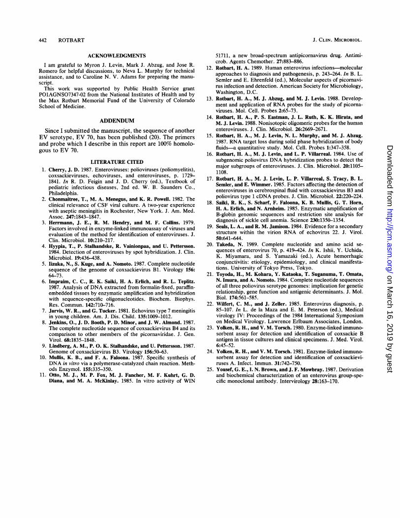
hybridization among the negative controls (not shown). Toaddress the sensitivity of this RNA amplification method, logdilutions of echovirus 11 were added to either CSF or PBS(Fig. 5). Amplified RNA was detected at all dilutions testedin PBS (103 through 106 TCID50) by both gel electrophoresisand hybridization. In CSF, the lowest titer of echovirus 11(103 TCID50) was detectable only by hybridization, but allother dilutions were detectable by both assays.
C.
G
VOL. 28, 1990 439
.... ..
on March 16, 2019 by guest
http://jcm.asm
.org/D
ownloaded from
440 ROTBART
A 13 - ;P'
< oe'l Pl
504
IM A 16
184154
124 oEli
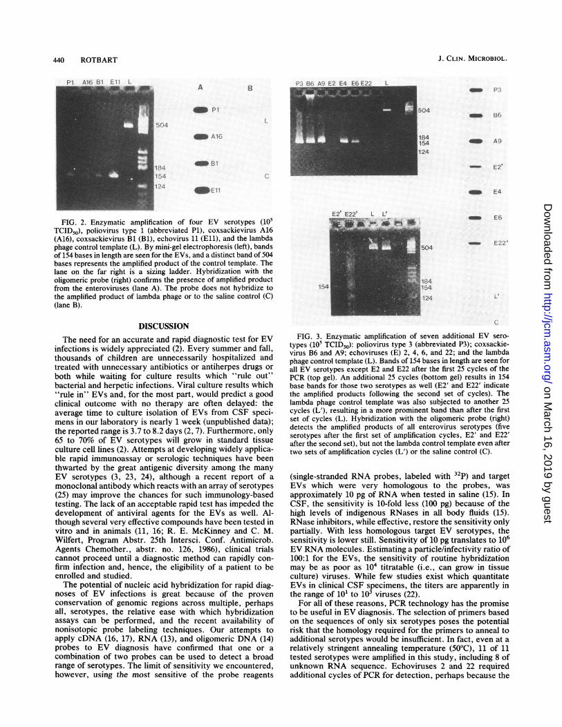
FIG. 2. Enzymatic amplification of four EV serotypes (105TCID50), poliovirus type 1 (abbreviated Pi), coxsackievirus A16(A16), coxsackievirus Bi (Bi), echovirus 11 (E11), and the lambdaphage control template (L). By mini-gel electrophoresis (left), bandsof 154 bases in length are seen for the EVs, and a distinct band of 504bases represents the amplified product of the control template. Thelane on the far right is a sizing ladder. Hybridization with theoligomeric probe (right) confirms the presence of amplified productfrom the enteroviruses (lane A). The probe does not hybridize tothe amplified product of lambda phage or to the saline control (C)(lane B).
DISCUSSION
The need for an accurate and rapid diagnostic test for EVinfections is widely appreciated (2). Every summer and fall,thousands of children are unnecessarily hospitalized andtreated with unnecessary antibiotics or antiherpes drugs orboth while waiting for culture results which "rule out"bacterial and herpetic infections. Viral culture results which"rule in" EVs and, for the most part, would predict a goodclinical outcome with no therapy are often delayed: theaverage time to culture isolation of EVs from CSF speci-mens in our laboratory is nearly 1 week (unpublished data);the reported range is 3.7 to 8.2 days (2, 7). Furthermore, only65 to 70% of EV serotypes will grow in standard tissueculture cell lines (2). Attempts at developing widely applica-ble rapid immunoassay or serologic techniques have beenthwarted by the great antigenic diversity among the manyEV serotypes (3, 23, 24), although a recent report of amonoclonal antibody which reacts with an array of serotypes(25) may improve the chances for such immunology-basedtesting. The lack of an acceptable rapid test has impeded thedevelopment of antiviral agents for the EVs as well. Al-though several very effective compounds have been tested invitro and in animals (11, 16; R. E. McKinney and C. M.Wilfert, Program Abstr. 25th Intersci. Conf. Antimicrob.Agents Chemother., abstr. no. 126, 1986), clinical trialscannot proceed until a diagnostic method can rapidly con-firm infection and, hence, the eligibility of a patient to beenrolled and studied.The potential of nucleic acid hybridization for rapid diag-
noses of EV infections is great because of the provenconservation of genomic regions across multiple, perhapsall, serotypes, the relative ease with which hybridizationassays can be performed, and the recent availability ofnonisotopic probe labeling techniques. Our attempts toapply cDNA (16, 17), RNA (13), and oligomeric DNA (14)probes to EV diagnosis have confirmed that one or acombination of two probes can be used to detect a broadrange of serotypes. The limit of sensitivity we encountered,however, using the most sensitive of the probe reagents
504- fIfl
184154124
A9
- E2r
E4
- E6
- E.22<
L t
154
FIG. 3. Enzymatic amplification of seven additional EV sero-types (105 TCID50): poliovirus type 3 (abbreviated P3); coxsackie-virus B6 and A9; echoviruses (E) 2, 4, 6, and 22; and the lambdaphage control template (L). Bands of 154 bases in length are seen forall EV serotypes except E2 and E22 after the first 25 cycles of thePCR (top gel). An additional 25 cycles (bottom gel) results in 154base bands for those two serotypes as well (E2' and E22' indicatethe amplified products following the second set of cycles). Thelambda phage control template was also subjected to another 25cycles (L'), resulting in a more prominent band than after the firstset of cycles (L). Hybridization with the oligomeric probe (right)detects the amplified products of all enterovirus serotypes (fiveserotypes after the first set of amplification cycles, E2' and E22'after the second set), but not the lambda control template even aftertwo sets of amplification cycles (L') or the saline control (C).
(single-stranded RNA probes, labeled with 32P) and targetEVs which were very homologous to the probes, wasapproximately 10 pg of RNA when tested in saline (15). InCSF, the sensitivity is 10-fold less (100 pg) because of thehigh levels of indigenous RNases in ail body fluids (15).RNase inhibitors, while effective, restore the sensitivity onlypartially. With less homologous target EV serotypes, thesensitivity is lower still. Sensitivity of 10 pg translates to 106EV RNA molecules. Estimating a particle/infectivity ratio of100:1 for the EVs, the sensitivity of routine hybridizationmay be as poor as 104 titratable (i.e., can grow in tissueculture) viruses. While few studies exist which quantitateEVs in clinical CSF specimens, the titers are apparently inthe range of 101 to 103 viruses (22).For all of these reasons, PCR technology has the promise
to be useful in EV diagnosis. The selection of primers basedon the sequences of only six serotypes poses the potentialrisk that the homology required for the primers to anneal toadditional serotypes would be insufficient. In fact, even at arelatively stringent annealing temperature (500C), 11 of 11tested serotypes were amplified in this study, including 8 ofunknown RNA sequence. Echoviruses 2 and 22 requiredadditional cycles of PCR for detection, perhaps because the
J. CLIN. MICROBIOL.
on March 16, 2019 by guest
http://jcm.asm
.org/D
ownloaded from
ENZYMATIC RNA AMPLIFICATION OF ENTEROVIRUSES
f .
Af3~-~~~~~~~~~~~~~~~~~~~~1.241541241
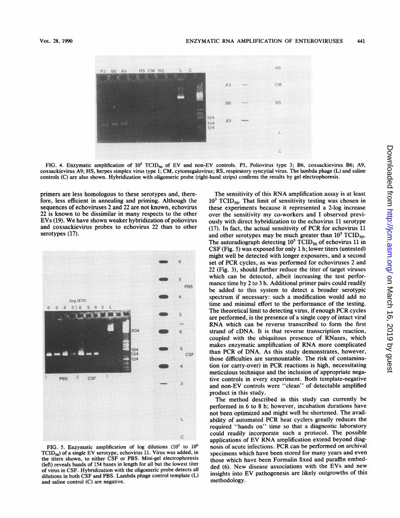
FIG. 4. Enzymatic amplification of 105 TCID50 of EV and non-EV controls. P3, Poliovirus type 3; B6, coxsackievirus B6; A9,coxsackievirus A9; HS, herpes simplex virus type 1; CM, cytomegalovirus; RS, respiratory syncytial virus. The lambda phage (L) and salinecontrols (C) are also shown. Hybridization with oligomeric probe (right-hand strips) confirms the results by gel electrophoresis.
primers are less homologous to these serotyfore, less efficient in annealing and primingsequences of echoviruses 2 and 22 are not kn22 is known to be dissimilar in many respe
EVs (19). We have shown weaker hybridizatiand coxsackievirus probes to echovirus 2,serotypes (17).
Iog [E11
6 S 4 3i 6 5 4 3 L
__ ~~~~~~~504
184_ 1~~54
124
PBS " CSF
FIG. 5. Enzymatic amplification of log diluTCID50) of a single EV serotype, echovirus 11. Vthe titers shown, to either CSF or PBS. Mini-g(left) reveals bands of 154 bases in length for all tof virus in CSF. Hybridization with the oligomeridilutions in both CSF and PBS. Lambda phage ccand saline control (C) are negative.
ypes and, there- The sensitivity of this RNA amplification assay is at leastg. Although the 103 TCID50. That limit of sensitivity testing was chosen iniown, echovirus these experiments because it represented a 2-log increasects to the other over the sensitivity my co-workers and I observed previ-ion of poliovirus ously with direct hybridization to the echovirus 11 serotype2 than to other (17). In fact, the actual sensitivity of PCR for echovirus 11
and other serotypes may be much greater than 103 TCID50.The autoradiograph detecting 103 TCID50 of echovirus 11 inCSF (Fig. 5) was exposed for only 1 h; lower titers (untested)might well be detected with longer exposures, and a second
_ 6 set of PCR cycles, as was performed for echoviruses 2 and22 (Fig. 3), should further reduce the titer of target viruses
_> which can be detected, albeit increasing the test perfor-PBS mance time by 2 to 3 h. Additional primer pairs could readily
be added to this system to detect a broader serotypic4 spectrum if necessary: such a modification would add no
time and minimal effort to the performance of the testing._3 The theoretical limit to detecting virus, if enough PCR cycles
are performed, is the presence of a single copy of intact viral- RNA which can be reverse transcribed to form the first
_ e6 strand of cDNA. It is that reverse transcription reaction,coupled with the ubiquitous presence of RNases, which
_5 makes enzymatic amplification of RNA more complicatedCSF than PCR of DNA. As this study demonstrates, however,
those difficulties are surmountable. The risk of contamina-_D 4 tion (or carry-over) in PCR reactions is high, necessitating
meticulous technique and the inclusion of appropriate nega-tive controls in every experiment. Both template-negative
- 3 and non-EV controls were "clean" of detectable amplifiedproduct in this study.The method described in this study can currently be
performed in 6 to 8 h; however, incubation durations havenot been optimized and might well be shortened. The avail-
L ability of automated PCR heat cyclers greatly reduces therequired "hands on" time so that a diagnostic laboratory
c could readily incorporate such a protocol. The possibleapplications of EV RNA amplification extend beyond diag-
tions (103 to 106 nosis of acute infections. PCR can be performed on archival'irus was added, in specimens which have been stored for many years and even,el electrophoresis those which have been Formalin fixed and paraffin embed-
cuprobeldetesctsitel ded (6). New disease associations with the EVs and newntrol template (L) insights into EV pathogenesis are likely outgrowths of this
methodology.
VOL. 28, 1990 441
on March 16, 2019 by guest
http://jcm.asm
.org/D
ownloaded from
J. CLIN. MICROBIOL.
ACKNOWLEDGMENTS
I am grateful to Myron J. Levin, Mark J. Abzug, and Jose R.Romero for helpful discussions, to Neva L. Murphy for technicalassistance, and to Caroline N. V. Adams for preparing the manu-script.
This work was supported by Public Health Service grantP01AGNS07347-02 from the National Institutes of Health and bythe Max Rotbart Memorial Fund of the University of ColoradoSchool of Medicine.
ADDENDUM
Since I submitted the manuscript, the sequence of anotherEV serotype, EV 70, has been published (20). The primersand probe which I describe in this report are 100% homolo-gous to EV 70.
LITERATURE CITED1. Cherry, J. D. 1987. Enteroviruses: polioviruses (poliomyelitis),
coxsackieviruses, echoviruses, and enteroviruses, p. 1729-1841. In R. D. Feigin and J. D. Cherry (ed.), Textbook ofpediatric infectious diseases, 2nd ed. W. B. Saunders Co.,Philadelphia.
2. Chonmaitree, T., M. A. Menegus, and K. R. Powell. 1982. Theclinical relevance of CSF viral culture. A two-year experiencewith aseptic meningitis in Rochester, New York. J. Am. Med.Assoc. 247:1843-1847.
3. Herrmann, J. E., R. M. Hendry, and M. F. Collins. 1979.Factors involved in enzyme-linked immunoassay of viruses andevaluation of the method for identification of enteroviruses. J.Clin. Microbiol. 10:210-217.
4. Hyypia, T., P. Stalhandske, R. Vainionpaa, and U. Pettersson.1984. Detection of enteroviruses by spot hybridization. J. Clin.Microbiol. 19:436-438.
5. Iizuka, N., S. Kuge, and A. Nomoto. 1987. Complete nucleotidesequence of the genome of coxsackievirus B1. Virology 156:64-73.
6. Impraim, C. C., R. K. Saiki, H. A. Erlich, and R. L. Teplitz.1987. Analysis of DNA extracted from formalin-fixed, paraffin-embedded tissues by enzymatic amplification and hybridizationwith sequence-specific oligonucleotides. Biochem. Biophys.Res. Commun. 142:710-716.
7. Jarvis, W. R., and G. Tucker. 1981. Echovirus type 7 meningitisin young children. Am. J. Dis. Child. 135:1009-1012.
8. Jenkins, O., J. D. Booth, P. D. Minor, and J. W. Almond. 1987.The complete nucleotide sequence of coxsackievirus B4 and itscomparison to other members of the picornaviridae. J. Gen.Virol. 68:1835-1848.
9. Lindberg, A. M., P. O. K. Stalhandske, and U. Pettersson. 1987.Genome of coxsackievirus B3. Virology 156:50-63.
10. Mullis, K. B., and F. A. Faloona. 1987. Specific synthesis ofDNA in vitro via a polymerase-catalyzed chain reaction. Meth-ods Enzymol. 155:335-350.
11. Otto, M. J., M. P. Fox, M. J. Fancher, M. F. Kuhrt, G. D.Diana, and M. A. McKinlay. 1985. In vitro activity of WIN
51711, a new broad-spectrum antipicornavirus drug. Antimi-crob. Agents Chemother. 27:883-886.
12. Rotbart, H. A. 1989. Human enterovirus infections-molecularapproaches to diagnosis and pathogenesis, p. 243-264. In B. L.Semler and E. Ehrenfeld (ed.), Molecular aspects of picornavi-rus infection and detection. American Society for Microbiology,Washington, D.C.
13. Rotbart, H. A., M. J. Abzug, and M. J. Levin. 1988. Develop-ment and application of RNA probes for the study of picorna-viruses. Mol. Cell. Probes 2:65-73.
14. Rotbart, H. A., P. S. Eastman, J. L. Ruth, K. K. Hirata, andM. J. Levin. 1988. Nonisotopic oligomeric probes for the humanenteroviruses. J. Clin. Microbiol. 26:2669-2671.
15. Rotbart, H. A., M. J. Levin, N. L. Murphy, and M. J. Abzug.1987. RNA target loss during solid phase hybridization of bodyfluids-a quantitative study. Mol. Cell. Probes 1:347-358.
16. Rotbart, H. A., M. J. Levin, and L. P. Viliarreal. 1984. Use ofsubgenomic poliovirus DNA hybridization probes to detect themajor subgroups of enteroviruses. J. Clin. Microbiol. 20:1105-1108.
17. Rotbart, H. A., M. J. Levin, L. P. Villarreal, S. Tracy, B. L.Semler, and E. Wimmer. 1985. Factors affecting the detection ofenteroviruses in cerebrospinal fluid with coxsackievirus B3 andpoliovirus type 1 cDNA probes. J. Clin. Microbiol. 22:220-224.
18. Saiki, R. K., S. Scharf, F. Faloona, K. B. Mullis, G. T. Horn,H. A. Erlich, and N. Arnheim. 1985. Enzymatic amplification ofB-globin genomic sequences and restriction site analysis fordiagnosis of sickle cell anemia. Science 230:1350-1354.
19. Seals, L. A., and R. M. Jamison. 1984. Evidence for a secondarystructure within the virion RNA of echovirus 22. J. Virol.50:641-644.
20. Takeda, N. 1989. Complete nucleotide and amino acid se-quences of enterovirus 70, p. 419-424. In K. Ishii, Y. Uchida,K. Miyamara, and S. Yamazaki (ed.), Acute hemorrhagicconjunctivitis: etiology, epidemiology, and clinical manifesta-tions. University of Tokyo Press, Tokyo.
21. Toyoda, H., M. Kohara, Y. Kataoka, T. Suganuma, T. Omata,N. Imura, and A. Nomoto. 1984. Complete nucleotide sequencesof all three poliovirus serotype genomes: implication for geneticrelationship, gene function and antigenic determinants. J. Mol.Biol. 174:561-585.
22. Wilfert, C. M., and J. Zeller. 1985. Enterovirus diagnosis, p.85-107. In L. de la Maza and E. M. Peterson (ed.), Medicalvirology IV: Proceedings of the 1984 International Symposiumon Medical Virology. Lawrence Erlbaum Associates, London.
23. Yolken, R. H., and V. M. Torsch. 1980. Enzyme-linked immuno-sorbent assay for detection and identification of coxsackie Bantigen in tissue cultures and clinical specimens. J. Med. Virol.6:45-52.
24. Yolken, R. H., and V. M. Torsch. 1981. Enzyme-linked immuno-sorbent assay for detection and identification of coxsackievi-ruses A. Infect. Immun. 31:742-750.
25. Yousef, G. E., I. N. Brown, and J. F. Mowbray. 1987. Derivationand biochemical characterization of an enterovirus group-spe-cific monoclonal antibody. Intervirology 28:163-170.
442 ROTBART
on March 16, 2019 by guest
http://jcm.asm
.org/D
ownloaded from