Embed Size (px)
Citation preview
Bahrain Medical Bulletin, Vol. 38, No. 2, June 2016
102
Wells Syndrome is a rare inflammatory disorder, characterized by the accumulation of eosinophils in the skin. It is a skin disorder with unknown etiology characterized by recurrent pruritic or tender skin lesions resembling cellulitis; cases often resolve spontaneously, although recurrences are common over several years.
Clinically, it has wide and variable presentations; however, painful erythematous plaque is a common presentation. It is often misdiagnosed as cellulitis due to the similarity in their presentation. Although the incidence and the number of the reported cases in the literature are increasing, still the exact etiology of the disease remain unknown and further studies are required to explore the pathogenesis. Several triggers have been proposed as potential contributors to the development of the disease. Misdiagnosis leads to a delay of proper management and inappropriate use of antibiotics.
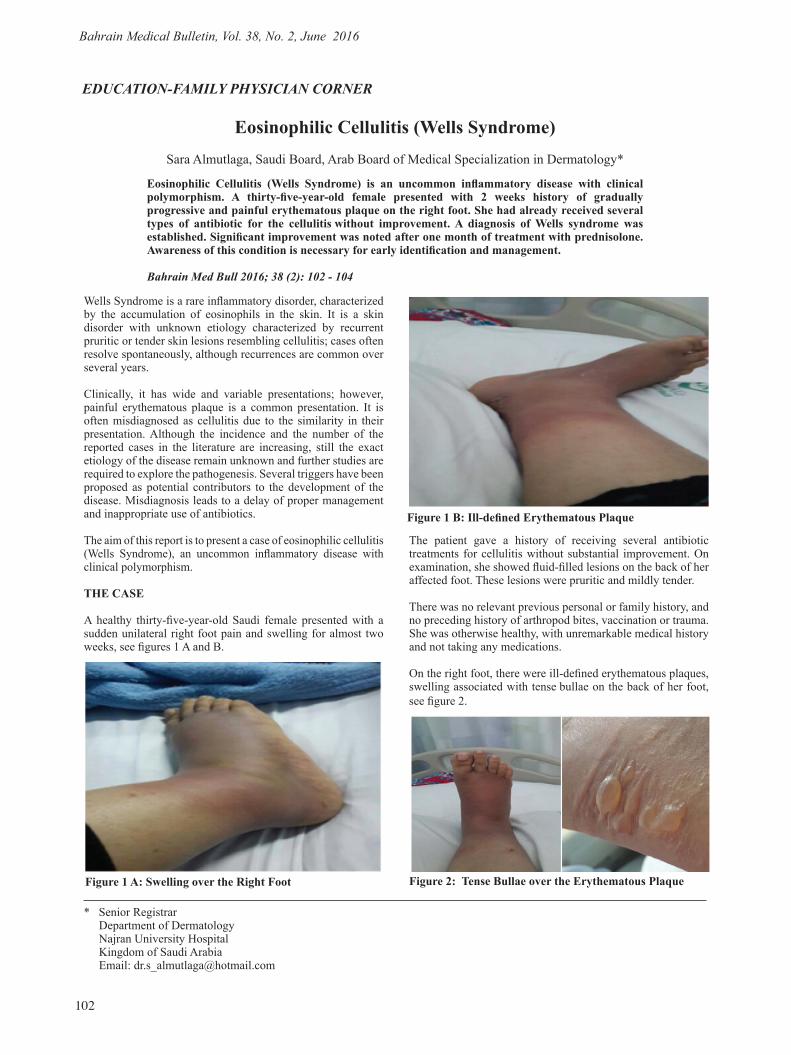
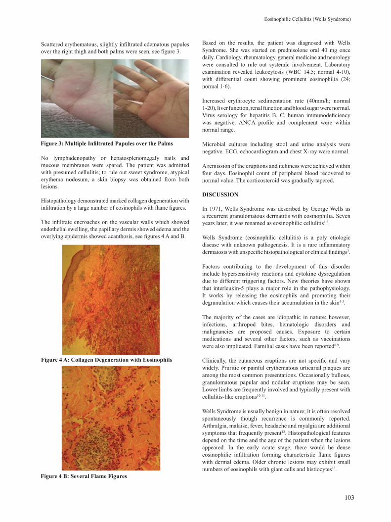
The aim of this report is to present a case of eosinophilic cellulitis (Wells Syndrome), an uncommon inflammatory disease with clinical polymorphism. THE CASE A healthy thirty-five-year-old Saudi female presented with a sudden unilateral right foot pain and swelling for almost two weeks, see figures 1 A and B.
EDUCATION-FAMILY PHYSICIAN CORNER
Eosinophilic Cellulitis (Wells Syndrome)Sara Almutlaga, Saudi Board, Arab Board of Medical Specialization in Dermatology*
Eosinophilic Cellulitis (Wells Syndrome) is an uncommon inflammatory disease with clinical polymorphism. A thirty-five-year-old female presented with 2 weeks history of gradually progressive and painful erythematous plaque on the right foot. She had already received several types of antibiotic for the cellulitis without improvement. A diagnosis of Wells syndrome was established. Significant improvement was noted after one month of treatment with prednisolone. Awareness of this condition is necessary for early identification and management.
Bahrain Med Bull 2016; 38 (2): 102 - 104
* Senior RegistrarDepartment of DermatologyNajran University HospitalKingdom of Saudi ArabiaEmail: [email protected]
The patient gave a history of receiving several antibiotic treatments for cellulitis without substantial improvement. On examination, she showed fluid-filled lesions on the back of her affected foot. These lesions were pruritic and mildly tender.
There was no relevant previous personal or family history, and no preceding history of arthropod bites, vaccination or trauma. She was otherwise healthy, with unremarkable medical history and not taking any medications.
On the right foot, there were ill-defined erythematous plaques, swelling associated with tense bullae on the back of her foot, see figure 2.
Figure 1 A: Swelling over the Right Foot
Figure 1 B: Ill-defined Erythematous Plaque
Figure 2: Tense Bullae over the Erythematous Plaque
Bahrain Medical Bulletin, Vol. 38, No. 2, June 2016
103
Scattered erythematous, slightly infiltrated edematous papules over the right thigh and both palms were seen, see figure 3.
No lymphadenopathy or hepatosplenomegaly nails and mucous membranes were spared. The patient was admitted with presumed cellulitis; to rule out sweet syndrome, atypical erythema nodosum, a skin biopsy was obtained from both lesions.
Histopathology demonstrated marked collagen degeneration with infiltration by a large number of eosinophils with flame figures.
The infiltrate encroaches on the vascular walls which showed endothelial swelling, the papillary dermis showed edema and the overlying epidermis showed acanthosis, see figures 4 A and B.
Based on the results, the patient was diagnosed with Wells Syndrome. She was started on prednisolone oral 40 mg once daily. Cardiology, rheumatology, general medicine and neurology were consulted to rule out systemic involvement. Laboratory examination revealed leukocytosis (WBC 14.5; normal 4-10), with differential count showing prominent eosinophilia (24; normal 1-6).
Increased erythrocyte sedimentation rate (40mm/h; normal 1-20), liver function, renal function and blood sugar were normal. Virus serology for hepatitis B, C, human immunodeficiency was negative. ANCA profile and complement were within normal range.
Microbial cultures including stool and urine analysis were negative. ECG, echocardiogram and chest X-ray were normal.
A remission of the eruptions and itchiness were achieved within four days. Eosinophil count of peripheral blood recovered to normal value. The corticosteroid was gradually tapered.
DISCUSSION
In 1971, Wells Syndrome was described by George Wells as a recurrent granulomatous dermatitis with eosinophilia. Seven years later, it was renamed as eosinophilic cellulitis1,2.
Wells Syndrome (eosinophilic cellulitis) is a poly etiologic disease with unknown pathogenesis. It is a rare inflammatory dermatosis with unspecific histopathological or clinical findings3.
Factors contributing to the development of this disorder include hypersensitivity reactions and cytokine dysregulation due to different triggering factors. New theories have shown that interleukin-5 plays a major role in the pathophysiology. It works by releasing the eosinophils and promoting their degranulation which causes their accumulation in the skin4-5.
The majority of the cases are idiopathic in nature; however, infections, arthropod bites, hematologic disorders and malignancies are proposed causes. Exposure to certain medications and several other factors, such as vaccinations were also implicated. Familial cases have been reported6-9.
Clinically, the cutaneous eruptions are not specific and vary widely. Pruritic or painful erythematous urticarial plaques are among the most common presentations. Occasionally bullous, granulomatous papular and nodular eruptions may be seen. Lower limbs are frequently involved and typically present with cellulitis-like eruptions10-11.
Wells Syndrome is usually benign in nature; it is often resolved spontaneously though recurrence is commonly reported. Arthralgia, malaise, fever, headache and myalgia are additional symptoms that frequently present12. Histopathological features depend on the time and the age of the patient when the lesions appeared. In the early acute stage, there would be dense eosinophilic infiltration forming characteristic flame figures with dermal edema. Older chronic lesions may exhibit small numbers of eosinophils with giant cells and histiocytes13.
Figure 3: Multiple Infiltrated Papules over the Palms
Figure 4 A: Collagen Degeneration with Eosinophils
Figure 4 B: Several Flame Figures
Eosinophilic Cellulitis (Wells Syndrome)
Bahrain Medical Bulletin, Vol. 38, No. 2, June 2016
104
Therapy with systemic corticosteroids considered the first line of treatment; mild cases may respond to topical steroid therapy only14. Other possible treatments include dapsone, systemic antihistamines, antimicrobial agents, colchicines, interferon-α, antimalarial drugs or immunosuppressive agents. New therapeutic options may be tyrosine kinase inhibitors, such as imatinib and anti–tumor necrosis factor alpha like adalimumab1,15-19.
Wells Syndrome usually has an excellent prognosis. Most cases resolve within weeks to months and the lesions tend to heal without scarring. Recurrence is also commonly reported14-19.
CONCLUSION
Wells Syndrome is a rare clinical entity, with unspecific presentation which could mimic a variety of diseases, especially cellulitis. If cellulitis is not responding to antibiotic treatment, Wells Syndrome should be suspected.__________________________________________________
Potential Conflicts of Interest: None
Competing Interest: None. Sponsorship: None
Submission Date: 18 March 2015.
Acceptance Date: 3 April 2016
Ethical Approval: Approved by Department of Dermatology, Najran University Hospital, Kingdom of Saudi Arabia.
REFERENCES
1. Wells GC. Recurrent Granulomatous Dermatitis with Eosinophilia. Trans St John’s Hosp Dermatol Soc 1971; 57(1):46-56.
2. Wells GC, Smith NP. Eosinophilic Cellulitis. Br J Dermatol 1979; 100(1):101-9.
3. Renner R, Kauer F, Treudler R, et al. Eosinophilic Cellulitis (Wells' Syndrome) in Association with Angioimmunoblastic Lymphadenopathy. Acta Derm Venereol 2007; 87(6):525-8.
4. Mould AW, Matthaei KI, Young IG, et al. Relationship Between Interleukin-5 and Eotaxin in Regulating Blood and Tissue Eosinophilia in Mice. J Clin Invest 1997; 99(5):1064-71.
5. España A, Sanz ML, Sola J, et al. Wells’ Syndrome (Eosinophilic Cellulitis): Correlation between Clinical Activity, Eosinophil Levels, Eosinophil Cation Protein and Interleukin-5. Br J Dermatol 1999; 140(1):127-30.
6. Kaufmann D, Pichler W, Beer JH. Severe Episode of High Fever with Rash, Lymphadenopathy, Neutropenia, and Eosinophilia after Minocycline Therapy for Acne. Arch Intern Med 1994; 154(17):1983-4.
7. Hirsch K, Ludwig RJ, Wolter M, et al. Eosinophilic Cellulitis (Wells’ Syndrome) Associated with Colon Carcinoma. J Dtsch Dermatol Ges 2005; 3(7):530-1.
8. Heelan K, Ryan JF, Shear NH, et al. Wells Syndrome (Eosinophilic Cellulitis): Proposed Diagnostic Criteria and a Literature Review of the Drug-Induced Variant. J Dermatol Case Rep 2013; 7(4):113-20.
9. Winfield H, Lain E, Horn T, et al. Eosinophilic Cellulitislike Reaction to Subcutaneous Etanercept Injection. Arch Dermatol 2006; 142(2):218-20.
10. Odia SG, Purschel W, Worret WI, et al. Hypereosinophilic Cellulitis (Wells› Syndrome) Resembling Urticarial. Acta Derm Venerol (Ljubljana) 1994; 6:193–195.
11. Spinelli M, Frigerio E, Cozzi A, et al. Bullous Wells› Syndrome Associated with Non-Hodgkin›s Lymphocytic Lymphoma. Acta Derm Venereol 2008; 88(5):530-1.
12. Mutasim DF, Cooper CH. A Case of Wells› Syndrome in a Patient with Lymphocytic Lymphoma. Geriatr Dermatol 1996; 4(1):11-14.
13. Steffen C. The Man behind the Eponym. George Crichton Wells: Eosinophilic Cellulitis (Wells Syndrome). Am J Dermatopathol 2002; 24(2):164-5.
14. Caputo R, Marzano AV, Vezzoli P, et al. Wells Syndrome in Adults and Children: A Report of 19 Cases. Arch Dermatol 2006; 142(9):1157-61.
15. Aberer W, Konrad K, Wolff K. Wells› Syndrome Is a Distinctive Disease Entity and Not a Histologic Diagnosis. J Am Acad Dermatol 1988; 18(1 Pt 1):105-14.
16. Odia SG, Puerschel W, Worret WI, et al. Hypereosinophilic Cellulitis (Wells' syndrome) Resembling Urticaria. Acta Dermatovenerologica Alpina Panomica et Adriatica (APA) 1994; 4:193–6.
17. Herr H, Koh JK. Eosinophilic Cellulitis (Wells' Syndrome) Successfully Treated with Low-dose Cyclosporine. J Korean Med Sci 2001; 16(5):664-8.
18. Husak R, Goerdt S, Orfanos CE. Interferon Alfa Treatment of a Patient with Eosinophilic Cellulitis and HIV Infection. N Engl J Med 1997; 337(9):641-2.
19. Davis RF, Dusanjh P, Majid A, et al. Eosinophilic Cellulitis as a Presenting Feature of Chronic Eosinophilic Leukaemia, Secondary to a Deletion on Chromosome 4q12 Creating the FIP1L1-PDGFRA Fusion Gene. Br J Dermatol 2006; 155(5):1087-9.


![ENVIRONMENTAL PROTECTION COMMISSION[567] · 2014-06-25 · wells, recreational-use wells, monitoring wells, heat pump supply wells or GHEX loop boreholes, industrial wells, and dewatering](https://img.pdfslide.net/doc/110x75/5f3f728939b254613866ae00/environmental-protection-commission567-2014-06-25-wells-recreational-use-wells.jpg)


![Case Report Eosinophilic dermatosis: Wells syndrome. A · PDF fileWells” [4]. CASO CLÍNICO Varón, 58 años, chapista, procedente de área urbana del Paraguay (Sudamérica) con](https://img.pdfslide.net/doc/110x75/5a9680227f8b9a451b8cd195/case-report-eosinophilic-dermatosis-wells-syndrome-a-4-caso-clnico-varn.jpg)