Embed Size (px)
Citation preview
Bahrain Medical Bulletin, Vol. 38, No. 1, March 2016
46
Pre-eclampsia is a pregnancy-specific disease characterized by hypertension and significant proteinuria in a previously healthy woman on or after the 20th week of gestation, occurring in about 2% to 8% of pregnancies1,2.
The underlying abnormality is thought to be severe generalized vasospasm occurring throughout the body, perhaps as a result of increased sensitivity to circulating prostaglandins. The visual system may be affected in 30% to 100% of patients with pre-eclampsia. Blurred vision, although infrequent, is the most common symptom. Exudative retinal detachment is an unusual but well-documented cause of visual loss in pre-eclampsia, affecting less than 1% of pre-eclamptic patients and slightly higher incidence in patients with eclampsia3.
The aim of this presentation is to report a rare complication of pre-eclampsia presenting as visual disturbance in late pregnancy and after delivery.
THE CASE
A nineteen-year-old primigravida with no known previous systemic illness was seen in the obstetrics clinic on 35th week of gestation for a routine check-up. On examination, the patient was found to have blood pressure of 179/102 mmHg with a mean arterial pressure of 130 mmHg. Urine dip stick showed 3+ albumin and clinical examination showed bilateral pitting pedal edema and brisk deep tendon reflexes. Abdominal examination revealed a gestational age of 35 weeks, longitudinal lie and cephalic presentation of the fetus. Ultrasonography confirmed a singleton, live pregnancy.
The patient was admitted to the hospital for blood pressure monitoring and further investigations. Complete blood count, renal function test, liver function test, coagulation profile and 24 hours collection of urine protein were performed; all were within normal range except for the 24 hours collection of protein which was high (900 mg/24 hours). During hospitalization, the patient’s blood pressure was within normal and she was kept off anti-hypertensive medications until the 4th day of admission where she was booked for Cesarean section delivery.
Bilateral Exudative Retinal Detachment in Pre-Eclampsia
Muneera Abunajma, SB* Haitham Abdulla, FA** Saad Alkhalifa, FRCSC***
Exudative retinal detachment (ERD) is a rare cause of visual loss in pre-eclampsia with only few reported cases in the medical literature. We report a case of a nineteen-year-old primigravida with pre-eclampsia who developed bilateral exudative retinal detachments following delivery. Spontaneous resorption of the subretinal fluid and complete resolution of the exudative detachment occurred after a few weeks of observation.
Bahrain Med Bull 2016; 38(1): 46 - 47
* Senior Resident** Junior Resident*** Consultant Ophthalmologist, Vitreoretinal Surgery Department of Ophthalmology Bahrain Defense Force Royal Medical Services Kingdom of Bahrain
Email: [email protected]
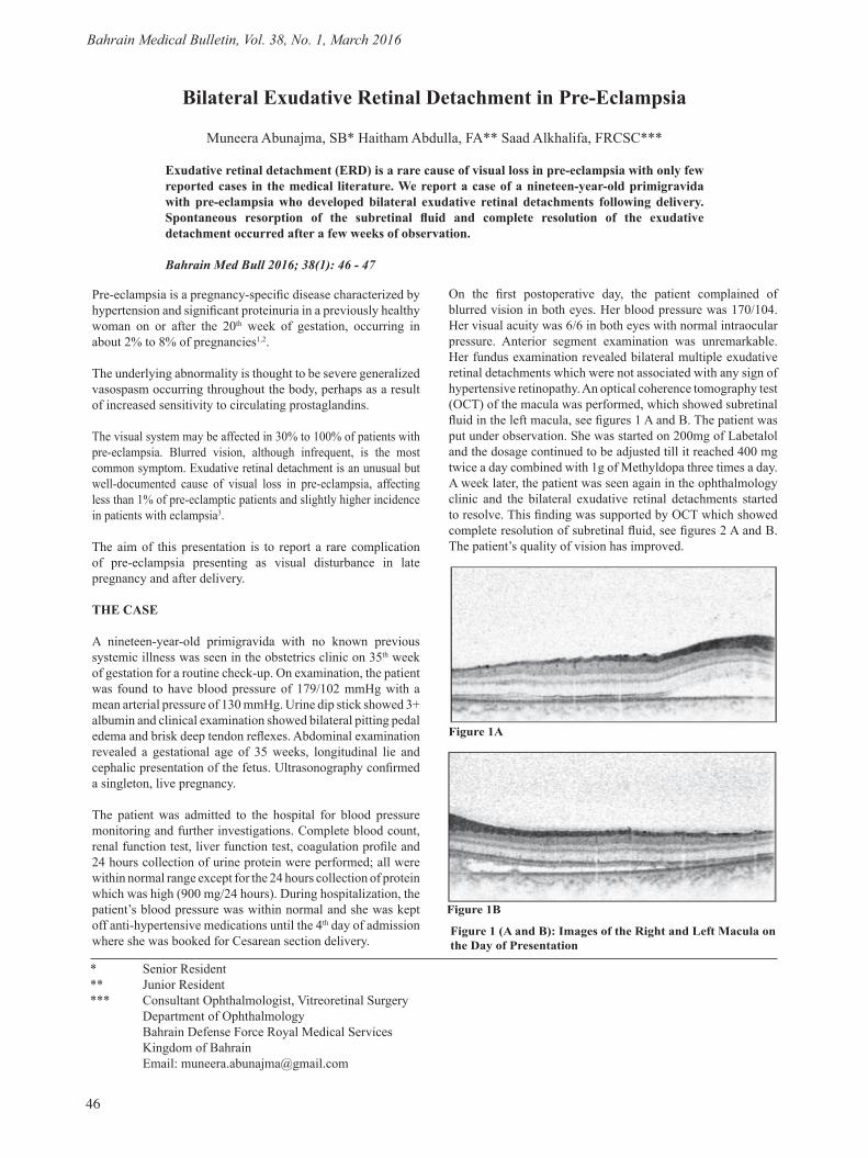
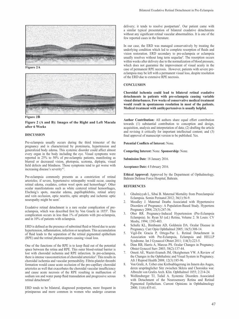
On the first postoperative day, the patient complained of blurred vision in both eyes. Her blood pressure was 170/104. Her visual acuity was 6/6 in both eyes with normal intraocular pressure. Anterior segment examination was unremarkable. Her fundus examination revealed bilateral multiple exudative retinal detachments which were not associated with any sign of hypertensive retinopathy. An optical coherence tomography test (OCT) of the macula was performed, which showed subretinal fluid in the left macula, see figures 1 A and B. The patient was put under observation. She was started on 200mg of Labetalol and the dosage continued to be adjusted till it reached 400 mg twice a day combined with 1g of Methyldopa three times a day. A week later, the patient was seen again in the ophthalmology clinic and the bilateral exudative retinal detachments started to resolve. This finding was supported by OCT which showed complete resolution of subretinal fluid, see figures 2 A and B. The patient’s quality of vision has improved.
Figure 1 (A and B): Images of the Right and Left Macula on the Day of Presentation
Figure 1A
Figure 1B
Bahrain Medical Bulletin, Vol. 38, No. 1, March 2016
47
DISCUSSION
Pre-eclampsia usually occurs during the third trimester of the pregnancy and is characterized by proteinuria, hypertension and generalized body edema. This systemic disorder could affect almost every organ in the body including the eye. Visual symptoms were reported in 25% to 50% of pre-eclamptic patients, manifesting as blurred or decreased vision, photopsia, scotoma, diplopia, visual field defects and blindness. Those symptoms tend to get worse with increasing disease’s severity4,5.
Pre-eclampsia commonly presents as a constriction of retinal arterioles; if severe, hypertensive retinopathy would occur, causing retinal edema, exudates, cotton wool spots and hemorrhage6. Other ocular manifestations such as white centered retinal hemorrhages, Elschnig’s spots, macular edema, papillophlebitis, retinal artery and vein occlusion, optic neuritis, optic atrophy and ischemic optic neuropathy might be seen7.
Exudative retinal detachment is a rare ocular complication of pre-eclampsia, which was described first by Von Graefe in 18558. This complication occurs in less than 1% of patients with pre-eclampsia, and in 10% of patients with eclampsia.
ERD is defined as the presence of subretinal fluid or blood due to acute hypertension, inflammation, infection or neoplasm. This accumulation of fluid leads to the separation of the retinal pigmented epithelium (RPE) and the retinal photoreceptors causing visual loss.
One of the functions of the RPE is to keep fluid out of the potential space between the retina and RPE. This outer blood-retinal barrier is lost with choroidal ischemia and RPE infarction. In pre-eclampsia, there is intense vasoconstriction of choroidal arterioles4. This results in choroidal ischemia and vascular permeability. Fibrin-platelet thrombi formation would cause acute occlusion of the pre-capillary choroidal arterioles as well that exacerbates the choroidal vascular insufficiency and cause acute necrosis of the RPE resulting in malfunction of sodium ion and water pump fluid transudation and progressive serous retinal detachment9.
ERD tends to be bilateral, diagnosed postpartum, more frequent in primiparous and more common in women who undergo cesarean
delivery; it tends to resolve postpartum5. Our patient came with a similar typical presentation of bilateral exudative detachments without any significant retinal vascular abnormalities. It is one of the few reported cases in the literature.
In our case, the ERD was managed conservatively by treating the underlying condition which led to complete resorption of fluids and vision restoration. ERD secondary to pre-eclampsia or eclampsia usually resolves without long term sequelae9. The resorption occurs within weeks after delivery due to the normalization of blood pressure, which does not guarantee the improvement of visual acuity in the case of permanent RPE necrosis. However, patients with severe pre-eclampsia may be left with a permanent visual loss, despite resolution of the ERD due to extensive RPE necrosis.
CONCLUSION
Choroidal ischemia could lead to bilateral retinal exudative detachments in patients with pre-eclampsia causing variable visual disturbances. Few weeks of conservative medical treatment would result in spontaneous resolution in most of the patients. Medical treatment with antihypertensives is usually helpful. _______________________________________________________
Author Contribution: All authors share equal effort contribution towards (1) substantial contribution to conception and design, acquisition, analysis and interpretation of data; (2) drafting the article and revising it critically for important intellectual content; and (3) final approval of manuscript version to be published. Yes.
Potential Conflicts of Interest: None.
Competing Interest: None. Sponsorship: None.
Submission Date: 18 January 2016.
Acceptance Date: 4 February 2016.
Ethical Approval: Approved by the Department of Opthalmology, Bahrain Defense Force Hospital, Bahrain.
REFERENCES
1. Ghulmiyyah L, Sibai B. Maternal Mortality from Preeclampsia/Eclampsia. Semin Perinatol 2012; 36(1):56-9.
2. Moodley J. Maternal Deaths Associated with Hypertensive Disorders of Pregnancy: A Population-Based Study. Hypertens Pregnancy 2004; 23(3):247-56.
3. Ober RR. Pregnancy-Induced Hypertension (Pre-Eclampsia Eclampsia). In: Ryan SJ (ed.) Retina, Volume 2. St Louis: CV Mosby, 1994; 1393-403.
4. Schultz KL, Birnbaum AD, Goldstein DA. Ocular Disease in Pregnancy. Curr Opin Ophthalmol 2005; 16(5):308-14.
5. Vigil-De Gracia P, Ortega-Paz L. Retinal Detachment in Association with Pre-Eclampsia, Eclampsia and HELLP Syndrome. Int J Gynaecol Obstet 2011; 114(3):223-5.
6. Dinn RB, Harris A, Marcus PS. Ocular Changes in Pregnancy. Obstet Gynecol Surv 2003; 58(2):137-44.
7. Omoti AE, Waziri-Erameh JM, Okeigbemen VW. A Review of the Changes in the Ophthalmic and Visual System in Pregnancy. Afr J Reprod Health 2008; 12(3):185-96.
8. Von Grafe, A. Ueber eine Krebsablagerung im Innern des Auges, deren ursprünglicher Sitz zwischen Sklera und Choroidea war. Albrecht von Grafes Arch. Klin. Ophthalmol 1855; 2:214-24.
9. Wolfensberger TJ, Tufail A. Systemic Disorders Associated with Detachment of the Neurosensory Retina and Retinal Pigmented Epithelium. Current Opinions in Ophthalmology 2000; 11(6):455-61.
Figure 2 (A and B): Images of the Right and Left Macula after 6 Weeks
Figure 2B
Figure 2A
Bilateral Exudative Retinal Detachment in Pre-Eclampsia