Embed Size (px)
Citation preview
REVIEW
Eph/ephrin molecules—a hub for signalingand endocytosis
Mara E. Pitulescu and Ralf H. Adams1
Department of Tissue Morphogenesis, Max-Planck-Institute for Molecular Biomedicine, and Faculty of Medicine, Universityof Munster, D-48149 Munster, Germany
The development, homeostasis, and regeneration of com-plex organ systems require extensive cell–cell communi-cation to ensure that different cells proliferate, migrate,differentiate, assemble, and function in a coordinated andtimely fashion. Eph receptor tyrosine kinases and theirephrin ligands are critical regulators of cell contact-de-pendent signaling and patterning. Eph/ephrin binding canlead to very diverse biological readouts such as adhesionversus repulsion, or increased versus decreased motility.Accordingly, depending on cell type and context, a limitedand conserved set of receptor–ligand interactions is trans-lated into a large variety of downstream signaling pro-cesses. Recent evidence indicates that the endocytosis ofEph/ephrin molecules, together with the internalizationof various associated tissue-specific effectors, might beone of the key principles responsible for such highlydiverse and adaptable biological roles. Here, we summa-rize recent insights into Eph/ephrin signaling and endo-cytosis in three biological systems; i.e., the brain,intestine, and vasculature.
Eph receptors, which have been divided into the sub-classes A and B, represent the largest family of receptortyrosine kinases (RTKs) in the animal kingdom. Inhumans, nine EphA (EphA1–8 and EphA10) and five EphB(EphB1–4 and EphB6) receptors are known. The extracel-lular part of Eph receptors contains a globular ligand-binding domain, a cysteine-rich region, and two fibronec-tin type III repeats. The intracellular, cytoplasmic partconsists of a short juxtamembrane region with severalconserved tyrosine residues, the tyrosine kinase domain,a sterile a motif (SAM) protein–protein interaction do-main, and a C-terminal PDZ-binding motif (Fig. 1A).Based on their structural features and binding affinityfor A- or B-type receptors, the ligands have also been di-vided into GPI-anchored ephrin-A (ephrin-A1–5 in mam-mals) and transmembranous ephrin-B (ephrin-B1–3) mol-ecules (Pasquale 2005). While EphA receptors typically
bind to ephrin-A proteins, and EphBs bind to ephrin-Bligands, there is also limited cross-binding between mem-bers of the two classes (Fig. 1A; Kullander and Klein 2002;Himanen et al. 2004; Klein 2004). In contrast to classicalgrowth factor receptors, Eph–ephrin binding leads to bi-directional signal transduction into both the receptor cell(a process termed ‘‘forward signaling’’) and the ligand cell(‘‘reverse signaling’’). For the B-ephrins, this active, re-ceptor-like signal transduction involves several highlyconserved tyrosine phosphorylation sites in the cytoplas-mic domain, a C-terminal PDZ motif, and the binding toseveral cytoplasmic adapter and PDZ domain proteins (Fig.1B). While ephrin-As lack a cytoplasmic tail, these ligandsare still capable of triggering downstream activation ofSrc family kinases (SFKs) and phosphoinositide 3-kinase(PI3K), which might involve a signal-transducing ‘‘core-ceptor’’ or the clustering of plasma membrane micro-domains (Davy et al. 1999; Davy and Robbins 2000; Holenet al. 2008). The neurotrophin receptors Trk/B and p75might serve as such coreceptors, and it has been shownthat their signaling is enhanced by interactions withephrin-A ligands in cis (Lim et al. 2008; Marler et al. 2008).
In many—but by no means all—settings, Eph/ephrinsignaling interactions generate repulsive signals that, forexample, help to guide growing neuronal axons and mi-grating cells to their appropriate targets (Fig. 1C). Thesorting and segregation of mixed Eph- and ephrin-express-ing cell subpopulations is another role that has beenobserved in a variety of biological processes. In this con-text, cells will move around with the aim of minimizingEph/ephrin interactions so that Eph-positive and ephrin-expressing cells preferentially end up in separate clustersor tissue domains (Fig. 1C; Xu et al. 1999, 2000; Batlleet al. 2002; Compagni et al. 2003; Davy et al. 2004, 2006;Kim et al. 2008; Passante et al. 2008; Jorgensen et al. 2009).
Comprehensive and up-to-date reviews of Eph/ephrinsignaling in cancer, structural features, and binding in-terfaces, and its role in the entry of Nipah and Hendraparamixoviruses have been published elsewhere (Himanenet al. 2007; Maisner et al. 2009; Pasquale 2010).
Eph/ephrin signaling modes and biological effects
In addition to the binding of Eph/ephrin molecules intrans, cis interactions between receptors and ligands
[Keywords: Eph; ephrin; receptor; signal transduction; patterning; endo-cytosis]1Corresponding author.E-MAIL [email protected]; FAX 49-251-70365-499.Article is online at http://www.genesdev.org/cgi/doi/10.1101/gad.1973910.Freely available online through the Genes & Development Open Accessoption.
2480 GENES & DEVELOPMENT 24:2480–2492 � 2010 by Cold Spring Harbor Laboratory Press ISSN 0890-9369/10; www.genesdev.org
Cold Spring Harbor Laboratory Press on September 16, 2018 - Published by genesdev.cshlp.orgDownloaded from
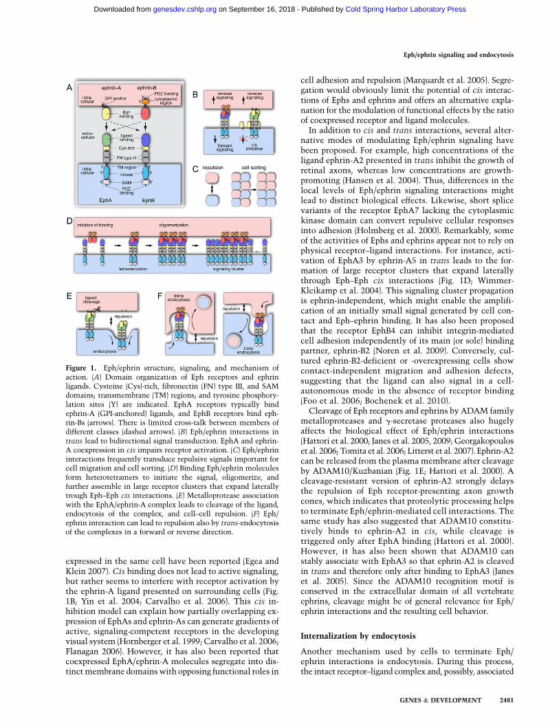
expressed in the same cell have been reported (Egea andKlein 2007). Cis binding does not lead to active signaling,but rather seems to interfere with receptor activation bythe ephrin-A ligand presented on surrounding cells (Fig.1B; Yin et al. 2004; Carvalho et al. 2006). This cis in-hibition model can explain how partially overlapping ex-pression of EphAs and ephrin-As can generate gradients ofactive, signaling-competent receptors in the developingvisual system (Hornberger et al. 1999; Carvalho et al. 2006;Flanagan 2006). However, it has also been reported thatcoexpressed EphA/ephrin-A molecules segregate into dis-tinct membrane domains with opposing functional roles in
cell adhesion and repulsion (Marquardt et al. 2005). Segre-gation would obviously limit the potential of cis interac-tions of Ephs and ephrins and offers an alternative expla-nation for the modulation of functional effects by the ratioof coexpressed receptor and ligand molecules.
In addition to cis and trans interactions, several alter-native modes of modulating Eph/ephrin signaling havebeen proposed. For example, high concentrations of theligand ephrin-A2 presented in trans inhibit the growth ofretinal axons, whereas low concentrations are growth-promoting (Hansen et al. 2004). Thus, differences in thelocal levels of Eph/ephrin signaling interactions mightlead to distinct biological effects. Likewise, short splicevariants of the receptor EphA7 lacking the cytoplasmickinase domain can convert repulsive cellular responsesinto adhesion (Holmberg et al. 2000). Remarkably, someof the activities of Ephs and ephrins appear not to rely onphysical receptor–ligand interactions. For instance, acti-vation of EphA3 by ephrin-A5 in trans leads to the for-mation of large receptor clusters that expand laterallythrough Eph–Eph cis interactions (Fig. 1D; Wimmer-Kleikamp et al. 2004). This signaling cluster propagationis ephrin-independent, which might enable the amplifi-cation of an initially small signal generated by cell con-tact and Eph–ephrin binding. It has also been proposedthat the receptor EphB4 can inhibit integrin-mediatedcell adhesion independently of its main (or sole) bindingpartner, ephrin-B2 (Noren et al. 2009). Conversely, cul-tured ephrin-B2-deficient or -overexpressing cells showcontact-independent migration and adhesion defects,suggesting that the ligand can also signal in a cell-autonomous mode in the absence of receptor binding(Foo et al. 2006; Bochenek et al. 2010).
Cleavage of Eph receptors and ephrins by ADAM familymetalloproteases and g-secretase proteases also hugelyaffects the biological effect of Eph/ephrin interactions(Hattori et al. 2000; Janes et al. 2005, 2009; Georgakopouloset al. 2006; Tomita et al. 2006; Litterst et al. 2007). Ephrin-A2can be released from the plasma membrane after cleavageby ADAM10/Kuzbanian (Fig. 1E; Hattori et al. 2000). Acleavage-resistant version of ephrin-A2 strongly delaysthe repulsion of Eph receptor-presenting axon growthcones, which indicates that proteolytic processing helpsto terminate Eph/ephrin-mediated cell interactions. Thesame study has also suggested that ADAM10 constitu-tively binds to ephrin-A2 in cis, while cleavage istriggered only after EphA binding (Hattori et al. 2000).However, it has also been shown that ADAM10 canstably associate with EphA3 so that ephrin-A2 is cleavedin trans and therefore only after binding to EphA3 (Janeset al. 2005). Since the ADAM10 recognition motif isconserved in the extracellular domain of all vertebrateephrins, cleavage might be of general relevance for Eph/ephrin interactions and the resulting cell behavior.
Internalization by endocytosis
Another mechanism used by cells to terminate Eph/ephrin interactions is endocytosis. During this process,the intact receptor–ligand complex and, possibly, associated
Figure 1. Eph/ephrin structure, signaling, and mechanism ofaction. (A) Domain organization of Eph receptors and ephrinligands. Cysteine (Cys)-rich, fibronectin (FN) type III, and SAMdomains; transmembrane (TM) regions; and tyrosine phosphory-lation sites (Y) are indicated. EphA receptors typically bindephrin-A (GPI-anchored) ligands, and EphB receptors bind eph-rin-Bs (arrows). There is limited cross-talk between members ofdifferent classes (dashed arrows). (B) Eph/ephrin interactions intrans lead to bidirectional signal transduction. EphA and ephrin-A coexpression in cis impairs receptor activation. (C) Eph/ephrininteractions frequently transduce repulsive signals important forcell migration and cell sorting. (D) Binding Eph/ephrin moleculesform heterotetramers to initiate the signal, oligomerize, andfurther assemble in large receptor clusters that expand laterallytrough Eph–Eph cis interactions. (E) Metalloprotease associationwith the EphA/ephrin-A complex leads to cleavage of the ligand,endocytosis of the complex, and cell–cell repulsion. (F) Eph/ephrin interaction can lead to repulsion also by trans-endocytosisof the complexes in a forward or reverse direction.
Eph/ephrin signaling and endocytosis
GENES & DEVELOPMENT 2481
Cold Spring Harbor Laboratory Press on September 16, 2018 - Published by genesdev.cshlp.orgDownloaded from

cytoplasmic proteins, together with the surroundingplasma membrane, can be internalized into the Eph- orephrin-expressing cell (Mann et al. 2003; Marston et al.2003; Zimmer et al. 2003; Lauterbach and Klein 2006).The exact mechanistic basis for this unusual process—termed trans-endocytosis—remains incompletely under-stood (Fig. 1F). Nevertheless, double-membrane-coatedintracellular structures, which one would predict for thecombined internalization of interacting plasma membraneregions, can be seen in the rat hippocampus by electronmicroscopy (Spacek and Harris 2004). Trans-endocytosisterminates adhesion and, like ligand proteolytic cleavage,enables cell retraction. It is noteworthy that the directionof endocytosis is determined by Eph/ephrin-mediatedsignal transduction. Signaling-deficient EphB2, lackingthe cytoplasmic region, directs the internalization of thereceptor–ligand complex into the adjacent, ephrin-B1-expressing cell. In contrast, the reverse scenario, full-lengthEphB2 and truncated ephrin-B1, leads to trans-endocytosisinto the receptor cell. Simultaneous truncation of EphB2and ephrin-B1 prevents internalization and strongly pro-longs cell adhesion (Zimmer et al. 2003). Both receptor–ligand complex internalization and cell retraction re-quire actin polymerization and activity of the smallGTPase Rac1 (Marston et al. 2003). Moreover, signalingfrom the internalized Eph/ephrin complex persists aftertrans-endocytosis, suggesting that active signal transduc-tion can be shifted into one or the other interacting cell(Marston et al. 2003).
The clathrin pathway has been linked to ephrin-B1endocytosis. Treatment of cells expressing green fluores-cent protein (GFP)-tagged ephrin-B1 with soluble, recom-binant EphB1/Fc fusion protein triggers ligand clusteringand internalization into clathrin-coated vesicles (Parkeret al. 2004). Internalized ephrin-B1 colocalizes with theearly endosome marker EEA1 (Early Endosome Antigen1), and the uptake of the ligand is blocked by dominant-negative dynamin (Parker et al. 2004). These featuressuggest that classical, clathrin-dependent endocytosis isresponsible for ephrin-B (reverse) internalization. How-ever, at least in the uptake of EphB receptors from the cellsurface, caveolae might also be involved. Caveolae areplasma membrane invaginations with a special lipidcomposition and roles in mechanosensing and endocyto-sis (Bruns and Palade 1968; Yu et al. 2006). Ephs are con-centrated in caveolae, and the receptor EphB1 associateswith the protein caveolin-1 (Vihanto et al. 2006). These fewreports indicate that further work is required to elucidatethe full molecular mechanism of Eph/ephrin endocytosis.
Eph/ephrin endocytosis—lessons from thenervous system
Ephs and ephrins are understood best as patterning andaxon guidance molecules in the nervous system (Flanaganand Vanderhaeghen 1998; Kullander and Klein 2002). Themotile growth cones at the distal ends of growing axonsrespond to repulsive or attractive cues in their environ-ment to ensure the proper wiring of the nervous system(Fig. 2A; Chisholm and Tessier-Lavigne 1999; Yu and
Bargmann 2001; Grunwald and Klein 2002). The activa-tion of Eph receptors in growing neurons typically, butnot always, leads to a growth cone collapse response andretraction from an ephrin-expressing substrate (Poliakovet al. 2004; Pasquale 2005; Klein 2009). Ephs and ephrinsare sometimes expressed in gradients so that neurons,
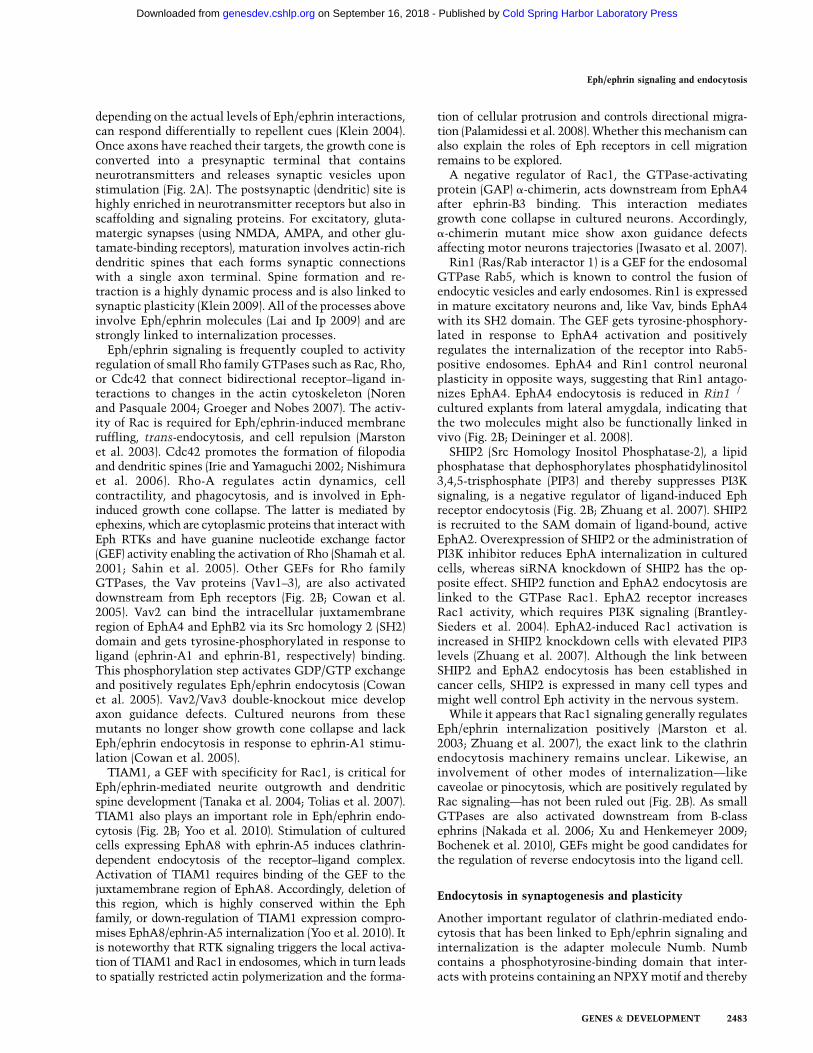
Figure 2. Eph/ephrin signaling and endocytosis in the nervoussystem. (A) The growth cones of extending axons are guided byattractive (+) and repulsive (�) cues. Follwing synapse formationand maturation, neurotransmitters are released from the pre-synaptic side and activate receptors (e.g., AMPAR or NMDARfor excitatory synapses) located in the postsynaptic terminal. (B)In the postsynaptic cell, Eph receptor endocytosis is clathrin-mediated and regulated by the activation of Vav2 (a GEF for Rhofamily GTPases), Rin1 (a GEF for endosomal Rab5 GTPase), andTIAM1 (a GEF for Rac1 GTPase). Receptor endocytosis andRac1 GTPase activity are inhibited by the lipid phosphataseSHIP2. Rac1 GTPase (which can get locally activated in endo-somes) modifies actin cytoskeleton and has been linked tocaveolar internalization or pinocytosis. A similar, Rac1 GTP-dependent mechanism might apply for ephrin-B reverse endo-cytosis in the presynaptic cell. Linked to forward Eph signaling,Numb (a clathrin adapter) regulates dendritic spine morphogen-esis by coupling activated Eph and intersectin (a GEF for Cdc42).Numb regulates not only spine growth, but also synapticplasticity, probably through Eph-dependent NMDA receptorendocytosis. EphB forward signaling and endocytosis regulatessynaptic plasticity by phosphorylation of synaptojanin andenhanced internalization of AMPAR. Postsynaptic ephrin-Breverse signaling leads to GRIP binding and increases AMPARsurface presentation. Some of the mechanisms of endocytosiswere derived from studies using soluble Fc fusion proteins andare not validated by cell–cell stimulation experiments.
Pitulescu and Adams
2482 GENES & DEVELOPMENT
Cold Spring Harbor Laboratory Press on September 16, 2018 - Published by genesdev.cshlp.orgDownloaded from
depending on the actual levels of Eph/ephrin interactions,can respond differentially to repellent cues (Klein 2004).Once axons have reached their targets, the growth cone isconverted into a presynaptic terminal that containsneurotransmitters and releases synaptic vesicles uponstimulation (Fig. 2A). The postsynaptic (dendritic) site ishighly enriched in neurotransmitter receptors but also inscaffolding and signaling proteins. For excitatory, gluta-matergic synapses (using NMDA, AMPA, and other glu-tamate-binding receptors), maturation involves actin-richdendritic spines that each forms synaptic connectionswith a single axon terminal. Spine formation and re-traction is a highly dynamic process and is also linked tosynaptic plasticity (Klein 2009). All of the processes aboveinvolve Eph/ephrin molecules (Lai and Ip 2009) and arestrongly linked to internalization processes.
Eph/ephrin signaling is frequently coupled to activityregulation of small Rho family GTPases such as Rac, Rho,or Cdc42 that connect bidirectional receptor–ligand in-teractions to changes in the actin cytoskeleton (Norenand Pasquale 2004; Groeger and Nobes 2007). The activ-ity of Rac is required for Eph/ephrin-induced membraneruffling, trans-endocytosis, and cell repulsion (Marstonet al. 2003). Cdc42 promotes the formation of filopodiaand dendritic spines (Irie and Yamaguchi 2002; Nishimuraet al. 2006). Rho-A regulates actin dynamics, cellcontractility, and phagocytosis, and is involved in Eph-induced growth cone collapse. The latter is mediated byephexins, which are cytoplasmic proteins that interact withEph RTKs and have guanine nucleotide exchange factor(GEF) activity enabling the activation of Rho (Shamah et al.2001; Sahin et al. 2005). Other GEFs for Rho familyGTPases, the Vav proteins (Vav1–3), are also activateddownstream from Eph receptors (Fig. 2B; Cowan et al.2005). Vav2 can bind the intracellular juxtamembraneregion of EphA4 and EphB2 via its Src homology 2 (SH2)domain and gets tyrosine-phosphorylated in response toligand (ephrin-A1 and ephrin-B1, respectively) binding.This phosphorylation step activates GDP/GTP exchangeand positively regulates Eph/ephrin endocytosis (Cowanet al. 2005). Vav2/Vav3 double-knockout mice developaxon guidance defects. Cultured neurons from thesemutants no longer show growth cone collapse and lackEph/ephrin endocytosis in response to ephrin-A1 stimu-lation (Cowan et al. 2005).
TIAM1, a GEF with specificity for Rac1, is critical forEph/ephrin-mediated neurite outgrowth and dendriticspine development (Tanaka et al. 2004; Tolias et al. 2007).TIAM1 also plays an important role in Eph/ephrin endo-cytosis (Fig. 2B; Yoo et al. 2010). Stimulation of culturedcells expressing EphA8 with ephrin-A5 induces clathrin-dependent endocytosis of the receptor–ligand complex.Activation of TIAM1 requires binding of the GEF to thejuxtamembrane region of EphA8. Accordingly, deletion ofthis region, which is highly conserved within the Ephfamily, or down-regulation of TIAM1 expression compro-mises EphA8/ephrin-A5 internalization (Yoo et al. 2010). Itis noteworthy that RTK signaling triggers the local activa-tion of TIAM1 and Rac1 in endosomes, which in turn leadsto spatially restricted actin polymerization and the forma-
tion of cellular protrusion and controls directional migra-tion (Palamidessi et al. 2008). Whether this mechanism canalso explain the roles of Eph receptors in cell migrationremains to be explored.
A negative regulator of Rac1, the GTPase-activatingprotein (GAP) a-chimerin, acts downstream from EphA4after ephrin-B3 binding. This interaction mediatesgrowth cone collapse in cultured neurons. Accordingly,a-chimerin mutant mice show axon guidance defectsaffecting motor neurons trajectories (Iwasato et al. 2007).
Rin1 (Ras/Rab interactor 1) is a GEF for the endosomalGTPase Rab5, which is known to control the fusion ofendocytic vesicles and early endosomes. Rin1 is expressedin mature excitatory neurons and, like Vav, binds EphA4with its SH2 domain. The GEF gets tyrosine-phosphory-lated in response to EphA4 activation and positivelyregulates the internalization of the receptor into Rab5-positive endosomes. EphA4 and Rin1 control neuronalplasticity in opposite ways, suggesting that Rin1 antago-nizes EphA4. EphA4 endocytosis is reduced in Rin1�/�
cultured explants from lateral amygdala, indicating thatthe two molecules might also be functionally linked invivo (Fig. 2B; Deininger et al. 2008).
SHIP2 (Src Homology Inositol Phosphatase-2), a lipidphosphatase that dephosphorylates phosphatidylinositol3,4,5-trisphosphate (PIP3) and thereby suppresses PI3Ksignaling, is a negative regulator of ligand-induced Ephreceptor endocytosis (Fig. 2B; Zhuang et al. 2007). SHIP2is recruited to the SAM domain of ligand-bound, activeEphA2. Overexpression of SHIP2 or the administration ofPI3K inhibitor reduces EphA internalization in culturedcells, whereas siRNA knockdown of SHIP2 has the op-posite effect. SHIP2 function and EphA2 endocytosis arelinked to the GTPase Rac1. EphA2 receptor increasesRac1 activity, which requires PI3K signaling (Brantley-Sieders et al. 2004). EphA2-induced Rac1 activation isincreased in SHIP2 knockdown cells with elevated PIP3levels (Zhuang et al. 2007). Although the link betweenSHIP2 and EphA2 endocytosis has been established incancer cells, SHIP2 is expressed in many cell types andmight well control Eph activity in the nervous system.
While it appears that Rac1 signaling generally regulatesEph/ephrin internalization positively (Marston et al.2003; Zhuang et al. 2007), the exact link to the clathrinendocytosis machinery remains unclear. Likewise, aninvolvement of other modes of internalization—likecaveolae or pinocytosis, which are positively regulated byRac signaling—has not been ruled out (Fig. 2B). As smallGTPases are also activated downstream from B-classephrins (Nakada et al. 2006; Xu and Henkemeyer 2009;Bochenek et al. 2010), GEFs might be good candidates forthe regulation of reverse endocytosis into the ligand cell.
Endocytosis in synaptogenesis and plasticity
Another important regulator of clathrin-mediated endo-cytosis that has been linked to Eph/ephrin signaling andinternalization is the adapter molecule Numb. Numbcontains a phosphotyrosine-binding domain that inter-acts with proteins containing an NPXY motif and thereby
Eph/ephrin signaling and endocytosis
GENES & DEVELOPMENT 2483
Cold Spring Harbor Laboratory Press on September 16, 2018 - Published by genesdev.cshlp.orgDownloaded from

links this cargo to the AP2 adapter complex and clathrin(Santolini et al. 2000). Numb and the related Numb-likecontrol neural progenitor maintenance, differentiation,and cortical morphogenesis (Petersen et al. 2002; Li et al.2003). In cultured hippocampal neurons, Numb accumu-lates in growing neurites and regulates axon guidance,which involves the endocytosis of the neuronal celladhesion molecule L1 (Nishimura et al. 2003). Stimula-tion of cultured neurons with soluble, recombinantephrin-B1 protein induces dendritic spine formation andmaturation, which depends on the presence of Numb.This effect is mediated by postsynaptic Eph receptor ac-tivation, as Numb forms a complex with NMDA receptorand ephrin-B1-bound EphB2 (Fig. 2B; Nishimura et al. 2006).Another critical step is binding of Numb to intersectin(a GEF for Cdc42), which leads to Cdc42 activation, theformation of dendritic protrusions, and spine elongation(Irie and Yamaguchi 2002; Nishimura et al. 2006). In linewith these findings, the Drosophila homolog of intersec-tin, Dap160, also has a role in synaptic development andendocytosis (Koh et al. 2004; Marie et al. 2004).
As mentioned above, the strength of functionalsynapses—i.e., their responsiveness to stimulation andquantity of neurotransmitter release—is modulated in pro-cesses such as learning and memory storage. While syn-aptic plasticity remains incompletely understood, impor-tant roles have been attributed to the modulation of theaxon terminal and the function of neurotransmitter recep-tors. Synaptojanin 1, a polyphosphoinositide phosphatase,controls the internalization of postsynaptic AMPA recep-tor (Gong and De Camilli 2008), a process that has beenlinked to Eph/ephrin signaling (Fig. 2B). Ephrin-B2 stimu-lation of EphB2 in cultured neuroblastoma-like cells orhippocampal neurons leads to tyrosine phosphorylation ofsynaptojanin 1 (Irie et al. 2005). This phosphorylation takesplace in the proline-rich domain (PRD) and inhibits theinteractions with the SH3 domain of endofilin, a presyn-aptic BAR (named after the proteins Bin–Amphiphysin–Rvs) domain-containing protein controlling membranecurvature (Hopper and O’Connor 2005). Binding of endo-phylin to the PRD domain stimulates the 59-phosphataseactivity of synaptojanin 1 and thereby triggers the hydro-lysis of phosphatidylinositol 4,5-biphosphate (PIP2) tophosphatidylinositol 4-phosphate (PI4P). Once this processis inhibited by EphB2 signaling, PIP2 levels go up, which inturn promotes clathrin-mediated endocytosis and trans-ferrin uptake. In line with these findings, mutations in theEph kinase or the PRD domain of synaptojanin 1 block theincrease of transferrin uptake and prevent the uptake ofAMPA receptor in hippocampal neurons (Irie et al. 2005).Interestingly, EphB2–ephrin-B2 interactions not only pro-mote endocytosis, but also impair the transfer of internal-ized cargo—in this case, the protein transferrin—intoendosomes (Irie et al. 2005). Thus, Eph signaling controlstwo distinct processes in the endocytic machinery in theopposite fashion, the early and the late phases of clathrin-mediated endocytosis.
While the findings above suggest that B-class ephrinsmainly act presynaptically, ephrin-B2 also controls den-dritic spine morphogenesis, synapse formation, and syn-
aptic plasticity on the postsynaptic side (Grunwald et al.2004; Segura et al. 2007). Some of these functions havebeen linked to the regulation of AMPA receptor traffick-ing (Fig. 2B; Essmann et al. 2008). Stimulation of culturedhippocampal neurons with soluble, recombinant EphB4fusion protein stabilizes AMPA receptor at the cell sur-face. In contrast, the receptor is constitutively internal-ized and synaptic transmission is reduced in neuronslacking ephrin-B2 (Essmann et al. 2008). Both ephrin-B2and AMPA receptor interact with and are bridged by themulti-PDZ domain protein GRIP (glutamate receptor-interacting protein). The binding of GRIP to ephrin-B2 isinduced by Eph receptor binding and involves the phos-phorylation of a serine residue in the proximity of thePDZ-binding motif at the C terminus of the ligand. Ac-cordingly, rescue of ephrin-B2 knockout neurons withwild-type ephrin-B2, but not a point mutant lacking theserine phosphorylation site, can restore AMPA receptorsurface presentation (Essmann et al. 2008). Besides GRIP,the PDZ domain proteins PICK1 and syntenin have beenidentified as binding partners of various glutamate re-ceptor subunits and Eph/ephrin molecules (Torres et al.1998; Bruckner et al. 1999; Hirbec et al. 2002, 2003;Essmann et al. 2008; McClelland et al. 2009). Interest-ingly, serine (Ser 880) phosphorylation of the AMPA re-ceptor subunit GluR2 by protein kinase C a also in-terferes with PDZ (PICK1 and GRIP) binding and therebydecreases the constitutive surface expression and recy-cling of internalized GluR2 (Lu and Ziff 2005). Ser 880phosphorylation seems to be suppressed by ephrin-B2 andis therefore increased in knockout hippocampal neuronslacking the transmembrane ligand (Essmann et al. 2008).
These examples show that Eph/ephrin molecules havevery versatile roles in the developing and adult nervoussystem. Many of these functions involve the positive ornegative regulation of endocytosis and trafficking. As weshow below, internalization might be a general mecha-nism by which Eph/ephrin molecules control complexand highly diverse biological processes.
Cell positioning in the gastrointestinal tract
In the gastrointestinal tract, processes such as cell di-vision, differentiation, migration, and patterning are crit-ical not only during development, but throughout adultlife due to the very high rate of cell replacement in theepithelial lining of the intestine (Crosnier et al. 2006).This epithelium forms during embryogenesis in the mouse,and proliferation becomes confined to the pockets betweenthe finger-like villi, which protrude into the lumen of theintestine. In postnatal life, the intervillus pockets developinto crypts, which contain stem cells together withtransient populations of yet undifferentiated progenitors(Fig. 3A). Differentiated epithelial cells display distinctpatterns of migration. Paneth cells, which provide anti-microbial defense in the small intestine, move toward thebottom of the crypt, whereas mucus-secreting Goblet,hormone-producing enterendocrine, and adsorptive cellsmigrate upward into the villi (Casali and Batlle 2009).Migratory behavior and proliferation are tightly coupled.
Pitulescu and Adams
2484 GENES & DEVELOPMENT
Cold Spring Harbor Laboratory Press on September 16, 2018 - Published by genesdev.cshlp.orgDownloaded from
Cells reaching the upper crypt boundary stop cycling,begin their differentiation, and enter the villus.
Stem cell maintenance and progenitor cell proliferationin the intestine are under the control of canonical Wntsignaling involving b-catenin and TCF/LEF family tran-scription factors (Crosnier et al. 2006; van der Flier andClevers 2009). Proliferation in the crypts is compromisedin mice lacking critical Wnt pathway components (van deWetering et al. 2002). In contrast, loss of the tumorsuppressor APC, which normally targets b-catenin fordestruction, increases b-catenin levels and leads to cryptenlargement and the amplification of undifferentiatedprogenitor cells in mouse models or in the vast majorityof human intestinal cancers (Sansom et al. 2004; Cleversand Batlle 2006).
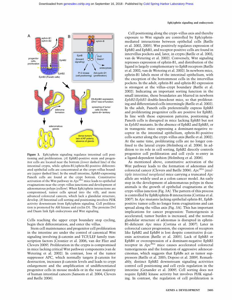
Cell positioning along the crypt–villus axis and therebyexposure to Wnt signals are controlled by Eph/ephrin-mediated interactions between epithelial cells (Batlleet al. 2002, 2005). Wnt positively regulates expression ofEphB2 and EphB3, and receptor-positive cells are found inintervillus pockets and, later, in crypts (Batlle et al. 2002;van de Wetering et al. 2002). Conversely, Wnt signalingrepresses expression of ephrin-B1, and distribution of theligand is largely complementary to EphB receptors (Batlleet al. 2002; van de Wetering et al. 2002). In newborn mice,ephrin-B1 labels most of the intestinal epithelium, withthe exception of the bottommost cells in the intervilluspockets. In the adult, ephrin-B1 and ephrin-B2 expressionis strongest at the villus–crypt boundary (Batlle et al.2002). Indicating an important sorting function in thesmall intestine, these boundaries are blurred in newbornEphB2/EphB3 double-knockout mice, so that proliferat-ing and differentiated cells intermingle (Batlle et al. 2002).In the adult, Paneth cells preferentially express EphB3and proliferating progenitor cells are positive for EphB2.In line with these expression patterns, positioning ofPaneth cells is disrupted in mice lacking EphB3 but notin EphB2 mutants. In the absence of EphB2 and EphB3, orin transgenic mice expressing a dominant-negative re-ceptor in the intestinal epithelium, ephrin-B1-positivecells scatter along the crypt–villus axis (Batlle et al. 2002).At the same time, proliferating cells are no longer con-fined to the lateral crypts (Holmberg et al. 2006). In ad-dition to its role in cell sorting, EphB2 directly controlsprogenitor cell proliferation and cell cycle re-entry ina ligand-dependent fashion (Holmberg et al. 2006).
As mentioned above, constitutive activation of theWnt pathway leads to the formation of adenomas andcolorectal cancer (Clevers and Batlle 2006). ApcMin (mul-tiple intestinal neoplasia) mice carrying a truncated Apcallele are widely used as a colon cancer model. An earlystep in the development of adenomatous polyps in theseanimals is the growth of epithelial evaginations at thecrypt–villus junction (Fig. 3A). The pattern of this processis controlled by EphB/ephrin-B interactions (Cortina et al.2007). In Apc mutants lacking epithelial ephrin-B1, EphB-positive tumor cells no longer form evaginations and canspread along the villus axis (Fig. 3A). This has importantimplications for cancer progression: Tumorigenesis isaccelerated, tumor burden is increased, and the normalglandular structure of adenomas is disrupted in ephrin-B1-deficient Apc mice (Cortina et al. 2007). Later incolorectal cancer progression, the expression of receptorslike EphB2 and EphB4 is lost despite constitutive b-cat-enin activation (Batlle et al. 2005). Lack of EphB3 orEphB4 or overexpression of a dominant-negative EphB2receptor in ApcMin mice causes accelerated colorectaltumorigenesis and the formation of aggressive adenocar-cinomas, which suggests that EphBs act as tumor sup-pressors (Batlle et al. 2005; Dopeso et al. 2009). Remark-ably, distinct EphB2 downstream signaling activitiescontrol cell positioning and cell cycle regulation in theintestine (Genander et al. 2009). Cell sorting does notrequire EphB2 kinase activity but involves PI3K signal-ing. In contrast, the regulation of cell proliferation is
Figure 3. Eph/ephrin signaling regulates intestinal cell posi-tioning and proliferation. (A) EphB2-positive stem and progen-itor cells are located near the bottom (lower dashed line) of theintestinal crypts, while ephrin-B1/ephrin-B2-positive differenti-ated epithelial cells are concentrated at the crypt–villus bound-ary (upper dashed line). In the small intestine, EphB3-expressingPaneth cells are found at the crypt bottom. Constitutiveactivation of the Wnt pathway in ApcMin mice leads to epithelialevaginations near the crypt–villus junctions and development ofadenomatous polyps (yellow). When Eph/ephrin interactions arecompromised, tumor cells spread into the villi, and moreadvanced colorectal cancers, which lack a glandular structure,develop. (B) Intestinal cell sorting and positioning involves PI3Kactivity downstream from Eph/ephrin signaling. Cell prolifera-tion is promoted by Abl kinase and cyclin D1. The proteins Dvland Daam link Eph endocytosis and Wnt signaling.
Eph/ephrin signaling and endocytosis
GENES & DEVELOPMENT 2485
Cold Spring Harbor Laboratory Press on September 16, 2018 - Published by genesdev.cshlp.orgDownloaded from
kinase-dependent and involves the downstream activa-tion of the Abelson tyrosine kinase and up-regulation ofthe cell cycle regulator cyclin D1. In carcinomas, cyclinD1 expression becomes uncoupled from EphB signalingso that high proliferation is maintained even when EphBexpression is lost (Genander et al. 2009).
Connections between Wnt and Eph/ephrin signalinghave been described in numerous tissues and morphoge-netic processes (Tanaka et al. 2003; Kida et al. 2007).Dishevelled proteins (Dvl), which are also essential com-ponents of the Wnt signaling pathway, form complexeswith EphBs and the ligand ephrin-B1, leading to bidirec-tional signal transduction, activation of RhoA and Rhokinase, and cell repulsion in Xenopus embryos (Tanakaet al. 2003). Daam1 (dishevelled-associated activator ofmorphogenesis), a protein involved in noncanonicalWnt signaling and the regulation of planar cell polarity,forms a complex with phosphorylated Eph receptor andDishevelled 2 (Kida et al. 2007). This complex getsinternalized in a dynamin-dependent fashion, a processthat is essential for convergent extension of the zebra-fish notochord and normal body axis formation. Futurework will have to address whether similar links betweenEph receptor endocytosis and Wnt signaling are alsorelevant in the gastrointestinal tract and, in particular,the sorting and organization of cells within the epithelialsheet.
Regulation of blood vessel morphogenesisby Eph/ephrin signaling
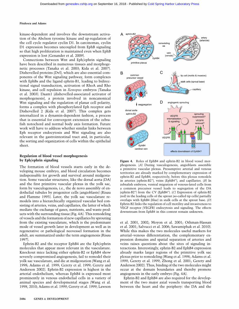
The formation of blood vessels starts early in the de-veloping mouse embryo, and blood circulation becomesindispensable for growth and survival around midgesta-tion. Some vascular structures, like the dorsal aorta (DA)and the first primitive vascular plexus in the yolk sac,form by vasculogenesis; i.e., the de novo assembly of en-dothelial tubules by progenitor cells (angioblasts) (Risauand Flamme 1995). Later, the yolk sac vasculature re-models into a hierarchically organized vascular bed con-sisting of arteries, veins, and capillaries, the latter of whichmediate the exchange of gases, nutrients, and waste prod-ucts with the surrounding tissue (Fig. 4A). This remodelingof vessels and the formation of new capillaries by sproutingfrom the existing vasculature, which is the predominantmode of vessel growth later in development as well as inregenerative or pathological neovessel formation in theadult, are summarized under the term angiogenesis (Risau1997).
Ephrin-B2 and the receptor EphB4 are the Eph/ephrinmolecules that appear most relevant in the vasculature.Knockout mice lacking either ephrin-B2 or EphB4 showseverely compromised angiogenesis, fail to remodel theiryolk sac vasculature, and die at midgestation (Wang et al.1998; Adams et al. 1999; Gerety et al. 1999; Gerety andAnderson 2002). Ephrin-B2 expression is highest in thearterial endothelium, whereas EphB4 is expressed mostprominently in venous endothelial cells in a variety ofanimal species and developmental stages (Wang et al.1998, 2010; Adams et al. 1999; Gerety et al. 1999; Lawson
et al. 2001, 2002; Moyon et al. 2001; Othman-Hassanet al. 2001; Salvucci et al. 2006; Sawamiphak et al. 2010).While this makes the two molecules useful markers forarterial–venous differentiation, the complementary ex-pression domains and spatial separation of arteries andveins raises questions about the sites of signaling in-teractions. Interestingly, ephrin-B2 and EphB4 expressionalready marks larger regions of the primitive yolk sacplexus prior to remodeling (Wang et al. 1998; Adams et al.1999; Gerety et al. 1999; Zhong et al. 2001; Gerety andAnderson 2002). Thus, binding of the two molecules mightoccur at the domain boundaries and thereby promoteangiogenesis in the early embryo (Fig. 4A).
Ephrin-B2 and EphB4 are also required for the develop-ment of the two major axial vessels transporting bloodbetween the heart and the periphery: the DA and the
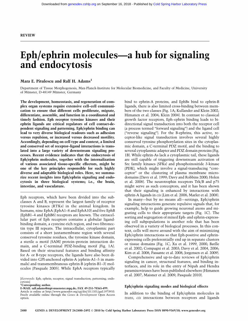
Figure 4. Roles of EphB4 and ephrin-B2 in blood vessel mor-phogenesis. (A) During vasculogenesis, angioblasts assemblea primitive vascular plexus. Presumptive arterial and venousterritories are already marked by complementary expression ofephrin-B2 and EphB4, respectively, before this plexus remodelsin arteries (ephrin-B2+), veins (EphB4+), and capillaries. (B) Inzebrafish embryos, ventral migration of venous-fated cells froma common precursor vessel leads to segregation of the DA(ephrin-B2+) from the CV (EphB4+). (C) Expression of ephrin-B2(red) in the leading cells of the sprout (so-called tip cells) partiallyoverlaps with EphB4 (blue) in stalk cells at the sprout base. (D)Ephrin-B2 links the regulation of cell motility and invasiveness toVEGF receptor (VEGFR) endocytosis and signaling. The effectsdownstream from EphB4 in this context remain unknown.
Pitulescu and Adams
2486 GENES & DEVELOPMENT
Cold Spring Harbor Laboratory Press on September 16, 2018 - Published by genesdev.cshlp.orgDownloaded from
cardinal vein (CV). It was previously thought that thesevessels form by the vasculogenic assembly of angioblasts.However, this view has been profoundly challenged bya study in zebrafish embryos that shows that angioblastsinitially assemble into only a single axial vessel, termedthe common precursor vessel, in the trunk (Herbert et al.2009). Ventral sprouting from the precursor vessel and themigration of venous-fated endothelial cells leads to a sep-aration of the DA and the CV. Thus, the CV is formed notby vasculogenesis but arterial–venous cell segregation(Fig. 4B). This process is controlled by Eph/ephrin in-teractions (Herbert et al. 2009). Endothelial cells express-ing ephrin-B2a (one of the two orthologs of mammalianephrin-B2) have limited ability to migrate ventrally,whereas those expressing the venous marker EphB4(EphB4a) preferentially move into the CV. Fewer cells areretained in the DA when ephrin-B2 expression is targetedwith morpholinos or when a C-terminally truncated,reverse signaling-incompetent version of the ligand isoverexpressed. Conversely, injection of morpholinos tar-geting EphB4 reduces the contribution of cells to the CV(Herbert et al. 2009).
Ephrin-B2 expression in the vasculature is positivelycontrolled by vascular endothelial growth factor (VEGF)signaling and the Notch pathway, both of which are crit-ical regulators of vessel growth and arterial differentia-tion (Torres-Vazquez et al. 2003; Lamont and Childs2006). VEGF-A; its receptor, Kdrl/VEGFR2; and Notch,all of which are known to upregulate the expression ofephrin-B2, promote DA formation and prevent excessiveventral sprouting. The ventral migration of venous-fated,EphB4-expressing cells and CV formation are positivelyregulated by VEGF-C (another member of the VEGFfamily), its receptor Flt4/VEGFR3, and the p110a cata-lytic PI3K subunit, all of which enhance endothelialmotility. Although it is not yet clear whether the majoraxial vessels in mammals also develop by segregationfrom a common precursor, the caliber of the DA and CVin the early mouse embryo is reciprocally balanced byNotch, ephrin-B2, and EphB4. Moreover, the anteriorparts of these axial vessels show conspicuous connec-tions that might correspond to migrating endothelialcells (Carlson et al. 2005; Benedito et al. 2008; Kimet al. 2008).
Endothelial sprouting and VEGF receptor endocytosis
Another site of overlapping EphB4 and ephrin-B2 expres-sion in the embryonic and early postnatal mouse isdeveloping lymphatic vessels. Unlike blood vessels,which carry circulating blood, the lymphatic vasculatureis a blind-ended network that transports protein-richlymph and immune cells unidirectionally from the pe-riphery through lymph nodes into certain veins. Growthof dermal lymphatic vessels involves endothelial sprout-ing from a primitive plexus, and this process is defectivein mutant mice lacking the C-terminal PDZ motif ofephrin-B2 (Makinen et al. 2005). Ephrin-B2 is also animportant regulator of endothelial sprouting from bloodvessels (Sawamiphak et al. 2010; Wang et al. 2010).
Expression of the ligand labels angiogenic capillaries inthe embryonic skin and postnatal retina, where it par-tially overlaps with EphB4 (Wang et al. 2010). Ephrin-B2levels are also increased at sites of physiological andpathological neoangiogenesis in the adult (Gale et al.2001; Shin et al. 2001).
Angiogenic sprouting involves important phenotypicchanges in a subset of endothelial cells, termed tip cells,which become motile and invasive, extend filopodialprotrusions, and lead the sprout from its distal end (Fig.4C). Other endothelial cells, the stalk cells, form the baseof the sprout and follow the tip cell (Gerhardt et al. 2003).Ephrin-B2, which strongly promotes endothelial cellmotility and invasive behavior in cultured cells andtransgenic mice, is an important positive regulator of thissprouting process (Bochenek et al. 2010; Sawamiphaket al. 2010; Wang et al. 2010). Physiological and patho-logical angiogenesis are impaired in inducible loss-of-function mutant mice or, albeit to a lesser extent, inanimals lacking the C-terminal PDZ-binding motif inephrin-B2 (Sawamiphak et al. 2010; Wang et al. 2010). Ex-pression of EphB4 in tumor cells enhances blood vesselgrowth through interactions with endothelial ephrin-B2(Noren et al. 2006), which further supports a proangio-genic role of ephrin-B2 reverse signaling.
The distinct behavior of tip versus stalk endothelialcells is strongly coupled to VEGF and Notch signaling.Gradients of VEGF-A in the tissue promote filopodiaformation and attract vascular sprouts (Ruhrberg et al.2002; Gerhardt et al. 2003). The combined action of twoNotch ligands, Delta-like 4 and Jagged1, with opposingfunctional roles in the vasculature confines Notch activa-tion preferentially to stalk cells. This, in turn, is thoughtto down-regulate the expression of VEGF receptors in thestalk so that these cells respond less to VEGF than thoseat the sprout tip (Siekmann et al. 2008; Benedito et al.2009). VEGF signaling has also been linked to traffickingof the receptor VEGFR2 and the dynamin-binding proteinsynectin/GIPC, which connects actin-based molecularmotors to endocytic vesicles (Lanahan et al. 2010). Inthe absence of synectin, trafficking of VEGFR2-contain-ing endosomes is delayed, the receptor gets selectivelydephosphorylated at a critical cytoplasmic tyrosine resi-due, and the downstream activation of mitogen-activatedprotein (MAP) kinase signaling is impaired (Lanahan et al.2010). These findings are consistent with previous evi-dence for VEGF receptor signaling in endosomes or in anautocrine fashion without VEGF secretion (Lampugnaniet al. 2006; Lee et al. 2007).
Interestingly, endothelial ephrin-B2 and its C-terminalPDZ-binding motif are also required for the endocytosis ofthe receptors VEGFR2 and VEGFR3 in cultured cells ormutant mice (Fig. 4D; Sawamiphak et al. 2010; Wang et al.2010). In the absence of the B-class ligand, stimulatedVEGF receptors are retained on the cell surface, which isaccompanied by reduced tyrosine phosphorylation of thesemolecules and impaired downstream activation of Rac1,Akt, and MAP kinase Erk (Sawamiphak et al. 2010; Wanget al. 2010). While the stimulation of EphB4 forward orephrin-B2 reverse signaling can trigger some VEGF receptor
Eph/ephrin signaling and endocytosis
GENES & DEVELOPMENT 2487
Cold Spring Harbor Laboratory Press on September 16, 2018 - Published by genesdev.cshlp.orgDownloaded from
internalization even in the absence of VEGF stimulation,this process fails to induce significant activation of theVEGF pathway (Fig. 4D; Sawamiphak et al. 2010; Wanget al. 2010). Although the exact molecular connection be-tween the Eph/ephrin system and VEGF receptor endocy-tosis remains to be resolved, the available evidence pointsto a transient interaction at the plasma membrane early inthe endocytosis process (Sawamiphak et al. 2010; Wanget al. 2010). The activation of small Rho family GTPasesdownstream from endothelial ephrin-B2 (Bochenek et al.2010) might be important in this context, similar to whathas been shown for Eph/ephrin-mediated internalizationprocesses in the nervous system. The link between ephrin-B2 and VEGF signaling might also be relevant for thesegregation of the DA and CV, which is differentiallycontrolled by VEGF-A, VEGF-C, and the corresponding re-ceptors (Herbert et al. 2009).
Outlook
Eph receptors and ephrins are highly versatile regulatorsof processes as diverse as axon guidance, the modulationof synaptic plasticity, cell sorting in the intestinal crypt,or the morphogenesis of the vascular system. These rolesare not confined to physiological processes, but are highlyrelevant for a variety of human pathologies and, in par-ticular, cancer. Thus, understanding the key mechanismscontrolling Eph/ephrin function would not only providedeeper insight into interesting biological principles, itmight also enable the development of new therapeuticreagents. For example, while antibodies against VEGF-Aare already used for the treatment of cancer, inhibition ofephrin-B2 might be useful to simultaneously interferewith the function of VEGFR2 and VEGFR3, which acttogether during angiogenesis (Tammela et al. 2008).
Unfortunately, factors such as the large number of li-gands and receptors, their dynamic expression patterns,the complexity of bidirectional signaling, the strength oftheir interactions, or the influence of cis versus transbinding complicate the research on Eph/ephrin moleculesand can lead to confusing, seemingly incompatible re-sults. For example, it has been shown that Eph/ephrinsignaling can modulate integrin activity positively aswell as negatively (Becker et al. 2000; Huai and Drescher2001; Noren et al. 2009; Yamazaki et al. 2009). Anotherpuzzle is the question of how a limited set of receptor–ligand interactions is translated into highly diverse bi-ological readouts depending on cell type and tissue. Thus,generic activities of the Eph/ephrin system, like the re-cruitment of specific cytoplasmic adapter and signalingmolecules, need to get coupled to cell type-specific mol-ecules with specialized functional roles. The emerginglinks between Eph/ephrin molecules and endocytosis,which are highlighted in this review, appear to resolvethis conundrum by connecting Ephs and ephrins withAMPA-type glutamatergic receptors in neurons or VEGFreceptors in endothelial cells. Likewise, it should beworthwhile to search for similar links to the internaliza-tion of the receptors for Wnts, FGF and EGF familygrowth factors, or other key players in the gastrointes-
tinal tract, or in other organ systems where little isknown about the role of Eph/ephrin-induced endocytosis.In the vasculature or the nervous system, where excitingconnections with receptor trafficking have already beenestablished, more insight is needed into the underlyingmolecular machinery and the association with clathrin,clathrin-binding adapters, molecular motors, and thecytoskeleton. Differential effects of Eph/ephrin mole-cules on integrins, which are also strongly regulated bytrafficking and control the internalization of other sur-face molecules (Caswell et al. 2009), need to be re-explored in the context of endocytosis.
The internalization of the Eph/ephrin complex or ofassociated surface receptors might also explain some ofthe puzzling variation in the experimental results pub-lished by different groups. Trans-endocytosis can convertadhesive interactions into repulsion. Likewise, the con-trolled removal of Ephs and ephrins from the cell surfacein response to certain stimuli could lead to fundamentalchanges in migratory behavior or cell repulsion andsorting processes. The fate of the internalized Eph/ephrinmolecules—e.g., degradation versus recycling to the cellsurface—and the regulation of the underlying traffickingprocesses remain to be resolved. Future work should ad-dress these and other important questions to resolvewhether the control of endocytosis is one of the generalmechanisms responsible for Eph/ephrin function in highlydiverse biological settings.
Acknowledgments
We thank the Max Planck Society and the University ofMuenster for financial support, and members of the Departmentof Tissue Morphogenesis for stimulating discussions.
References
Adams RH, Wilkinson GA, Weiss C, Diella F, Gale NW,
Deutsch U, Risau W, Klein R. 1999. Roles of ephrinB ligands
and EphB receptors in cardiovascular development: Demar-
cation of arterial/venous domains, vascular morphogenesis,
and sprouting angiogenesis. Genes Dev 13: 295–306.Batlle E, Henderson JT, Beghtel H, van den Born MM, Sancho E,
Huls G, Meeldijk J, Robertson J, van de Wetering M, Pawson
T, et al. 2002. b-Catenin and TCF mediate cell positioning in
the intestinal epithelium by controlling the expression of
EphB/ephrinB. Cell 111: 251–263.Batlle E, Bacani J, Begthel H, Jonkheer S, Gregorieff A, van de
Born M, Malats N, Sancho E, Boon E, Pawson T, et al. 2005.
EphB receptor activity suppresses colorectal cancer progres-
sion. Nature 435: 1126–1130.Becker E, Huynh-Do U, Holland S, Pawson T, Daniel TO,
Skolnik EY. 2000. Nck-interacting Ste20 kinase couples
Eph receptors to c-Jun N-terminal kinase and integrin
activation. Mol Cell Biol 20: 1537–1545.Benedito R, Trindade A, Hirashima M, Henrique D, da Costa LL,
Rossant J, Gill PS, Duarte A. 2008. Loss of Notch signalling
induced by Dll4 causes arterial calibre reduction by increas-
ing endothelial cell response to angiogenic stimuli. BMC
Dev Biol 8: 117. doi: 10.1186/1471-213X-8-117.Benedito R, Roca C, Sorensen I, Adams S, Gossler A, Fruttiger
M, Adams RH. 2009. The notch ligands Dll4 and Jagged1
have opposing effects on angiogenesis. Cell 137: 1124–1135.
Pitulescu and Adams
2488 GENES & DEVELOPMENT
Cold Spring Harbor Laboratory Press on September 16, 2018 - Published by genesdev.cshlp.orgDownloaded from
Bochenek ML, Dickinson S, Astin JW, Adams RH, Nobes CD.2010. Ephrin-B2 regulates endothelial cell morphology andmotility independently of Eph-receptor binding. J Cell Sci
123: 1235–1246.Brantley-Sieders DM, Caughron J, Hicks D, Pozzi A, Ruiz JC,
Chen J. 2004. EphA2 receptor tyrosine kinase regulatesendothelial cell migration and vascular assembly throughphosphoinositide 3-kinase-mediated Rac1 GTPase activa-tion. J Cell Sci 117: 2037–2049.
Bruckner K, Pablo Labrador J, Scheiffele P, Herb A, Seeburg PH,Klein R. 1999. EphrinB ligands recruit GRIP family PDZadaptor proteins into raft membrane microdomains. Neuron
22: 511–524.Bruns RR, Palade GE. 1968. Studies on blood capillaries. I.
General organization of blood capillaries in muscle. J Cell
Biol 37: 244–276.Carlson TR, Yan Y, Wu X, Lam MT, Tang GL, Beverly LJ,
Messina LM, Capobianco AJ, Werb Z, Wang R. 2005.Endothelial expression of constitutively active Notch4elicits reversible arteriovenous malformations in adult mice.Proc Natl Acad Sci 102: 9884–9889.
Carvalho RF, Beutler M, Marler KJ, Knoll B, Becker-Barroso E,Heintzmann R, Ng T, Drescher U. 2006. Silencing of EphA3through a cis interaction with ephrinA5. Nat Neurosci 9:322–330.
Casali A, Batlle E. 2009. Intestinal stem cells in mammals andDrosophila. Cell Stem Cell 4: 124–127.
Caswell PT, Vadrevu S, Norman JC. 2009. Integrins: Masters andslaves of endocytic transport. Nat Rev Mol Cell Biol 10: 843–853.
Chisholm A, Tessier-Lavigne M. 1999. Conservation and di-vergence of axon guidance mechanisms. Curr Opin Neuro-
biol 9: 603–615.Clevers H, Batlle E. 2006. EphB/EphrinB receptors and Wnt
signaling in colorectal cancer. Cancer Res 66: 2–5.Compagni A, Logan M, Klein R, Adams RH. 2003. Control of
skeletal patterning by ephrinB1–EphB interactions. Dev Cell5: 217–230.
Cortina C, Palomo-Ponce S, Iglesias M, Fernandez-Masip JL,Vivancos A, Whissell G, Huma M, Peiro N, Gallego L, JonkheerS, et al. 2007. EphB–ephrin-B interactions suppress colorectalcancer progression by compartmentalizing tumor cells. Nat
Genet 39: 1376–1383.Cowan CW, Shao YR, Sahin M, Shamah SM, Lin MZ, Greer PL,
Gao S, Griffith EC, Brugge JS, Greenberg ME. 2005. Vavfamily GEFs link activated Ephs to endocytosis and axonguidance. Neuron 46: 205–217.
Crosnier C, Stamataki D, Lewis J. 2006. Organizing cell renewalin the intestine: Stem cells, signals and combinatorialcontrol. Nat Rev Genet 7: 349–359.
Davy A, Robbins SM. 2000. Ephrin-A5 modulates cell adhesionand morphology in an integrin-dependent manner. EMBO
J 19: 5396–5405.Davy A, Gale NW, Murray EW, Klinghoffer RA, Soriano P,
Feuerstein C, Robbins SM. 1999. Compartmentalized signalingby GPI-anchored ephrin-A5 requires the Fyn tyrosine kinase toregulate cellular adhesion. Genes Dev 13: 3125–3135.
Davy A, Aubin J, Soriano P. 2004. Ephrin-B1 forward and reversesignaling are required during mouse development. Genes
Dev 18: 572–583.Davy A, Bush JO, Soriano P. 2006. Inhibition of gap junction
communication at ectopic Eph/ephrin boundaries underliescraniofrontonasal syndrome. PLoS Biol 4: e315. doi: 10.1371/journal.pbio.0040315.
Deininger K, Eder M, Kramer ER, Zieglgansberger W, Dodt HU,Dornmair K, Colicelli J, Klein R. 2008. The Rab5 guanylate
exchange factor Rin1 regulates endocytosis of the EphA4
receptor in mature excitatory neurons. Proc Natl Acad Sci
105: 12539–12544.Dopeso H, Mateo-Lozano S, Mazzolini R, Rodrigues P, Lagares-
Tena L, Ceron J, Romero J, Esteves M, Landolfi S, Hernandez-
Losa J, et al. 2009. The receptor tyrosine kinase EPHB4 has
tumor suppressor activities in intestinal tumorigenesis.
Cancer Res 69: 7430–7438.Egea J, Klein R. 2007. Bidirectional Eph-ephrin signaling during
axon guidance. Trends Cell Biol 17: 230–238.Essmann CL, Martinez E, Geiger JC, Zimmer M, Traut MH,
Stein V, Klein R, Acker-Palmer A. 2008. Serine phosphory-
lation of ephrinB2 regulates trafficking of synaptic AMPA
receptors. Nat Neurosci 11: 1035–1043.Flanagan JG. 2006. Neural map specification by gradients. Curr
Opin Neurobiol 16: 59–66.Flanagan JG, Vanderhaeghen P. 1998. The ephrins and Eph recep-
tors in neural development. Annu Rev Neurosci 21: 309–345.Foo SS, Turner CJ, Adams S, Compagni A, Aubyn D, Kogata N,
Lindblom P, Shani M, Zicha D, Adams RH. 2006. Ephrin-B2
controls cell motility and adhesion during blood-vessel-wall
assembly. Cell 124: 161–173.Gale NW, Baluk P, Pan L, Kwan M, Holash J, DeChiara TM,
McDonald DM, Yancopoulos GD. 2001. Ephrin-B2 selectively
marks arterial vessels and neovascularization sites in the
adult, with expression in both endothelial and smooth-muscle
cells. Dev Biol 230: 151–160.Genander M, Halford MM, Xu NJ, Eriksson M, Yu Z, Qiu Z,
Martling A, Greicius G, Thakar S, Catchpole T, et al. 2009.
Dissociation of EphB2 signaling pathways mediating pro-
genitor cell proliferation and tumor suppression. Cell 139:
679–692.Georgakopoulos A, Litterst C, Ghersi E, Baki L, Xu C, Serban G,
Robakis NK. 2006. Metalloproteinase/Presenilin1 processing
of ephrinB regulates EphB-induced Src phosphorylation and
signaling. EMBO J 25: 1242–1252.Gerety SS, Anderson DJ. 2002. Cardiovascular ephrinB2 func-
tion is essential for embryonic angiogenesis. Development
129: 1397–1410.Gerety SS, Wang HU, Chen ZF, Anderson DJ. 1999. Symmetrical
mutant phenotypes of the receptor EphB4 and its specific
transmembrane ligand ephrin-B2 in cardiovascular develop-
ment. Mol Cell 4: 403–414.Gerhardt H, Golding M, Fruttiger M, Ruhrberg C, Lundkvist A,
Abramsson A, Jeltsch M, Mitchell C, Alitalo K, Shima D,
et al. 2003. VEGF guides angiogenic sprouting utilizing
endothelial tip cell filopodia. J Cell Biol 161: 1163–1177.Gong LW, De Camilli P. 2008. Regulation of postsynaptic AMPA
responses by synaptojanin 1. Proc Natl Acad Sci 105: 17561–
17566.Groeger G, Nobes CD. 2007. Co-operative Cdc42 and Rho
signalling mediates ephrinB-triggered endothelial cell retrac-
tion. Biochem J 404: 23–29.Grunwald IC, Klein R. 2002. Axon guidance: Receptor com-
plexes and signaling mechanisms. Curr Opin Neurobiol 12:
250–259.Grunwald IC, Korte M, Adelmann G, Plueck A, Kullander K,
Adams RH, Frotscher M, Bonhoeffer T, Klein R. 2004.
Hippocampal plasticity requires postsynaptic ephrinBs. Nat
Neurosci 7: 33–40.Hansen MJ, Dallal GE, Flanagan JG. 2004. Retinal axon response
to ephrin-as shows a graded, concentration-dependent tran-
sition from growth promotion to inhibition. Neuron 42: 717–
730.
Eph/ephrin signaling and endocytosis
GENES & DEVELOPMENT 2489
Cold Spring Harbor Laboratory Press on September 16, 2018 - Published by genesdev.cshlp.orgDownloaded from
Hattori M, Osterfield M, Flanagan JG. 2000. Regulated cleavageof a contact-mediated axon repellent. Science 289: 1360–1365.
Herbert SP, Huisken J, Kim TN, Feldman ME, Houseman BT,Wang RA, Shokat KM, Stainier DY. 2009. Arterial–venoussegregation by selective cell sprouting: An alternative modeof blood vessel formation. Science 326: 294–298.
Himanen JP, Chumley MJ, Lackmann M, Li C, Barton WA,Jeffrey PD, Vearing C, Geleick D, Feldheim DA, Boyd AW,et al. 2004. Repelling class discrimination: ephrin-A5 bindsto and activates EphB2 receptor signaling. Nat Neurosci 7:501–509.
Himanen JP, Saha N, Nikolov DB. 2007. Cell–cell signaling viaEph receptors and ephrins. Curr Opin Cell Biol 19: 534–542.
Hirbec H, Perestenko O, Nishimune A, Meyer G, Nakanishi S,Henley JM, Dev KK. 2002. The PDZ proteins PICK1, GRIP,and syntenin bind multiple glutamate receptor subtypes.Analysis of PDZ binding motifs. J Biol Chem 277: 15221–15224.
Hirbec H, Francis JC, Lauri SE, Braithwaite SP, Coussen F, MulleC, Dev KK, Coutinho V, Meyer G, Isaac JT, et al. 2003. Rapidand differential regulation of AMPA and kainate receptors athippocampal mossy fibre synapses by PICK1 and GRIP.Neuron 37: 625–638.
Holen HL, Shadidi M, Narvhus K, Kjosnes O, Tierens A,Aasheim HC. 2008. Signaling through ephrin-A ligand leadsto activation of Src-family kinases, Akt phosphorylation, andinhibition of antigen receptor-induced apoptosis. J Leukoc
Biol 84: 1183–1191.Holmberg J, Clarke DL, Frisen J. 2000. Regulation of repulsion
versus adhesion by different splice forms of an Eph receptor.Nature 408: 203–206.
Holmberg J, Genander M, Halford MM, Anneren C, Sondell M,Chumley MJ, Silvany RE, Henkemeyer M, Frisen J. 2006.EphB receptors coordinate migration and proliferation in theintestinal stem cell niche. Cell 125: 1151–1163.
Hopper NA, O’Connor V. 2005. Ephrin tempers two-facedsynaptojanin 1. Nat Cell Biol 7: 454–456.
Hornberger MR, Dutting D, Ciossek T, Yamada T, HandwerkerC, Lang S, Weth F, Huf J, Wessel R, Logan C, et al. 1999.Modulation of EphA receptor function by coexpressed eph-rinA ligands on retinal ganglion cell axons. Neuron 22: 731–742.
Huai J, Drescher U. 2001. An ephrin-A-dependent signalingpathway controls integrin function and is linked to thetyrosine phosphorylation of a 120-kDa protein. J Biol Chem
276: 6689–6694.Irie F, Yamaguchi Y. 2002. EphB receptors regulate dendritic
spine development via intersectin, Cdc42 and N-WASP. NatNeurosci 5: 1117–1118.
Irie F, Okuno M, Pasquale EB, Yamaguchi Y. 2005. EphrinB–EphB signalling regulates clathrin-mediated endocytosisthrough tyrosine phosphorylation of synaptojanin 1. NatCell Biol 7: 501–509.
Iwasato T, Katoh H, Nishimaru H, Ishikawa Y, Inoue H, SaitoYM, Ando R, Iwama M, Takahashi R, Negishi M, et al. 2007.Rac-GAP a-chimerin regulates motor-circuit formation asa key mediator of EphrinB3/EphA4 forward signaling. Cell
130: 742–753.Janes PW, Saha N, Barton WA, Kolev MV, Wimmer-Kleikamp
SH, Nievergall E, Blobel CP, Himanen JP, Lackmann M,Nikolov DB. 2005. Adam meets Eph: An ADAM substraterecognition module acts as a molecular switch for ephrincleavage in trans. Cell 123: 291–304.
Janes PW, Wimmer-Kleikamp SH, Frangakis AS, Treble K,Griesshaber B, Sabet O, Grabenbauer M, Ting AY, Saftig P,
Bastiaens PI, et al. 2009. Cytoplasmic relaxation of activeEph controls ephrin shedding by ADAM10. PLoS Biol 7:e1000215. doi: 10.1371/journal.pbio.1000215.
Jorgensen C, Sherman A, Chen GI, Pasculescu A, Poliakov A,Hsiung M, Larsen B, Wilkinson DG, Linding R, Pawson T.2009. Cell-specific information processing in segregatingpopulations of Eph receptor ephrin-expressing cells. Science
326: 1502–1509.Kida YS, Sato T, Miyasaka KY, Suto A, Ogura T. 2007. Daam1
regulates the endocytosis of EphB during the convergentextension of the zebrafish notochord. Proc Natl Acad Sci
104: 6708–6713.Kim YH, Hu H, Guevara-Gallardo S, Lam MT, Fong SY, Wang
RA. 2008. Artery and vein size is balanced by Notch andephrin B2/EphB4 during angiogenesis. Development 135:3755–3764.
Klein R. 2004. Eph/ephrin signaling in morphogenesis, neuraldevelopment and plasticity. Curr Opin Cell Biol 16: 580–589.
Klein R. 2009. Bidirectional modulation of synaptic functions byEph/ephrin signaling. Nat Neurosci 12: 15–20.
Koh TW, Verstreken P, Bellen HJ. 2004. Dap160/intersectin actsas a stabilizing scaffold required for synaptic developmentand vesicle endocytosis. Neuron 43: 193–205.
Kullander K, Klein R. 2002. Mechanisms and functions ofEph and ephrin signalling. Nat Rev Mol Cell Biol 3: 475–486.
Lai KO, Ip NY. 2009. Synapse development and plasticity: Rolesof ephrin/Eph receptor signaling. Curr Opin Neurobiol 19:275–283.
Lamont RE, Childs S. 2006. MAPping out arteries and veins. SciSTKE 2006: pe39. doi: 10.1126/stke.3552006pe39.
Lampugnani MG, Orsenigo F, Gagliani MC, Tacchetti C, DejanaE. 2006. Vascular endothelial cadherin controls VEGFR-2internalization and signaling from intracellular compart-ments. J Cell Biol 174: 593–604.
Lanahan AA, Hermans K, Claes F, Kerley-Hamilton JS, ZhuangZW, Giordano FJ, Carmeliet P, Simons M. 2010. VEGFreceptor 2 endocytic trafficking regulates arterial morpho-genesis. Dev Cell 18: 713–724.
Lauterbach J, Klein R. 2006. Release of full-length EphB2 re-ceptors from hippocampal neurons to cocultured glial cells.J Neurosci 26: 11575–11581.
Lawson ND, Scheer N, Pham VN, Kim CH, Chitnis AB,Campos-Ortega JA, Weinstein BM. 2001. Notch signaling isrequired for arterial–venous differentiation during embry-onic vascular development. Development 128: 3675–3683.
Lawson ND, Vogel AM, Weinstein BM. 2002. sonic hedgehogand vascular endothelial growth factor act upstream of theNotch pathway during arterial endothelial differentiation.Dev Cell 3: 127–136.
Lee S, Chen TT, Barber CL, Jordan MC, Murdock J, Desai S,Ferrara N, Nagy A, Roos KP, Iruela-Arispe ML. 2007. Auto-crine VEGF signaling is required for vascular homeostasis.Cell 130: 691–703.
Li HS, Wang D, Shen Q, Schonemann MD, Gorski JA, Jones KR,Temple S, Jan LY, Jan YN. 2003. Inactivation of Numb andNumblike in embryonic dorsal forebrain impairs neurogen-esis and disrupts cortical morphogenesis. Neuron 40: 1105–1118.
Lim YS, McLaughlin T, Sung TC, Santiago A, Lee KF, O’LearyDD. 2008. p75(NTR) mediates ephrin-A reverse signalingrequired for axon repulsion and mapping. Neuron 59: 746–758.
Litterst C, Georgakopoulos A, Shioi J, Ghersi E, Wisniewski T,Wang R, Ludwig A, Robakis NK. 2007. Ligand binding andcalcium influx induce distinct ectodomain/g-secretase-pro-cessing pathways of EphB2 receptor. J Biol Chem 282:16155–16163.
Pitulescu and Adams
2490 GENES & DEVELOPMENT
Cold Spring Harbor Laboratory Press on September 16, 2018 - Published by genesdev.cshlp.orgDownloaded from
Lu W, Ziff EB. 2005. PICK1 interacts with ABP/GRIP to regulateAMPA receptor trafficking. Neuron 47: 407–421.
Maisner A, Neufeld J, Weingartl H. 2009. Organ- and endothe-
liotropism of Nipah virus infections in vivo and in vitro.Thromb Haemost 102: 1014–1023.
Makinen T, Adams RH, Bailey J, Lu Q, Ziemiecki A, Alitalo K,Klein R, Wilkinson GA. 2005. PDZ interaction site in
ephrinB2 is required for the remodeling of lymphatic vascu-lature. Genes Dev 19: 397–410.
Mann F, Miranda E, Weinl C, Harmer E, Holt CE. 2003. B-typeEph receptors and ephrins induce growth cone collapse
through distinct intracellular pathways. J Neurobiol 57:323–336.
Marie B, Sweeney ST, Poskanzer KE, Roos J, Kelly RB, Davis
GW. 2004. Dap160/intersectin scaffolds the periactive zoneto achieve high-fidelity endocytosis and normal synaptic
growth. Neuron 43: 207–219.Marler KJ, Becker-Barroso E, Martinez A, Llovera M, Wentzel C,
Poopalasundaram S, Hindges R, Soriano E, Comella J,Drescher U. 2008. A TrkB/EphrinA interaction controls
retinal axon branching and synaptogenesis. J Neurosci 28:12700–12712.
Marquardt T, Shirasaki R, Ghosh S, Andrews SE, Carter N,
Hunter T, Pfaff SL. 2005. Coexpressed EphA receptors andephrin-A ligands mediate opposing actions on growth conenavigation from distinct membrane domains. Cell 121: 127–
139.Marston DJ, Dickinson S, Nobes CD. 2003. Rac-dependent
trans-endocytosis of ephrinBs regulates Eph–ephrin contact
repulsion. Nat Cell Biol 5: 879–888.McClelland AC, Sheffler-Collins SI, Kayser MS, Dalva MB.
2009. Ephrin-B1 and ephrin-B2 mediate EphB-dependentpresynaptic development via syntenin-1. Proc Natl Acad
Sci 106: 20487–20492.Moyon D, Pardanaud L, Yuan L, Breant C, Eichmann A. 2001.
Plasticity of endothelial cells during arterial–venous differ-
entiation in the avian embryo. Development 128: 3359–3370.
Nakada M, Drake KL, Nakada S, Niska JA, Berens ME. 2006.Ephrin-B3 ligand promotes glioma invasion through activa-
tion of Rac1. Cancer Res 66: 8492–8500.Nishimura T, Fukata Y, Kato K, Yamaguchi T, Matsuura Y,
Kamiguchi H, Kaibuchi K. 2003. CRMP-2 regulates polarized
Numb-mediated endocytosis for axon growth. Nat Cell Biol
5: 819–826.Nishimura T, Yamaguchi T, Tokunaga A, Hara A, Hamaguchi T,
Kato K, Iwamatsu A, Okano H, Kaibuchi K. 2006. Role of
numb in dendritic spine development with a Cdc42 GEFintersectin and EphB2. Mol Biol Cell 17: 1273–1285.
Noren NK, Pasquale EB. 2004. Eph receptor–ephrin bidirectionalsignals that target Ras and Rho proteins. Cell Signal 16: 655–
666.Noren NK, Foos G, Hauser CA, Pasquale EB. 2006. The EphB4
receptor suppresses breast cancer cell tumorigenicity
through an Abl–Crk pathway. Nat Cell Biol 8: 815–825.Noren NK, Yang NY, Silldorff M, Mutyala R, Pasquale EB. 2009.
Ephrin-independent regulation of cell substrate adhesion bythe EphB4 receptor. Biochem J 422: 433–442.
Othman-Hassan K, Patel K, Papoutsi M, Rodriguez-Niedenfuhr
M, Christ B, Wilting J. 2001. Arterial identity of endothelialcells is controlled by local cues. Dev Biol 237: 398–409.
Palamidessi A, Frittoli E, Garre M, Faretta M, Mione M, Testa I,Diaspro A, Lanzetti L, Scita G, Di Fiore PP. 2008. Endocytic
trafficking of Rac is required for the spatial restriction ofsignaling in cell migration. Cell 134: 135–147.
Parker M, Roberts R, Enriquez M, Zhao X, Takahashi T, Pat
Cerretti D, Daniel T, Chen J. 2004. Reverse endocytosis of
transmembrane ephrin-B ligands via a clathrin-mediated
pathway. Biochem Biophys Res Commun 323: 17–23.Pasquale EB. 2005. Eph receptor signalling casts a wide net on
cell behaviour. Nat Rev Mol Cell Biol 6: 462–475.Pasquale EB. 2010. Eph receptors and ephrins in cancer: Bi-
directional signalling and beyond. Nat Rev Cancer 10: 165–
180.Passante L, Gaspard N, Degraeve M, Frisen J, Kullander K, De
Maertelaer V, Vanderhaeghen P. 2008. Temporal regulation of
ephrin/Eph signalling is required for the spatial patterning of
the mammalian striatum. Development 135: 3281–3290.Petersen PH, Zou K, Hwang JK, Jan YN, Zhong W. 2002.
Progenitor cell maintenance requires numb and numblike
during mouse neurogenesis. Nature 419: 929–934.Poliakov A, Cotrina M, Wilkinson DG. 2004. Diverse roles of
eph receptors and ephrins in the regulation of cell migration
and tissue assembly. Dev Cell 7: 465–480.Risau W. 1997. Mechanisms of angiogenesis. Nature 386: 671–
674.Risau W, Flamme I. 1995. Vasculogenesis. Annu Rev Cell Dev
Biol 11: 73–91.Ruhrberg C, Gerhardt H, Golding M, Watson R, Ioannidou S,
Fujisawa H, Betsholtz C, Shima DT. 2002. Spatially restricted
patterning cues provided by heparin-binding VEGF-A control
blood vessel branching morphogenesis. Genes Dev 16: 2684–
2698.Sahin M, Greer PL, Lin MZ, Poucher H, Eberhart J, Schmidt S,
Wright TM, Shamah SM, O’Connell S, Cowan CW, et al.
2005. Eph-dependent tyrosine phosphorylation of ephexin1
modulates growth cone collapse. Neuron 46: 191–204.Salvucci O, de la Luz Sierra M, Martina JA, McCormick PJ,
Tosato G. 2006. EphB2 and EphB4 receptors forward signal-
ing promotes SDF-1-induced endothelial cell chemotaxis and
branching remodeling. Blood 108: 2914–2922.Sansom OJ, Reed KR, Hayes AJ, Ireland H, Brinkmann H,
Newton IP, Batlle E, Simon-Assmann P, Clevers H, Nathke
IS, et al. 2004. Loss of Apc in vivo immediately perturbs Wnt
signaling, differentiation, and migration. Genes Dev 18:
1385–1390.Santolini E, Puri C, Salcini AE, Gagliani MC, Pelicci PG,
Tacchetti C, Di Fiore PP. 2000. Numb is an endocytic
protein. J Cell Biol 151: 1345–1352.Sawamiphak S, Seidel S, Essmann CL, Wilkinson GA, Pitulescu
ME, Acker T, Acker-Palmer A. 2010. Ephrin-B2 regulates
VEGFR2 function in developmental and tumour angiogene-
sis. Nature 465: 487–491.Segura I, Essmann CL, Weinges S, Acker-Palmer A. 2007. Grb4
and GIT1 transduce ephrinB reverse signals modulating
spine morphogenesis and synapse formation. Nat Neurosci
10: 301–310.Shamah SM, Lin MZ, Goldberg JL, Estrach S, Sahin M, Hu L,
Bazalakova M, Neve RL, Corfas G, Debant A, et al. 2001.
EphA receptors regulate growth cone dynamics through the
novel guanine nucleotide exchange factor ephexin. Cell 105:
233–244.Shin D, Garcia-Cardena G, Hayashi S, Gerety S, Asahara T,
Stavrakis G, Isner J, Folkman J, Gimbrone MA Jr, Anderson
DJ. 2001. Expression of ephrinB2 identifies a stable genetic
difference between arterial and venous vascular smooth
muscle as well as endothelial cells, and marks subsets of
microvessels at sites of adult neovascularization. Dev Biol
230: 139–150.
Eph/ephrin signaling and endocytosis
GENES & DEVELOPMENT 2491
Cold Spring Harbor Laboratory Press on September 16, 2018 - Published by genesdev.cshlp.orgDownloaded from
Siekmann AF, Covassin L, Lawson ND. 2008. Modulation of
VEGF signalling output by the Notch pathway. Bioessays 30:
303–313.Spacek J, Harris KM. 2004. Trans-endocytosis via spinules in
adult rat hippocampus. J Neurosci 24: 4233–4241.Tammela T, Zarkada G, Wallgard E, Murtomaki A, Suchting S,
Wirzenius M, Waltari M, Hellstrom M, Schomber T, Peltonen
R, et al. 2008. Blocking VEGFR-3 suppresses angiogenic
sprouting and vascular network formation. Nature 454:
656–660.Tanaka M, Kamo T, Ota S, Sugimura H. 2003. Association of
Dishevelled with Eph tyrosine kinase receptor and ephrin
mediates cell repulsion. EMBO J 22: 847–858.Tanaka M, Ohashi R, Nakamura R, Shinmura K, Kamo T, Sakai
R, Sugimura H. 2004. Tiam1 mediates neurite outgrowth
induced by ephrin-B1 and EphA2. EMBO J 23: 1075–1088.Tolias KF, Bikoff JB, Kane CG, Tolias CS, Hu L, Greenberg ME.
2007. The Rac1 guanine nucleotide exchange factor Tiam1
mediates EphB receptor-dependent dendritic spine develop-
ment. Proc Natl Acad Sci 104: 7265–7270.Tomita T, Tanaka S, Morohashi Y, Iwatsubo T. 2006. Presenilin-
dependent intramembrane cleavage of ephrin-B1. Mol Neuro-
degener 1: 2.Torres R, Firestein BL, Dong H, Staudinger J, Olson EN,
Huganir RL, Bredt DS, Gale NW, Yancopoulos GD. 1998.
PDZ proteins bind, cluster, and synaptically colocalize with
Eph receptors and their ephrin ligands. Neuron 21: 1453–
1463.Torres-Vazquez J, Kamei M, Weinstein BM. 2003. Molecular
distinction between arteries and veins. Cell Tissue Res 314:
43–59.van de Wetering M, Sancho E, Verweij C, de Lau W, Oving I,
Hurlstone A, van der Horn K, Batlle E, Coudreuse D,
Haramis AP, et al. 2002. The b-catenin/TCF-4 complex
imposes a crypt progenitor phenotype on colorectal cancer
cells. Cell 111: 241–250.van der Flier LG, Clevers H. 2009. Stem cells, self-renewal,
and differentiation in the intestinal epithelium. Annu Rev
Physiol 71: 241–260.Vihanto MM, Vindis C, Djonov V, Cerretti DP, Huynh-Do U.
2006. Caveolin-1 is required for signaling and membrane
targeting of EphB1 receptor tyrosine kinase. J Cell Sci 119:
2299–2309.Wang HU, Chen ZF, Anderson DJ. 1998. Molecular distinction
and angiogenic interaction between embryonic arteries and
veins revealed by ephrin-B2 and its receptor Eph-B4. Cell 93:
741–753.Wang Y, Nakayama M, Pitulescu ME, Schmidt TS, Bochenek
ML, Sakakibara A, Adams S, Davy A, Deutsch U, Luthi U,
et al. 2010. Ephrin-B2 controls VEGF-induced angiogenesis
and lymphangiogenesis. Nature 465: 483–486.Wimmer-Kleikamp SH, Janes PW, Squire A, Bastiaens PI,
Lackmann M. 2004. Recruitment of Eph receptors into
signaling clusters does not require ephrin contact. J Cell
Biol 164: 661–666.Xu NJ, Henkemeyer M. 2009. Ephrin-B3 reverse signaling
through Grb4 and cytoskeletal regulators mediates axon
pruning. Nat Neurosci 12: 268–276.Xu Q, Mellitzer G, Robinson V, Wilkinson DG. 1999. In vivo
cell sorting in complementary segmental domains mediated
by Eph receptors and ephrins. Nature 399: 267–271.Xu Q, Mellitzer G, Wilkinson DG. 2000. Roles of Eph receptors
and ephrins in segmental patterning. Philos Trans R Soc
Lond B Biol Sci 355: 993–1002.
Yamazaki T, Masuda J, Omori T, Usui R, Akiyama H, Maru Y.2009. EphA1 interacts with integrin-linked kinase and regu-lates cell morphology and motility. J Cell Sci 122: 243–255.
Yin Y, Yamashita Y, Noda H, Okafuji T, Go MJ, Tanaka H. 2004.EphA receptor tyrosine kinases interact with co-expressedephrin-A ligands in cis. Neurosci Res 48: 285–296.
Yoo S, Shin J, Park S. 2010. EphA8–ephrinA5 signaling andclathrin-mediated endocytosis is regulated by Tiam-1, aRac-specific guanine nucleotide exchange factor. Mol Cells29: 603–609.
Yu TW, Bargmann CI. 2001. Dynamic regulation of axonguidance. Nat Neurosci 4: 1169–1176.
Yu J, Bergaya S, Murata T, Alp IF, Bauer MP, Lin MI, Drab M,Kurzchalia TV, Stan RV, Sessa WC. 2006. Direct evidence forthe role of caveolin-1 and caveolae in mechanotransductionand remodeling of blood vessels. J Clin Invest 116: 1284–1291.
Zhong TP, Childs S, Leu JP, Fishman MC. 2001. Gridlocksignalling pathway fashions the first embryonic artery.Nature 414: 216–220.
Zhuang G, Hunter S, Hwang Y, Chen J. 2007. Regulation ofEphA2 receptor endocytosis by SHIP2 lipid phosphatase viaphosphatidylinositol 3-kinase-dependent Rac1 activation.J Biol Chem 282: 2683–2694.
Zimmer M, Palmer A, Kohler J, Klein R. 2003. EphB–ephrinBbi-directional endocytosis terminates adhesion allowingcontact mediated repulsion. Nat Cell Biol 5: 869–878.
Pitulescu and Adams
2492 GENES & DEVELOPMENT
Cold Spring Harbor Laboratory Press on September 16, 2018 - Published by genesdev.cshlp.orgDownloaded from
10.1101/gad.1973910Access the most recent version at doi: 24:2010, Genes Dev.
Mara E. Pitulescu and Ralf H. Adams
a hub for signaling and endocytosis−−Eph/ephrin molecules
References
http://genesdev.cshlp.org/content/24/22/2480.full.html#ref-list-1
This article cites 138 articles, 47 of which can be accessed free at:
License Freely available online through the Genes & Development Open Access option.
ServiceEmail Alerting
click here.right corner of the article or
Receive free email alerts when new articles cite this article - sign up in the box at the top
Copyright © 2010 by Cold Spring Harbor Laboratory Press
Cold Spring Harbor Laboratory Press on September 16, 2018 - Published by genesdev.cshlp.orgDownloaded from