Embed Size (px)
Citation preview
Epidemiological studies of the relationship between handheld cellular telephone use
and brain tumours: a review of the evidence
June 16, 2008
Prepared for
The National Collaborating Centre for Environmental Health, BC Centre for Disease Control, Vancouver, British Columbia
by
Michelle C. Turner MSc
Brian Habbick MB ChB, FRCPC, FRCP(Glas),MRCP(Lond)
Daniel Krewski PhD, MHA
Risk Sciences International Inc.
Contact: Greg Paoli
1 Stewart Street Room 318B, Ottawa, ON, Canada K1N 6N5
Tel: 613-260-1424, Email : [email protected] Production of this document has been made possible through a financial contribution from the Public Health Agency of Canada through the National Collaborating Centre for Environmental Health. The views expressed herein do not necessarily represent the views of the Public Health Agency of Canada or the National Collaborating Centre for Environmental Health.
2
ABBREVIATIONS
AGNIR: National Radiation Protection Bureau Advisory Group on Non-Ionizing Radiation
CAPI: computer-assisted personal interview
CI: confidence interval
CT: computed tomography
EMF: electromagnetic field
IARC: International Agency for Cancer Research
ICNIRP: International Commission on Non-Ionizing Radiation Protection
IEGMP: Independent Expert Group on Mobile Phones
IFN: intratemporal facial nerve
MMSE: Mini-Mental State Examination
MRI: magnetic resonance imaging
NCCEH: National Collaborating Centre for Environmental Health
OR: odds ratio
RFR: radiofrequency radiation
RR: relative risk
SAR: specific absorption rate
SEER: Surveillance, Epidemiology and End Results
SIR: Standardized incidence ratios
SMP: software-modified phone
SSI: Swedish Radiation Protection Authority
3
TABLE OF CONTENTS
FACT SHEET .................................................................................................................... 5 ABSTRACT ....................................................................................................................... 7 INTRODUCTION.............................................................................................................. 9
Background .................................................................................................................... 9 Project Plan ................................................................................................................. 12
METHODS ...................................................................................................................... 14 Search Strategy ........................................................................................................... 14 Selection Criteria ........................................................................................................ 15 Analysis ........................................................................................................................ 15
RESULTS ........................................................................................................................ 17 Synthesis of Results ..................................................................................................... 17 Meta-Analysis .............................................................................................................. 21 Cohort Studies ............................................................................................................. 24 INTERPHONE ............................................................................................................ 28
Pooled INTERPHONE studies .............................................................................. 29 Individual INTERPHONE study centres ............................................................. 35
Population-Based Case-Control Studies ................................................................... 53 Pooled studies .......................................................................................................... 54 Individual studies .................................................................................................... 57
Hospital-Based Case-Control Studies ....................................................................... 66 Ecologic Studies ........................................................................................................... 77
DISCUSSION .................................................................................................................. 82 Consistency .................................................................................................................. 82 Temporality ................................................................................................................. 83 Dose-Response ............................................................................................................. 84 Exposure Assessment .................................................................................................. 88 Outcome Assessment .................................................................................................. 94 Sample Size .................................................................................................................. 95 Participant Selection and Recruitment ..................................................................... 96 Confounding ................................................................................................................ 98 Biological Mechanisms ............................................................................................... 98
IMPLICATIONS FOR FURTHER RESEARCH ..................................................... 100 CONCLUSION ............................................................................................................. 105 ACKNOWLEDGEMENTS ......................................................................................... 106 REFERENCES .............................................................................................................. 107 APPENDIX 1 ................................................................................................................. 153
4
LIST OF TABLES
Table 1. List of journals handsearched. Table 2. Cohort studies. Table 3a. Ascertainment of study participants for INTERPHONE studies. Table 3b. Number of participants and response rate for INTERPHONE studies. Table 3c. Exposure assessment for INTERPHONE studies. Table 3d. Statistical analysis for INTERPHONE studies. Table 4a. Ascertainment of study participants for population-based case-control studies. Table 4b. Number of participants and response rate for population-based case-control studies. Table 4c. Exposure assessment for population-based case-control studies. Table 4d. Statistical analysis for population-based case-control studies. Table 5a. Ascertainment of study participants for hospital-based case-control studies. Table 5b. Number of participants and response rate for hospital-based case-control studies. Table 5c. Exposure assessment for hospital-based case-control studies. Table 5d. Statistical analysis for hospital-based case-control studies. Table 6a. Relative risk estimates for glioma associated with handheld cellular telephone use overall. Table 6b. Relative risk estimates for glioma associated with handheld cellular telephone use according to laterality. Table 6c. Relative risk estimates for glioma associated with handheld cellular telephone use according to type of phone used. Table 7a. Relative risk estimates for meningioma associated with handheld cellular telephone use overall. Table 7b. Relative risk estimates for meningioma associated with handheld cellular telephone use according to laterality. Table 7c. Relative risk estimates for meningioma associated with handheld cellular telephone use according to type of phone used. Table 8a. Relative risk estimates for acoustic neuroma associated with handheld cellular telephone use overall. Table 8b. Relative risk estimates for acoustic neuroma associated with handheld cellular telephone use according to laterality. Table 8c. Relative risk estimates for acoustic neuroma associated with handheld cellular telephone use according to type of phone used. Table 9a. Relative risk estimates for other tumour types associated with handheld cellular telephone use overall. Table 9b. Relative risk estimates for other tumour types associated with handheld cellular telephone use according to laterality. Table 9c. Relative risk estimates for other tumour types associated with handheld cellular telephone use according to type of phone used.
5
FACT SHEET - Cellular Telephones and Brain Tumours
In 2006, over 18 million cellular telephones were in use in Canada.
There are concerns that use of these telephones may cause brain cancers.
Cellular telephones emit radiofrequency radiation (RFR). RFR is part of the
electromagnetic spectrum, and falls between visible light and extremely low frequency
fields.
Exposure to RFR from wireless telecommunications devices in Canada, including all
cellular telephones, is governed by Health Canada’s Safety Code 6.
Power output levels from cellular telephones have been declining over time, particularly
with the shift from analog to digital handsets.
Epidemiological studies of cellular telephones and brain tumours have reported conflicting
results. Although some studies have provided suggestions of a possible association
between cell phone use and cancer risk, the overall weight of evidence from the studies
completed to date does not provide a clear indication of such an association.
Previous studies are subject to a variety of methodological limitations. These include
limitations in the assessment of previous cellular telephone use, participation selection and
recruitment, and limited numbers of long-term cellular telephone users.
A large multinational study involving 13 countries, the INTERPHONE study, is currently
exploring the potential relationship between cellular telephone use and brain tumours. The
results of the full INTERPHONE study, the largest study of potential cancer risks
associated with cellular telephone use to date, are expected later this year.
6
Authoritative reviews of the current epidemiological evidence on potential cancer risks
related to cellular telephone use conducted by national and international expert groups,
including the Royal Society of Canada (www.rsc.ca), have consistently concluded that the
current data do not provide clear evidence of increased risk.
The US National Research Council recently made recommendations for further research
to clarify the potential health effects of cellular telephone use (www.nas.edu).
Since children have not been the focus of epidemiological research to date, a large scale
epidemiological study of cellular telephone use among children, who may be particulary
susceptible to RFR, was included in these recommendations.
7
ABSTRACT
INTRODUCTION: As of 2006, it was estimated that Canadian wireless phone
subscribers numbered 18.5 million. The extensive use of cellular telephones has caused
concern surrounding the possibility of adverse health effects amongst users, including
potential carcinogenic effects from exposure to radiofrequency radiation. The current
review assesses the epidemiologic evidence to examine the question: Is there an
increased risk of brain tumours from the use of handheld cellular telephones?
METHODS: A variety of electronic databases, peer-reviewed scientific journals, web
resources and other sources (including governmental and non-governmental reports) were
searched through to May 31, 2008 in order to identify relevant studies. Eligible studies
were summarized and evaluated according to a number of scientific criteria
RESULTS: A total of 48 eligible studies were identified. Ecologic studies examining time
trends in the incidence of or mortality from brain tumours with number of cellular telephone
subscriptions provided no evidence for an association. Hospital-based case-control studies
revealed few significant findings. Population-based case-control studies conducted by Hardell
et al. were suggestive of a potential positive association between long-term cellular telephone
use and acoustic neuroma, although these studies are subject to methodological limitations.
National results from the multinational INTERPHONE study published to date, have provided
little clear evidence of a positive association between cellular telephone use and brain
tumours. Although there is some evidence of a positive association between long periods of
cellular telephone use and acoustic neuroma, particularly on the ipsilateral side of the head,
8
the strength of the evidence is weak. Major limitations of existing studies include potential
biases due to exposure misclassification and participant selection and recruitment, as well as
limited numbers of long-term users of cellular telephones.
CONCLUSION: Overall, epidemiological studies conducted to date provide little clear
evidence of an association between cellular telephone use and brain cancer risk. Although a
few positive associations have been reported, they subject to methodological limitations.
Further epidemiological research is needed to clarify the possible association between cellular
telephone use and brain cancer risk.
9
INTRODUCTION
Background
In 2006, it was estimated that Canadian cellular telephone subscribers numbered
18.5 million (CWTA, 2007). Although cellular telephone use varies considerably by
region (Statistics Canada, 2007), it is estimated that cellular telephone penetration
approaches 80% in some metropolitan areas (CWTA, 2007). The extensive use of
cellular telephones has caused concern surrounding the possibility of adverse health
effects amongst users, including potential carcinogenic effects (Schuz et al., 2006a).
Radiofrequency radiation (RFR) is emitted from a cellular telephone during
operation and can penetrate 4-6 cm into the human brain (Rothman et al., 1996a). RFR is
part of the electromagnetic spectrum, and falls between that of visible light and extremely
low frequency fields. Exposure to RFR from wireless telecommunications devices in
Canada, including all cellular telephones, is governed by Health Canada’s Safety Code 6.
Widespread publicity has been given to previous reports of a positive association between
brain tumours and cellular telephone use. This review will assess the epidemiological
evidence to specifically examine the question: Is there an increased risk of brain
tumours from the use of handheld cellular telephones?
Evaluation of the potential association between cellular telephone use and brain
tumours is of direct relevance to environmental health practitioners or policymakers.
Although brain cancer is a relatively rare condition, with an annual incidence rate in
Canada of the order of 8 cases per 100,000 males and 6 cases per 100,000 females
(Canadian Cancer Society/National Cancer Institute of Canada, 2008), even a small
10
increase in risk due to cellular telephone use could have a significant impact on
population health, in view of the now widespread use of cellular telephones.
Brain tumours represent a heterogeneous group of malignancies. The two broad
groupings of gliomas, or tumours of neuroepithelial tissue, and meningiomas (benign)
constitute the majority of brain tumour cases (Savitz and Trichopoulos, 2002; Fisher et al.
2007). It is estimated that there will be 2,600 new cases and 1,750 deaths from brain
cancer in Canada in 2008 (Canadian Cancer Society/National Cancer Institute of Canada,
2008), defined as a malignant neoplasm of the meninges, brain, or other part of the
central nervous system. Brain cancer has a relatively poor survival rate, with only 23%
of cases alive 5 years following diagnosis (Canadian Cancer Society/National Cancer
Institute of Canada, 2008). The epidemiology of brain tumours varies greatly according
to type of brain tumour. There are also tumours of the cranial and spinal nerves (such as
acoustic neuromas arising on the auditory nerve) and tumours of the sellar region
(pituitary, craniopharyngioma). Relatively little known about the etiology of brain
tumours, with ionizing radiation the only well established risk factor for this neoplasm
(Savitz and Trichopoulos, 2002).
The nature and extent of RFR emitted from cellular telephones depnds on a
number of different factors. Different types of cellular telephones emit RFR at different
frequencies and signal power. Safety limits for cellular telephones according to the rate
at which RFR is absorbed by the tissue (called the specific absorption rate, or SAR), have
been developed. In Canada, the SAR limit for cellular telephones is 1.6 W/kg averaged
over 1 g of tissue. The majority of RFR from cellular telephone use is received in a small
area of the head nearest to the handset (Takebayashi et al., 2008). Characteristics of
11
cellular telephones themselves, such as type and angle of antenna, also affect the nature
of RFR exposure received (Rothman et al. 1996a). Cellular telephones have also evolved
over time, with the shift from analog to digital technology resulting in a reduction in the
levels of RFR exposure (Mild et al. 2005).
Previous reviews have concluded that epidemiological findings were not
consistent with an increased risk of cancer, but that further research was needed (Elwood
1999, 2003; Moulder et al., 1999; 2005; Jauchem, 2003; Kundi et al., 2004; Ahlbom et
al., 2004; 2005; Krewski et al., 2007). Kundi et al. (2004) acknolwedged that previous
studies are subject to certain methodological limitations, but concluded that: "...all studies
approaching reasonable latency found an increased cancer risk associated with mobile
phone use". All of these reviews have been published before the results from the
INTERPHONE study were available.
The International Agency for Cancer Research (IARC), which is part of the
World Health Organization, is coordinating the multinational INTERPHONE study,
which is a series of national case-control studies that commenced in the year 2000
(Cardis and Kilkenny, 1999; Cardis et al., 2007). A number of papers presenting results
from individual study centres, or combined results from up to five study centres, have
now been published. Recently, a BioInitiatives report summarizing the state of the
scientific evidence base (Carpenter and Sage, 2007) concluded that “people who have
used a cell phone for ten years or more have higher rates of malignant brain tumour and
acoustic neuromas” and called for increased safety standards. Here, we will review all of
the epidemiological studies conducted to date that examined the potential association
12
between cellular telephone use and risk of brain tumours. The specific objectives of this
review are to:
1) summarize the epidemiological literature for environmental health
practitioners and policymakers;
2) provide a basis for general statements to be made about the potential
association between cellular telephones and risk of brain tumours based on
epidemiological studies;
3) consider reasons for conflicting evidence;
4) identify gaps in research; and
5) serve as a reference document, detailing the current state of the scientific
literature.
Project Plan
The major steps taken in conducting the current review are listed below:
1. Enlistment of project collaborators;
2. Conduct of literature searches (according to the search strategy detailed below);
3. Application of inclusion and exclusion criteria (outlined below) to identify the
epidemiologic studies of interest;
4. Prepared a first draft of our review to submit to the National Collaborating Centre
for Environmental Health (NCCEH) by March 31, 2007;
5. Prepared a revised draft of our review to send to Robert Bradley,
Federal/Provincial/Territorial Radiation Protection Committee, to enlist
comments on the draft report from policymakers at both the provincial and
13
territorial level. We also sent our draft report to the Canadian Federation of
Municipalities for comment (July 13, 2007);
6. Submisison of a revised review to NCCEH (August 15, 2007);
7. Address peer-reviewer comments and submit the final version of the review to
NCCEH (June 16, 2008).
14
METHODS
Search Strategy
In order to identify epidemiological studies of relevance, we searched a variety of
electronic databases, peer-reviewed scientific journals, web resources and other sources
up to May 31, 2008. PUBMED (http://www.ncbi.nlm.nih.gov/sites/entrez) is a service of
the US National Library of Medicine that includes over 16 million citations from life
sciences journals. PUBMED was the primary resource used to identify relevant
epidemiological studies. According to the MESH database on this site, "telephone" was
used from 1991 until 2002, and "cellular phone" was introduced in 2003. The following
key words were used grouped by the Boolean operators AND and OR: telephone,
cellular phone, brain neoplasms, acoustic neuroma, glioma, meningioma, salivary gland
neoplasms. Reference lists of relevant articles were hand-searched for additional
references. Relevant journals were also hand-searched in order to identify any further
citations (Table 1).
Additionally, we searched the databases of the websites www.rfcom.ca, a
resource devoted to the health issues related to wireless communications, the
International EMF project (www.who.int/peh/), and the University Hospital of Aachen
University (www.femu.rwth-aachen.de/) (see Appendix 1 for additional detail).
Although a number of other websites were also examined (Table A1), no new references
were found. Lastly, we searched several high-profile documents issued by governmental
and non-governmental agencies in the area of RFR from 1999 onwards (Royal Society of
Canada, 1999; Independent Expert Group on Mobile Phones (IEGMP), 2000; Health
15
Council of the Netherlands, 2002; National Radiation Protection Bureau Advisory Group
on Non-Ionizing Radiation (AGNIR), 2003; Swedish Radiation Protection Authority
(SSI), 2003; Nordic Competent Authorities, 2004). The reports did not reveal any
references that were not apparent in early searches.
Selection Criteria
Studies were included in the current review if they were: peer-reviewed original
epidemiologic studies, meta-analyses, or pooled-analyses published prior to May 31,
2008; studies with an analytic study design that examined risk for brain and other
tumours of the head and neck in relation to personal (including occupational) use of
handheld cellular telephones; and written in either English or French. Studies were
excluded if they evaluated other exposures (such as base stations) to RFR (besides
cellular telephones). All laboratory and animal studies were also excluded.
Analysis
All eligible studies were gathered and the key information extracted into tabular
format according to study design (Tables 2-5) and cancer site (Tables 6-9). The strengths
and limitations of each study were evaluated according to a number of scientific criteria
relevant to the present review, including:
1) consistency of findings across studies, in order to ensure that a particular feature
of a specific study is not responsible for the association observed;
2) temporality, that is, the exposures of interest occur in the appropriate, biologically
relevant time period, before the onset of disease;
16
3) evidence of a dose-response relationship (if a true association exists, we may
expect that the strength of the association increases with increasing exposure).
Taking into account temporality and latency considerations, as well as the presumed
tumour promoting effect (as opposed to an initiating effect) of RFR exposure, we might
expect to observe an increase in brain tumour risk, should one exist, some 5-10 years from
the start of cellular telephone use (IEGMP, 2000). Additionally, in relation to dose-
response and exposure assessment concerns, it is expected that the most relevant RFR
exposures for brain tumours occurs on the ipsilateral (same side) as opposed to the
contralateral (opposite) side of the head. Since RFR exposures are also likely higher from
analog, as compared to digital telephones, we might also expect to see stronger effects,
should one exist, with analog use. Similarly, stronger effects may also be expected with
cellular telephone use in a rural area as opposed to an urban area, where the density of base
stations is less. Other specific methodological features of importance in previous studies
include exposure assessment, sample size (both in overall and subgroup analyses), and
participant selection and recruitment.
17
RESULTS
A synthesis of relevant studies is presented below, highlighting both study
methodology and findings. This is followed by detailed descriptions of the individual
studies in chronological order by study design beginning with prior meta-analyses,
followed by cohort studies, population-based studies, hospital-based studies, and
ecological studies. Studies from the INTERPHONE group are discussed separately from
other population-based studies. Some of the main methodological considerations in
interpreting the evidence are also discussed in brief below and in further detail in the
Discussion section.
Synthesis of Results
A total of 48 eligible publications were identified for the current review. Of
these, four were meta-analyses, three were cohort studies (Table 2), three were
publications pooling data from individual INTERPHONE study centres, eleven were
from an individual INTERPHONE study centre (Table 3), sixteen employed a
population-based case-control design (Table 4), five used a hospital-based case-control
design (Table 5), and six were ecologic. Due to the rarity of brain tumours, the case-
control design has been used most often. Studies were conducted in the U.S., Japan,
Israel, and throughout Europe. There were also many publications, including several
multiple publications, arising out of Sweden. All of the publications identified here were
published in the peer-reviewed literature. Some publications evaluated risk for individual
tumour types, while others presented results for multiple tumour types in the same
18
publication. While the majority of publications examined gliomas, meningiomas, and
acoustic neuromas, other tumour types, such as tumours of the eye, and salivary gland for
example, were also studied. All studies were conducted among adults.
Study sizes varied greatly. Case-control studies ranged from a total of 18 cases of
intratemporal facial nerve tumour (IFN) in the study conducted by Warren et al. (2003) to
a total of 966 glioma cases in the study undertaken by Hepworth et al. (2006) and 905
malignant and 1,254 benign brain tumour cases in the study done by Hardell et al.
(2006b; 2006c). Cohort studies included up to 420,000 participants, including 580
individuals with tumours of the brain/nervous system (Schuz et al., 2006b). Response
rates among cases were generally higher than those of controls. Case response rates
varied from a low of 51% in the study of Hepworth et al. (2006) up to rates over 90% in
several studies by Hardell et al. (1999; 2000; 2001; 2002a; 2002b; 2003a; 2003b; 2004a;
2004b; 2005a) and others (Inskip et al. 2001a; Lonn et al. 2004b). Control response rates
were 20 - 30% lower than that in cases (Christensen et al. 2004a, 2005; Takebayashi et al.
2006; Schoemaker et al. 2005). Although information was not provided in all studies,
studies of glioma generally relied on a greater proportion of proxy interviews than did
those of other tumour types, ranging up to 16% of interviews conducted by proxy in the
study of Inskip et al. (2001a). Cellular telephone exposure history was collected mainly
via interview with a variety of data related to use, duration of use, and frequency of use
reported by study participants. There were four studies (Dreyer et al. 1999; Johansen et
al. 2001; Auvinen et al. 2002; Schuz et al. 2006b) − mainly cohort studies − that collected
cellular telephone exposure history through use of billing records of the service provider.
One study, the Japanese arm of INTERPHONE, estimated the SAR inside the tumour
19
accounting for spatial relations between the tumour site and RFR exposure (Takebayashi
et al. 2008).
Tables 6a-6c summarize the findings for studies of glioma. In general, no clear
evidence for a positive relation was found between regular use, or use of increasing
duration and glioma. Although there were some elevated point estimates reported in
studies of Auvinen et al. (2002), Christensen et al. (2005), Scuhz et al. (2006a), and
Hours et al. (2007), they were based on a small number of participants in the highest use
category. The meta-analysis of Lahkola et al. (2006) reported a summary odds ratio (OR)
of 0.96 (95% confidence interval (CI) 0.78-1.18) for glioma among those with more than
5 years of cellular telephone use. A subsequent meta-analysis by Hardell et al. (2007a;
2008), reported a summary OR of 1.2 (95% CI 0.8-1.9) among subjects with at least 10
years of use. There were conflicting restuls for laterality and type of telephone (analog vs
digital). Although studies by Hardell et al. (2002b; 2006a; 2006c) tend to report positive
associations with ipsilateral cellular telephone use and both analog and digital telephones,
these findings were not replicated among individual INTERPHONE sites, nor in a larger
study combining the results among several of these sites (Lahkola et al. 2007). Results
from the Japanese INTERPHONE group reported no association between glioma and
both self-reported cellular telephone use and the SAR inside the tumour (Takebayashi et
al. 2008).
Studies focussing on meningioma have tended not to report any evidence for a
positive association with cellular telephone use, including the study of Takebayashi et al.
(2008) in Japan where SAR within the tumour site was estimated (Tables 7a-7c). Some
20
positive associations among analog users however were reported in studies of Hardell et
al. (2005b; 2006b).
Studies of acoustic neuroma are summarized in Tables 8a-8c. Although there
were some elevated point estimates reported among those with the highest years of use in
studies of Inskip et al. (2001a), Muscat et al. (2002), and Lonn et al. (2004b), overall, the
majority of studies reported no relation. There were also some suggestions of a positive
relation between ipsilateral cellular telephone use and acoustic neuroma. Both Lonn et
al. (2004b) in the Swedish INTERPHONE site and Schoemaker et al. (2005) in a pooling
of data from five INTERPHONE sites reported significantly elevated relative risk
estimates for ipsilateral use and no association with contralateral use. Studies by Hardell
et al. and Lonn et al. (2004b) also reported elevated relative risk estimates, some
significantly so, for acoustic neuroma in relation to analog cellular telephone use. No
association was reported with analog telephone use however in the pooling of data from
five INTERPHONE study sites (Schoemaker et al. 2005) and there remain a variety of
methodological considerations of concern. We are also awaiting the results of the full
INTERPHONE study.
Results for other tumours of the head and neck are summarized in Tables 9a-9c.
Although there were some positive associations reported for uveal melanoma (Stang et al.
2001) and salivary gland tumours (Auvinen et al. 2002), these results are based on few
study subjects and therefore subject to imprecision.
21
Meta-Analysis
Meta-analysis represents a statistical combination of the relative risk estimates
reported in previous studies in order to obtain an overall summary measure of effect.
Results from four recent meta-analyses are described here. Although meta-analysis can
be a powerful tool, quantitatively summarizing the scientific literature, the usefulness of
such summary measures of effect is directly related to the strength of the individual
contributing studies.
Lahkola et al. (2006)
Lahkola et al. (2006) conducted a meta-analysis of previous studies of intracranial
tumours and cellular telephone use published up to December 1, 2005. This analysis
focused on participants who had been cellular telephone users for the longest period of
time (usually > 5 years). Data on 1,352 cases of glioma, 527 cases of meningioma, and
605 cases of acoustic neuroma were analyzed in previous studies. A summary OR of
0.98 (95% CI 0.83-1.16) was obtained using a random effects model for all intracranial
tumours. The corresponding estimate associated with the category of highest cumulative
hours of use was similar (OR = 0.98, 95% CI 0.73-1.30). Results for the separate tumour
types were 0.96 (95% CI 0.78-1.18) for glioma using a random effects model, 0.87 (95%
CI 0.72-1.05) for meningioma using a fixed effects model, and 1.07 (95% CI 0.89-1.30)
for acoustic neuroma using a fixed effects model. No overall association was reported
with use of an analog (OR random effects model = 1.17, 95% CI 0.91-1.49) or digital
phone (OR random effects model = 1.04, 95% CI 0.80-1.35). There was an elevated
summary relative risk estimate among ipsilateral users (OR random effects model = 1.36,
22
95% CI 0.99-1.87) with a corresponding OR for contralateral use of 1.02 (95% CI 0.78-
1.35). The OR for temporal tumours was 1.02 (95% CI 0.68-1.52), based on a random
effects model. The risk for intracranial tumours was not found to increase with length of
cellular telephone use in regression analysis (regression coefficient = 0.0072, p = 0.41).
It was concluded that cellular telephone use was not associated with risk for intracranial
tumours with use for a period of at least 5 years. However, it was suggested that studies
of longer-term use may be more relevant for intracranial tumour etiology.
Hardell et al. (2007a ; 2008)
Hardell et al. (2007a) reviewed the literature examining the potential association
between cellular telephone use and risk of brain tumours with a focus on long-term
exposure. Meta-analyses were conducted using a random effects model for studies
examining cellular telephone use of at least 10 years in duration. Summary ORs for
glioma, meningioma, and acoustic neuroma were 1.2 (95% CI 0.8-1.9), 1.3 (95% CI 0.9-
1.8), and 1.3 (95% CI 0.6-2.8) overall respectively. Summary ORs were seen to increase
to 2.0 (95% CI 1.2-3.4), 1.7 (95% CI 0.99-3.1), and 2.4 (95% CI 1.1-5.3), respectively,
when considering ipsilateral phone use only. The authors concluded that there was a
positive association between long-term ipsilateral cellular telephone use and glioma and
acoustic neuroma, however, that further research with larger groups of long-term users is
still required. In 2008, Hardell et al. published an update to their paper to include studies
that were published in the year 2007, with point estimates and CIs remaining virtually
unchanged.
23
Kan et al. (2008)
A third meta-analysis was recently published by Kan et al. (2008), focusing on
case-control studies of brain tumours and cellular telephone use published through April
2006. In total, nine studies containing data on 5,259 cases and 12,074 controls were
analyzed. Random effects models were used to combine ORs reported in previous
studies. Summary ORs for regular use of a cellular telephone and risk of high-glioma,
low-grade glioma, meningioma, and acoustic neuroma were 0.86 (95% CI 0.70-1.05),
1.14 (95% CI 0.91-1.43), 0.64 (95% CI 0.56-0.74), and 0.96 (95% CI 0.83-1.10)
respectively. Upon examination of risk for all brain tumours with use of a cellular
telephone for at least 10 years, a summary OR of 1.25 (1.01-1.54) was reported. No
association with brain tumours was reported with use of a digital (OR = 0.86, 95% CI
0.68-1.09) or analog (OR = 1.13, 95% CI 0.83-1.54) phone. The authors concluded that
their results were not suggestive of an association between cellular telephone use and
brain tumours. The significant finding with long-term cellular telephone use, although
suggestive, also requires confirmation.
24
Cohort Studies
Three cohort studies of cellular telephone users examined brain tumours and other
tumours of the head and neck as endpoints (Dreyer et al., 1999; Johansen et al., 2001;
Schuz et al., 2006b) (Table 5). Although a prospective study was also conducted by
Rothman et al. (1996b), it is excluded here since the authors conducted an assessment of
overall mortality only. Although cohort studies are generally preferred methodologically
over case-control studies (for reasons including less potential for selection biases and
differential recall biases), the cohort studies identified here are subject to a number of
limitations, rendering them of limited utility for clarifying a potential association.
Overall, the cohort studies conducted to date are not suggestive of a positive association
between cellular telephone use and brain tumours.
Dreyer et al. (1999)
Dreyer et al. (1999) formed a cohort of over 285,000 analog cellular telephone
users from the records of two service providers in the US. Non-corporate telephone users
were followed for a period of one year and linked to the National Death Index to
ascertain cause of death. Since only two brain cancer deaths were observed among
cohort members, this study provides little information of use regarding the association of
interest. Other limitations, including the fact that the average duration of use of a cellular
telephone was less than two years and that participants were followed up for only one
year, decreased the biological relevance of the exposure information obtained. The
exclusion of corporate customers also may have excluded those with the greatest usage
history. The use of record linkage to ascertain both exposure and outcome information
25
also precluded an assessment of laterality of phone use. It is also unknown if the
subscriber was the sole user of the telephone.
Johansen et al. (2001)
Johansen et al. (2001) retrospectively formed a cohort of over 420,000 non-
corporate (28% of subscribers excluded since they were corporate users) cellular
telephone users using billing record information from the period 1982-1995 in Denmark.
Participants were linked to the Danish Cancer Registry from 1982-1996 to ascertain
information regarding incident cancers. Cellular telephone providers provided a variety
of demographic and cellular telephone use data including the type of telephone (analog or
digital) and the date of subscription. Nearly 20% of the subscribers originally identified
(n = 522,914) by the network providers were excluded from the mortality analysis due to
a variety of reasons such as record linkage errors, duplicate records, and subscriptions not
in the eligibility period. Standardized incidence ratios (SIR) were calculated according to
cancer site and by length of digital telephone use, age, and type of phone used for
tumours of the brain and nervous system. SIRs were also calculated according to tumour
morphology and topography for intracranial tumours.
No association was found between cellular telephone use and cancers of the
brain/nervous system (SIR = 0.95, 95% CI 0.81-1.12) or salivary gland (SIR = 0.72, 95%
CI 0.29-1.49). No association was found for tumours of the brain and nervous system
(n=154) with increasing years of use of a cellular telephone (p = 0.16), use of an analog
(SIR = 1.0, 95% CI 0.8-1.3) or digital (SIR = 0.9, 95% CI 0.7-1.2) phone, or years of a
digital phone subscription (p = 0.19). The SIRs for glioma (n=66), meningioma (n=16),
26
and nerve sheath tumours (n=7) were 0.94 (95% CI 0.72-1.20), 0.86 (95% CI 0.49-1.40),
and 0.64 (95% CI 0.26-1.32) respectively. No associations were observed by glioma
topography, although an elevated point estimate for gliomas of the occipital lobe was
found based however on only 5 observed cases (SIR = 1.79, 95% CI 0.58-4.17).
Although record linkage methodology was used to ascertain both exposure and
outcome information, this study is associated with many of the same limitations as that of
Dreyer et al. (1999). Cohort members used a cellular telephone for an average of 3.5
years (analog users) (digital users were followed for an average of 1.9 years), limiting the
biological relevance of the exposure history obtained. The majority of subscriptions
(69%) also only began in the later exposure period (1994-1995).
Schuz et al. (2006b)
Schuz et al. (2006b) extended follow-up of the Danish cohort through to 2002 for
cancer incidence, thereby increasing the numbers of cancer cases observed among
cellular telephone subscribers as well as increasing the length of exposure history (mean
length of cellular telephone use 8.5 years in the extended analysis). In the extended
follow-up, no excess in cancers of the brain/nervous system (n=580, SIR = 0.97), the
salivary gland (n=26, SIR = 0.77), or of the eye (n=44, SIR = 0.96) was again observed.
SIRs for glioma (n=257), meningioma (n=68), and nerve sheath tumours (n=32) were
1.01 (95% CI 0.89-1.14), 0.86 (95% CI 0.67-1.09), and 0.73 (95% CI 0.50-1.03),
respectively. No association was reported when examining risk for glioma by
topographic site; however there was an elevated, non-significant, relative risk estimate
reported for temporal lobe tumours (n=54, SIR = 1.21, 95% CI 0.91-1.58). Risks for
27
tumours of the brain and nervous system were not found to increase with increasing time
since first subscription (SIR >= 10 years 0.66, 95% CI 0.44-0.95, p trend = 0.51). It was
concluded that cellular telephone use was not associated with brain tumour risk, although
further studies with prolonged exposure periods were recommended. Limitations are
similar to those of the original analysis by Johansen et al. (2001). Although no new
billing record information was collected post 1995, it was suggested that more historical
exposure information may be of greater biological relevance. Interestingly, a subsample
of the cohort examined here were also included in the Danish arm of the INTERPHONE
study (described below). Among the 85 participants who had a telephone subscription,
only 61% of them reported in the INTERPHONE investigation that they were cellular
telephone users. This suggests the potential for exposure misclassification, as the
subscriber may not be the main user of the phone (Ahlbom et al., 2007).
28
INTERPHONE
The INTERPHONE study is an international multicentre population-based case-
control study coordinated by IARC (Cardis and Kilkenny, 1999; Cardis et al., 2007)
(Table 4). Thirteen countries around the world are participating in this effort including
Canada, which has three study sites: Montreal, Ottawa, and Vancouver. The
INTERPHONE study was designed to be a large, well powered, and methodologically
improved study, particularly with respect to participant selection and exposure
assessment. Data on 2,765 cases of glioma, 2,425 cases of meningioma, 1,121 cases of
acoustic neuroma, 109 cases of malignant parotid gland tumours, and 7,658 controls were
collected. A common protocol was used in all participating study countries and detailed
information regarding cellular telephone use was collected. At several study sites,
validation of self-reported cellular telephone use was conducted using billing records.
Software modified phones (SMPs) were also used in some sites to evaluate varation in
power output levels of cellular telephones (and hence variation in RFR exposure).
To date, results from several individual INTERPHONE study centres have been
published, along with combined analyses of restuls from several INTERPHONE sites.
Overall, results to date have not demonstrated a positive association between cellular
telephone use and brain tumours. There was some evidence of a positive association with
acoustic neuroma following long periods of use, particularly on the ipsilateral side of the
head, although the strength of the evidence is weak. A number of considerations remain,
including low response rates, low statistical power among the individual study sites, low
numbers of highly exposed participants, and potential biases associated with recall. (This
29
latter source of bias is being well studied by INTERPHONE investigators.) Indeed, we
are awaiting the results of the full INTERPHONE study, which will present results from
all study centres combined.
Pooled INTERPHONE studies
Results from studies combining primary data from several INTERPHONE study
centres are presented here. Results from publications from individual study centres are
then described.
Schoemaker et al. (2005)
As part of the INTERPHONE study, a pooled-analysis of data on acoustic
neuroma cases and population-based controls from five North European countries
(Denmark, Finland, Norway, Sweden, UK) was presented by Schoemaker et al. (2005),
the largest study of this tumour type. Risk for acoustic neuroma was evaluated in 678
cases and 3,553 matched controls from 1999 to 2004. Patients diagnosed during the
study period with ages ranging from 18 to 69 years (depending on study centre) were
included. Cases were identified through contact with medical centres and cancer
registries. Controls were frequency matched to cases according to 5-year age groups,
gender, and region. Controls were mainly identified through population registries,
although in the UK study centres, controls were recruited through general practitioners’
lists.
30
Participants were interviewed face-to-face by trained interviewers using a
computer-assisted interview tool (with the exception of Finland, where recorded
responses were entered into the database following the interview). Five percent of cases
and 4% of controls were interviewed by telephone. A total of 83-84% of cases (with
participation rates varying by study centre) and 51-61% controls participated.
Conditional logistic regression models were stratified by centre, region, age, and gender,
and adjusted for education, interview year and interview lag time. Trends according to
increasing cellular telephone use were evaluated according to < median, median <= third
quartile, and > third quartile of use among controls. Laterality was assessed by the
method of Inskip et al. (2001a) and of Lonn et al. (2004b). A total of 53% of cases and
54% of controls were regular cellular telephone users (an average of once per week for at
least 6 months to 1 year prior to the referent date). Overall, no association was reported
for regular use of a cellular telephone (OR = 0.9, 95% CI 0.7-1.1). No association was
reported with increasing number of years of use (p for trend = 0.7), years since first use
(OR >= 10 years 1.0, 95% CI 0.7-1.5, p trend = 0.9), cumulative number of calls (OR >
8000 calls 1.0, 95% CI 0.7-1.3, p = 0.5), or cumulative hours of use (p = 0.5). ORs for
analog and digital phone use were 0.9 (95% CI 0.7-1.2) and 0.9 (95% CI 0.7-1.1)
respectively. Similar findings were reported among sites of higher and lower control
response rates. No association was found for ipsilateral use (OR=0.9, 95% CI 0.7-1.1)
overall, although a significantly elevated relative risk estimate was found with ipsilateral
use for 10 years of more (OR = 1.8, 95% CI 1.1-3.1) (OR for contralateral use of 10 years
or more = 0.9, 95% CI 0.5-1.8). An alternate laterality analysis revealed a RR of 0.9
(Fisher’s exact test p = 0.4) with ipsilateral use overall increasing to 1.8 (p = 0.09) with
31
10 years or more of cumulative ipsilateral use. Overall, it was concluded that there is no
association between acoustic neuroma and cellular telephone use after one decade of use
with results for longer term use remaining to be clarified.
Lonn et al. (2006)
Lonn et al. (2006) present results of a study of 60 malignant parotid gland tumour
cases, 112 benign pleomorphic adenoma cases (the first study to examine this type of
tumour), and 681 controls from the Swedish and Danish INTERPHONE study sites
combined. Cases aged from 20 to 69 years were identified from medical centres and
cancer registries from 2000-2002. Controls were matched (individually-matched in
Denmark (3:1) and frequency-matched in Sweden to cases based on 5-year age groups
and gender. Matching based on region was also performed in Sweden. Cases of benign
pleomorphic adenoma tumours were ascertained only from the region of the Göteborg
cancer registry, Sweden in order to maximize case recruitment since benign tumours are
not captured by regional cancer registries.
The majority of study participants (92% of cases and 90% of controls) were
interviewed in person with a computer-assisted personal interview (CAPI) tool. The
remaining participants completed an interview by telephone or completed a mailed
questionnaire. Unconditional logistic regression models were adjusted for age, gender,
region, country, and education. Trends in cumulative number of hours and cumulative
number of calls were evaluated using cutpoints at the 25th and 75th percentile. Laterality
was assessed by the method of Lonn et al. (2004b). Regular use of a cellular telephone
32
was reported by 42% of malignant parotid gland tumour cases (59% of controls) and 69%
of benign pleomorphic adenoma cases (63% of controls).
Lonn et al. (2006) reported no overall association between cellular telephone use
and risk of malignant (OR = 0.7, 95% CI 0.4-1.3) or benign tumours (OR = 0.9, 95% CI
0.5-1.5). No association was observed when results were stratified by years of use, years
since first use (OR >=10 years since first use: 0.4, 95% CI 0.1-2.6 malignant; 1.4, 95%
CI 0.5-3.9 benign), hours of cumulative use, or cumulative number of calls (OR >= 7,350
calls: 0.7, 95% CI 0.3-2.0 malignant; 1.0, 95% CI 0.5-2.1 benign). Assessment of tumour
laterality revealed no association between malignant tumours and ipsilateral cellular
telephone use (OR = 1.2, 95% CI 0.6-2.6). The corresponding relative risk estimate for
contralateral phone use was 0.5 (95% CI 0.2-1.1). For benign tumours, no significant
overall association was reported with ipsilateral use (OR = 1.4, 95% CI 0.9-2.2), although
relative risk estimates increased with increasing length of ipsilateral phone use (OR >=
10 years ipsilateral regular use = 2.0, 95% CI 0.5-7.0; OR >=10 years since first
ipsilateral regular use = 2.6, 95% CI 0.9-7.9). However, the number of study subjects in
such categories was small and thus the relative risk estimates are unstable. The
corresponding relative risk estimates for contralateral use showed inverse associations
ranging from 30-70% suggesting recall bias is likely. No association was reported
according to type of phone (analog versus digital) or urban versus rural use (relative risk
estimates were not presented). Overall, the authors concluded that cellular telephone use
was not associated with risk for parotid gland tumours. A potential limitation was a
possible incomplete ascertainment of benign tumour cases, although it was suggested that
this may not lead to biases if ascertainment was unrelated to cellular telephone usage.
33
Bias may also result from non-participation by controls since non-participating controls
were less likely to be cellular telephone users in a small sample of questionnaires
completed by non-participants.
Lahkola et al. (2007)
Lahkola et al. (2007) reported a pooled-analysis of data on 1,521 glioma cases
and 3,301 population-based controls from five North European INTERPHONE countries
(Denmark, Finland, Norway, Sweden, UK) in the largest study of this tumour. Patients
identified from medical departments and cancer registries ranging in age from 18 to 69
years were evaluated (age range varied by study centre). Controls from population
registers (general practitioners lists in the UK) were frequency matched to cases based on
age, gender, and region.
Trained interviewers interviewed study participants using a CAPI tool (with the
exception of Finland, where recorded responses were entered into the database following
the interview). The majority of case interviews were conducted in person either in the
hospital (44%) or at home (40%). Eleven percent of case interviews were conducted by
telephone (mainly in Norway). The median time from diagnosis to interview of glioma
cases was 92 days. Participation rates were relatively low with 60% of cases and 50% of
controls with completed interviews. Participation rates varied considerably by study
centre. Twelve percent of cases (<1% of controls) were interviewed by proxy.
Conditional logistic regression models were stratified by country, region, gender, and 5-
year age groups. Cumulative cellular telephone use was categorized according to the
distribution of use among controls and adjusted for hands-free device use. Laterality was
34
assessed by the method of Inskip et al. (2001a) and of Lonn et al. (2004b). Overall, 58%
of cases and 59% of controls were regular cellular telephone users.
The OR for glioma in relation to regular cellular telephone use was 0.78 (95% CI
0.68-0.91). No association was reported with increasing years of use (p trend = 0.67),
years since first use (OR >=10 years since first use = 0.95, 95% CI 0.74-1.23; p trend =
0.28), cumulative number of calls (OR > 7,792 calls = 0.92, 95% CI 0.74-1.12; p trend =
0.93), or cumulative hours of use (p trend = 0.98). When examined as a continuous
variable, there was a significant increasing trend with cumulative hours of use (OR =
1.006, 95% CI 1.002-1.010 adjusted for hands-free device use). Gliomas were not
associated with digital or analog cellular telephone use. A significantly elevated relative
risk estimate was reported for glioma associated with >= 10 years since first ipsilateral
phone use (OR 1.39, 95% CI 1.01-1.92, p trend = 0.04) using the method of Lonn et al.
(2004b). The corresponding OR for >= 10 years since first contralateral phone use was
0.98 (95% CI 0.71-1.37; p trend = 0.11). No significant findings were found for
increasing lifetime years or cumulative hours of ipsilateral phone use. Using the method
of Inskip et al. (2001a), a RR for ipsilateral phone use of 1.24 (Fisher’s exact test: p <
0.001) was reported. However, no association was observed using this method for
increased exposure histories (RR >=10 years lifetime use = 1.01; p = 1.00, RR >=10
years since first use = 1.09; p = 0.53). It remains unclear if this significant finding
reflects a real assocation. No overall association was reported for glioblastoma (OR
regular use = 0.77, 95% CI 0.64-0.93), or with any category of increasing cellular
telephone use. The authors concluded that cellular telephone use for less than 10 years
35
was not associated with glioma and that further studies evaluating longer-term users were
needed.
Individual INTERPHONE study centres
A number of individual INTERPHONE study centres have published study findings. The
results of these studies are described below.
Christensen et al. (2004a; 2005)
Christensen et al. (2004a; 2005) were among one of the first to report country-
specific findings from the INTERPHONE study. The Danish study examined risk among
252 glioma, 175 meningioma, and 106 acoustic neuroma cases compared to 1,034
matched population controls from 2000-2002. Cases ranged in age from 20 to 69 years at
diagnosis and were identified from relevant hospital departments in Denmark.
Investigators were notified by the hospital when glioma and meningioma cases were
admitted and when acoustic neuroma cases were referred to the hospital for verification
and treatment. The Danish Population Register was used to ascertain controls who were
frequency-matched (1:1) for glioma and meningioma and individually-matched (2:1) for
acoustic neuroma. Matching was based on 5-year age groups and gender. Cases were
verified by magnetic resonance imaging (MRI), CT scan, or histology.
A research nurse or medical student administered a computerized personal
questionnaire in face-to-face interviews to collect detailed information on cellular
telephone history and other demographic characteristics. Additionally, a variety of
36
socioeconomic information was obtained from Statistics Denmark and used to compared
socioeconomic status between cases and controls. No major differences were reported in
socioeconomic status, except for gender where nonparticipants were more likely female.
The information could not be used to adjust ORs however, since it was in anonymous
form. For the glioma and meningioma analysis, all participants also completed the
Folstein Mini-Mental State Examination (MMSE) for memory. Limited data were also
provided from cellular telephone providers regarding call history. A total of 19
interviews for glioma cases and 3 interviews for meningioma cases were performed with
a proxy. Glioma/meningioma cases were interviewed both in the hospital before surgery
or at home after surgery. Cumulative cellular telephone use was adjusted according to
hands-free device use.
A total of 71% of glioma cases, 74% of meningioma cases, and 52% of controls
agreed to participate (Erratum in: Neurology 2005; 65: 1324). Unconditional logistic
regression models were used stratified by gender and 5-year age groups and adjusted for
education, region, and marital status. No overall association was reported between
glioma (high-grade OR = 0.58, 95% CI 0.37-0.90; low-grade OR = 1.08, 95% CI 0.58-
2.00) or meningioma (OR = 0.83, 95% CI 0.54-1.28) and ever use of a cellular telephone.
No association was reported for years since first regular use, cumulative hours of use, or
cumulative number of calls (ORs for > 8,921 calls: high-grade glioma = 0.51, 95% CI
0.24-1.08; low-grade glioma = 1.14, 95% CI 0.45-2.89; meningioma = 0.70, 95% CI
0.26-1.87). Accounting for a history of exposure to ionizing radiation did not appreciably
alter relative risk estimates. MMSE scores were lower for patients than controls. The
OR for high-grade glioma increased slightly when excluding participants with a poor
37
MMSE score and when stratifying by level of educational attainment, rather than with
adjustment (OR = 0.71, 95% CI 0.38-1.32). Larger size tumours were not associated
with cellular telephone use (OR = 0.77, 95% CI 0.52-1.14). No association was reported
with tumour laterality. Kappa coefficients for the agreement between billing information
and self-reported number and duration of calls were relatively low (0.31 and 0.28
respectively).
For acoustic neuroma, the response rate among cases was 82% and was lower for
controls (64%). Conditional logistic regression models were used and adjusted for
education, region, marital status, and use of hands-free devices. Regular use of a cellular
telephone was reported by 42% of cases and 46% of controls. Eighteen percent of cases
and 24% of controls reported cellular telephone use for 5 years or more. Overall, no
association was reported between regular use of a cellular telephone and acoustic
neuroma (OR = 0.90, 95% CI 0.51-1.57). ORs tended to decrease with increasing length
of time since first regular use and with different indices of cumulative use. ORs for
acoustic neuroma associated with the highest category of years since first use (>=10),
lifetime number of calls (>11,550), lifetime hours of use (>654), and cumulative use (>=
5 years and > 81.7 hours) were 0.22 (95% CI 0.04-1.11), 0.72 (95% CI 0.28-1.87), 0.66
(95% CI 0.25-1.74), and 0.72 (95% CI 0.28-1.88) respectively. Among the higher
exposure categories however there were often less than 10 exposed cases. The OR for
first use of an analog operating system was 0.26 (95% CI 0.08-0.83). Results were not
presented on further operating systems used (Hardell and Mild, 2004). No positive
association was found with cellular telephone use considering handedness (RR = 0.68, p
= 0.02) or mean tumour size. The authors suggested that hearing loss by patients may
38
have partially accounted for the inverse associations found (Kundi, 2004; Christensen et
al. 2004b). Restriction of cases to only those who had not developed hearing problems
resulted in an OR of 0.96 (95% CI 0.40-2.26). Overall, the authors concluded that their
study did not support an association between cellular telephone use and risk for glioma,
meningioma, or acoustic neuroma.
Lonn et al. (2004b; 2005a)
The Swedish INTERPHONE study group evaluated risk for 148 acoustic neuroma
cases, 371 glioma cases, and 273 meningioma cases associated with cellular telephone
use from 2000-2002 (acoustic neuroma 1999-2002). Eligible cases ranged in age from 20
to 69 years and resided in an area covered by the Stockholm, Lund and Göteborg cancer
registries (although the majority of the case ascertainment was conducted through
collaboration with relevant clinics and hospitals). Controls were frequency matched (1:1
for brain tumours, and 2:1 for acoustic neuroma) to cases based on 5-year age groups,
gender, and region. Cases ascertained in the first year of the study were ascertained
retrospectively. Glioma cases were interviewed a median time of 56 days following
diagnosis and meningioma cases 69 days post-diagnosis. Histopathology data and MRI
reports were used to confirm tumours in cases.
Participants were interviewed in person by a health professional using a
computer-assisted interview tool (5% of acoustic neuroma cases and controls were
interviewed by telephone, 4% of glioma/meningioma cases and controls were
interviewed by telephone). Information was collected regarding cellular telephone use as
well as other factors including hearing loss or tinnitus, family history of cancer, and
39
exposure to ionizing radiation. Unconditional logistic regression models were adjusted
for age, gender, region, and education. Cumulative time and number of calls were
categorized according to the 25th and 75th percentile. Cumulative number of hours was
adjusted for hands-free device usage. Laterality was assessed by dividing cases into two
groups according to the side of the tumour and randomly assigning controls (by age,
gender, and region) to each group. Ipsilateral phone use (or use on both sides) was
considered exposed and contralateral phone use was considered unexposed. RRs were
calculated for each side and then pooled.
The response rate for acoustic neuroma cases was 93% whereas 72% of controls
agreed to participate. Sixteen percent of non-participants answered questions about
regular cellular telephone use. Regular use of a cellular telephone was reported by 60%
of cases and 59% of controls. Thirty-three percent of non-participants reported regular
use of a cellular telephone (thus potentially biasing relative risk estimates downward,
although a small proportion of non-participants responded to the non-participant survey).
Overall, no association between cellular telephone use and acoustic neuroma (OR = 1.0,
95% CI 0.6-1.5) was reported. Relative risk estimates however tended to increase when
stratified by number of years since first use, with an OR of 1.9 (95% CI 0.9-1.4) reported
for acoustic neuroma with at least 10 years since first use and increasing further to 3.9
(95% CI 1.6-9.5) with ipsilateral phone use. No association was found with contralateral
phone use (OR > 10 years of use = 0.9, 95% CI 0.2-3.1). The elevated relative risk
estimates however were based on small numbers of exposed cases. No associations were
reported for acoustic neuroma with cumulative use (hours, number of calls) or digital
phone use, although relative risk estimates tended to be elevated for analog phone use
40
(regular use OR = 1.6, 95% CI 0.9-2.8). Adjustment for hearing loss did not appreciably
affect relative risk estimates. Relative risk estimates for cellular telephone use in rural
and urban areas were 0.7 (95% CI 0.3-1.6) and 1.4 (95% CI 0.9-2.3) respectively. It was
concluded that long-term cellular telephone use was associated with risk for acoustic
neuroma, although studies with increased numbers of participants with long-term
exposure are required.
Regular use of a cellular telephone was reported by 58% of glioma cases, 43% of
meningioma cases, and 59% of controls. No associations were observed for glioma (OR
= 0.8, 95% CI 0.6-1.0) or meningioma (OR = 0.7, 95% CI 0.5-0.9) overall, or according
to duration of use (ORs >= 10 years of regular use: 0.9, 95% CI 0.5-1.6 glioma; 0.7, 95%
CI 0.3-1.6 meningioma), time since first use (ORs >= 10 years since first regular use: 0.9,
95% CI 0.5-1.5 glioma; 0.9, 95% CI 0.4-1.9 meningioma), hours of cumulative use (ORs
>=500 hours of lifetime use: 0.6, 95% CI 0.4-1.0 glioma; 0.7, 95% CI 0.4-1.2
meningioma), or lifetime number of calls (ORs >=8,550 calls: 0.7, 95% CI 0.4-1.0
glioma; 0.8, 95% CI 0.5-1.3 meningioma). No association was reported for either digital
or analog cellular telephone use. Parietal/temporal lobe tumours were not associated with
cellular telephone use overall (OR glioma = 0.8, 95% CI 0.6-1.1; OR meningioma = 0.5,
95% CI 0.3-0.8) or with increasing duration of use (OR >= 10 years of regular use: 0.8,
95% CI 0.4-1.7 glioma; 0.2, 95% CI 0.0-1.8 meningioma). Upon stratification according
to low- or high- grade tumours or glioblastoma no associations were observed (ORs
regular use: 0.6, 95% CI 0.3-1.0 low-grade; 0.9, 95% CI 0.6-1.2 high-grade; 0.8, 95% CI
0.5-1.2 glioblastoma). There was a slight elevation of relative risk estimates associated
with long-term ipsilateral cellular telephone use. However, they were not significant and
41
decreased ORs were observed among contralateral users, possibly suggesting bias in the
recall of cellular telephone usage (Milham, 2005; Lonn et al., 2005b). There were also
small numbers observed in many of the high exposure categories. No association was
observed for any tumour type when stratifying results by urban or rural area usage (ORs
urban use: 0.8, 95% CI 0.6-1.2 glioma and 0.8, 95% CI 0.5-1.1 meningioma; ORs rural
use: 0.8, 95% CI 0.5-1.3 glioma and 0.8, 95% CI 0.4-1.4 meningioma). It was concluded
that glioma and meningioma is not related with cellular telephone use. Non-participation
of controls who are non-cellular telephone users was also highlighted as a potential
source of bias here.
Hepworth et al. (2006)
In a UK study of glioma, the largest study of glioma to date, Hepworth et al.
(2006) examined risk among 966 cases and 1, 716 controls from 2000 to 2004. Cases
ranged in age between 18 to 69 years, lived in one of five areas of the UK, and were
recruited from medical centres and cancer registries. Controls were recruited from
general practitioners’ lists and in the southeast were frequency-matched according to age,
gender, and region whereas in the northern study regions they were individually-matched
based on age, gender, and practice. Physicians’ lists were used since there exists no
population register (as in other INTERPHONE study centres) and it was estimated that
approximately 98% of the UK population are registered with a general practitioner. Scan
and pathology reports were used to confirm site, laterality and tumour grade.
Participants were interviewed by a trained interviewer using a CAPI tool. A small
proportion of glioma case interviews were conducted with a proxy respondent (69
42
patients, 7%). Response rates were low with 51% of patients and 45% of controls
participating. Thirty percent of cases were either too ill or had died prior to the interview.
A large proportion of controls either did not respond to the invitation letter (21%) or
refused (29%). Unconditional logistic regression models were adjusted for region, age,
gender, deprivation (Townsend score), interview year and lag time. For cumulative
cellular telephone use, categories were constructed based on the median and 75th
percentile of number of calls and duration of calls based on the control population.
Laterality was examined by both the method of Lonn et al. (2004b) and of Inskip et al.
(2001a). Over half of cases (53%) and controls (52%) reported regular cellular telephone
use.
Overall no association was reported for regular cellular telephone use (OR = 0.94,
95% CI 0.78-1.13). Similarly, no association was reported with increasing lifetime years
of use, years since first use (OR >=10 years since first use 0.90, 95% CI 0.63-1.28),
cumulative hours of use, or cumulative number of calls (OR >6909 0.97, 95% CI 0.71-
1.23). The OR for first use of a cellular telephone in a mainly urban area was 0.83 (95%
CI 0.66-1.03) and the corresponding estimate for rural use was 0.89 (95% CI 0.66-1.46).
Upon evaluation by tumour grade no increase in risk was observed with regular cellular
telephone use (OR high grade 0.95, 95% CI 0.77-1.17; OR low grade 0.85, 95% CI 0.63-
1.13). Regular use of an analog telephone resulted in an OR of 0.87 (95% CI 0.66-1.15).
Only use of a digital telephone resulted in an OR of 0.96 (95% CI 0.79-1.16). According
to the method of Lonn et al. (2004b), a significant positive association was found with
ipsilateral phone use (OR = 1.24, 95% CI 1.02-1.52). However a significant inverse
association was also reported for contralateral use (OR = 0.75, 95% CI 0.61-0.93). It was
43
suggested therefore that the findings for laterality likely were due to reporting biases
where cases tended to over-report ipsilateral and under-report contralateral use due to
their tumour. The Inskip et al. (2001a) method resulted in an overall RR of 1.3 (Fisher’s
exact p < 0.001) for ipsilateral use. It is unknown to what extent selection bias due to low
response may have influenced the results. Overall, it was concluded that short and
medium term use of a cellular telephone was not associated with glioma and that longer-
term studies are warranted.
Schuz et al. (2006a)
In the German component of the INTERPHONE study, Schuz et al. (2006a)
evaluated risk for 366 glioma cases, 381 meningioma cases, and 1,494 controls
associated with cellular telephone use from 2000-2003. Cases with histologically
confirmed incident tumours ranged in age from 30 to 69 years and were ascertained from
neurosurgical clinics in Bielefeld, Heidelberg, Mainz, and Mannheim. Controls were
frequency-matched to cases based on gender, age, and region.
Study participants were interviewed in person using a computer-assisted interview
tool. A small proportion of glioma (11%) and meningioma (1%) cases and controls
(0.4%) were interviewed with a proxy respondent. Participation rates of 80%, 88%, and
63% respectively were achieved. Non-participating controls were more likely to be of
lower socioeconomic status and among men, non-cellular telephone users. Conditional
logistic regression models were stratified by gender and study centre and adjusted for
age, socioeconomic status, and place of residence (based on the number of inhabitants).
Proxy data were excluded in analyses of number or duration of calls. Hands-free device
44
use was considered in calculations of the number and duration of cellular telephone use.
A total of 38% of glioma cases (39% of controls) and 27% of meningioma cases (31% of
controls) were regular cellular telephone users.
Overall, no association was observed between regular use of a cellular telephone
and risk of glioma (OR = 0.98, 95% CI 0.74-1.29) or meningioma (OR = 0.84, 95% CI
0.62-1.13). Upon stratification, the relative risk estimate for glioma increased to 2.20
(95% CI 0.94-5.11) associated with 10 or more years since first regular use of a cellular
telephone. There were however only 12 cases and 11 controls in this exposure category
and the results were sensitive to the 10-year cut-off point. No associations were reported
for either glioma or meningioma with lifetime number of calls (OR >4,350 calls 1.34,
95% CI 0.86-2.07 glioma; 0.76, 95% CI 0.44-1.34 meningioma), lifetime duration of
calls, or intensity of use (OR >=30 minutes/day 1.54, 95% CI 0.75-3.15 glioma; 0.97,
95% CI 0.44-2.17 meningioma). Temporal tumours were observed somewhat less
frequently for cellular telephone users compared to nonusers (p = 0.54 low-grade glioma;
p = 0.35 high-grade glioma; p = 0.43 meningioma). Upon stratification by gender,
generally no associations were reported. However, a significantly elevated relative risk
estimate was reported for female cellular telephone users for high-grade glioma (OR =
1.96, 95% CI 1.10-3.50). The authors suggested that this finding by gender may in fact
represent a chance finding due to differences observed in cellular telephone use among
the randomly assigned high-grade female controls compared to the other control groups.
Bias may have also been introduced into the study results due to the relatively low
response rate among controls and likely recall biases in the exposure data. It was
45
concluded that glioma and meningioma were not associated with cellular telephone use in
the current study, but additional studies with long-term users were required.
Takebayashi et al. (2006; 2008)
The Japanese arm of the INTERPHONE study evaluated the risk of incident
acoustic neuroma based on 97 cases and 330 matched controls recruited from 2000 to
2004. Hospitalized patients from neurosurgery departments in the Tokyo area were
recruited and ranged in age from 30 to 69 years. Cases were confirmed through
histopathology and MRI. Controls were individually matched (2:1) to cases according to
5-year age groups, gender, and region and identified through random digit dialing. Cases
were interviewed on average 25 weeks prior to controls.
Participants were interviewed in person by a health professional using a CAPI
system. Cases were interviewed in the hospital while controls were interviewed at home
or at work. Conditional logistic regression models were adjusted for education and
marital status. Laterality was assessed using two methods. Regular use of a cellular
telephone was reported by 53% of cases and 58% of controls. Participation rates of 84%
for cases and 52% of controls were reported.
No association was reported between cellular telephone use and acoustic neuroma
(OR = 0.73, 95% CI 0.43-1.23). No associations were also reported with stratification of
results by cumulative use (years, hours), type of phone use (analog and digital), or with
laterality of phone use. When modeled as a continuous variable the OR for each one-year
increase in use was 0.998 (95% CI 0.991-1.006, p = 0.652) and for each 300 hour
increase in use was 1.000 (95% CI 0.999-1.002, p = 0.541). According to the method of
46
Inskip et al. (2001a) the RR for acoustic neuroma on the ipsilateral side of the head was
0.72 (p = 0.01). The authors concluded that mobile phone use in Japan was not
associated with acoustic neuroma. Small numbers of participants were found in the
highest exposure categories, limiting the power and precision of the study. Other
potential biases that may have influenced the study findings include latent disease bias
and recall bias as well as low participation rates by controls.
A second publication by Takebayashi et al. (2008) examined risk for glioma,
meningioma, and pituitary adenoma based on 88, 132, and 102 cases, respectively, and
683 matched controls. In an attempt to more precisely measure RFR exposure in the
brain, investigators here categorized cellular telephones into four categories of mean
maxSAR, cumulative maxSAR-year, and cumulative maxSAR-hour estimated on the
basis of SAR data on 76 phones on the market in 2001. SAR values were found to be
low, with maximum SAR values of less than 0.1 W/kg reported.
For self-reported cellular telephone use, no association was reported for any
cancer site. ORs of 1.22 (95% CI 0.63-2.37), 0.70 (95% CI 0.42-1.16), and 0.90 (95% CI
0.50-1.61) were reported for glioma, meningioma, and pituitary adenoma with regular
use of a cellular telephone, respectively. No association was found according to
cumulative years of use (p values for trend: 0.743 glioma, 0.800 meningioma, 0.885
pituitary adenoma), cumulative hours of use (p values for trend: 0.483 glioma, 0.356
meningioma, 0.865 pituitary adenoma), use of an analog or digital phone, or with
laterality of phone use. Although there was an elevated relative risk estimate for glioma
associated with the highest number of cumulative hours of cellular telephone use (OR =
1.74, 95% CI 0.71-4.26), the authors concluded that it was likely due to recall bias.
47
Similar findings were reported when using estimated SAR values instead of self-
reported data for glioma and meningioma. ORs for the highest category of mean
maxSAR, cumulative maxSAR-year, and cumulative maxSAR-hour respectively were
1.04 (95% CI 0.37-2.93), 1.75 (95% CI 0.63-4.85), and 1.55 (95% CI 0.57-4.19) for
glioma and 1.10 (95% CI 0.50-2.41), 1.07 (95% CI 0.48-2.36), and 0.70 (95% CI 0.30-
1.63) for meningioma with no evidence for any trend associated with increasing
categories of exposure. The authors concluded that there was no evidence for a relation
between cellular telephone use and glioma, meningioma, or pituitary adenoma.
Klaeboe et al. (2007)
Incident cases of glioma (289), meningioma (207), acoustic neuroma (45), and
358 matched controls were captured during the period 2001 - 2002 through neurosurgery
clinics (cases) and the population register (controls) in Norway. Cases were 19 to 69
years of age and lived in the south/east and western/middle parts of the country. Controls
were frequency matched according to age, gender, and region. Cases were confirmed by
histology, CT scan, or MRI.
Participants were interviewed in person by a health professional or experienced
interviewer. Cases were interviewed at hospitalization, at home, or the majority by
telephone. Proxy respondents were required for 36% of glioma cases. Unconditional
logistic regression models were adjusted for age, gender, region, and education. Cut
points for cumulative cellular telephone use were based on the 25th and 75th percentile of
phone use among controls. Laterality was examined by the method of Lonn et al.
(2004b). Sensitivity analyses were conducted to adjust cellular telephone usage
48
according to reported hands-free device use or excluding proxy respondents with little
difference found. Regular use of a cellular telephone was reported by 56% of glioma
cases, 46% of meningioma cases, 49% of acoustic neuroma cases, and 63% of controls.
There was no association of regular cellular telephone use with glioma (OR = 0.6,
95% CI 0.4-0.9), meningioma (OR = 0.8, 95% CI 0.5-1.1), or acoustic neuroma (OR =
0.5, 95% CI 0.2-1.0). Upon stratification, no association was found for any tumour type
and years of regular use, time since first regular use (OR >= 6 years since first regular use
= 0.8, 95% CI 0.5-1.2 glioma; 1.0, 95% CI 0.6-1.8 meningioma; 0.5, 95% CI 0.2-1.4
acoustic neuroma), hours of use, or number of calls (OR >= 7000 calls = 0.7, 95% CI 0.4-
1.1 glioma; 1.0, 95% CI 0.5-1.9 meningioma; 0.7, 95% CI 0.2-1.9 acoustic neuroma). No
associations were observed for any tumour type and use of a digital or analog telephone.
ORs for ipsilateral (contralateral) phone use were 1.0 (95% CI 0.7-1.4) (0.7, 95% CI 0.5-
1.1) for glioma, 0.9 (95% CI 0.6-1.3) (0.9, 95% CI 0.6-1.3) for meningioma, and 0.7
(95% CI 0.3-1.4) (0.9, 95% CI 0.5-1.9) for acoustic neuroma. The corresponding relative
risk estimates associated with ipsilateral (contralateral) phone use or >= 6 years of
duration were 1.2 (95% CI 0.7-2.1) (0.9, 95% CI 0.5-1.5) for glioma, 1.4 (95% CI 0.7-
2.9) (1.4, 95% CI 0.7-2.9) for meningioma, and 0.7 (95% CI 0.2-2.5) (0.8, 95% CI 0.3-
2.6) for acoustic neuroma. Results tended to be fairly unstable particularly at high levels
of exposure for acoustic neuroma, due to the small number of cases recruited in the
current study.
Hours et al. (2007)
49
The French INTERPHONE study group reported on results of brain tumours and
acoustic neuroma in 2007. Hours et al. (2007) collected data on a total of 96 cases of
glioma, 145 cases of meningioma, 109 cases of acoustic neuroma, and 455 matched
controls. Cases were recruited between the years 2000-2003 in Paris or Lyon from
relevant hospital departments. Cases were also recruited from a specialized treatment
centre in Marseilles, since patients from these two study regions may seek treatment
there. Although full participation of hospital departments was realized in Lyon, only
partial participation was realized in Paris. Cases ranged in age between 30-59 years and
were confirmed by histology or radiology. Controls were recruited from voters lists,
matched to cases according to age, sex, and region.
Participants were interviewed by a trained interviewer mainly at home (55.4%
cases and 52.1% controls), with an additional 24.0% of case interviews conducted in the
hospital and 15.2% of control interviews at work. A small proportion were interviewed
by telephone (4.9% of cases and 11.2% of controls). Only 4% of case interviews were
conducted using a proxy respondent. Conditional logistic regression models were used
adjusted by occupational category and smoking status for all tumours, and well as by
marital status for gliomas and exposure to loud noises for acoustic neuromas. Cut points
for cumulative cellular telephone use were based on quartiles of use among controls.
Sensitivity analysis taking into account hands-free device use were also performed in the
calculation of cumulative cellular telephone use. A total of 53.7% of cases and 56.5% of
controls were regular cellular telephone users.
No association was reported between regular use of a cellular telephone and
glioma (OR = 1.15, 95% CI 0.65-2.05), meningioma (OR = 0.74, 95% CI 0.43-1.28), or
50
acoustic neuroma (OR = 0.92, 95% CI 0.53-1.59). Although there was a tendency for
elevated point estimates to be observed for glioma with increasing cumulative cellular
telephone use (ORs were non-significant and ranged from 1.53-1.96), such estimates
were based on a small number of study subjects. No relation was observed between
meningioma or acoustic neuroma and cumulative cellular telephone use. Taking into
account use of hands-free devices, did not materially affect relative risk estimates.
Analysis according to laterality did not reveal an association between cellular telephone
use and any tumour type.
Sadetzki et al. (2008)
The Israel INTERPHONE study group recently reported on benign and malignant
parotid gland tumours. A total of 460 parotid gland tumours (402 benign and 58
malignant) and 1,266 individually-matched controls were ascertained from
otolaryngology departments (cases) and the population register (controls) in Israel from
2001-2003. Cases were 18 years old or above. Up to seven controls were matched to
cases according to gender, date of interview, age, and continent of birth. Case status was
confirmed according to histology or cytology.
The majority of participants were interviewed in person, with a small number
interviewed by telephone (19 cases and 49 controls). Proxy respondents were also used
for a small number of cases (18) and controls (8). Conditional logistic regression models
were used for the main analysis while unconditional logistic regression models, adjusted
for age, gender, and year of interview, were used for the laterality analysis. Cut points
for cumulative cellular telephone use for the main analysis were as follows: ≤ median, >
51
median but ≤ the third quartile, and > third quartile of use among controls. For the
laterality analysis, cut points of ≤ median and > median were used. Both the method of
Inskip et al. (2001a) and Lonn et al. (2004b) were used to assess laterality. Sensitivity
analyses adjusting for smoking status and using regular cellular telephone users in the
low-exposure category as the reference group were performed. Sixty-two percent of
cases and 55% of controls were regular users of a cellular telephone. Among users,
cumulative cellular telephone use was high in the Israel INTERPHONE arm compared to
other study sites.
In the overall analysis, there was no association reported between cellular
telephone use and benign (OR = 0.85, 95% CI 0.64-1.12) or malignant (OR = 1.06, 95%
CI 0.54-2.10) parotid gland tumours. Nor was an association reported with increasing
duration or cumulative use. In the analysis of laterality according to the method of
Inskip, a RR of 1.32 (p = 0.001) was reported. Using the methodology of Lonn et al.
(2004b) , significantly increased relative risks were reported for ipsilateral use among
those in the highest categories of cumulative use. ORs ranged from 1.47 to 1.80,
depending on the precise definition of the cumulative use category. The corresponding
findings for contralateral use ranged from 0.63-0.84 and were not significant. Positive
associations were also reported for use mainly in rural areas with no relation observed for
use in mainly urban areas, particularly among those in the highest use categories.
Although the authors concluded that their findings were supportive of a positive relation
between cellular telephone use and parotid gland tumours, there remain methodological
concerns due to several factors including potential selection biases in controls by cellular

52
telephone status, potential differential recall bias in cases, and limited numbers of highly
exposed cases.
53
Population-Based Case-Control Studies
A number of non-INTERPHONE population-based case-control studies have
been conducted, primarily in Sweden (Hardell et al., 1999; 2000; 2001; 2002a; 2002b;
2003a; 2003b; 2004a; 2004b; 2005a; 2005b; 2006a; 2006b; 2006c; Mild et al., 2007;
Auvinen et al., 2002) (Table 3). Population-based case-control studies are generally
preferred over hospital-based case-control studies due to the elimination of selection
biases associated with using hospital patients (see below). Using controls drawn from the
general population is also an advantage since they likely better reflect the true study base
of interest. A summary of studies by Hardell was published in 2006 (Hardell et al.,
2006d).
Overall, non-INTERPHONE population-based case-control studies are suggestive
of a potential positive association between long-term cellular telephone use and acoustic
neuroma. However, the strength of the evidence is weak due to a variety of
methodological limitations. Studies by Hardell et al., which tend to report such positive
associations, have been criticized (Elwood, 2003; Moulder et al., 2005; Ahlbom, 2004;
2005) for using prevalent, as opposed to incident, case recruitment, a lack of information
regarding control selection and recruitment, potential interviewer biases, and the
reporting of results in an unclear manner. Another limitation, both here and elsewhere, is
the fact that there were often low numbers of participants in the highest exposure
categories – where the positive associations tended to be observed. Replication of these
findings in further studies is required in order that firm inferences can be drawn.
54
Pooled studies
The population-based case-control studies described here represent studies
combining data from previous individual studies originally conducted over different time
periods (1997-2000 and 2000-2003). The individual studies are described below.
Hardell et al. (2006b; 2006c); Mild et al. (2007)
A combined total of 916 meningioma cases and 243 acoustic neuroma cases were
analyzed by Hardell et al. (2006b), along with 905 malignant brain tumours by Hardell et
al. (2006c). Unconditional logistic regression models were used, adjusted for age,
gender, socio-economic index, and year of diagnosis. Elevated relative risk estimates
were reported for acoustic neuroma (OR analog phone use = 2.9, 95% CI 2.0- 4.3; OR
digital phone use = 1.5, 95% CI 1.1-2.1) but less so for meningioma. Relative risk
estimates for acoustic neuroma also tended to increase with increasing cumulative use
(OR > 1000 hours of analog phone use = 5.1, 95% CI 1.9-14; OR > 1000 hours of digital
phone use = 3.1, 95% CI 1.5-6.4), with increasing latency (for analog phone use only),
and with ipsilateral analog (OR = 3.0, 95% CI 1.9-5.0) and digital (OR = 1.7, 95% CI
1.1-2.6) phone use. However, there were small numbers of cases in the highest exposed
and most latent time periods and increased relative risk estimates were also observed in
time periods of shorter latency and with contralateral phone use (see discussion below).
Few significant findings were reported for meningioma although significantly elevated
relative risk estimates were reported for analog phone use for greater than 10 years (OR =
1.6, 95% CI 1.02-2.5) and for ipsilateral use of a digital phone (OR = 1.4, 95% CI 1.01-
1.8).
55
For malignant brain tumours, Hardell et al. (2006c) reported a significant positive
association between analog and digital cellular telephone use, increasing with increasing
latency period and cumulative hours of use. Upon evaluation by histologic subtype, ORs
for high-grade astrocytoma were 1.7 (95% CI 1.3-2.3) for analog phone use and 2.2 (95%
CI 1.5-3.2) for digital phone use. Upon evaluation by latency period, ORs associated
with a >10 year latency period for high-grade astrocytoma were 2.7 (95% CI 1.8-4.2) for
analog phone use and 3.8 (95% CI 1.8-8.1) for digital phone use. Malignant brain
tumours (OR = 2.1, 95% CI 1.5-2.9 analog; 1.8, 95% CI 1.4-2.4 digital) and high-grade
astrocytoma (OR = 2.4, 95% CI 1.6-3.6 analog; 2.3, 95% CI 1.7-3.1 digital) were
positively associated with ipsilateral phone use. Overall, the authors concluded that
cellular telephone is positively associated with malignant brain tumours, and that cellular
and cordless telephone use may be responsible for 15% of such tumours.
Mild et al. (2007), in the most recent pooled analysis of benign and malignant
brain tumours, reported that each 100 hours of use of an analog telephone was associated
with a significant 5% increase (95% CI 2-9%) in risk for acoustic neuroma. A similar
finding was reported for malignant tumours (OR analog = 1.05, 95% CI 1.02-1.07; OR
digital = 1.03, 95% CI 1.01-1.05). Risk for brain tumours was found to increase with
each year of use of an analog phone (OR meningioma = 1.05, 95% CI 1.01-1.09; OR
acoustic neuroma = 1.12, 95% CI 1.06-1.17; OR malignant tumours = 1.08, 95% CI 1.04-
1.11). Elevated relative risk estimates were particularly observed for high-grade
astrocytomas with each year of analog or digital phone use (ORs = 1.10, 95% CI 1.06-
1.14 and 1.11, 95% CI 1.06-1.16 respectively). Similar results were reported with each
year of latency period associated with analog phone use. Elevated relative risk estimates
56
were also reported for malignant tumours among the exposure period of the greatest
latency (> 10 years) and cumulative exposure (>2,000 hours) (OR analog = 9.6, 95% CI
3.5-27; OR digital = 5.9, 95% CI 1.01-34). However due to small numbers among this
highest exposure category, relative risk estimates are imprecise.
57
Individual studies
Hardell et al. (1999; 2000; 2001)
Hardell et al. (1999; 2000; 2001) collected data on 209 brain tumour cases,
including 136 malignant and 62 benign cases, identified from medical records at
oncology centres in the Uppsala-Örebro region (1994-1995) and the Stockholm region
(1995-1996), who were 20 to 80 years of age at diagnosis, with a primary brain tumour.
All cases were histopathologically confirmed and alive at the study start. It is unclear
what proportion of potentially eligible participants may have died prior to participant
recruitment due to the retrospective case ascertainment. Thirty seven cases were
considered unable to participate. A total of 425 controls from the Swedish population
register were matched to cases (2:1) on gender, age (born in the same year), and region.
No information on interview time was provided, such as the length of time between
diagnosis and interview.
Eligible participants were mailed a questionnaire following surgery to ascertain
information on cellular telephone usage and other relevant exposures (such as X-ray
exposure, and chemical and occupational exposures with a focus on aspartame). The
mailed questionnaire was then supplemented by a telephone interview, conducted by a
research nurse, in the event of any unclear responses. Additionally, all positive responses
to cellular telephone use were also verified by phone. Questionnaires were blinded as to
case or control status. A response rate among eligible approached cases of 90% was
reported, and a similar response rate was reported for controls (91%). It is unclear if all
participants completed the study questionnaire themselves, or if a proportion received
58
help to complete the questionnaire or were proxy respondents. Conditional logistic
regression analysis was used to estimate ORs for all brain tumours together with a 1-year
latency period. Analyses according to tumour localization and laterality of exposure
were presented. A total of 37% of cases and 38% of controls reported being a cellular
telephone user for a mean number of hours of 511 and 428, respectively.
Overall, no association was reported between cellular telephone use and brain
tumours (OR = 0.98, 95% CI 0.69-1.41). Relative risk estimates associated with analog
and digital phone use were 0.94 (95% CI 0.62-1.44) and 0.97 (95% CI 0.61-1.56)
respectively. No association was reported with increasing latency or hours of use. An
elevated OR was reported for temporal, temporoparietal, and occipital lobe tumours with
ipsilateral phone use (OR = 2.42, 95% CI 0.97-6.05) with no association reported for
contralateral use (OR = 1.06, 95% CI 0.42-2.70). The OR increased in a multivariate
analysis that included other occupational risk factors to 2.62 (95% CI 1.02- 6.71). This
finding was calculated however with 13 exposed cases only. The authors concluded that
cellular telephone use increased risk for brain tumours in the area of the brain with the
highest dose received, but that further studies are necessary.
Auvinen et al. (2002)
A Finnish registry-based study assessed risk for brain and salivary gland tumours
associated with cellular telephone use in 1996. All 398 cases of brain tumours (198
gliomas, 129 meningiomas, 72 other or unspecified) and 34 cases of salivary gland
tumours in Finland, between the ages of 20 and 69 years were identified from the Finnish
Cancer Registry. The Population Registry was used to match 5 controls to each case
59
according to age and gender. Controls that were found to have a previous brain tumour
diagnosis were excluded. The majority of brain (88%) and salivary gland tumours (97%)
were microscopically confirmed.
Information on cellular telephone use was obtained from cellular network
providers for the cases and controls. Information regarding analog or digital subscription
and the start and end date of the subscription was obtained. Information on select social
and demographic confounders was also obtained from the Population Registry and
Statistics Finland. Conditional logistic regression was used to obtain ORs and 95% CIs.
Analyses were conducted according to type and duration of subscription by tumour type.
A total of 13% of brain tumour, 12% of salivary gland tumour cases, and 11% of controls
were identified as being a subscriber to cellular telephone service. The average duration
of a cellular service subscription was 2-3 years for analog service (digital users had their
subscription for an average of less than one year).
No overall association between salivary gland tumours (OR = 1.3, 95% CI 0.4-
4.7) or brain tumours (OR = 1.3, 95% CI 0.9-1.8) was reported with cellular telephone
use. An elevated relative risk estimate was reported for glioma in relation to analog
phone use (OR = 2.1, 95% CI 1.3-3.4). An OR of 1.2 (95%CI 1.1-1.5) was found for
glioma associated with each year of analog phone service. No associations were found
according to histologic subtype or tumour location. Although the registry-based
approach used here may avoid potential recall and selection biases of previous studies, a
major limitation is the fact that it is unclear whether the subscriber was the sole user of
the telephone. No information on factors such as laterality of use or on corporate users
was obtained.
60
Hardell et al. (2002a; 2002b; 2003a; 2003b; 2004a; 2005a)
A further study in Sweden resulted in the collection of data on 1,429 brain tumour
cases and 1,470 controls from cancer and population registries. Histopathologically
confirmed brain tumour cases from 1997-2000 ranging in age from 20 to 80 years and
alive at the start of the study were sought. Controls were matched (1:1) to cases
according to 5-year age groups, gender, and region.
Potential study participants were mailed a 21-page questionnaire that collected
data on a variety of factors including cellular telephone use, occupation, chemical
exposures and reproductive history in women. Where answers were unclear a research
nurse clarified the responses with a telephone interview. A small proportion of
respondents completed a telephone interview only (12 cases and 13 controls). All
responses were self-reported, although 32% of cases and 9% of controls received help
from a relative to complete the questionnaire. Participation rates of 88% and 91% were
reported for cases and controls respectively, although 21% of patients diagnosed during
this period were deceased, and hence were not considered in the study. Conditional
logistic regression was used to obtain ORs and 95% CIs. Analyses were performed
according to type of phone (analog, digital), with increasing latency period (>1, >5, >10
years), tumour location and histopathology, and laterality. Analog and digital phone use
(a user was defined as ever use at least 1 year prior to diagnosis) was reported by 17%
(15%) and 30% (30%) of cases (controls).
Further detail regarding the exposure profile of cases and controls was provided in
the manuscript on malignant brain tumours (Hardell et al., 2002b). Among the 588
61
malignant brain tumour cases and the 581 controls, analog phone use was reported by
19% and 18% of participants respectively (35% and 33% for digital phone use). The
mean number of hours of cumulative cellular telephone use was 415 hours for cases and
236 hours for controls for analog phones. The figures for digital phone use are 279 hours
for cases and 298 hours for controls.
A variety of comparisons were reported. Risk was significantly increased overall
for all brain tumours among analog telephone users (OR = 1.3, 95% CI 1.02-1.6), but not
among digital (OR = 1.0, 95% CI 0.8-1.2) users (Hardell et al., 2002a). Relative risk
estimates for all brain tumours increased with increasing latency of analog phone use
(OR = 1.8, 95% CI 1.1-2.9 for a > 10 year latency period) and with ipsilateral analog
phone use (OR = 1.8, 95% CI 1.3-2.5) (OR for ipsilateral digital phone use = 1.3, 95% CI
0.99-1.8). The ORs for contralateral use were 0.9 (95% CI 0.6-1.3) and 0.8 (0.6-1.1)
respectively. Elevated relative risk estimates were also observed for temporal lobe
tumours with analog use (OR = 2.0, 95% CI 1.3-3.1) (increasing with a > 10 year latency
period OR = 2.6, 95% CI 0.9-7.3 and with ipsilateral use OR = 2.5, 95% CI 1.3-4.9) (OR
contralateral use = 1.5, 95% CI 0.8-2.7). For benign brain tumours, ORs of 1.4 (95% CI
1.05-1.9) and 0.9 (95% CI 0.7-1.1) were found in relation to analog and digital phone use
respectively. The corresponding ORs for meningioma were 1.1 (95% CI 0.7-1.5) and 0.8
(95% CI 0.6-1.03) and for acoustic neuroma were 3.5 (95% CI 1.8-6.8) and 1.2 (0.7-2.2).
A re-analysis of study data reported in Hardell et al. (2003a) using unconditional logistic
regression produced similar results.
An analysis according to age group at diagnosis found the highest ORs in the 20-
29 year age group and the 70-80 year age group (Hardell et al., 2004a). ORs of 1.68
62
(95% CI 0.60-4.74) and 1.53 (95% CI 0.76-3.07) were reported for 20-29 years olds with
analog and digital phone use. ORs were 1.56 (95% CI 0.54-4.55) and 1.56 (95% CI 0.60-
4.10) for the 70-80 year age group with analog and digital phone use respectively. In the
separate manuscript on malignant brain tumours, no association was reported with analog
(OR = 1.13, 95% CI 0.82-1.57) or digital phone use (OR = 1.13, 95% CI 0.86-1.48)
overall (Hardell et al., 2002b). Risk was found to increase with ipsilateral phone use (OR
analog = 1.85, 95% CI 1.16-1.57, OR digital = 1.59, 95% CI 1.05-2.41). The
corresponding ORs for contralateral use were 0.62 (95% CI 0.35-1.11) and 0.86 (95% CI
0.53-1.39) respectively. Separate relative risk estimates for astrocytoma of 1.29 (95%CI
0.88-1.91) and 1.13 (95% CI 0.82-1.56) were reported for analog and digital phone use
increasing to 1.95 (95% CI 1.12-3.39) and 1.62 (95% CI 0.99-2.63) with ipsilateral phone
use. ORs for contralateral use were 0.81 (95% CI 0.40-1.65) and 0.87 (95% CI 0.48-
1.57) respectively. No trend was found with increasing cumulative exposure.
Hardell et al. (2005a) also suggested that risk for brain tumours tended to be
greater among rural digital phone users (OR > 1 year latency = 1.4, 95% CI 0.98-2.0, OR
> 5 year latency = 3.2, 95% CI 1.2-8.4) as compared to urban digital phone users (OR > 1
year latency = 1.0, 95% CI 0.9-1.3, OR > 5-year latency 1.1, 95% CI 0.8-1.6),
particularly with a > 5 year latency period. However, small numbers in many of the
analysis categories yield the findings relatively unstable.
Hardell et al. (2004b)
In 2004, Hardell et al. reported on the findings of a study of the association
between salivary gland tumours and cellular telephone use. The methodology used for
63
the study was similar to that of previous studies. However, this study made use of an
extended data collection period, due to the rarity of the tumours of the salivary gland.
Regional cancer registries throughout Sweden reported on cases of salivary gland
tumours from 1994-2000 (although the precise data collection period varied slightly by
medical region). Patients diagnosed over the study period and alive at recruitment were
included in the study. This resulted in the exclusion of 96 out of a possible 415 cases
reported during this time. Controls were matched (4:1) to cases based on 5-year age
groups, gender, and region from the national population register. Histopathological and
tumour localization data was obtained from the cancer registry and medical records.
Unconditional logistic regression models were adjusted for age and gender and
used to obtain ORs and 95% CIs. A total of 12% (13%) of cases (controls) were users of
an analog telephone and 17% (16%) were digital telephone users. Cases used an analog
telephone for an average of 6 years and a digital telephone for an average of 3 years.
Similar data for controls were not provided.
No association between salivary gland tumours and analog (OR = 0.92, 95% CI
0.58-1.44) or digital cellular telephone use (OR = 1.01, 95% CI 0.68-1.50) was reported
overall. Upon stratification by cumulative use (hours) or with increasing latency period,
no associations were reported. ORs for the greatest cumulative use of analog telephones
were 0.90 (95% CI 0.49-1.66) associated with >91hours of use and 1.07 (95% CI 0.64-
1.80) associated with digital telephone use of > 64 hours. Upon evaluation by tumour
location, some elevated relative risk estimates were reported for tumours of the
submaxillary gland (OR analog use = 2.06, 95% CI 0.66-6.46, OR digital use = 1.31,
95% CI 0.35-4.92), although the results are based on only 32 cases with few of them
64
cellular telephone users. Upon stratification by histopathological subtype, the number of
cases was small and ORs were relatively imprecise. It was concluded that was no
association between cellular telephone use and salivary gland tumours, although further
studies with long-term cellular telephones are warranted.
Hardell et al. (2005b; 2006a)
The population-based case-control study was further extended in Sweden from
2000-2003 (Hardell et al., 2005b; 2006a). Methodology remained fairly constant over
the different time periods of data collection. Histopathologically confirmed cases of
brain tumours from the Uppsala/Örebro and Linköping cancer registries aged from 20 to
80 years at diagnosis were mailed a questionnaire to complete regarding cellular
telephone use, chemical exposures and occupation history. Only patients alive at the
commencement of the study were considered eligible. Controls from the population
register were matched (1:1) to cases by 5-year age groups and region. Cases were mailed
the questionnaire a median length of 68 days post date of diagnosis for malignant brain
tumours and 79 days for benign brain tumours.
The participation rate was 88% for malignant brain tumours, 89% for benign
brain tumours, and 84% for controls. A total of 205 out of a possible 1168 identified
brain tumour cases were excluded since they were deceased at the study start.
Unconditional logistic regression models were adjusted for gender, age, socioeconomic
status (based on most recent occupation), and year at diagnosis. Analyses were
conducted according to type of phone, cumulative hours of exposure, increasing latency
periods, histology, tumour location, and laterality of phone usage.

65
Elevated relative risk estimates were reported in relation to analog phone use for
all brain tumour types, and significantly so for malignant brain tumours (OR = 2.6, 95%
CI 1.5-4.3) and acoustic neuroma (OR = 4.2, 95% CI 1.8-10). Relative risk estimates
were also elevated in relation to digital phone use (OR malignant brain tumours = 1.9,
95% CI 1.3-2.7, OR acoustic neuroma = 2.0, 95% CI 1.05-3.8). Risk for malignant brain
tumours was also found to increase with increasing latency period to 3.5 (95% CI 2.0-
6.4) with a latency of > 10 years for analog phone use and 3.6 (95% CI 1.7-7.5) for
digital phone use. Significantly elevated ORs for malignant brain tumours were also
reported with increasing cumulative use of analog (OR > 80 hours = 4.0, 95% CI 2.2-7.3)
and digital phones (OR > 64 hours = 2.4, 95% CI 1.6-3.7). Risk for acoustic neuroma
also increased with increasing cumulative hours of use (OR > 80 hours = 6.0, 95% CI
2.2-17). Elevated relative risk estimates were reported for all malignant brain tumour
locations. A significant four-fold increase in risk was also reported for high-grade
astrocytoma with greater than 10 years of use for both analog and digital phones. There
was also a tendency for the greatest relative risk estimates for brain tumours to be found
for ipsilateral users, although there were also some reported for contralateral use.
Although the results were not reported, no difference in risk ORs for malignant brain
tumours were found between urban and rural users in this study period.
66
Hospital-Based Case-Control Studies
A number of hospital-based case-control studies were conducted in the US and
Germany (Muscat et al., 2000; 2002; Inskip et al., 2001a; Stang et al., 2001; Warren et
al., 2003). In the studies listed here, case and control subjects were selected from
particular hospital or clinic populations (Rothman and Greenland, 1998). Studies of this
type, may be associated with biases due to hospital-based sampling (possibly relating to
differences in illness severity or referral patterns) or if use of a cellular telephone was
associated with the condition of the hospital control (Muscat et al., 2000). Here, as in all-
case control studies, there may also be response biases from brain cancer cases. Overall,
few significant findings were observed among studies discussed below and they provide
limited information regarding a potential association. They are also limited by the small
proportion of cellular telephone users, and, particularly, the small numbers of users with
an etiologically relevant time period.
Muscat et al. (2000)
In a US hospital-based case-control study, Muscat et al. (2000) examined risk for
primary brain tumours among 469 cases and 422 matched controls from 1994 to 1998.
English-speaking patients between 18 and 80 years of age diagnosed with a malignant
primary brain tumour in the past year were identified in 5 academic medical centres in
New York, NY and Boston, Mass. Controls were frequency matched (1:1) to cases
according to 5-year age groups, gender, race, and month of admission. The control group
consisted of patients who had been admitted for a benign condition or other cancer
(excluding lymphoma and leukemia, because the authors considered these conditions to
67
be possibly linked to exposure from RFR) and were identified from daily admission
rosters. Seventy percent of cases were interviewed within 2 months of their diagnosis, an
average of 5 months earlier than controls. Pathology data and MRI reports were used to
confirm the histology and location of the brain tumours in cases.
Participants were interviewed by a health professional or health professional in
training with a structured questionnaire, designed to collect a variety of data on cellular
telephone use and other demographic and lifestyle data. Cases were interviewed on
average only 1-2 days following surgery for their condition, possibly resulting in
reporting biases in exposure ascertainment (Hardell and Mild, 2001; Muscat, 1999).
With the exception of some participants identified in Years 1 and 2, all interviews were
completed with the study participant themselves. A total of 8% (or 55) of eligible cases
were not approached due to illness, possibly resulting in the exclusion of patients with
advanced disease. Unconditional logistic regression models were adjusted for age,
education, gender, race, study centre, proxy subject, month and year of interview. Trends
according to increasing cellular telephone usage were evaluated using the median
quantile value in logistic regression models. Laterality was assessed in cases who were
cellular telephone users according to the χ2 test where the proportions of sidedness of
brain tumours and hand used were compared.
Only 14% of brain cancer cases and 18% of controls were regular cellular
telephone users (defined as being a subscriber to a service provider). The mean duration
of use of a cellular telephone by both cases and controls was less than 3 years (2.8 years
cases, 2.7 years controls). The median monthly number of hours of use was 2.5 for cases
and 2.2 for controls. An examination of cellular telephone use according to control
68
disease category revealed little differences according to disease with the exception of the
cancer control group where there were fewer cellular telephone users, possibly due to
older age. A moderate correlation was found between monthly self-reported cellular
telephone use and self-reported monthly cellular telephone bill (r = 0.58, p < 0.01).
No association was reported for regular use of a cellular phone (OR = 0.85, 95%
CI 0.6-1.2) or with increasing duration (number of years) (p for trend = 0.54), frequency
(hours/month) (p = 0.27), or cumulative use (hours) (p = 0.30). Upon stratification by
anatomic location no significant findings were observed. An OR of 1.1 (95% CI 0.7-2.0)
for tumours of the frontal lobe was reported with ORs for the remaining sites evaluated
all less than 1.0. An elevated OR was found for neuroepitheliomatous tumours (OR =
2.1, 95% CI 0.9-4.7). However, recent changes in diagnostic criteria require that this
finding be interpreted with caution. There was also some uncertainty related to the
accuracy of diagnosis of gangliogliomas (a neuroepitheliomatous cancer) in relation to a
certain form of glioma (with entrapped neurons). No association was reported for
astrocytic tumours (OR = 0.8, 95% CI 0.5-1.2) or oligodendrogliomas/mixed gliomas
(OR = 0.9, 95% CI 0.4-2.1). There was a tendency for cerebral tumours to be diagnosed
more often on the same side of the head as hand used to hold the phone (p = 0.06).
However for temporal tumours, the opposite finding was observed (p = 0.33). It was
concluded that short-term use of a cellular telephone was not associated with malignant
brain cancer.
Inskip et al. (2001a)
69
A second US hospital-based case-control study evaluated risk for intracranial
tumours of the nervous system associated with cellular telephone use from 1994 to 1998.
A total of 489 glioma cases, 197 meningioma cases, and 96 acoustic neuroma cases were
ascertained from major referral hospitals in three US cities. A total of 799 controls were
ascertained who were admitted to the same hospital as the case for a non-malignant
condition. Cases were English or Spanish-speaking patients, 18 years of age or older
who received treatment at one of the participating study hospitals, resided within 50 miles
of the hospital, and were diagnosed within an 8 week period prior to hospitalization.
Both glioma and meningioma cases were microscopically confirmed cases, and acoustic
neuromas were confirmed by MRI or computed tomography (CT) scan. Tumour
localization was determined by MRI, CT scan, or surgical reports. Controls were
frequency matched (1:1) to cases according to 10-year age groups, hospital, gender,
race/ethnic group, and proximity of residence to the hospital. Eighty percent of cases
were interviewed within 3 weeks of diagnosis an average of 4 months earlier than
controls.
Participants were interviewed by a research nurse using a computer-assisted,
personal interview tool to collect data on cellular telephone usage as well as a variety of
individual level sociodemographic data. Address information was also used to obtain a
census tract level indicator of household income. The majority of interviews were direct
interviews and were audiotaped in order to resolve any data discrepancies. High
participation rates for both cases (92%) and controls (86%) were reported. Conditional
logistic regression models were used to estimate the relative risk of intracranial tumours
of the nervous system associated with cellular telephone usage compared to controls.
70
Models were adjusted for matching variables (age, hospital, gender, race/ethnic group,
proximity of residence to the hospital), education, household income, date of interview,
respondent type (direct interview, proxy interview). Analyses for acoustic neuroma were
also adjusted for household income at the census tract level. In order to assess laterality,
the RR associated with cellular telephone use for cases was calculated according to the
formula ( OR + 1) /2, where the OR is the odds ratio estimated from a 2 x 2 table for
laterality. Here it is assumed that brain tumours occur with equal probability on either
side of the head in the absence of cellular telephone exposure and that cellular telephone
use on one side of the head does not result in radiation exposure to the other side of the
head (see also “Dose-Response Assessment” in the Discussion and Appendix to Inskip et
al. (2001) for further details). P values were calculated according to Fisher’s exact test.
A total of 18% of all brain tumour cases and 22% of controls reported using a cellular
telephone regularly (two or more calls per week). Few participants reported use of a
cellular telephone for greater than 5 years (22 (3%) of cases and 31 (4%) of controls).
No association was reported between regular use of a hand-held cellular telephone
and brain tumours overall (OR = 0.8, 95% CI 0.6-1.1) or for any of the specific tumour
types (glioma OR = 0.8, 95% CI 0.6-1.2; meningioma OR = 0.8, 95% CI 0.4-1.3;
acoustic neuroma OR = 1.0, 95% CI 0.5-1.9). Upon evaluation by quartiles of minutes of
average daily use, years of use, hours of cumulative use, as well as the year use began, no
significant positive associations were observed. Although an elevated relative risk
estimate was observed for acoustic neuroma with use of a cellular telephone for more
than 5 years (OR = 1.9, 95% CI 0.6-5.9), this result was based on only 5 cases.
71
Upon examination of glioma risk by grade, no associations were observed for
high (OR = 0.9, 95% CI 0.6-1.4) or low (OR = 1.0, 95% CI 0.5-1.7) grade glioma. Upon
evaluation by histologic tumour type there was an elevated OR reported for anaplastic
astrocytomas (OR = 1.8, 95% CI 0.7-5.1). For neuroepitheliomatous tumours, an OR of
0.5 (95% CI 0.1-2.0) was found. ORs according to lobe affected (frontal, temporal,
parietal, and occipital) were all non-significant and of the magnitude 1.1 or lower. The
RRs (p values) associated with cellular telephone use for six months or more considering
laterality were: glioma 0.9 (0.77), meningioma 0.9 (1.00), and acoustic neuroma 0.9
(0.63). Similar to the previous studies described, limitations including the fact that there
were few regular cellular phone users among the case group, and few users for longer
than 5 years of duration, limit the ability of the study by Inskip et al. (2001a) to reveal a
meaningful finding. It was concluded that short-term recent use of a cellular telephone
was not associated with brain tumours.
Stang et al. (2001)
A German case-control study evaluated risk for uveal melanoma, a malignancy of
the eye, associated with occupational cellular telephone use. Although RFR exposure to
the eye is generally thought to be low, it has been suggested recently that high RFR
exposures to the eye may occur at higher frequencies, and certain antenna angles
(Moneda et al., 2003). A total of 118 cases and 475 controls were ascertained in a
population and hospital-based control study and then combined. The population-based
study sought to identify primary incident cases of uveal melanoma ranging in age
between 35 and 69 years through active reporting of hospital departments and from the
72
Hamburg cancer registry from 1995-1997. Cases were reviewed by a pathologist.
Population controls identified by lists of residence were frequency-matched to cases
according to 5-year age group, gender, and region. The hospital-based study sought to
capture primary incident uveal melanoma cases ranging in age from 35 to 74 years who
were treated at the University of Essen from 1996-1998. They were identified by the
active reporting system of the hospital. Controls diagnosed with a benign eye disease
(excluding occupational accidents) were frequency-matched to cases based on 5-year age
groups, gender, and region.
Based on the results of an interviewer-administered questionnaire, occupational
exposure to ‘mobile phones’ for at least several hours each day was ascertained.
Questions to ascertain duration of exposure were also posed. Two experts then reviewed
the blinded questionnaire responses and assigned participants as ‘possibly’ or
‘probably/certainly’ exposed to mobile phones. Response rates were >80% for cases but
for population-based controls only a 48% response rate was reported. Conditional
logistic regression models were used matched on age, gender, and region. Overall, only 6
(5%) cases and 15 (3%) controls were ‘probably/certainly’ exposed to mobile phones
occupationally. None of the population-based cases were ‘probably/certainly’ exposure
to mobile phones for a period of at least 3 years in duration.
Results combining both the population and hospital-based study components
revealed elevated ORs for ‘probable/certain’ occupational exposure to mobile phones
(OR = 4.2, 95% CI 1.2-14.5), increasing with >= 5 years of exposure prior to diagnosis
(OR = 4.9, 95% CI 0.5-51.0). Estimates were imprecise however, due to the low number
of exposed cases. Although the study by Stang et al. (2001) is suggestive of a potential
73
association between cellular phone use and uveal melanoma, limitations diminish the
strength of the investigation. These include the small number of exposed cases, the
evaluation of occupational exposure only, lack of information on the nature of the mobile
phone exposure, and no data on other potentially relevant confounders (such as
ultraviolet radiation exposure) (Inskip, 2001b).
Muscat et al. (2002)
A subsequent study conducted by Muscat et al. (2002) examined risk for acoustic
neuroma in 90 cases who were 18 years or older and 86 non-malignant disease controls in
hospitals in New York, NY from 1997-1999. Prevalent cases were approached following
surgery (and as in the Muscat et al. 2000 study, this may potentially result in reporting
biases from cases) and their case status was confirmed using pathology and MRI data.
In-patient controls were frequency-matched (1:1) to cases according to 5-year age group,
gender, race, and hospital. The control group consisted mainly of patients with
musculoskeletal disorders and were identified from admission lists.
Study data were collected from participants using a structured questionnaire. A
variety of data on cellular telephone use and other personal lifestyle, medical, and
occupational data was collected. Nearly all the interviews were direct interviews with the
study participant themselves. No data on response rates were reported in the publication.
Unconditional logistic regression models were adjusted for age, education, gender, and
study centre. Trends were evaluated according to the midpoint value of each exposure
category. Laterality was assessed according to the method of Inskip et al. (2001a). As in
previous studies, few participants were cellular telephone users (18 cases and 23 controls)
74
(having had a cellular telephone subscription) and fewer had used a cellular telephone for
more than 3 years (11 cases and 6 controls). The mean duration of cellular telephone use
differed by case and control status (cases 4.1 years and controls 2.2 years). Cases
reported using a cellular telephone for an average of 4.6 hours per month and controls
reported usage for 6.6 hours per month. A moderate correlation was found between
monthly self-reported cellular telephone use and self-reported monthly cellular telephone
bill (r = 0.44).
No associations were reported with use (RR = 0.9) or with increasing frequency
of use (hours/month) (p for trend = 0.40), duration of use (years) (p = 0.84), or
cumulative use (hours) (p = 0.53) and acoustic neuroma. An elevated OR was reported
for use of a cellular telephone for 3-6 years (OR = 1.7, 95% CI 0.5-5.1), although these
participants also used their cellular telephone infrequently. Tumours were found to be
more likely to occur on the contralateral side of the head (RR for cellular telephone use =
0.65, Boice and McLaughlin, 2002, p = 0.07). It is unclear if such finding is related to
hearing loss in the affected ear. Overall the authors concluded that the study did not
support an association between cellular telephone use and acoustic neuroma, although
further studies including participants with a longer history of usage are needed.
Warren et al. (2003)
The final hospital-based case-control study identified is that of IFN tumours by
Warren et al. (2003). It is suggested that the IFN may receive higher levels of radiation
than intracranial sites from cellular telephone use. A total of 18 cases diagnosed from
1995-2000 were identified from a hospital fiscal database. Twelve controls were
75
matched to each case by 6-year age groups, gender, and race. Two control groups were
formed: 1) a nontumour group consisting of rhinosinusitis and dysphonia or
gastroesophageal reflux patients (n=141) and 2) acoustic neuroma patients (n=51) who
also served as a second case group. No information was provided regarding length of
time from diagnosis to interview.
Participants were interviewed by telephone by a health care professional using a
structured questionnaire to collect data on medical history, occupation, social habits, and
cellular telephone use. Although information was not explicitly provided, it appears that
all interviews were direct interviews, and that proxies were not required. No information
was given regarding participation rates. Regular use of a cellular telephone (more than
one call per week) was reported by 11% of IFN cases, 22% of acoustic neuroma cases,
and 22% of nontumour controls. IFN cases reported an average of 209 hours of cellular
telephone use/month while nontumour controls reported 60 hours of use/month. Acoustic
neuroma cases reported using a cellular telephone an average of 130.84 hours/month.
Both IFN cases and nontumour controls reported an average of 1 year of cellular
telephone use compared to 5.67 years for those with acoustic neuroma. No association
was reported between IFN tumours and use (OR = 0.6, 95% CI 0.2-1.9) or regular use
(OR = 0.4, 95% CI 0.1-2.1) of a cellular telephone compared to non-tumour controls.
Similarly, no association was reported between acoustic neuroma and use (OR = 1.2,
95% CI 0.6-2.2) or regular use (OR = 1.0, 95% CI 0.4-2.2) of a cellular telephone
compared to non-tumour controls. Laterality of use was not evaluated due to the small
study sample. It was concluded that both risk for IFN tumours and acoustic neuroma was

76
not associated with short-term cellular telephone use, although further studies with
greater amounts of cellular telephone exposure are needed.
77
Ecologic Studies
Several ecologic studies have examined, at the population level, time trends in the
incidence of (or mortality from) tumours of the head and neck with number of cellular
telephone subscriptions (Johansen et al., 2002; Cook et al., 2003; Lonn et al., 2004a;
Muscat et al., 2006; Nelson et al., 2006; Roosli et al., 2007). Although some studies
observed increases in rates of incident intracerebral tumours (Lonn et al., 2004a),
acoustic neuroma (Nelson et al., 2006), or brain tumour mortality (Roosli et al., 2007)
over time, no relation was observed with rates of cellular telephone use. Rather the
increases observed were suggested to be due to changes in diagnosis and treatment over
time. Further studies with the ability to examine time trends over longer periods of time
were recommended.
Johansen et al. (2002)
In Denmark, the incidence rates for ocular malignant melanomas from the Danish
Cancer Registry from 1943 to 1996 were evaluated against the number of subscribers to
cellular telephone service from the National Board of Telecommunication from 1982 to
1996. Age-standardized incidence rates were calculated according to 5-year age groups
and 5-year time intervals. Overall, incidence rates for ocular malignant melanoma
remained relatively unchanged (ranging from incidence rates of 0.62 to 0.79 per
1000,000 population among different time periods). The number of cellular telephone
subscribers, in contrast, increased exponentially (from 13,586 subscribers in 1982 to over
one million in 1996). It was concluded that the study does not support an association
between ocular malignant melanoma and cellular telephone use.
78
Cook et al. (2003)
In New Zealand, incidence rates for malignant tumours of the head and neck from
the New Zealand Cancer Registry from 1986 to 1998 among males and females between
20 and 59 years of age were examined in relation to the number of cellular telephone
subscribers provided by the service providers. Incident tumours were classified into
regions of high, medium, and low exposure to RFR and lower frequency electromagnetic
energy from cellular telephone use. Excluded were benign tumours (including benign
meningioma and acoustic neuroma), tumours occurring in an ‘unspecified’ site,
lymphomas and leukemias, and metastatic tumours. Age-standardized incidence rates
were calculated by gender. Results suggested no evidence for an increase in the
incidence of tumours of the head and neck since the introduction of cellular telephones,
including sites potentially receiving higher levels of RFR exposure (malignant tumours of
the temporal lobe, parietal lobe, meningionas, and salivary glands). The limitations of
the ecologic study were acknowledged, and it was suggested that although no evidence
for a positive relation was observed, it is possible that if a true association exists, that the
strength of the effect may be small or require longer periods of evaluation to ascertain.
Lonn et al. (2004a)
Data from the national cancer registries of Denmark, Finland, Norway, and
Sweden on incident intracerebral tumours from 1969 to 1998 were examined in relation
to cellular telephone use. Age-standardized incidence rates for intracerebral tumours
were calculated for men and women ranging in age from 20 to 79 years as 2 or 3- year
79
moving averages. Excluded here were neurinomas, meningiomas, lymphomas and pineal
gland tumours. Poisson regression was used to analyze time trends. Overall, a total of
43,120 cases of intracerebral tumours were captured. From the beginning of the study
period, an overall annual average increase for intracerebral tumours of 0.6% (95% CI 0.4-
0.7) and 0.9% (95% CI 0.7-1.0) for men and women respectively was reported. Similar
results were reported for glioma (0.7%, 95% CI 0.5-0.9 men, 0.6%, 95% CI 0.4-0.8
women). When analyses examined the time period after 1983, or the time period since
the introduction of cellular telephones, average annual incidence rates were found to
lower slightly (-0.6%, 95% CI -1.0 to -0.2 men, -0.4%, 95% CI -0.8 to 0.0 women).
Little change was reported for glioma incidence over the similar time period (-0.1%, 95%
CI -0.6 to 0.3 men, 0.2%, 95% CI -0.3 to 0.7 women). The authors concluded that the
overall increase in incidence of intracerebral tumours was likely due to changes in
diagnosis in the 1970s and 1980s and no evidence for a relation with cellular telephone
use was found. It was acknowledged that potential longer-term effects of cellular
telephone use would not be able to be identified in the current study.
Muscat et al. (2006)
In the US, incidence rates for neuronal brain cancers (gangliogliomas and other
similar types) from the Surveillance, Epidemiology and End Results (SEER) program
from 1973 to 2002 were compared in the time period prior to and post the introduction of
cellular telephones. In 2005, it was estimated that there were over 200 million cellular
telephone subscribers in the US. Age-adjusted incidence rates for neuronal brain cancers
were calculated for men and women who were 20 years old or greater. Overall,
80
incidence rates were unchanged from the time period prior (1973-1985) (0.01 cases, 95%
CI 0.00-0.02, per 100,000) to post the introduction of cellular telephones (1986-2002)
(0.01 cases, 95% CI 0.01-0.01, per 100,000). It was concluded that cellular telephone use
was not related to neuronal brain cancers.
Nelson et al. (2006)
In England and Wales, incidence rates for acoustic neuroma and other benign
cranial nerve neoplasms from the National Cancer Registry from 1979 to 2001 were
compared with the number of telephone subscriptions from 1984 to 2004. Three-year
moving averages of age-standardized incidence rates were calculated. Registrations of
acoustic neuroma cases increased from 1980 to 1997 (3-year moving average = 2.4 per
million and 7.6 per million respectively). Following the peak in 1997, rates declined to
5.5 per million in the year 2000. The increasing trend in acoustic neuroma registrations,
however, did not follow that of cellular telephone use. It was suggested that the increase
was likely due to changes in diagnosis and registration of these tumours over time. The
authors acknowledged that longer periods of follow-up are warranted.
Roosli et al. (2007)
In Switzerland, brain tumour mortality rates obtained from the national mortality
registry from 1969 to 2002 were examined in relation to the number of cellular telephone
subscribers provided from the governmental telecommunication statistic. Age-
standardized mortality rates were calculated each year for men and women and by 15-
year age groups. Nine different predicted scenarios were compared with actual mortality
81
rates. Brain tumour mortality rates were found to increase during the study period,
particularly in the 1970s and 1980s and less so in the 1990s. The largest increase was
observed in the age group of at least 75 years old. Among those ages less than 60 years,
little change in brain tumour mortality rates were observed. Using Poisson regression,
from 1969 to 1986 brain tumour mortality rates were found to increase from -1.7 (95% CI
-5.0 to 1.6) to 7.5 (95% CI 4.5-10.6) in different age groups in men and from -2.0 (95%
CI -5.9 to 1.9) to 7.0 (95% CI 4.0-9.9) among women. From 1987 to 2002, increases
ranging from -2.2 (95% CI -6.2 to 1.8) to 1.9 (95% CI 0.1-3.7) and -0.7 (95% CI -6.3 to
4.9) to 3.6 (95% CI 1.9-5.3) in men and women respectively were observed. It was
concluded that the observed increases were likely due to changes in diagnosis and
treatment since little evidence was found to suggest a potential relation to cellular
telephone use. It was acknowledged that the current study was likely limited in its ability
to detect an effect should its magnitude be small with a long latency period.
82
DISCUSSION
Overall, epidemiological studies of cellular telephones and brain tumours provide
little clear evidence for an association between cell phone use and increased cancer risk.
Although there are some that are suggestive of a potential positive association for
acoustic neuroma, particularly with longer periods of use, a number of methodological
issues limit the strength of the currently available epidemiological evidence. Major
methodological issues are discussed in detail below.
Consistency
Consistency of findings across studies and population groups examining the
potential association between cellular telephone use and brain tumours is critical in order
to ensure that findings reported in a particular study/group are not solely due to certain
study specific factors or biases. Most studies have reported no association between brain
tumours and cellular telephone use. (Some studies from the INTERPHONE study group
have in fact reported inverse associations.) Exceptions are studies conducted in Sweden
by Hardell and colleagues that have reported positive associations (ranging from
approximately 2 to 5 fold increases in risk) with ispilateral and analog cellular telephone
use for malignant and benign brain tumours. There have also been some elevated point
estimates in subanalyses – such as those for specific tumour histological subtypes or with
increasing cumulative use, including some individual INTERPHONE study centres
(Schuz et al., 2006a; Lonn et al., 2004b). However, in the majority of cases, little weight
can be placed on such findings due to various methodological factors including the
limited numbers of participants included in such subanalyses and the need to adjust for
83
multiple comparisons. Indeed, with multiple statistical comparisons being conducted in
individual studies according to different exposure and outcome categories, it is
reasonable to expect that some positive findings to emerge simply by chance. It is also
unclear to what extent some positive findings reported for individual INTERPHONE
study sites will be confirmed in the global pooling.
Temporality
Temporality, or the requirement for the observed timing of cellular telephone use
to occur in a biologically relevant time period for tumour promotion, has been criticized
in previous studies. A particular aspect of this is latency, defined as the length of time
between a given exposure and an associated health outcome. Although the appropriate
length of a latency period for a possible cancer promoting effect of cellular telephone use
for brain tumours is unknown, it is generally thought that prolonged exposures only a few
years prior to tumour diagnosis may be less relevant than exposures that occurred 5 to 10
years ago (IEGMP, 2000). Indeed, early studies are limited by the fact that cellular
telephone use occurred just prior to tumour diagnosis. More recent studies reporting
positive associations between glioma and acoustic neuroma with use of a cellular
telephone for at least 10 years relied on small numbers of cases in analysis (Schuz et al.,
2006a, Lonn et al., 2004b). Although recent analyses pooling data from multiple
INTERPHONE sites (Schoemaker et al., 2005; Lahkola et al., 2007) capture higher
numbers of longer-term cellular telephone users and improve on the exposure period of
early studies with some positive findings reported, methodological concerns remain.
Results from the entire INTERPHONE study of 13 countries are also not yet available,
84
but will represent the largest and most definitive study of cellular telephone use and brain
cancer risk conducted to date.
Dose-Response
If cellular telephone use is positively associated with brain tumours, it is generally
assumed that risk would increase with increasing exposure, reflected by increasing
duration or intensity of exposure. Cumulative exposure, which reflects both duration and
intensity of exposure may be used as an exposure metric, although the possibility of dose
rate effects need to be considered when information on duration and intensity is
aggregated in this manner. At this point, the most relevant exposure pattern with respect
to potential cancer risk remains unclear (Inyang et al., 2007). Early studies were limited
by the small number of such high users of cellular phones, and cohort studies have tended
to exclude corporate users. Although some positive associations were reported in
individual case-control studies among higher exposed individuals, they were also based
on small numbers, and few significant trends emerged.
Ipsilateral cellular telephone exposures are also more relevant than contralateral
exposures. Laterality, or the side of the head most often used by a telephone user, is an
issue of particular relevance in case-control studies. In general, should an effect exist, it
would be expected that risk would be elevated on the side of the head on which the phone
is most often used (the ipsilateral side), with no increased risk on the contralateral side.
Cohort studies using record linkage methodology have been unable to examine laterality.
Case-control studies are potentially limited due to certain reporting biases associated with
laterality of cellular telephone use. For example, cases might over-report ipsilateral
85
telephone use, particularly if they felt that the use of a cellular telephone in some way
played a part in the development of the cancer. This possibility may have been enhanced
by the extensive media coverage of potential cancer risks associated with cellular
telephone use. Reports of laterality may be biased in studies of acoustic neuroma,
because of hearing loss in the affected ear. This could lead the user to change use to the
other ear, even before the tumour is diagnosed. This would lead to an underestimation of
risk for the ipsilateral side, and overestimation of risk for the contralateral side. Hearing
loss in acoustic neuroma cases could also potentially confound results as the loss may
reduce cellular telephone use. Another potential bias is that the tumour could be detected
earlier in those who use the telephone on the same side as the tumour, because they
notice the hearing loss sooner than those who use the telephone on the other side. This
could increase the relative risk estimates among short-term users as well as in long-term
users (Schoemaker et al., 2005).
Two main methods have been used in the examination of laterality. The first
method introducted by Inskip et al. (2001a), and subsequenlty adopted by other
investigators, used an analysis restricted to cases only. This type of analysis requires the
assumption that brain tumours are equally likely to occur on the left and the right side of
the head in the absence of cellular telephone exposure, in order to obtain the relative risk
estimate associated with cellular telephone use from the laterality risk ratio, although not
in the calculation of the level of significance (Tarone and Inskip, 2005). Cases who
reported using their cellular telephones on both sides of the head are excluded from this
analysis. The second method of Lonn et al. (2004b) divides cases into a left-sided and a
right-sided group, depending on the localization of the tumour, and randomly assigns
86
controls to either the left or the right group. For both cases and controls, exposure is
defined as ipsilateral use or use on both sides, while contralateral use is considered
unexposed. Side specific RRs are calculated and pooled into one RR estimate. The
authors tested for recall bias (where cases may overestimate their ipsilateral use and
underestimate their contralateral use) by repeating the analyses with both contralateral
use and use on both sides considered as unexposed.
For glioma, the majority of previous studies using the methodology of Lonn et al.
(2004b) have reported either no positive association between celluar telephone use and
brain cancer risk, or an inverse contralateral association where a positive ipsilateral
association was reported (Table 6b). Hepworth et al. (2006) and Lahkola et al. (2007)
reported significant positive associations for ipsilateral phone use among cases using the
methodology of Inskip et al. (2001a); however, these findings may be due to recall bias in
cases. Hardell and colleagues reported significant positive associations for glioma with
ipsilateral phone use, with corresponding inverse, positive, and null contralateral findings
reported. With the exception of studies by Hardell and colleagues, studies of acoustic
neuroma have not provided evidence of elevated RRs among ipsilateral users. Hardell
and colleagues reported elevated RR estimates for both ipsilateral and contralateral users
for acoustic neuroma in the different study periods, suggesting the presence of some form
of recall bias. In their calculation of laterality of exposure, Hardell and colleagues
assigned the same anatomical location to the matched control as the corresponding case.
Boice and McLaughlin (2002) suggested that in the studies by Hardell et al., instead of
using separate calculations for those who used the telephone on both ears, that rather
these participants should have been included with the ipsilateral group. In most cases, it
87
appears that this would have the effect of reducing the magnitude of the ORs for
ipsilateral use. Due to the nature of the results reported to date, and the methodological
difficulties of the evaluation of laterality, the overall conclusions that can be drawn from
currently available studies remain unclear.
Few positive associations have been reported in relation to the anatomic location
of brain tumours. RFR exposure due to cellular telephone use is greatest in the frontal,
temporal, and parietal region of the head (Balzano et al., 1995; Rothman et al., 1996a;
Cardis et al., 2007; 2008). Previous studies presenting results for brain tumours stratified
by anatomic location have not reported any consistent evidence for an association
between a frontal, temporal, or parietal tumour and cellular telephone use (Muscat et al.,
2000; Inskip et al., 2001a; Johansen et al., 2001; Schuz et al., 2006a; 2006b; Lonn et al.,
2005a; Christensen et al., 2005). Hardell and colleagues reported an elevated risk for
brain tumours of the temporal region as well as other regions of the head (Hardell et al.,
1999; 2005b; 2006a). The most recent papers by Hardell et al. (2006b; 2006c) have not
presented results for brain tumours according to anatomic location. The study of
Takebayashi et al. (2008), which estimated SAR within the tumour, reported no
association with brain tumours.
The type of cellular telephone used (digital or analog), as well as other factors
discussed in ‘exposure assessment’, can also greatly affect the cumulative dose of RFR
received. Use of analog telephones lead to higher exposures to RFR than does use of
digital telephones (Mild et al., 2005). Analyses evaluating cancer risk according to type
of phone used have generally shown no association among analog or digital phone users,
with the exception of studies by Hardell and colleagues, where elevated relative risk
88
estimates are reported for both types of phone used, but tend to be greater among analog
users for glioma and acoustic neuroma. In recent years, a shift towards digital telephones
has occurred, resulting in lower exposures to RFR.
Exposure Assessment
It is very difficult to obtain accurate assessments of the amount of RFR exposure
that an individual has experienced from use of a handheld cellular telephone. RFR
exposure is dependent on a range of factors including the duration of use, the number and
length of individual calls, and other ‘individual habits of use’ including factors such as
the angle at which the phone is held and laterality of use (discussed above) (Rothman et
al., 1996a). Detailed characteristics of the cellular telephone itself, such as flip versus no
flip, antenna, make and model are also important predictors of RFR exposure.
Characteristics of the environment where calls are placed also affect power absorption
from the antenna of a handheld cellular telephone, and depend on a number of physical
factors related to the power level of the RF signal transmitted from the base station.
These include the distance of the user from the base station, the interference of the signal
by buildings or other structures, and the direction the user is moving in relation to the
antenna (ICNIRP, 1996, Ahlbom et al., 2004; Erdreich et al., 2007). Lonn et al. (2004c)
reported that power output was higher in rural areas than in urban areas. They deduced
that this was due to a lower density of base stations in rural areas, although they
acknowledge that other factors, e.g. the presence of physical factors discussed above,
may also have an effect. Hillert et al. (2006), in a study conducted in Sweden and the
UK, also found that high cellular telephone output power was more frequent in rural as
89
compared to urban areas. Additional factors examined including length of call,
moving/stationary, indoor/outdoor were found to be of less importance in predicting
output power. More recent models of cellular telephones also have adopted adaptive
power management technology whereby output power is maintained at the minimum
level needed to maintain an acceptable signal. Another issue is that most individuals
experience some level of background exposure to electromagnetic fields (EMF),
depending on their use of electrical devices at home and work, and proximal location to
telecommunications transmitters or electrical power distribution sources.
It is clear that many factors can affect an individual's exposure to RFR associated
with cellular telephone use. This renders exposure assessment in epidemiological studies
based on self-report difficult to interpret. In experimental situations, the SAR is used.
This is the amount of energy that is deposited in tissue, and is measured in W/kg. SAR
has been developed for quantification of thermal effects of RFR. It is assumed that it
may serve as an adequate measure of other effects, although no biological mechanism has
been established by which possible health effects could be induced (Auvinen et al.,
2006). In nearly all previous epidemiological studies, similar SAR levels have been
assumed for all cellular telephone models, although results are often presented separately
for users of analog and digital phones (above). Typically, cumulative exposure is used as
an overall measure of dose, with no account taken of variations in the signal for the
reasons discussed above. In long-term studies, estimation of a dose-response relationship
is important for assessment of causality (discussed above). The INTERPHONE study
group has developed a model of absorbed RF energy that incorporates information on the
distribution of SAR (Cardis et al., 2007; Takebayashi et al. 2008).
90
Exposure assessment in epidemiological studies of cellular telephone use is
primarily conducted by self-report. The case-control design is the most common study
design used, and presents additional difficulties in exposure assessment. There may be
biases in the reporting of past cellular telephone use in cases compared to controls where
cases may preferentially recall previous cellular telephone use leading to inflated relative
risk estimates. These problems may be accentuated if different procedures are used to
obtain information from cases and controls. Different interviewers might be used, or the
location of the interview (often the home or hospital) may be different. Potential
cognitive impairment among brain tumour cases is an important consideration in
exposure misclassification, and difficult to assess. For glioma cases in particular, proxy
respondents may be used to report exposure information on behalf of the patient due to
the lethality of the tumour. Studies have tried to limit the use of proxy respondents by
using a rapid ascertainment of cases following diagnosis, and some have conducted
sensitivity analyses in order to examine if respondent-type influenced the results. The
INTEPHONE study reported a median delay of 3 months from glioma diagnosis to
interview (Cardis et al., 2007). It is difficult to evaluate studies by Hardell and
colleagues. At first glance, it appears that Hardell’s studies were designed to specifically
collect all data directly from the participant (due to the criteria of alive at study start and
including ‘only people who were thought to be able to answer the questionnaire
themselves’ (Hardell et al., 2001)). However, a later publication (Hardell et al., 2002a)
acknowledges that nearly one third of brain cancer cases as part of the following study
required assistance from a relative to complete the questionnaire. Indeed, questionnaires
91
were completed post-surgery, which may introduce additional biases in the reporting of
previous cellular telephone use.
Two main types of questionnaires have been used in previous studies: an
interviewer administered questionnaire and a mailed questionnaire (supplemented by a
telephone interview), as was used by Hardell and colleagues. The merits of both types of
questionnaires have been debated in the literature (Hardell and Mild 2006; Christensen et
al., 2004b; Boice and McLaughlin, 2002; Mild et al., 2003). Indeed, potential biases
associated with the different questionnaire methodology may account for some of the
discrepant results reported. Other reviews have questioned the nature of the
supplementary telephone interviews conducted in studies by Hardell, stating that
interviewer biases may be present (Boice and McLaughlin, 2003). The INTERPHONE
study group has conducted a number of studies to examine the influence of reporting
biases on study findings (see below).
Among validation studies of self-reported cellular telephone use, Parslow et al.
(2003) found that users of cellular telephones in a UK prospective study tended to over-
report their use (number of calls by 1.7 times and duration of calls by 2.8 times); however
the participation rate in this study was low. Researchers from Germany recently assessed
the validity of self-reported cellular telephone use from a questionnaire used in the
INTERPHONE study (Samkange-Zeeb et al., 2004; Berg et al., 2005). A correlation of
0.62 (95% CI 0.45-0.75) was found between self-reported use and network provider data
in terms of the number of calls per day. A correlation of 0.56 (95% CI 0.38-0.70) was
reported with regards to cumulative hours of use over a three month period. Average
duration of each cellular telephone call was less well reported (r = 0.34, 95% CI 0.11-
92
0.54). Schuz and Johansen (2007) compared self-reported cellular telephone use with
subscriber data obtained in separate studies. They found "fair" agreement between the
two data sources, and contended that both measures have limitations and may lead to a
potential underestimation of an association. Another INTERPHONE study group carried
out a validation study of short-term recall of telephone use (Vrijheid et al., 2006a). There
were moderate to high correlations between recalled and actual use, as measured by
operators or through the use of SMPs. The authors found that there was moderate
systematic error and substantial random error and that over-reporting of previous cellular
telephone use by 50-100% is common. The main outcome of such exposure
misclassification is a tendency for bias of the resulting relative risk estimate towards the
null value (Vrijheid et al., 2006b; Schuz et al., 2007). The use of company billing
records to assess cellular telephone use may also be problematic, since large proportions
of corporate participants may need to be excluded (and are frequently high users), and
since the subscriber may not be the sole user of the telephone. The use of billing records
also limits the extent of the data collected since no interview is performed.
Auvinen et al. (2006) suggested that an appropriate measure of exposure would be
a weighted average of the cumulative time of cellular telephone use, with weighting by
power, stratified by side, and excluding hands-free device use. Indeed, the use of hands-
free equipment reduces the amount of absorbed energy in the head by > 90% (Bit-Babik
et al., 2003). In an attempt to account for factors that may reduce actual exposure of the
head and neck to RFR, some studies accounted for use of hands free devices in all
analyses or in analyses of cumulative use (see the studies by Hardell et al., as well as
Christensen et al. 2004; 2005; Schuz et al. 2006a; Lahkola et al. 2007; Sadetzki et al.
93
2008). Some studies reported results for cumulative use both with and without
consideration of hands-free device use with little difference in results reported (Lonn et
al. 2005a; Schoemaker et al. 2005; Klaeboe et al. 2007; Lonn et al. 2004b; Hepworth et
al. 2006; Hours et al. 2007). Auvinen et al. (2006) further suggest that power can be
estimated from the hours of use by adjusting for characteristics of the telephone and
network. It is interesting to note that in Japan, however, Takebayashi et al. (2008)
reported little difference in results obtained for gliomas or meningiomas with exposure
measured using either self-reported cellular telephone use or SAR estimated within the
tumour.
Cooper et al. (2004) and Ardoino et al. (2004) described the development of
specially adapted cellular telephones that were able to measure various aspects of long-
term use. Technology such as this may help to overcome the difficulties of determining
RFR exposure from cellular telephones based solely on self-reported data. Morrissey
(2007) used SMPs that recorded length of call and changing transmit power levels.
Motorola employees were enlisted in different sites around the world to use the SMPs for
two weeks. Each volunteer was then sent a questionnaire within two weeks of use that
included questions on their usage history. Considerable variability in transmit power
within a single call was found as well as between separate calls, between individuals in
the same study region, and between averaged values from different study groups.
Significant inaccuracies (45-60%) were also reported in recall of length of use.
Mild et al. (2005) proposed a method that would enable combining the use of
different cellular (e.g. analogue and digital) and cordless telephones by using weighting
factors. Weighting factors would take account of the fact that analog telephones operate
94
with a maximum power greater than digital telephones, which in turn operate at a greater
power than cordless ones. Kim et al. (2006) proposed a new method to estimate
quantitative and relative RF exposure levels using a neural network model. The
parameters that were used to develop this model were average usage time per day, total
period of usage in years, SAR of the specific phone, hands-free usage, antenna extraction,
and the type of phone (flip or folder). Bürgi et al. (2007) developed a geospatial model
that allowed for the estimation of ambient high-frequency EMF strengths with spatial
resolution. They included cellular telephone base-stations and broadcast transmitters in
their model, which considers the location and transmission patterns of the transmitters,
the three-dimensional topography, and shielding effects of buildings. In an evaluation of
their method in the region of Basel in Switzerland, a good correlation between modeling
and measurements was found. Inyang et al. (2007) reviewed the different methods of
exposure assessment in epidemiological studies of cell phones. They concluded that
hardware-modified phones may offer advantages for future studies since they record call
duration and number of calls, and thus avoid the potential for recall biases by study
participants. These phones also capture the various tilts and rotations that occur in
everyday use, and record power fluctuations of each call. Overall, limitations associated
with exposure assessment, including the reliance on recall in previous epidemiological
studies, limit the strength of the findings reported to date.
Outcome Assessment
There is a possibility of misclassification if the diagnosis of cancer is not based on
histological evaluation. There may be errors in distinguishing between a neoplastic and
95
non-neoplastic lesion, or a metastatic cancer from another site could be labelled as a
primary brain tumour. It is generally accepted that medical imaging is sufficient for cases
of acoustic neuroma. Nearly all previous epidemiological studies of cellular telephone
use and tumours of the head and neck relied on established histological or imaging
criteria for diagnosis including the INTERPHONE study (Cardis et al., 2007).
Sample Size
Epidemiological studies must have an adequate number of study participants
overall, as well as in subgroup analyses, in order that relative risk estimates are stable and
adequately powered to detect an association, should one exist. Statistical power
effectively relates to the ability of the study to detect a true effect. Previous studies were
likely limited by inadequate study sizes to detect potential small increases in risk.
Another limitation of previous epidemiological studies is that in subgroup analyses
according to cumulative exposure metrics or other related factors, the sample size among
the most highly exposed is usually quite small, often less than 10 exposed cases (see also
‘Dose-Response’). The majority of relative risk estimates reported for high users were
null. However, there were a few suggestions of elevated risks in the INTERPHONE
study, including those for glioma (Schuz et al., 2006a) and acoustic neuroma (Lonn et al.,
2004b), and in studies by Hardell and colleagues. Although these highly exposed
individuals may represent those with the most biologically relevant exposure history, it is
difficult to draw any firm conclusions due to the instability of relative risk estimates
reported among the small numbers of such high users. Results from the global pooling of
INTERPHONE data have not yet been published; however, data on over six thousand
96
cases of head and neck tumours and over seven thousand controls were collected (Cardis
et al., 2007).
Participant Selection and Recruitment
Biases in study results may be introduced by the methodology used to select and
recruit participants as well as by the rates of participation among eligible subjects.
Differences associated with hospital- and population- based study designs are described
above. Bias in the selection of study participants may influence the study findings if
certain groups (for example, high users of cellular telephones) are excluded, as occurred
in previous record linkage studies. Incident cancer cases are also preferred, as the use of
prevalent cancer cases may also bias study findings. Whereas incident case recruitment
seeks to collect data on all new cases that are diagnosed prospectively over time,
prevalent case recruitment involves a retrospective component in that all cases currently
alive at study start that were diagnosed at some point in the past are captured. Cases that
die at any point between diagnosis and study start are therefore excluded. Indeed, studies
by Hardell that have relied on prevalent cases have tended exclude large numbers of
potential study participants that were deceased prior to study start. Although the
influence of the use of prevalent cases would likely result in more conservative
inferences if cellular telephone use is indeed associated with increased tumour severity,
there may also be other influences which may be acting jointly on the cellular telephone
use and brain tumour mortality experience. Due to the high numbers of cases potentially
excluded, there may also be other types of selection biases that may be acting to influence
study findings. Indeed, it is preferred to obtain a case study population that is the most
97
representative of the entire case population as possible. Hardell and colleagues have also
required that all participating cases have a histologic confirmation of diagnosis. Indeed,
for acoustic neuroma, this may occur up to several years following detection (Lonn et al.,
2005c), and, as such, may also result in potential selection biases among cases. It is also
unclear to what extent other selection biases may have influenced results by Hardell et al.
(1999), as a large number of cancer cases may not be captured by the study (Ahlbom and
Feychting, 1999).
Another important issue related to previous studies is the participation rate. Some
studies, notably the recent INTERPHONE studies, have been associated with low rates of
participation, particularly among controls. Participation rates for the overall
INTERPHONE study are 65% for glioma cases, 78% for meningioma cases, 82% for
acoustic neuroma cases, 75% for malignant parotid gland cases and 53% for controls
(Cardis et al., 2007). There is the possibility that this may introduce bias, particularly if
study participation is somehow related to cellular telephone use. Cellular telephone users
have been found to be more likely to participate than non-users among both cases and
controls (Lonn et al., 2004b; Lahkola et al., 2005). It has been suggested that this may be
due to more common use of cellular telephones by people with a high level of education
and socio-economic status, who are also more willing to participate in research (Lahkola
et al., 2007). Overestimation of exposure among controls due to selective participation
may cause an underestimation of the true effect. In Finland, a slight bias of the results
below unity was reported (Lahkola et al., 2005). Vrijheid et al. (2006b) found that
selection bias from under-selection of unexposed controls led to J-shaped exposure-
response patterns, with risk apparently decreasing at low to moderate levels. It is
98
possible that inverse associations reported in previous individual INTERPHONE study
sites may be due to such biases in participation recruitment.
Confounding
Confounding factors can bias study findings. For brain tumours, relatively little is
known about their etiology, rendering control of such factors particularly difficult. In the
majority of studies examined, control for important demographic factors such as age,
gender, residential area, and educational level were achieved by matching or adjustment
of statistical analyses. The INTERPHONE study collected detailed data on other
potential risk factors for brain tumours including medical factors, demographic factors,
and occupational exposures (Blettner et al., 2007; Cardis et al., Schlehofer et al., 2007),
as did other case-control studies. Cohort studies, since they relied upon record linkage,
were limited in the extent of capture of such information.
Biological Mechanisms
If cellular telephone use is associated with tumours of the head and neck, the
precise mechanism by which this may occur is unclear. In contrast to ionizing radiation,
RFR does not have enough energy to break chemical bonds or damage DNA (Royal
Society of Canada, 1999). This view has been challenged by some laboratory studies that
have suggested that RFR exposure can lead to DNA damage (Lai and Singh, 1995; 1996;
Diem et al., 2005; Zotti-Martelli et al., 2005). The majority of studies, however, found no
99
evidence of DNA damage following RFR exposure (Malyapa et al., 1997a; 1997b;
Vijayalaxmi et al., 2000; 2001; 2006; McNamee et al., 2002a; 2002b; 2003).
Several laboratory-based studies have reported an increased incidence of tumours
as a result of exposure to RFR (Szmigielski et al., 1982; Chou et al., 1992, Repacholi et
al., 1997). In contrast, other studies using SARs at moderate levels have shown no
increase in tumour rates (Toler et al., 1997; Frei et al., 1998a; 1989b; Adey et al., 1999;
2000; Zook and Simmens, 2001; Utteridge et al., 2002; La Regina et al., 2003; Anderson
et al., 2004; Sommer et al., 2004; Tillmann et al., 2007). Most of the evidence from these
animal studies suggests that RFR exposure does not promote or enhance tumour
development. Studies that have shown an effect on tumour growth have had unusual
features. Some have been associated with high SARs and possible thermal effects
(Szmigielski et al., 1982; Repacholi et al., 1997). The study by Chou et al. (1992) had an
unusually low tumour incidence in control animals and no decrease in longevity. The
study by Repacholi et al. (1997) was repeated by Utteridge et al. (2002), but failed to
replicate the increased incidence of lymphoma originally reported by Repacholi and
colleagues. The others studies have not yet been replicated (IEGMP, 2000). French et al.
(2001) have hypothesized that RFR from chronic exposure to mobile phones could
induce or promote cancer by causing a heat shock response and the chronic expression of
heat shock proteins. Krewski et al. (2001a; 2001b; 2007) and Habash et al. (2008)
provide a further review of the scientific literature of the animal and laboratory evidence
of adverse health effects associated with exposure to RFR. The U.S. National Research
Council (2008) presents detailed research recommendations as they relate to animal and
cell biology.
100
IMPLICATIONS FOR FURTHER RESEARCH
The question explored in this review - Is there an increased risk of brain tumours
from the use of handheld cellular telephones? - is a significant one for public health. The
number of cellular telephone users throughout the world is vast, and even a small
increase in risk of a condition such as brain tumours would have major implications.
Although a significant number of papers have now been published that explore the
relationship between cellular telephone use and brain tumours, there is no clear answer to
the question at this point in time. A number of issues remain to be resolved, and more
research is needed. Major methodological deficiencies are apparent in the published
studies, including imprecise exposure assessment, low participation rates, and small
numbers of long-term users. These limitations will need to be overcome in future studies
in order to obtain information that is most relevant to the question at hand.
A primary consideration for future research is the possibility for additional case-
control studies. It is difficult to see however how the limitations associated with
exposure assessment, including recall bias, and potential selection biases could be
overcome in such studies. In addition, future case-control studies would become
increasingly difficult to conduct as the prevalence of cellular telephone use increases.
Rapidly changing technology may also limit the future utility of these studies. Indeed,
we are awaiting the results of the global pooling of INTERPHONE study centres.
Although this study will be the largest, most authoritative study to date; a number of such
potential limitations will likely remain. Indeed, the majority of previous studies have
examined tumours of the head and neck as outcomes; however, there have been other
case-control studies that have examined cancer at other sites including non-Hodgkin’s
101
lymphoma and testicular cancer, with no clear results (Linet et al., 2006; Hardell et al.,
2005c; 2007b). The possibility for additional case-control studies for other cancer sites,
beyond tumours of the head and neck, remains.
Technological advances have led to cellular telephones that can record call time,
duration, and power used. These SMPs may lead to improvements in exposure
assessment, but would likely be most relevant for a cohort study. Indeed, initiation of a
large-scale prospective cohort study might enable the use of such SMPs, as well as other
methodologies (billing records with adjustment for multiple users and hands-free devices
and personal diaries), to be considered for exposure ascertainment. Advances in
exposure ascertainment may also allow for improved characterization of a potential dose-
response relationship. A prospective cohort study would allow for the evaluation of
multiple cancer and non-cancer outcomes as well as the evaluation of new technologies
as they emerged. Major limitations to a large-scale prospective study would likely
include a high cost, a long-time before results are obtained, the requirement for a
substantial number of study participants to be recruited, and likely poor power for rare
outcomes.
Beyond prospective cohort studies, there exist a number of alternative study
designs that deserve further consideration. A retrospective cohort study using billing
records and/or recall, although being more time efficient, would also likely result in many
uncertainties in exposure measurement. A retrospective/prospective cohort study using
some prospective validation of historical exposure measurements would also likely be
more time efficient, and result in fewer exposure measurement uncertainties. Linking a
102
future study to an ongoing prospective study would also likely result in significant cost
savings.
There also exist opportunities for further methodological research in the area.
Further studies should be undertaken to characterize the extent of systematic and random
exposure measurement error and examine the effectiveness of adjustments for such error.
Similar work should be undertaken to understand and adjust for selection bias in existing
studies. The development of detailed RFR exposure gradients for the head and neck
associated with handheld cellular telephone use would also represent an important area
for further work, as is further linking such exposure gradients to tumour localization data
in epidemiological studies (Cardis et al., 2007; Takebayashi et al. 2008). Lastly,
additional studies should consider the potential human health risks associated with future
RFR emitting technologies.
Children represent a population subgroup about whom there is significant
concern, given their apparent high use of cellular telephones, developing organ and tissue
systems, a longer period for the development of chronic diseases, as well as potential
anatomical considerations (IEGMP, 2000; Soderqvist et al., 2007). Little research in
children exists. In a recent editorial, Repacholi et al. (2005) state: "…there is no direct
evidence that children are more vulnerable to EMF. However, ….. there is little research
that addresses this question” (Kheifets et al., 2005). The U.S. National Research Council
(2008) has identified epidemiological studies of cellular telephone use among children as
a priority research need. Future studies should consider the evaluation of potential
human health risks associated with handheld cellular telephone use in children as well as
other relevant population subgroups that may be particularly vulnerable, such as those
103
who may be predisposed to brain cancer for genetic factors for example (Savitz and
Trichopoulos, 2002).
The following represents the specific research recommendations relating to the
potential adverse health effects of wireless communications reported by the U.S. National
Research Council (2008):
• Characterization of exposure to juveniles, children, pregnant women, and
fetuses from personal wireless devices and RF fields from base stations
antennas.
• Characterization of radiated electromagnetic fields for typical multiple-
element base station antennas and exposure to affected individuals.
• Characterization of the dosimetry of evolving antenna configurations for
cell phones and text messaging devices.
• Prospective epidemiologic cohort studies of children and pregnant women.
• Epidemiologic case-control studies and childhood cancers, including brain
cancer.
• Prospective epidemiological cohort studies of adults in a general
population and retrospective cohorts with medium to high occupational
exposures.
• Human laboratory studies that focus on possible adverse effects on
electroencephalography activity and that include a sufficient number of
subjects.
• Investigation of the effect of RF electromagnetic fields on neural
networks.
104
• Evaluation of doses occurring on the microscopic level.
• Additional experimental research focused on the identification of potential
biophysical and biochemical/molecular mechanisms of RF action.
105
CONCLUSION
In conclusion, currently available epidemiological studies of brain tumours
provide little clear evidence for an association with cellular telephone use. Few
positive associations have been reported, and, where they have, they are subject to a
variety of methodological limitations. Previous studies, although of differing design,
may be limited by biases in exposure assessment and participant selection and have
limited numbers of long-term cellular telephone users. Although over forty previous
studies have been conducted, the strength of the evidence for a potential association is
weak. The public has embraced cellular telephones as important telecommunications
advancement and have adopted their widespread use. Further epidemiological
research is needed to clarify whether or not the use of cellular telephones is associated
with an increased risk of brain cancer.
106
ACKNOWLEDGEMENTS
The authors gratefully acknowledge Robert Bradley, Brian Phillips, and Franco
Momoli for helpful comments on earlier versions of the report. M Turner holds a
Canada Graduate Scholarship from the Canadian Institutes of Health Research. D
Krewski is Chief Risk Scientist with Risk Sciencs International, a Canadian
controlled private corporation established in partnership with the University of
Ottawa in 2006. He also holds the NSERC/SSHRC/McLaughlin Chair in Population
Health Risk Assessment at the University of Ottawa, established in 2002 as a
university-industry industrial research chair by the Natural Sciences and Engineering
Research Council of Canada, with the Canadian Wireless Telecommunications
Association (CWTA) as one of the industrial sponsors. This work was conducted
independently of all industrial sponsors; the views and conclusions presented in this
report are those of the authors.
107
REFERENCES
Adey WR, Byus CB, Cain CD, Higgins RJ, Jones RA, Kean CJ, Kuster N, MacMurray A, Stagg RB, Zimmerman G, Phillips JL, Haggren W. Spontaneous and nitrosourea-induced primary tumors of the central nervous system in Fischer 344 rats chronically exposed to 836 MHz modulated microwaves. Radiat Res 1999;152:293-302.
Adey WR, Byus CV, Cain CD, Higgins RJ, Jones RA, Kean CJ, Kuster N, MacMurray A, Stagg RB, Zimmerman G. Spontaneous and nitrosourea-induced primary tumors of the central nervous system in Fischer 344 rats exposed to frequency-modulated microwave fields. Cancer Res 2000;60:1857-1863.
AGNIR (Independent Advisory Group on Non-Ionizing Radiation). Health Effects from Radiofrequency Electromagnetic Fields. Documents of the NRPB 2003;14:177.
Ahlbom A, Feychting M. Re: “Use of cellular phones and the risk of brain tumours: a case-control study”. Int J Oncol 1999;15:1045.
Ahlbom A, Green A, Kheifets L, Savitz D, Swerdlow A. Epidemiology of health effects of radiofrequency exposure. Environ Health Perspect 2004;112:1741-1754.
Ahlbom A, Feychting M, Lonn S. Mobile phones and tumor risk: Interpretation of recent results. Radio Science Bulletin 2005;314:30-33.
Ahlbom A, Feychting M, Cardis E, Elliott P. Re: “Cellular telephone use and cancer risk: update of a nationwide Danish cohort study”. JNCI 2007;99:655.
Anderson LE, Sheen DM, Wilson BW, Grumbein SL, Creim JA, Sasser LB. Two-year chronic bioassay study of rats exposed to a 1.6 GHz radiofrequency signal. Radiat Res 2004;162:201-210.
Ardoino L, Barbieri E, Vecchia P. Determinants of exposure to electromagnetic fields from mobile phones. Radiat Prot Dosim 2004;111:403-406.
Auvinen A, Hietanen M, Luukkonen R, Koskela R. Brain tumors and salivary gland cancers among cellular telephone users. Epidemiology 2002;13:356-359.
Auvinen A, Toivo T, Tokola K. Epidemiological risk assessment of mobile phones and cancer: where can we improve? Eur J Cancer Prev 2006;15:516-523.
Balzano Q, Garay O, Manning TG Jr. Electromagnetic energy exposure of simulated users of portable cellular telephones. IEEE Trans Veh Technol 1995;44:390-403.
Berg G, Schuz J, Samkange-Zeeb F, Blettner M. Assessment of radiofrequency exposure from cellular telephone daily use in an epidemiological study: German validation study of the international case-control study of cancers of the brain-INTERPHONE-Study. J Expo Anal Environ Epidemiol 2005;15:217-224.
Bit-Babik G, Chou CK, Faraone A, Gessner A, Kanda M, Balzano Q. Estimation of the SAR in the human head and body due to radiofrequency radiation exposure from handheld mobile phones with hands-free accessories. Radiat Res 2003;159:550-557.
Blettner M, Schlehofer B, Samkange-Zeeb F, Berg G, Schlaefer K, Schuz J. Medical exposure to ionizing radiation and the risk of brain tumours: Interphone study group, Germany. Eur J Cancer 2007;EPub.
108
Boice JD, McLaughlin JK. Epidemiologic Studies of Cellular Telephones and Cancer Risk – A Review. SSI report 2002:16. 2002. Available from: http://www.ssi.se/ssi_rapporter/pdf/ssi_rapp_2002_16.pdf
Bürgi A, Theis G, Siegenthaler A, Röösli M. Exposure modeling of high-frequency electromagnetic fields. J Expo Anal Environ Epidemiol 2007; doi:10.1038/sj.jes.7500575.
Canadian Cancer Society/ National Cancer Institute of Canada. Canadian Cancer Statistics, 2008. www.cancer.ca
Cardis E, Kilkenny M. International case-control study of adult brain, head and neck tumors: results of the feasibility study. Radiat Prot Dosimetry 1999;83:179-183.
Cardis E, Richardson L, Deltour I, Armstrong B, Feychting M, Johansen C, Kilkenny M, McKinney P, Modan B, Sadetzki S, Schuz J, Swerdlow A Vrijheid M, Auvinen An, Berg G, Blettner M, Bowman J, Brown J, Chetrit A, Christensen HC, Cook A, Hepworth S, Giles G, Hours M, Iavarone I, Jarus-Hakak A, Klaeboe L, Krewski D, Lagorio S, Lonn S, Mann S, McBride M, Muir K, Nadon L, Parent M-E, Pearce N, Salminen T, Schoemaker M, Schlehofer B, Siemiatycki J, Taki M, Takebayashi T, Tynes T, van Tongeren M, Vecchia P, Wiart J, Woodward A, Yamaguchi N. The INTERPHONE study: design, epidemiological methods, and description of the study population. Eur J Epidemiol 2007;DOI 10.1007/s10654-007-9152-z.
Cardis E, Deltour I, Mann S, Moissonnier M,Taki M, Varsier N, Wake K, Wiart J. Distribution of RF energy emitted by mobile phones in anatomical structures of the brain. Phys Med Biol 2008; In press.
Chou C-K, Guy AW, Kunz LL, Johnson RB, Crowley JJ, Krupp JH. Long-term, low-level microwave irradiation of rats. Bioelectromagnetics 1992;13:469-496.
Christensen H, Schuz J, Kosteljanetz M, Poulsen H, Thomsen J, Johansen C. Cellular telephone use and risk of acoustic neuroma. Am J Epidemiol 2004a;159:277-283.
Christensen H, Schuz J, Kosteljanetz M, Poulsen HS, Thomsen J, Cardis E, Johansen C. The authors and Dr. Cardis reply. Am J Epidemiol 2004b;160:924-925.
Christensen H, Schuz J, Kosteljanetz M, Poulsen H, Boice JJ, McLaughlin J, Johansen C. Cellular telephones and risk for brain tumors: a population-based, incident case-control study. Neurology 2005;64:1189-1195.
Cook A, Woodward A, Pearce N, Marshall C. Cellular telephone use and time trends for brain, head and neck tumours. NZ Med J 2003;116:U457.
Cooper TG, Allen SG, Blackwell RP, Litchfield I, Mann SM, Pope JM, van Tongeren MJ. Assessment of occupational exposure to radiofrequency fields and radiation. Radiat Prot Dosim 2004;111:191-203.
CWTA. 2007. Canada's Wireless Industry: A Global Success Story Continues. Available from: http://cwta.ca/CWTASite/english/index.html. Accessed: March 29, 2007.
Diem E, Schwarz C, Adlkofer F, Jahn O, Rudiger H. Non-thermal DNA breakage by mobile-phone radiation (1800 MHz) in human fibroblasts and in transformed GFSH-R17 rat granulosa cells in vitro. Mutat Res 2005;583:178-183.
Dreyer N, Loughlin J, Rothman K. Cause-specific mortality in cellular telephone users. JAMA 1999;282:1814-1816.
Elwood JM. A critical review of epidemiologic studies of radiofrequency exposure and human cancers. Environ Health Perspect 1999;107 (Suppl 1):155-168.
109
Elwood ME. Epidemiological studies of radio frequency exposures and human cancer. Bioelectromagnetics 2003 (Supplement 6):S63-73.
Erdreich LS, Van Kerkhove MD< Scrafford CG, Barraj L, McNeely M, Shum M, Sheppard AR, Kelsh M. Factors that influence the radiofrequency power output of GSM mobile phones. Radiat Res 2007;168:253-261.
Fisher JL, Schwartzbaum JA, Wrensch M, Wiemels JL. 2007. Epidemiology of brain tumors. Neurol Clin 25:867-890. Frei MR, Berger RE, Dusch SJ, Guel V, Jauchem JR, Merritt JH, Stedham MA. Chronic exposure of cancer-prone mice to low-level 2450 MHz radiofrequency radiation. Bioelectromagnetics 1998a;19:20-31.
Frei MR, Jauchem JR, Dusch SJ, Merritt JH, Berger RE, Stedham MA. Chronic, low-level (1.0W/kg) exposure of mice prone to mammary cancer to 2450 MHz microwaves. Radiat Res 1998b;150:568-576.
French PW, Penny R, Laurence JA, McKenzie DR. Mobile phones, heat shock proteins and cancer. Differentiation 2001;67:93-97.
Habash RWY, Byus CV, Ellwood M, Krewski D, Lotz WG, McBride ML, McNamee JP, Prato FS. Recent advances in research on radiofrequency fields and health: 2004-2007. J Toxicol Environ Health B 2008; Submitted.
Hardell L, Mild KH. Handheld cellular telephones and brain cancer risk. JAMA 2001;285:1838.
Hardell L, Mild KH. Re: “Cellular telephone use and risk of acoustic neuroma”. Am J Epidemiol 2004;160:923.
Hardell L, Mild K. Re “Mobile phone use and risk of acoustic neuroma: results of the Interphone case-control study in five North European countries”. Br J Cancer 2006;94:1348-1349.
Hardell L, Nasman A, Pahlson A, Hallquist A, Hansson A, Hansson M. Use of cellular telephones and the risk for brain tumours: A case-control study. Int J Oncol 1999;15:113-116.
Hardell L, Nasman A, Pahlson A, Hallquist A. Case-control study on radiology work, medical x-ray investigations, and use of cellular telephones as risk factors for brain tumors. Med Gen Med 2000;2:E2.
Hardell L, Hansson Mild K, Pahlson A, Hallquist A. Ionizing radiation, cellular telephones and the risk for brain tumours. Eur J Cancer Prev 2001;10:523-529.
Hardell L, Hallquist A, Mild K, Carlberg M, Pahlson A, Lilja A. Cellular and cordless telephones and the risk for brain tumours. Eur J Cancer Prev 2002a;11:377-386.
Hardell L, Mild K, Carlberg M. Case-control study on the use of cellular and cordless phones and the risk for malignant brain tumours. Int J Radiat Biol 2002b;78:931-936.
Hardell L, Mild K, Carlberg M. Further aspects on cellular and cordless phones and brain tumours. Int J Oncol 2003a;22:399-407.
Hardell L, Mild K, Carlberg M, Hallquist A, Pahlson A. Vestibular schwannoma, tinnitus and cellular telephones. Neuroepidemiology 2003b;22:124-129.
Hardell L, Mild K, Carlberg M, Hallquist A. Cellular and cordless telephone use and the association with brain tumors in different age groups. Arch Environ Health 2004a;132-137.
110
Hardell L, Hallquist A, Mild K, Carlberg M, Gertzen H, Schildt E-B, Dahlqvist A. No association between the use of cellular or cordless telephones and salivary gland tumours. Occup Environ Med 2004b;61:675-679.
Hardell L, Carlberg M, Hansson Mild K. Use of cellular telephones and brain tumour risk in urban and rural areas. Occup Environ Med 2005a;62:390-394.
Hardell L, Carlberg M, Mild K. Case-control study in cellular and cordless telephones and the risk for acoustic neuroma or meningioma in patients diagnosed 2002-2003. Neuroepidemiology 2005b;25:120-128.
Hardell L, Eriksson M, Carlberg M, Sundstrom C, Hansson Mild K. Use of cellular or cordless telephones and the risk for non-Hodgkin’s lymphoma. Int Arch Occup Environ Health 2005c;78:625-632.
Hardell L, Carlberg M, Mild K. Case-control study of the association between the use of cellular and cordless telephones and malignant brain tumours diagnosed during 2000-2003. Environ Res 2006a;100:232-241.
Hardell L, Carlberg M, Mild K. Pooled analysis of two case-control studies on the use of cellular and cordless telephones and the risk of benign brain tumours diagnosed during 1997-2003. Int J Oncol 2006b;28:509-518.
Hardell L, Carlberg M, Mild KH. Pooled analysis of two case-control studies on the use of cellular and cordless telephones and the risk for malignant brain tumours diagnosed in 1997-2003. Int Arch Occup Environ Health 2006c;79:630-639.
Hardell L, Mild K, Carlberg M, Söderqvist F. Tumour risk associated with use of cellular telephones or cordless desktop telephones. World J Surg Oncol 2006d;4:74.
Hardell L, Carlberg M, Soderqvist F, Hansson Mild K, Morgan LL. Long-term use of cellular phones and brain tumours – increased risk associated with use for >= 10 years. Occup Environ Med 2007a;64:626-632.
Hardell L, Carlberg M, Ohlson CG, Westberg H, Eriksson M, Hansson Mild K. Use of cellular and cordless telephones and risk of testicular cancer. Int J Androl 2007b;30:115-122.
Hardell L, Carlberg M, Soderqvist F, Hansson Mild K. Meta-analysis of long-term mobile phone use and the association with brain tumours. Int J Oncol 2008;32:1097-1103.
Health Council of the Netherlands. Mobile Telephones; An Evaluation of Health Effects. The Hague: Health Council of the Netherlands, 2002;publication no.2002/OIE.
Hepworth S, Schoemaker M, Muir K, Swerdlow A, van Tongeren M, McKinney P. Mobile phone use and risk of glioma in adults: case-control study. BMJ 2006;332:1035.
Hillert L, Ahlbom A, Neasham D, Feychting M, Jarup L, Navin R, Elliott P. Call-related factors influencing power output from mobile phones. J Expo Anal Environ Epidemiol 2006;16:507-514.
Hours M, Bernard M, Montestrucq L, Arslan M, Bergeret A, Deltour I, Cardis E. [Cell Phones and Risk of brain and acoustic nerve tumours: the French INTERPHONE case-control study] Rev Epidemiol Sante Publique. 2007 55:321-332. IEGMP (Independent Expert Group on Mobile Phones). Mobile Phones and Health. 2000. National Radiological Protection Board, Chilton, UK.
111
Inskip P, Tarone R, Hatch E, Wilcosky T, Shapiro W, Selker R, Fine HA, Black PM, Loeffler JS, Linet MS. Cellular-telephone use and brain tumors. N Engl J Med 2001a;344:79-86.
Inskip P. Frequent radiation exposures and frequency-dependent effects: the eyes have it. Epidemiology 2001b;12:1-4.
International Commission on Non-Ionizing Radiation Protection (ICNIRP). Health issues related to the use of hand-held radiotelephones and base transmitters. Health Phys 1996;70:587-593.
Inyang I, Benke G, McKenzie R, Abramson M. Comparison of measuring instruments for radiofrequency radiation from mobile telephones in epidemiological studies: Implications for exposure assessment. J Expo Anal Environ Epidemiol 2007;doi:10.1038/sj.jes.7500555.
Jauchem JR. A literature review of medical side effects from radio-frequency energy in the human environment: involving cancer, tumours, and problems of the central nervous system. J Microw Power Electromagn Energy 2003;38:103-123.
Johansen C, Boice J, McLaughlin J, Oslen J. Cellular telephones and cancer-a nationwide cohort study in Denmark. J Natl Cancer Inst 2001;93:203-207.
Johansen C, Boice Jr JD, McLaughlin JK, Christensen HC, Olsen JH. Mobile phones and malignant melanoma of the eye. Br J Cancer 2002;86:348-349.
Kan P, Simonsen SE, Lyon JL, Kestle JRW. Cellular phone use and brain tumor: a meta-analysis. J Neurooncol 2008; 86:71-78.
Kheifets L, Repacholi M, Saunders R, van Deventer E. Sensitivity of children to electromagnetic fields. Pediatrics 2005;116:e303-e313.
Kim SC, Nam KC, Kim DW. Estimation of relative exposure levels for cellular telephone users using a neural network. Bioelectromagnetics 2006;27:440-444.
Klaeboe L, Blaasaas K, Tynes T. Use of mobine phones in Norway and risk of intracranial tumours. Eur J Cancer Prev 2007;16:158-164.
Krewski D, Byus CV, Glickman BW, Lotz WG, Mandeville R, McBride ML, Prato FS, Weaver DF. 2001a. Potential health risks of radiofrequency fields from wireless telecommunication devices. J Toxicol Environ Health B 2007 4:1-143.
Krewski D, Byus CV, Glickman BW, Lotz WG, Mandeville R, McBride ML, Prato FS, Weaver DF. 2001b. Recent advances in research on radiofrequency fields and health. J Toxicol Environ Health B 2001; 4:145-159.
Krewski D, Byus CV, Glickman BW, Habash RWY, Habbick B, Lotz WG, Mandeville R, McBride ML, Prato FS, Salam T, Weaver DF. Recent advances in research on radiofrequency fields and health: 2001-2003. J Toxicol Environ Health B 2007;10:287-318.
Kundi M. Re: “Cellular telephone use and risk of acoustic neuroma. Am J Epidemiol 2004;160:923-924.
Kundi M, Mild K, Hardell L, Mattson M-O. Mobile telephones and cancer - a review of epidemiological evidence. J Toxicol Environ Health Part B 2004;7:351-384.
La Regina M, Moros EG, Pickard WF, Straube WL, Baty J, Roti Roti JL. The effect of chronic exposure to 835.62 MHz FDMA or 847.74 MHz CDMA radiofrequency radiation on the incidence of spontaneous tumours in rats. Radiat Res 2003;160:143-151.
112
Lahkola A, Salminen T, Auvinen A. Selection bias due to differential participation in a case-control study of mobile phone use and brain tumors. Ann Epidemiol 2005;15:321-325.
Lahkola A, Tokola K, Auvinen A. Meta-analysis of mobile phone use and intracranial tumors. Scand J Work Environ Health 2006;32:171-177.
Lahkola A, Auvinen A, Raitanen J, Schoemaker M, Christensen HC, Feychting M, Johansen C, Klaeboe L, Lonn S, Swerdlow AJ, Tynes T, Salminen T. Mobile phone use and risk of glioma in 5 North European countries. Int J Cancer 2007;120:1769-1775.
Lai H, Singh NP. Acute low-intensity microwave exposure increases DNA single strand breaks in rat brain. Bioelectromagnetics 1995;16:207-210.
Lai H, Singh NP. Single- and double-strand DNA breaks in rat brain cells after acute exposure to radiofrequency electromagnetic radiation. Int J Radiation Biol 1996;69:513-521.
Linet MS, Taggart T, Severson RK, Cerhan JR, Cozen W, Hartge P, Colt J. Cellular telephones and non-Hodgkin lymphoma. Int J Cancer 2006; 119:2382-2388.
Lonn S, Klaeboe L, Hall P, Mathiesen T, Auvinen A, Christensen HC, Johansen C, Salminen T, Tynes T, Feychting M. Incidence trends of adult primary intracerebral tumors in four Nordic countries. Int J Cancer 2004a;108:450-455.
Lonn S, Ahlbom A, Hall P, Feychting M. Mobile phone use and the risk of acoustic neuroma. Epidemiology 2004b;15:653-659.
Lonn S, Forssen U, Vecchia P, Ahlbom A, Feychting M. Output power levels from mobile phones in different geographical areas; implications for exposure assessment. Occup Environ Med 2004c;61:769-772.
Lonn S, Ahlbom A, Hall P, Feychting M, Swedish Interphone Study Group. Long-term mobile phone use and brain tumor risk. Am J Epidemiol 2005a;161:526-535.
Lonn S, Ahlbom A, Hall P, Feychting M. The authors reply. Am J Epidemiol 2005b;162:601.
Lonn S, Ahlbom A, Hall P, Feychting M. The authors respond. Epidemiology 2005c;16:417-418.
Lonn S, Ahlbom A, Christensen H, Johansen C, Schuz J, Edstrom S, Henriksson G, Lundgren J, Wennerberg J, Feychting M. Mobile phone use and risk of parotid gland tumor. Am J Epidemiol 2006;164:637-643.
Malyapa RS, Ahern EW, Straube WL, Moros EG, Pickard WF, Roti Roti JL. Measurement of DNA damage after exposure to 2450 MHz electromagnetic radiation. Radiat Res 1997a;148:608-617.
Malyapa RS, Ahern EW, Straube WL, Moros EG, Pickard WF, Roti Roti JL. Measurement of DNA damage after exposure to electromagnetic radiation in the cellular communications frequency band (835.62 and 847.74 MHz). Radiat Res 1997b;148:618-627.
McNamee JP, Bellier PV, Gajda GB, Miller SM, Lemay EP, Lavallee BF, Marro L, Thansandote A. DNA damage and micronucleus induction in human leukocytes after acute in vitro exposure to a 1.9 GHz continuous-wave radiofrequency field. Radiat Res 2002a;158:523-533.
McNamee JP, Bellier PV, Gajda GB, Lavallée BF, Lemay EP, Marro L, Thansandote A. DNA damage in human leukocytes after acute in vitro exposure to a 1.9 GHz pulse-modulated radiofrequency field. Radiat Res 2002b;158:534-537.
113
McNamee JP, Bellier PV, Gajda GB, Lavallée BF, Marro L, Lemay EP, Thansandote A. No evidence for genotoxic effects from 24 hr exposure of human leukocytes to 1.9 GHz radiofrequency fields. Radiat Res 2003;159:693-697.
Mild KH, Hardell L, Kundi M, Mattsson M-O. Mobile telephones and cancer: Is there really no evidence of an association? (Review). Int J Mol Med 2003;12:67-72.
Mild KH, Carlberg M, Wilen J, Hardell L. How to combine the use of different mobile and cordless telephones in epidemiological studies on brain tumours? Eur J Cancer Prev 2005;14:285-288.
Mild KH, Hardell L, Carlberg M. Pooled analysis of two Swedish case-control studies on the use of mobile and cordless telephones and the risk of brain tumours diagnosed during 1997-2003. Int J Occup Saf Ergon 2007;13:63-71.
Milham S. Re “Long-term mobile phone use and brain tumor risk”. Am J Epidemiol 2005;162:599.
Moneda A, Ioannidou M, Chrissoulidis D. Radio-wave exposure of the human head: analytical study based on a versatile eccentric spheres model including a brain core and a pair of eyeballs. IEEE Trans Biomed Eng 2003;50:667-676.
Morrissey J. Radio frequency exposure in mobile phone users: Implications for exposure assessment in epidemiological studies. Rad Prot Dosim 2007; doi:10.1093/rpd/ncl547.
Moulder JE, Erdreich LS, Malyapa RS, Merritt J, Pickard WF, Vijayalaxmi. Cell phones and cancer: What is the evidence for a connection? Radiat Res 1999;151:513-531.
Moulder JE, Foster KR, Erdreich LS, McNamee JP. Mobile phones, mobile phone base stations and cancer: a review. Int J Radiat Biol 2005;81:189-203.
Muscat JE. In: Program of the International Workshop on Mobile Phone and Tumour of Brain, Head and Neck; November 13, 1999; Neidelberg, Germany.
Muscat J, Malkin M, Thompson S, Shore R, Stellman S, McRee D, Neugut AI, Wynder EL. Handheld cellular telephone use and risk of brain cancer. JAMA 2000;284:3001-3007.
Muscat J, Malkin M, Shore R, Thompson S, Neugut A, Stellman S, Bruce J. Handheld cellular telephones and risk of acoustic neuroma. Neurology 2002;58:1304-1306.
Muscat JE, Hinsvark M, Malkin M. Mobile telephones and rates of brain cancer. Neuroepidemiology 2006;27:55-56.
Nelson PD, Toledano MB, McConville J, Quinn MJ, Cooper N, Elliott P. Trends in acoustic neuroma and cellular phones : Is there a link ? Neurology 2006;66:284-285.
Nordic Competent Authorities. Mobile Telephony and Health, 2004. www.ssi.se/ickejoniserande_stralning/mobiltele/NordicMobilPress2004.pdf
Parslow RC, Hepworth SJ, McKinney PA. Recall of past use of mobile phone handsets. Radiat Prot Dosim 2003;106:233-240.
Repacholi MH, Basten A, Gebski V, Noonan D, Finnie J, Harris AW. Lymphomas in El-Pim1 transgenic mice exposed to pulsed 900 MHz electromagnetic fields. Radiat Res 1997;147:631-640.
Repacholi M, Saunders R, van Deventer E, Kheifets L. Guest editors' introduction: Is EMF a potential environmental risk for children? Bioelectromagnetics 2005;26(S7):S2-S4.
114
Roosli M, Michel G, Kuehni CE, Spoerri A. Cellular telephone use and time trends in brain tumour mortality in Switzerland from 1969 to 2002. Eur J Cancer Prev 2007;16:77-82.
Rothman KJ, Chou CK, Morgan R, Balzano Q, Guy AW, Funch DP, Preston-Martin S, Mandel J, Steffens R, Carlo G. Assessment of cellular telephone and other radio frequency exposure for epidemiological research. Epidemiology 1996a;7:291-298.
Rothman K, Loughlin J, Funch D, Dreyer N. Overall mortality of cellular telephone customers. Epidemiology 1996b;7:303-305.
Royal Society of Canada. A Review of the Potential Health Risks of Radiofrequency Fields from Wireless Telecommunication Devices. 1999. Expert Panel Report, Ottawa, Canada.
Sadetzki S, Chetrit A, Jarus-Hakak A, Cardis E, Deutch Y, Duvdevani S, Zultan A, Novikow I, Freedman L, Wolf M. Cellular phone use and risk of benign and malignant parotid gland tumors--a nationwide case-control study. Am J Epidemiol 2008 167:457-467.
Samkange-Zeeb F, Berg G, Blettner M. Validation of self-reported cellular phone use. J Expo Anal Environ Epidemiol 2004;14:245-248.
Savitz D, Trichopoulos D. Brain Cancer. In Textbook of Cancer Epidemiology, Adami H-O, Hunter D, Trichopoulos D (eds.). 2002. New York: Oxford University Press. pp. 486-503.
Schlehofer B, Schlaefer K, Blettner M, Berg G, Bohler E, Hettinger I, Kunna-Grass K, Wahrendorf J, Schuz J. Environmental risk factors for sporadic acoustic neuroma (Interphone Study Group, Germany). Eur J Cancer 2007;43:1741-1747.
Schoemaker M, Swerdlow A, Ahlbom A, Auvinen A, Blaasaas K, Cardis E, Christensen HC, Feychting M, Hepworth SJ, Johansen C, Klaeboe L, Lonn S, McKinney PA, Muir K, Raitanen J, Salminen T, Thomsen J, Tynes T. Mobile phone use and risk of acoustic neuroma: results of the Interphone case-control study in five North European countries. Br J Cancer 2005;93:842-848.
Schuz J, Bohler E, Berg G, Schlehofer B, Hettinger I, Schlaefer K, Wahrendorf J, Kunna-Grass K, Blettner M. Cellular phones, cordless phones, and the risks of glioma and meningioma (Interphone Study Group, Germany). Am J Epidemiol 2006a;163:512-520.
Schuz J, Jacobsen R, Oslen J, Boice J, McLaughlin J, Johansen C. Cellular telephone use and cancer risk: update of a nationwide Danish cohort. J Natl Cancer Inst 2006b;98:1707-1713.
Schuz J, Johansen C. A comparison of self-reported cellular telephone use with subscriber data: Agreement between the two methods and implications for risk estimation. Bioelectromagnetics 2007;28:130-136.
Soderqvist F, Hardell L, Carlberg M, Hansson Mild K. Ownership and use of wireless telephones: a population-based study of Swedish children aged 7-14 years. BMC Public Health 2007;7:105.
Sommer AM, Streckert J, Bitz AK, Hansen VW, Lerchl A. No effects of GSM-modulated 900 MHz electromagnetic fields on survival rate and spontaneous development of lymphoma in female AKR/J mice. BMC Cancer 2004;4:77.
Stang A, Anastassiou G, Ahrens W, Bromen K, Bornfeld N, Jockel K. The possible role of radiofrequency radiation in the development of uveal melanoma. Epidemiology 2001;12:7-12.
Statistics Canada. Residential telephone service survey. The Daily, Friday May 4, 2007. Available from: http://www.statcan.ca/Daily/English/070504/d070504a.htm
115
Swedish Radiation Protection Authority (SSI). Recent research on mobile telephony and cancer and other selected biological effects: First annual report from SSI's Independent Expert Group on Electromagnetic Fields, 2003. Dnr 00/1854/02.
Szmigielski S, Szudzinski A, Pietraszek A, Bielec M, Janiak M, Wrembel JK. Accelerated development of spontaneous and benzopyrene-induced skin cancer in mice exposed to 2450 MHz microwave irradiation. Bioelectromagnetics 1982;3:179-191.
Takebayashi T, Akiba S, Kikuchi Y, Taki M, Wake K, Watanabe S, Yamaguchi N. Mobile phone use and acoustic neuroma risk in Japan. Occup Environ Med 2006;63:802-807.
Takebayashi T, Varsier N, Kikuchi Y, Wake K, Taki M, Watanabe S, Akiba S, Yamaguchi N. Mobile phone use, exposure to radiofrequency electromagnetic field, and brian tumour: a case-control study. Br J Cancer 2008;98:652-659.
Tarone RE, Inskip PD. Re: “Mobile phone use and acoustic neuromas”. Epidemiology 2005;16:414.
Tillmann T, Ernst H, Ebert S, Kuster N, Behnke W, Rittinghausen S, Dasenbrock C. Carcinogenicity study of GSM and DCS wireless communication signals in B6C3F1 mice. Bioelectromagnetics 2007;28:173-187.
Toler JC, Shelton WW, Frei MR, Merritt JH, Stedham, MA. Long-term, low-level exposure of mice prone to mammary tumors to 435 MHz radiofrequency radiation. Rad Res 1997;148:227-234.
U.S. National Research Council (2008). Identification of Research Needs Relating to Potential Biological or Adverse Health Effects of Wireless Communicatin. National Academy Press, Washington, D.C.
Utteridge TD, Gebski V, Finnie JW, Vernon-Roberts B, Kuchel TR. Long-term exposure of Eμ-Pim1 transgenic mice to 898.4 MHz microwaves does not increase lymphoma incidence. Radiat Res 2002;158:357-364.
Vijayalaxmi, Leal BZ, Szilagyi M, Prihoda TJ, Meltz ML. Primary DNA damage in human blood lymphocytes exposed in vitro to 2450 MHz radiofrequency radiation. Radiat Res 2000;153:479-486.
Vijayalaxmi, Bisht KS, Pickard WE, Meltz ML, Roti Roti JL, Moros EG. Chromosome damage and micronucleus formation in human blood lymphocytes exposed in vitro to radiofrequency radiation at a cellular telephone frequency (847.74 MHz, CDMA). Radiat Res 2001;156:430-433.
Vijayalaxmi. Cytogenetics studies in human blood lymphocytes exposed in vitro to 2.45 GHz or 8.2 GHz radiofrequency radiation. Radiat Res 2006;166:532-538.
Vrijheid M, Cardis E, Armstrong BK, Auvinen A, Berg G, Blaasaas KG, Brown J, Carroll M, Chetrit A, Christensen HC, Deltour I, Feychting M, Giles GG, Hepworth SJ, Hours M, Iavarone I, Johansen C, Klaeboe L, Kurttio P, Lagorio S, Lonn S, McKinney PA, Montestrucq L, Parslow RC, Richardson L, Sadetzki S, Salminen T, Schuz J, Tynes T, Woodward A, Interphone Study Group. Validation of short term recall of mobile phone use for the Interphone study. Occup Environ Med 2006a;63:237-243.
Vrijheid M, Deltour I, Krewski D, Sanchez M, Cardis E. The effects of recall errors and of selection bias in epidemiologic studies of mobile phone use and cancer risk. J Expo Anal Environ Epidemiol 2006b;16:371-384.
Warren H, Prevatt A, Daly K, Antonelli P. Cellular telephone use and risk of intratemporal facial nerve tumour. Laryngoscope 2003;113:663-667.

116
Zotti-Martelli L, Peccatori M, Maggini V, Ballardin M, Barale R. Individual responsiveness to induction of micronuclei in human lymphocytes after exposure in vitro to 1800 MHz microwave radiation. Mutat Res 2005;582:42-52.
Zook BC, Simmens SJ. The effects of 860 MHz radiofrequency radiation on the induction or promotion of brain tumours and other neoplasms in rats. Radiat Res 2001;155:572-583.
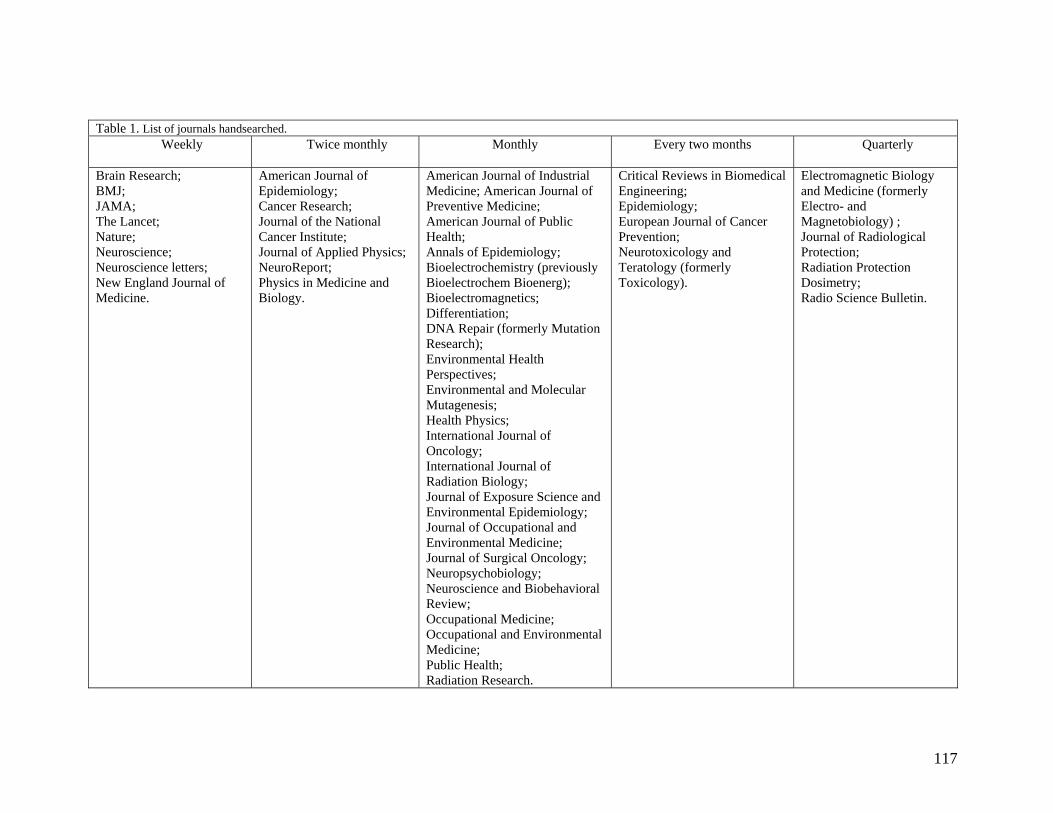
117
Table 1. List of journals handsearched. Weekly Twice monthly
Monthly Every two months Quarterly
Brain Research; BMJ; JAMA; The Lancet; Nature; Neuroscience; Neuroscience letters; New England Journal of Medicine.
American Journal of Epidemiology; Cancer Research; Journal of the National Cancer Institute; Journal of Applied Physics; NeuroReport; Physics in Medicine and Biology.
American Journal of Industrial Medicine; American Journal of Preventive Medicine; American Journal of Public Health; Annals of Epidemiology; Bioelectrochemistry (previously Bioelectrochem Bioenerg); Bioelectromagnetics; Differentiation; DNA Repair (formerly Mutation Research); Environmental Health Perspectives; Environmental and Molecular Mutagenesis; Health Physics; International Journal of Oncology; International Journal of Radiation Biology; Journal of Exposure Science and Environmental Epidemiology; Journal of Occupational and Environmental Medicine; Journal of Surgical Oncology; Neuropsychobiology; Neuroscience and Biobehavioral Review; Occupational Medicine; Occupational and Environmental Medicine; Public Health; Radiation Research.
Critical Reviews in Biomedical Engineering; Epidemiology; European Journal of Cancer Prevention; Neurotoxicology and Teratology (formerly Toxicology).
Electromagnetic Biology and Medicine (formerly Electro- and Magnetobiology) ; Journal of Radiological Protection; Radiation Protection Dosimetry; Radio Science Bulletin.
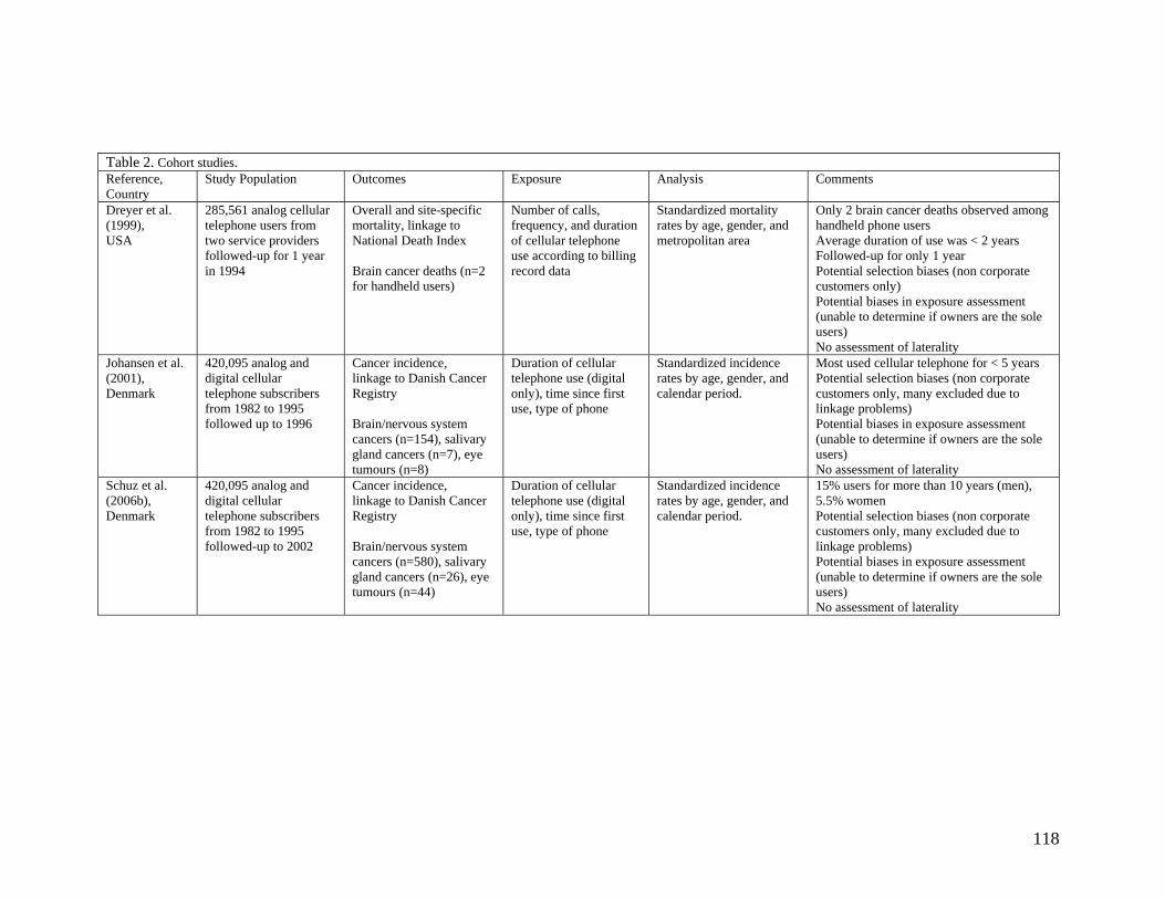
118
Table 2. Cohort studies. Reference, Country
Study Population Outcomes Exposure Analysis Comments
Dreyer et al. (1999), USA
285,561 analog cellular telephone users from two service providers followed-up for 1 year in 1994
Overall and site-specific mortality, linkage to National Death Index Brain cancer deaths (n=2 for handheld users)
Number of calls, frequency, and duration of cellular telephone use according to billing record data
Standardized mortality rates by age, gender, and metropolitan area
Only 2 brain cancer deaths observed among handheld phone users Average duration of use was < 2 years Followed-up for only 1 year Potential selection biases (non corporate customers only) Potential biases in exposure assessment (unable to determine if owners are the sole users) No assessment of laterality
Johansen et al. (2001), Denmark
420,095 analog and digital cellular telephone subscribers from 1982 to 1995 followed up to 1996
Cancer incidence, linkage to Danish Cancer Registry Brain/nervous system cancers (n=154), salivary gland cancers (n=7), eye tumours (n=8)
Duration of cellular telephone use (digital only), time since first use, type of phone
Standardized incidence rates by age, gender, and calendar period.
Most used cellular telephone for < 5 years Potential selection biases (non corporate customers only, many excluded due to linkage problems) Potential biases in exposure assessment (unable to determine if owners are the sole users) No assessment of laterality
Schuz et al. (2006b), Denmark
420,095 analog and digital cellular telephone subscribers from 1982 to 1995 followed-up to 2002
Cancer incidence, linkage to Danish Cancer Registry Brain/nervous system cancers (n=580), salivary gland cancers (n=26), eye tumours (n=44)
Duration of cellular telephone use (digital only), time since first use, type of phone
Standardized incidence rates by age, gender, and calendar period.
15% users for more than 10 years (men), 5.5% women Potential selection biases (non corporate customers only, many excluded due to linkage problems) Potential biases in exposure assessment (unable to determine if owners are the sole users) No assessment of laterality
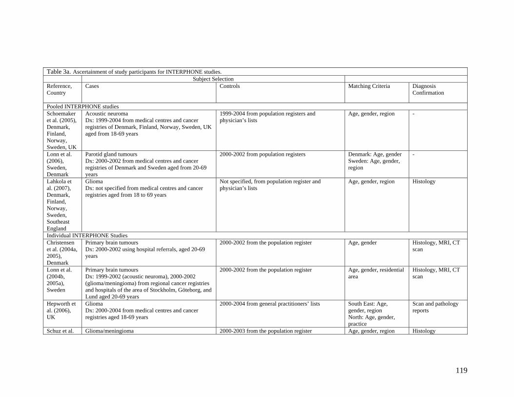
119
Table 3a. Ascertainment of study participants for INTERPHONE studies. Subject Selection Reference, Country
Cases Controls Matching Criteria Diagnosis Confirmation
Pooled INTERPHONE studies Schoemaker et al. (2005), Denmark, Finland, Norway, Sweden, UK
Acoustic neuroma Dx: 1999-2004 from medical centres and cancer registries of Denmark, Finland, Norway, Sweden, UK aged from 18-69 years
1999-2004 from population registers and physician’s lists
Age, gender, region -
Lonn et al. (2006), Sweden, Denmark
Parotid gland tumours Dx: 2000-2002 from medical centres and cancer registries of Denmark and Sweden aged from 20-69 years
2000-2002 from population registers Denmark: Age, gender Sweden: Age, gender, region
-
Lahkola et al. (2007), Denmark, Finland, Norway, Sweden, Southeast England
Glioma Dx: not specified from medical centres and cancer registries aged from 18 to 69 years
Not specified, from population register and physician’s lists
Age, gender, region Histology
Individual INTERPHONE Studies Christensen et al. (2004a, 2005), Denmark
Primary brain tumours Dx: 2000-2002 using hospital referrals, aged 20-69 years
2000-2002 from the population register Age, gender Histology, MRI, CT scan
Lonn et al. (2004b, 2005a), Sweden
Primary brain tumours Dx: 1999-2002 (acoustic neuroma), 2000-2002 (glioma/meningioma) from regional cancer registries and hospitals of the area of Stockholm, Göteborg, and Lund aged 20-69 years
2000-2002 from the population register Age, gender, residential area
Histology, MRI, CT scan
Hepworth et al. (2006), UK
Glioma Dx: 2000-2004 from medical centres and cancer registries aged 18-69 years
2000-2004 from general practitioners’ lists South East: Age, gender, region North: Age, gender, practice
Scan and pathology reports
Schuz et al. Glioma/meningioma 2000-2003 from the population register Age, gender, region Histology
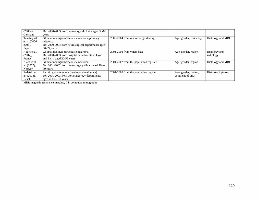
120
(2006a), Germany
Dx: 2000-2003 from neurosurgical clinics aged 30-69 years
Takebayashi et al. (2006, 2008), Japan
Glioma/meningioma/acoustic neuroma/pituitary adenoma Dx: 2000-2004 from neurosurgical departments aged 30-69 years
2000-2004 from random-digit dialing Age, gender, residency Histology and MRI
Hours et al. (2007), France
Glioma/meningioma/acoustic neuroma Dx: 2000-2003 from hospital departments in Lyon and Paris, aged 30-59 years
2001-2003 from voters lists Age, gender, region Histology and radiology
Klaeboe et al. (2007), Norway
Glioma/meningioma/acoustic neuroma Dx: 2001-2002 from neurosurgery clinics aged 19 to 69 years
2001-2002 from the population register Age, gender, region Histology and MRI
Sadetzki et al. (2008), Israel
Parotid gland tumours (benign and malignant) Dx: 2001-2003 from otolaryngology departments aged at least 18 years
2001-2003 from the population register Age, gender, region, continent of birth
Histology/cytology
MRI: magnetic resonance imaging, CT: computed tomography
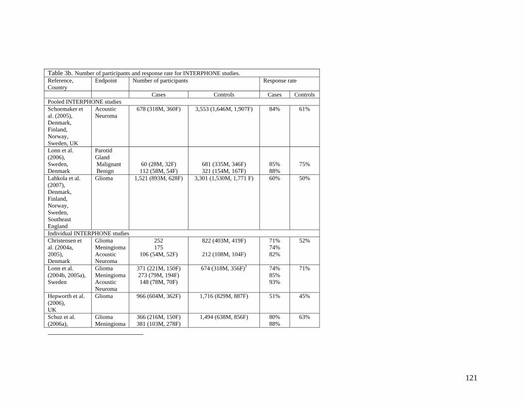
121
Table 3b. Number of participants and response rate for INTERPHONE studies. Reference, Country
Endpoint Number of participants Response rate
Cases Controls Cases Controls Pooled INTERPHONE studies Schoemaker et al. (2005), Denmark, Finland, Norway, Sweden, UK
Acoustic Neuroma
678 (318M, 360F) 3,553 (1,646M, 1,907F)
84% 61%
Lonn et al. (2006), Sweden, Denmark
Parotid Gland Malignant Benign
60 (28M, 32F) 112 (58M, 54F)
681 (335M, 346F) 321 (154M, 167F)
85% 88%
75%
Lahkola et al. (2007), Denmark, Finland, Norway, Sweden, Southeast England
Glioma 1,521 (893M, 628F)
3,301 (1,530M, 1,771 F)
60% 50%
Individual INTERPHONE studies Christensen et al. (2004a, 2005), Denmark
Glioma Meningioma Acoustic Neuroma
252 175
106 (54M, 52F)
822 (403M, 419F)
212 (108M, 104F)
71% 74% 82%
52%
Lonn et al. (2004b, 2005a), Sweden
Glioma Meningioma Acoustic Neuroma
371 (221M, 150F) 273 (79M, 194F) 148 (78M, 70F)
674 (318M, 356F)1 74% 85% 93%
71%
Hepworth et al. (2006), UK
Glioma 966 (604M, 362F)
1,716 (829M, 887F)
51% 45%
Schuz et al. (2006a),
Glioma Meningioma
366 (216M, 150F) 381 (103M, 278F)
1,494 (638M, 856F)
80% 88%
63%
122
Germany Takebayashi et al. (2006, 2008), Japan
Glioma Meningioma Acoustic Neuroma Pituitary Adenoma
83 (44M, 39F) 128 (29M, 99F) 97 (45M, 52F)
102 (62M, 39F)
163 (85M, 78F) 229 (48M, 181F)
330 (132M, 198F)
161 (101M, 60F)
59% 78% 84%
76%
52% 52% 52%
49%
Hours et al. (2007), France
Glioma Meningioma Acoustic Neuroma
96 (59M, 37F) 145 (26M, 119F) 109 (53M, 56F)
455 (187M, 268F) 60% 78% 81%
75%
Klaeboe et al. (2007), Norway
Glioma Meningioma Acoustic Neuroma
289 (170M, 119F) 207 (51M, 156F) 45 (22M, 23F)
358 (176M, 182F)
77% 71% 68%
69%
Sadetzki et al. (2008), Israel
Parotid Gland
460 (254M, 206F) 1,266 (551M, 715F) 87% 66%
M: male, F: female
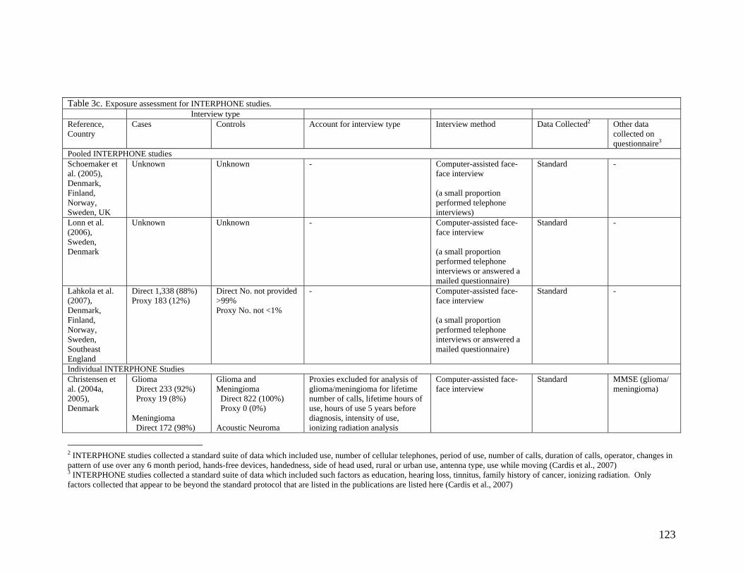
123
Table 3c. Exposure assessment for INTERPHONE studies. Interview type Reference, Country
Cases Controls Account for interview type Interview method
Data Collected2
Other data collected on questionnaire3
Pooled INTERPHONE studies Schoemaker et al. (2005), Denmark, Finland, Norway, Sweden, UK
Unknown Unknown - Computer-assisted face-face interview (a small proportion performed telephone interviews)
Standard -
Lonn et al. (2006), Sweden, Denmark
Unknown Unknown - Computer-assisted face-face interview (a small proportion performed telephone interviews or answered a mailed questionnaire)
Standard -
Lahkola et al. (2007), Denmark, Finland, Norway, Sweden, Southeast England
Direct 1,338 (88%) Proxy 183 (12%)
Direct No. not provided >99% Proxy No. not <1%
- Computer-assisted face-face interview (a small proportion performed telephone interviews or answered a mailed questionnaire)
Standard -
Individual INTERPHONE Studies Christensen et al. (2004a, 2005), Denmark
Glioma Direct 233 (92%) Proxy 19 (8%) Meningioma Direct 172 (98%)
Glioma and Meningioma Direct 822 (100%) Proxy 0 (0%) Acoustic Neuroma
Proxies excluded for analysis of glioma/meningioma for lifetime number of calls, lifetime hours of use, hours of use 5 years before diagnosis, intensity of use, ionizing radiation analysis
Computer-assisted face-face interview
Standard MMSE (glioma/ meningioma)
2 INTERPHONE studies collected a standard suite of data which included use, number of cellular telephones, period of use, number of calls, duration of calls, operator, changes in pattern of use over any 6 month period, hands-free devices, handedness, side of head used, rural or urban use, antenna type, use while moving (Cardis et al., 2007) 3 INTERPHONE studies collected a standard suite of data which included such factors as education, hearing loss, tinnitus, family history of cancer, ionizing radiation. Only factors collected that appear to be beyond the standard protocol that are listed in the publications are listed here (Cardis et al., 2007)
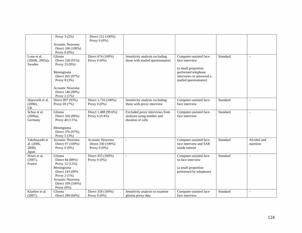
124
Proxy 3 (2%) Acoustic Neuroma Direct 106 (100%) Proxy 0 (0%)
Direct 212 (100%) Proxy 0 (0%)
Lonn et al. (2004b, 2005a), Sweden
Glioma Direct 338 (91%) Proxy 33 (9%) Meningioma Direct 265 (97%) Proxy 8 (3%) Acoustic Neuroma Direct 146 (99%) Proxy 2 (1%)
Direct 674 (100%) Proxy 0 (0%)
Sensitivity analysis excluding those with mailed questionnaires
Computer-assisted face-face interview (a small proportion performed telephone interviews or answered a mailed questionnaire)
Standard
-
Hepworth et al. (2006), UK
Direct 897 (93%) Proxy 69 (7%)
Direct 1,716 (100%) Proxy 0 (0%)
Sensitivity analysis excluding those with proxy interview
Computer-assisted face-face interview
Standard
-
Schuz et al. (2006a), Germany
Glioma Direct 326 (89%) Proxy 40 (11%) Meningioma Direct 376 (97%) Proxy 5 (3%)
Direct 1,488 (99.6%) Proxy 6 (0.4%)
Excluded proxy interviews from analyses using number and duration of calls
Computer-assisted face-face interview
Standard -
Takebayashi et al. (2006, 2008), Japan
Acoustic Neuroma Direct 97 (100%) Proxy 0 (0%)
Acoustic Neuroma Direct 330 (100%) Proxy 0 (0%)
- Computer-assisted face-face interview and SAR inside tumour
Standard
Alcohol and nutrition
Hours et al. (2007), France
Glioma Direct 84 (88%) Proxy 12 (12%) Meningioma Direct 143 (99% Proxy 2 (1%) Acoustic Neuroma Direct 109 (100%) Proxy (0%)
Direct 455 (100%) Proxy 0 (0%)
- Computer-assisted face-to-face interview (a small proportion performed by telephone)
Standard -
Klaeboe et al. (2007),
Glioma Direct 289 (64%)
Direct 358 (100%) Proxy 0 (0%)
Sensitivity analysis to examine glioma proxy data
Computer-assisted face-face interview
Standard
-
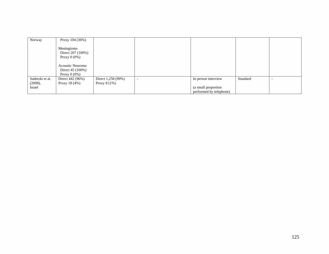
125
Norway Proxy 104 (36%) Meningioma Direct 207 (100%) Proxy 0 (0%) Acoustic Neuroma Direct 45 (100%) Proxy 0 (0%)
Sadetzki et al. (2008), Israel
Direct 442 (96%) Proxy 18 (4%)
Direct 1,258 (99%) Proxy 8 (1%)
- In person interview (a small proportion performed by telephone)
Standard -
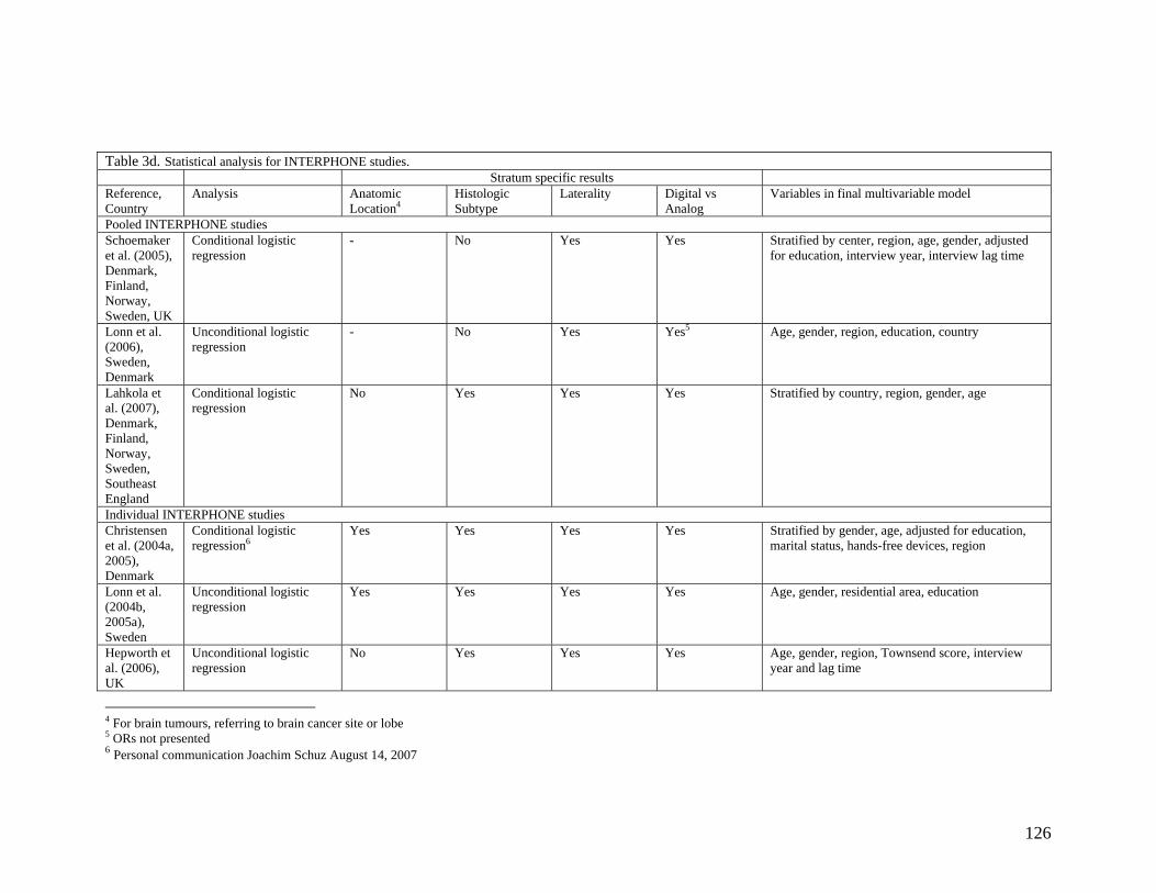
126
Table 3d. Statistical analysis for INTERPHONE studies. Stratum specific results Reference, Country
Analysis Anatomic Location4
Histologic Subtype
Laterality Digital vs Analog
Variables in final multivariable model
Pooled INTERPHONE studies Schoemaker et al. (2005), Denmark, Finland, Norway, Sweden, UK
Conditional logistic regression
- No Yes Yes Stratified by center, region, age, gender, adjusted for education, interview year, interview lag time
Lonn et al. (2006), Sweden, Denmark
Unconditional logistic regression
- No Yes Yes5 Age, gender, region, education, country
Lahkola et al. (2007), Denmark, Finland, Norway, Sweden, Southeast England
Conditional logistic regression
No Yes Yes Yes Stratified by country, region, gender, age
Individual INTERPHONE studies Christensen et al. (2004a, 2005), Denmark
Conditional logistic regression6
Yes Yes
Yes Yes Stratified by gender, age, adjusted for education, marital status, hands-free devices, region
Lonn et al. (2004b, 2005a), Sweden
Unconditional logistic regression
Yes Yes Yes Yes Age, gender, residential area, education
Hepworth et al. (2006), UK
Unconditional logistic regression
No Yes Yes Yes Age, gender, region, Townsend score, interview year and lag time
4 For brain tumours, referring to brain cancer site or lobe 5 ORs not presented 6 Personal communication Joachim Schuz August 14, 2007
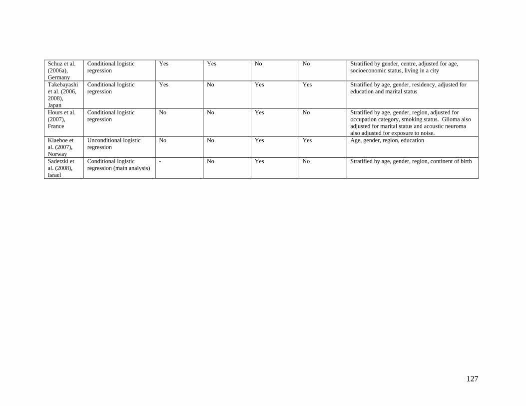
127
Schuz et al. (2006a), Germany
Conditional logistic regression
Yes Yes No No Stratified by gender, centre, adjusted for age, socioeconomic status, living in a city
Takebayashi et al. (2006, 2008), Japan
Conditional logistic regression
Yes No Yes Yes Stratified by age, gender, residency, adjusted for education and marital status
Hours et al. (2007), France
Conditional logistic regression
No No Yes No Stratified by age, gender, region, adjusted for occupation category, smoking status. Glioma also adjusted for marital status and acoustic neuroma also adjusted for exposure to noise.
Klaeboe et al. (2007), Norway
Unconditional logistic regression
No No Yes Yes Age, gender, region, education
Sadetzki et al. (2008), Israel
Conditional logistic regression (main analysis)
- No Yes No Stratified by age, gender, region, continent of birth
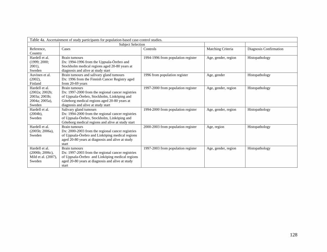
128
Table 4a. Ascertainment of study participants for population-based case-control studies. Subject Selection Reference, Country
Cases Controls Matching Criteria Diagnosis Confirmation
Hardell et al. (1999; 2000; 2001), Sweden
Brain tumours Dx: 1994-1996 from the Uppsala-Örebro and Stockholm medical regions aged 20-80 years at diagnosis and alive at study start
1994-1996 from population register Age, gender, region
Histopathology
Auvinen et al. (2002), Finland
Brain tumours and salivary gland tumours Dx: 1996 from the Finnish Cancer Registry aged from 20-69 years
1996 from population register Age, gender Histopathology
Hardell et al. (2002a; 2002b; 2003a; 2003b; 2004a; 2005a), Sweden
Brain tumours Dx: 1997-2000 from the regional cancer registries of Uppsala-Örebro, Stockholm, Linköping and Göteborg medical regions aged 20-80 years at diagnosis and alive at study start
1997-2000 from population register Age, gender, region
Histopathology
Hardell et al. (2004b), Sweden
Salivary gland tumours Dx: 1994-2000 from the regional cancer registries of Uppsala-Örebro, Stockholm, Linköping and Göteborg medical regions and alive at study start
1994-2000 from population register Age, gender, region Histopathology
Hardell et al. (2005b; 2006a), Sweden
Brain tumours Dx: 2000-2003 from the regional cancer registries of Uppsala-Örebro and Linköping medical regions aged 20-80 years at diagnosis and alive at study start
2000-2003 from population register Age, region Histopathology
Hardell et al. (2006b; 2006c), Mild et al. (2007), Sweden
Brain tumours Dx: 1997-2003 from the regional cancer registries of Uppsala-Örebro and Linköping medical regions aged 20-80 years at diagnosis and alive at study start
1997-2003 from population register Age, gender, region
Histopathology
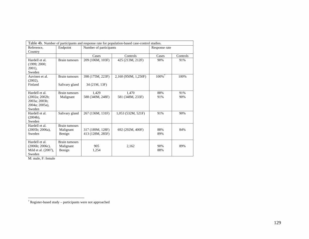
129
Table 4b. Number of participants and response rate for population-based case-control studies. Reference, Country
Endpoint Number of participants Response rate
Cases Controls Cases Controls Hardell et al. (1999; 2000; 2001), Sweden
Brain tumours 209 (106M, 103F)
425 (213M, 212F) 90% 91%
Auvinen et al. (2002), Finland
Brain tumours Salivary gland
398 (175M, 223F)
34 (21M, 13F)
2,160 (950M, 1,250F) 100%7 100%
Hardell et al. (2002a; 2002b; 2003a; 2003b; 2004a; 2005a), Sweden
Brain tumours Malignant
1,429 588 (340M, 248F)
1,470 581 (348M, 233F)
88% 91%
91% 90%
Hardell et al. (2004b), Sweden
Salivary gland 267 (136M, 131F)
1,053 (532M, 521F) 91% 90%
Hardell et al. (2005b; 2006a), Sweden
Brain tumours Malignant Benign
317 (189M, 128F) 413 (128M, 285F)
692 (292M, 400F)
88% 89%
84%
Hardell et al. (2006b; 2006c), Mild et al. (2007), Sweden
Brain tumours Malignant Benign
905
1,254
2,162
90% 88%
89%
M: male, F: female
7 Register-based study – participants were not approached
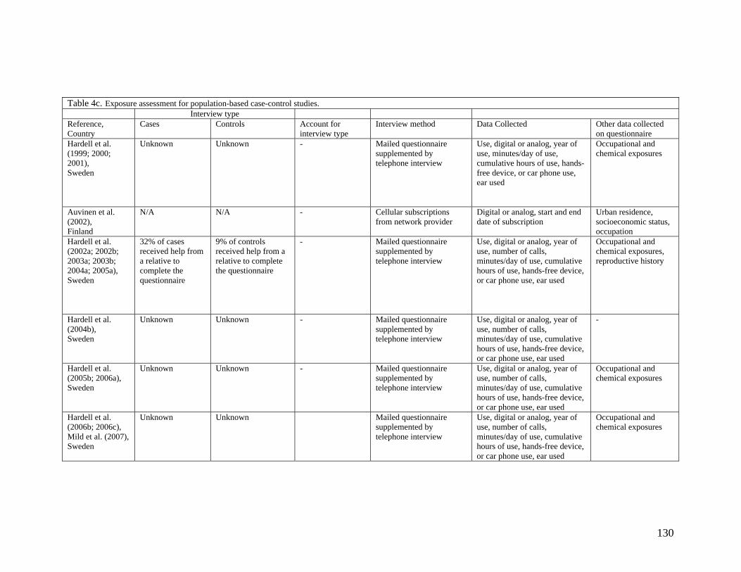
130
Table 4c. Exposure assessment for population-based case-control studies. Interview type Reference, Country
Cases Controls Account for interview type
Interview method
Data Collected
Other data collected on questionnaire
Hardell et al. (1999; 2000; 2001), Sweden
Unknown Unknown - Mailed questionnaire supplemented by telephone interview
Use, digital or analog, year of use, minutes/day of use, cumulative hours of use, hands-free device, or car phone use, ear used
Occupational and chemical exposures
Auvinen et al. (2002), Finland
N/A N/A - Cellular subscriptions from network provider
Digital or analog, start and end date of subscription
Urban residence, socioeconomic status, occupation
Hardell et al. (2002a; 2002b; 2003a; 2003b; 2004a; 2005a), Sweden
32% of cases received help from a relative to complete the questionnaire
9% of controls received help from a relative to complete the questionnaire
- Mailed questionnaire supplemented by telephone interview
Use, digital or analog, year of use, number of calls, minutes/day of use, cumulative hours of use, hands-free device, or car phone use, ear used
Occupational and chemical exposures, reproductive history
Hardell et al. (2004b), Sweden
Unknown Unknown - Mailed questionnaire supplemented by telephone interview
Use, digital or analog, year of use, number of calls, minutes/day of use, cumulative hours of use, hands-free device, or car phone use, ear used
-
Hardell et al. (2005b; 2006a), Sweden
Unknown Unknown - Mailed questionnaire supplemented by telephone interview
Use, digital or analog, year of use, number of calls, minutes/day of use, cumulative hours of use, hands-free device, or car phone use, ear used
Occupational and chemical exposures
Hardell et al. (2006b; 2006c), Mild et al. (2007), Sweden
Unknown Unknown Mailed questionnaire supplemented by telephone interview
Use, digital or analog, year of use, number of calls, minutes/day of use, cumulative hours of use, hands-free device, or car phone use, ear used
Occupational and chemical exposures
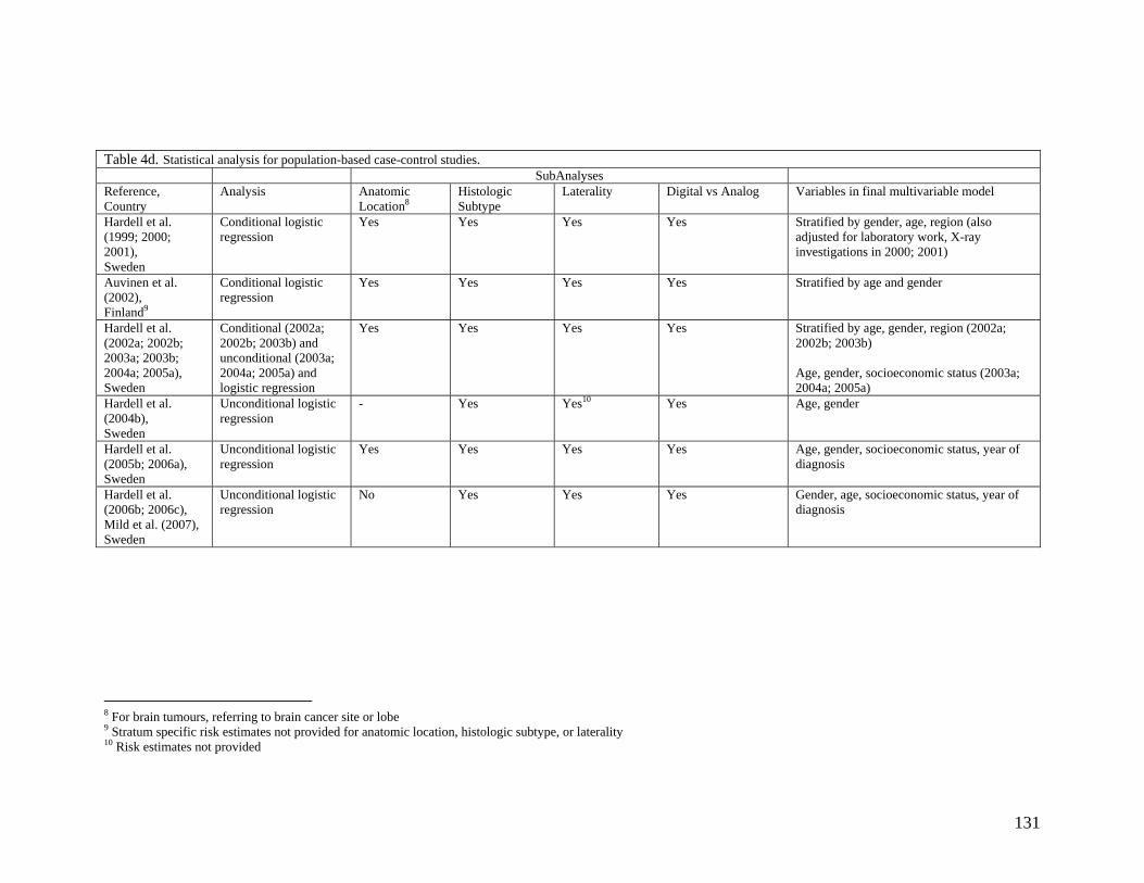
131
Table 4d. Statistical analysis for population-based case-control studies. SubAnalyses Reference, Country
Analysis Anatomic Location8
Histologic Subtype
Laterality Digital vs Analog Variables in final multivariable model
Hardell et al. (1999; 2000; 2001), Sweden
Conditional logistic regression
Yes Yes Yes Yes Stratified by gender, age, region (also adjusted for laboratory work, X-ray investigations in 2000; 2001)
Auvinen et al. (2002), Finland9
Conditional logistic regression
Yes Yes Yes Yes Stratified by age and gender
Hardell et al. (2002a; 2002b; 2003a; 2003b; 2004a; 2005a), Sweden
Conditional (2002a; 2002b; 2003b) and unconditional (2003a; 2004a; 2005a) and logistic regression
Yes Yes Yes Yes Stratified by age, gender, region (2002a; 2002b; 2003b) Age, gender, socioeconomic status (2003a; 2004a; 2005a)
Hardell et al. (2004b), Sweden
Unconditional logistic regression
- Yes Yes10 Yes Age, gender
Hardell et al. (2005b; 2006a), Sweden
Unconditional logistic regression
Yes Yes Yes Yes Age, gender, socioeconomic status, year of diagnosis
Hardell et al. (2006b; 2006c), Mild et al. (2007), Sweden
Unconditional logistic regression
No Yes Yes Yes Gender, age, socioeconomic status, year of diagnosis
8 For brain tumours, referring to brain cancer site or lobe 9 Stratum specific risk estimates not provided for anatomic location, histologic subtype, or laterality 10 Risk estimates not provided
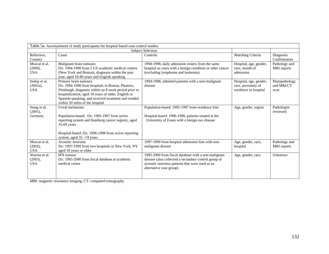
132
Table 5a. Ascertainment of study participants for hospital-based case-control studies. Subject Selection Reference, Country
Cases Controls Matching Criteria Diagnosis Confirmation
Muscat et al. (2000), USA
Malignant brain tumours Dx: 1994-1998 from 5 US academic medical centres (New York and Boston), diagnosis within the past year, aged 18-80 years and English speaking
1994-1998, daily admission rosters from the same hospital as cases with a benign condition or other cancer (excluding lymphoma and leukemia)
Hospital, age, gender, race, month of admission
Pathology and MRI reports
Inskip et al. (2001a), USA
Primary brain tumours Dx: 1994-1998 from hospitals in Boston, Phoenix, Pittsburgh, diagnosis within an 8 week period prior to hospitalization, aged 18 years of older, English or Spanish-speaking, and received treatment and resided within 50 miles of the hospital
1994-1998, admitted patients with a non-malignant disease
Hospital, age, gender, race, proximity of residence to hospital
Histopathology and MRI/CT scan
Stang et al. (2001), Germany
Uveal melanoma Population-based: Dx: 1995-1997 from active reporting system and Hamburg cancer registry, aged 35-69 years Hospital-based: Dx: 1996-1998 from active reporting system, aged 35 –74 years
Population-based: 1995-1997 from residence lists Hospital-based: 1996-1998, patients treated at the University of Essen with a benign eye disease
Age, gender, region
Pathologist reviewed
Muscat et al. (2002), USA
Acoustic neuroma Dx: 1997-1999 from two hospitals in New York, NY aged 18 years or older
1997-1999 from hospital admission lists with non-malignant disease
Age, gender, race, hospital
Pathology and MRI reports
Warren et al. (2003), USA
IFN tumour Dx: 1995-2000 from fiscal database at academic medical centre
1995-2000 from fiscal database with a non-malignant disease (also collected a secondary control group of acoustic neuroma patients that were used as an alternative case group)
Age, gender, race Unknown
MRI: magnetic resonance imaging, CT: computed tomography
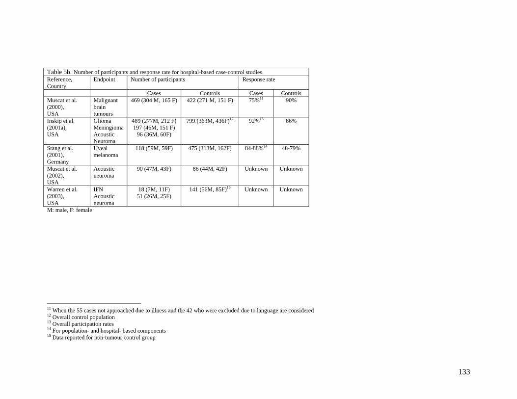
133
Table 5b. Number of participants and response rate for hospital-based case-control studies. Reference, Country
Endpoint Number of participants Response rate
Cases Controls Cases Controls Muscat et al. (2000), USA
Malignant brain tumours
469 (304 M, 165 F)
422 (271 M, 151 F)
75%11
90%
Inskip et al. (2001a), USA
Glioma Meningioma Acoustic Neuroma
489 (277M, 212 F) 197 (46M, 151 F)
96 (36M, 60F)
799 (363M, 436F)12
92%13 86%
Stang et al. (2001), Germany
Uveal melanoma
118 (59M, 59F)
475 (313M, 162F)
84-88%14 48-79%
Muscat et al. (2002), USA
Acoustic neuroma
90 (47M, 43F)
86 (44M, 42F)
Unknown Unknown
Warren et al. (2003), USA
IFN Acoustic neuroma
18 (7M, 11F) 51 (26M, 25F)
141 (56M, 85F)15 Unknown Unknown
M: male, F: female
11 When the 55 cases not approached due to illness and the 42 who were excluded due to language are considered 12 Overall control population 13 Overall participation rates 14 For population- and hospital- based components 15 Data reported for non-tumour control group
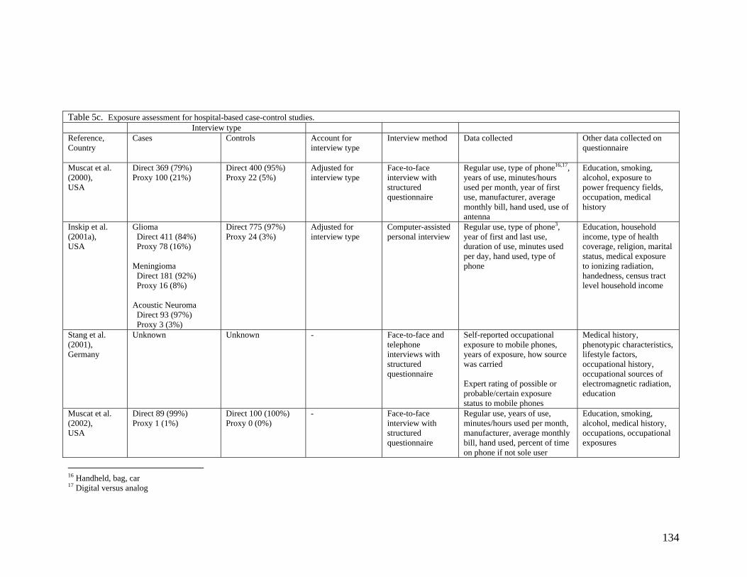
134
Table 5c. Exposure assessment for hospital-based case-control studies. Interview type Reference, Country
Cases Controls Account for interview type
Interview method
Data collected
Other data collected on questionnaire
Muscat et al. (2000), USA
Direct 369 (79%) Proxy 100 (21%)
Direct 400 (95%) Proxy 22 (5%)
Adjusted for interview type
Face-to-face interview with structured questionnaire
Regular use, type of phone16,17, years of use, minutes/hours used per month, year of first use, manufacturer, average monthly bill, hand used, use of antenna
Education, smoking, alcohol, exposure to power frequency fields, occupation, medical history
Inskip et al. (2001a), USA
Glioma Direct 411 (84%) Proxy 78 (16%) Meningioma Direct 181 (92%) Proxy 16 (8%) Acoustic Neuroma Direct 93 (97%) Proxy 3 (3%)
Direct 775 (97%) Proxy 24 (3%)
Adjusted for interview type
Computer-assisted personal interview
Regular use, type of phone3, year of first and last use, duration of use, minutes used per day, hand used, type of phone
Education, household income, type of health coverage, religion, marital status, medical exposure to ionizing radiation, handedness, census tract level household income
Stang et al. (2001), Germany
Unknown Unknown - Face-to-face and telephone interviews with structured questionnaire
Self-reported occupational exposure to mobile phones, years of exposure, how source was carried Expert rating of possible or probable/certain exposure status to mobile phones
Medical history, phenotypic characteristics, lifestyle factors, occupational history, occupational sources of electromagnetic radiation, education
Muscat et al. (2002), USA
Direct 89 (99%) Proxy 1 (1%)
Direct 100 (100%) Proxy 0 (0%)
- Face-to-face interview with structured questionnaire
Regular use, years of use, minutes/hours used per month, manufacturer, average monthly bill, hand used, percent of time on phone if not sole user
Education, smoking, alcohol, medical history, occupations, occupational exposures
16 Handheld, bag, car 17 Digital versus analog
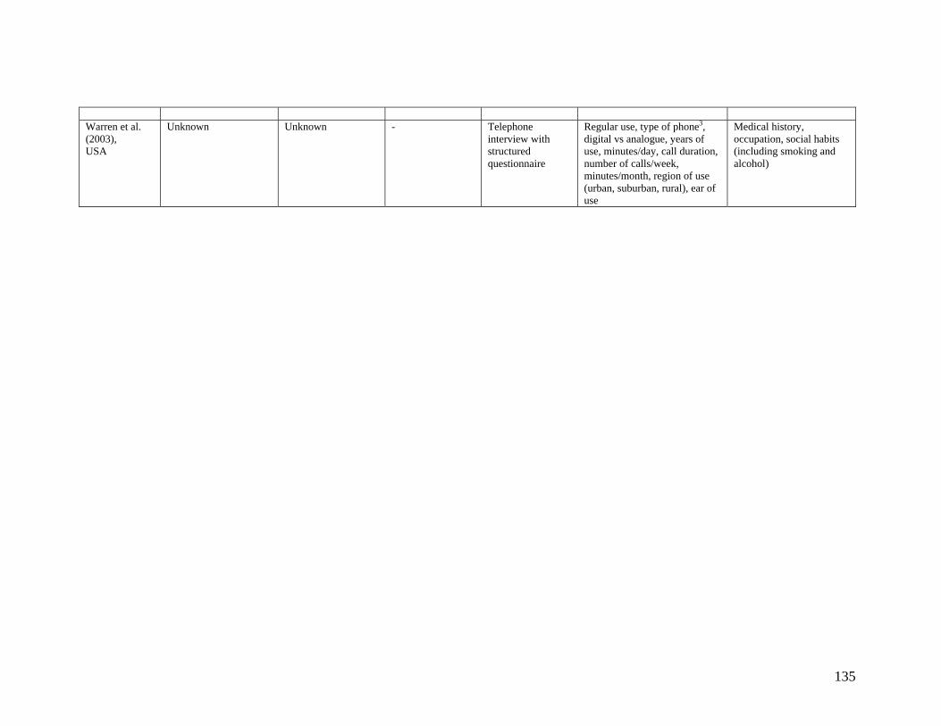
135
Warren et al. (2003), USA
Unknown Unknown - Telephone interview with structured questionnaire
Regular use, type of phone3, digital vs analogue, years of use, minutes/day, call duration, number of calls/week, minutes/month, region of use (urban, suburban, rural), ear of use
Medical history, occupation, social habits (including smoking and alcohol)
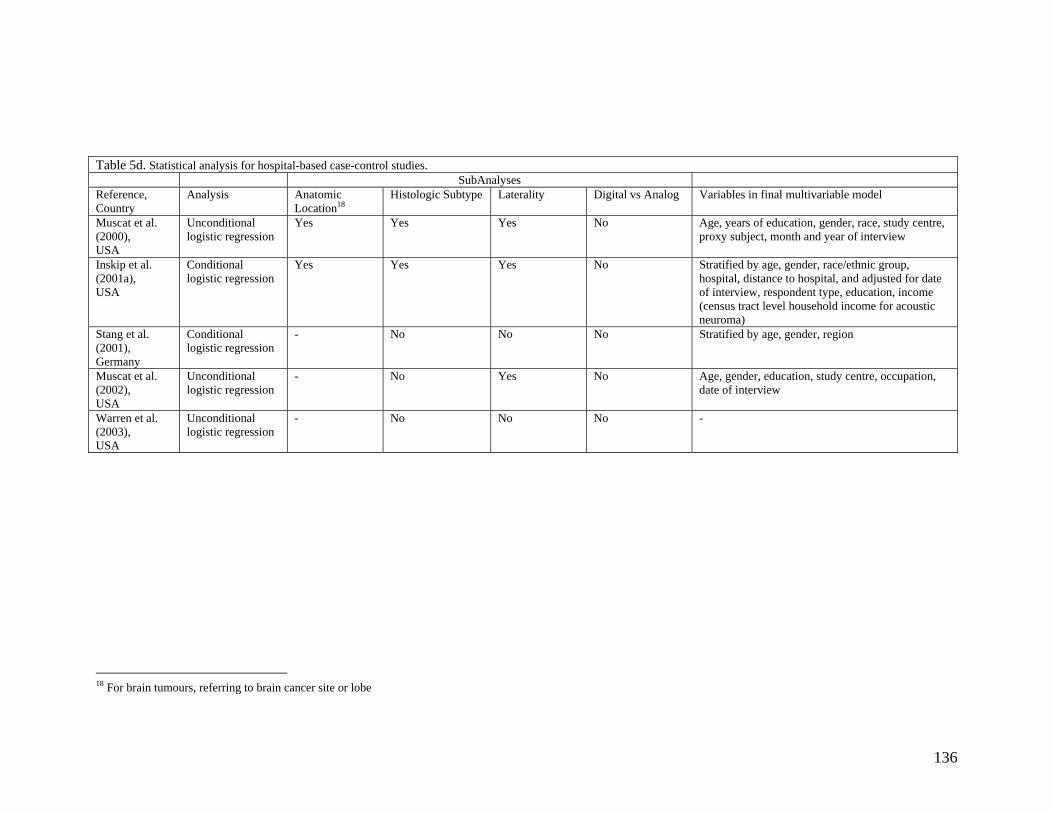
136
Table 5d. Statistical analysis for hospital-based case-control studies. SubAnalyses Reference, Country
Analysis Anatomic Location18
Histologic Subtype Laterality Digital vs Analog Variables in final multivariable model
Muscat et al. (2000), USA
Unconditional logistic regression
Yes Yes Yes No Age, years of education, gender, race, study centre, proxy subject, month and year of interview
Inskip et al. (2001a), USA
Conditional logistic regression
Yes Yes Yes No Stratified by age, gender, race/ethnic group, hospital, distance to hospital, and adjusted for date of interview, respondent type, education, income (census tract level household income for acoustic neuroma)
Stang et al. (2001), Germany
Conditional logistic regression
- No No No Stratified by age, gender, region
Muscat et al. (2002), USA
Unconditional logistic regression
- No Yes No Age, gender, education, study centre, occupation, date of interview
Warren et al. (2003), USA
Unconditional logistic regression
- No No No -
18 For brain tumours, referring to brain cancer site or lobe
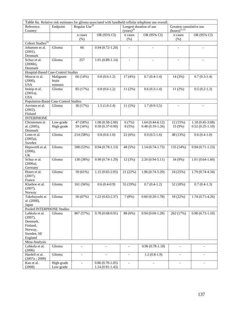
137
Table 6a. Relative risk estimates for glioma associated with handheld cellular telephone use overall. Reference, Country
Endpoint Regular Use19 Longest duration of use (years)20
Greatest cumulative use (hours)21,22
n cases (%)
OR (95% CI) n cases (%)
OR (95% CI) n cases (%)
OR (95% CI)
Cohort Studies23 Johansen et al. (2001), Denmark
Glioma 66 0.94 (0.72-1.20) - - - -
Schuz et al. (2006b), Denmark
Glioma 257 1.01 (0.89-1.14) - - - -
Hospital-Based Case-Control Studies Muscat et al. (2000), USA
Malignant brain tumours
66 (14%) 0.8 (0.6-1.2) 17 (4%) 0.7 (0.4-1.4) 14 (3%) 0.7 (0.3-1.4)
Inskip et al. (2001a), USA
Glioma 85 (17%)
0.8 (0.6-1.2)
11 (2%)
0.6 (0.3-1.4) 11 (2%)
0.5 (0.2-1.3)
Population-Based Case-Control Studies Auvinen et al. (2002), Finland
Glioma 36 (17%) 1.5 (1.0-2.4) 11 (5%) 1.7 (0.9-3.5)
- -
INTERPHONE Christensen et al. (2005), Denmark
Low-grade High-grade
47 (58%) 59 (34%)
1.08 (0.58-2.00) 0.58 (0.37-0.90)
6 (7%) 8 (5%)
1.64 (0.44-6.12) 0.48 (0.19-1.26)
12 (15%) 15 (9%)
1.18 (0.45-3.08) 0.52 (0.25-1.10)
Lonn et al. (2005a), Sweden
Glioma 214 (58%) 0.8 (0.6-1.0)
22 (6%) 0.9 (0.5-1.6)
48 (13%)
0.6 (0.4-1.0)
Hepworth et al. (2006), UK
Glioma 508 (53%) 0.94 (0.78-1.13) 48 (5%) 1.14 (0.74-1.73) 135 (14%) 0.94 (0.71-1.23)
Schuz et al. (2006a), Germany
Glioma
138 (38%) 0.98 (0.74-1.29)
12 (3%)
2.20 (0.94-5.11)
34 (9%)
1.01 (0.64-1.60)
Hours et al. (2007), France
Glioma 59 (61%) 1.15 (0.65-2.05) 21 (22%) 1.96 (0.74-5.20) 24 (25%) 1.79 (0.74-4.34)
Klaeboe et al. (2007), Norway
Glioma
161 (56%) 0.6 (0.4-0.9)
55 (19%) 0.7 (0.4-1.2)
52 (18%) 0.7 (0.4-1.3)
Takebayashi et al. (2008), Japan
Glioma 56 (67%) 1.22 (0.63-2.37) 7 (8%) 0.60 (0.20-1.78) 18 (22%) 1.74 (0.71-4.26)
Pooled INTERPHONE Studies Lahkola et al. (2007), Denmark, Finland, Norway, Sweden, SE England
Glioma 867 (57%) 0.78 (0.68-0.91) 88 (6%) 0.94 (0.69-1.28) 262 (17%) 0.90 (0.73-1.10)
Meta-Analysis Lahkola et al. (2006)
Glioma - - - 0.96 (0.78-1.18) - -
Hardell et al. (2007a ; 2008)
Glioma - - - 1.2 (0.8-1.9) - -
Kan et al. (2008)
High-grade Low-grade
- 0.86 (0.70-1.05) 1.14 (0.91-1.43)
- - - -
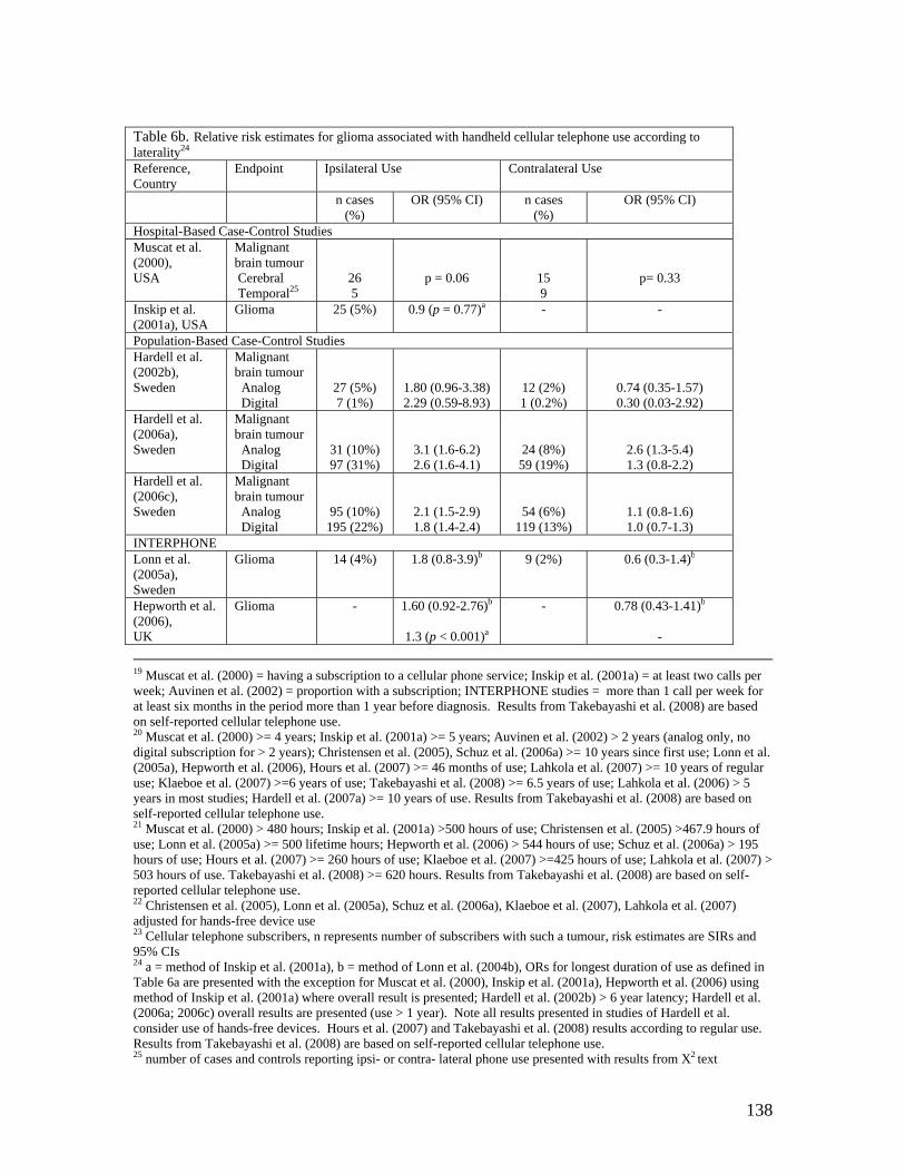
138
Table 6b. Relative risk estimates for glioma associated with handheld cellular telephone use according to laterality24 Reference, Country
Endpoint Ipsilateral Use Contralateral Use
n cases (%)
OR (95% CI) n cases (%)
OR (95% CI)
Hospital-Based Case-Control Studies Muscat et al. (2000), USA
Malignant brain tumour Cerebral Temporal25
26 5
p = 0.06
15 9
p= 0.33
Inskip et al. (2001a), USA
Glioma 25 (5%) 0.9 (p = 0.77)a - -
Population-Based Case-Control Studies Hardell et al. (2002b), Sweden
Malignant brain tumour Analog Digital
27 (5%) 7 (1%)
1.80 (0.96-3.38) 2.29 (0.59-8.93)
12 (2%) 1 (0.2%)
0.74 (0.35-1.57) 0.30 (0.03-2.92)
Hardell et al. (2006a), Sweden
Malignant brain tumour Analog Digital
31 (10%) 97 (31%)
3.1 (1.6-6.2) 2.6 (1.6-4.1)
24 (8%) 59 (19%)
2.6 (1.3-5.4) 1.3 (0.8-2.2)
Hardell et al. (2006c), Sweden
Malignant brain tumour Analog Digital
95 (10%) 195 (22%)
2.1 (1.5-2.9) 1.8 (1.4-2.4)
54 (6%) 119 (13%)
1.1 (0.8-1.6) 1.0 (0.7-1.3)
INTERPHONE Lonn et al. (2005a), Sweden
Glioma 14 (4%) 1.8 (0.8-3.9)b 9 (2%) 0.6 (0.3-1.4)b
Hepworth et al. (2006), UK
Glioma - 1.60 (0.92-2.76)b
1.3 (p < 0.001)a
-
0.78 (0.43-1.41)b
-
19 Muscat et al. (2000) = having a subscription to a cellular phone service; Inskip et al. (2001a) = at least two calls per week; Auvinen et al. (2002) = proportion with a subscription; INTERPHONE studies = more than 1 call per week for at least six months in the period more than 1 year before diagnosis. Results from Takebayashi et al. (2008) are based on self-reported cellular telephone use. 20 Muscat et al. (2000) >= 4 years; Inskip et al. (2001a) >= 5 years; Auvinen et al. (2002) > 2 years (analog only, no digital subscription for > 2 years); Christensen et al. (2005), Schuz et al. (2006a) >= 10 years since first use; Lonn et al. (2005a), Hepworth et al. (2006), Hours et al. (2007) >= 46 months of use; Lahkola et al. (2007) >= 10 years of regular use; Klaeboe et al. (2007) >=6 years of use; Takebayashi et al. (2008) >= 6.5 years of use; Lahkola et al. (2006) > 5 years in most studies; Hardell et al. (2007a) >= 10 years of use. Results from Takebayashi et al. (2008) are based on self-reported cellular telephone use. 21 Muscat et al. (2000) > 480 hours; Inskip et al. (2001a) >500 hours of use; Christensen et al. (2005) >467.9 hours of use; Lonn et al. (2005a) >= 500 lifetime hours; Hepworth et al. (2006) > 544 hours of use; Schuz et al. (2006a) > 195 hours of use; Hours et al. (2007) >= 260 hours of use; Klaeboe et al. (2007) >=425 hours of use; Lahkola et al. (2007) > 503 hours of use. Takebayashi et al. (2008) >= 620 hours. Results from Takebayashi et al. (2008) are based on self-reported cellular telephone use. 22 Christensen et al. (2005), Lonn et al. (2005a), Schuz et al. (2006a), Klaeboe et al. (2007), Lahkola et al. (2007) adjusted for hands-free device use 23 Cellular telephone subscribers, n represents number of subscribers with such a tumour, risk estimates are SIRs and 95% CIs 24 a = method of Inskip et al. (2001a), b = method of Lonn et al. (2004b), ORs for longest duration of use as defined in Table 6a are presented with the exception for Muscat et al. (2000), Inskip et al. (2001a), Hepworth et al. (2006) using method of Inskip et al. (2001a) where overall result is presented; Hardell et al. (2002b) > 6 year latency; Hardell et al. (2006a; 2006c) overall results are presented (use > 1 year). Note all results presented in studies of Hardell et al. consider use of hands-free devices. Hours et al. (2007) and Takebayashi et al. (2008) results according to regular use. Results from Takebayashi et al. (2008) are based on self-reported cellular telephone use. 25 number of cases and controls reporting ipsi- or contra- lateral phone use presented with results from X2 text
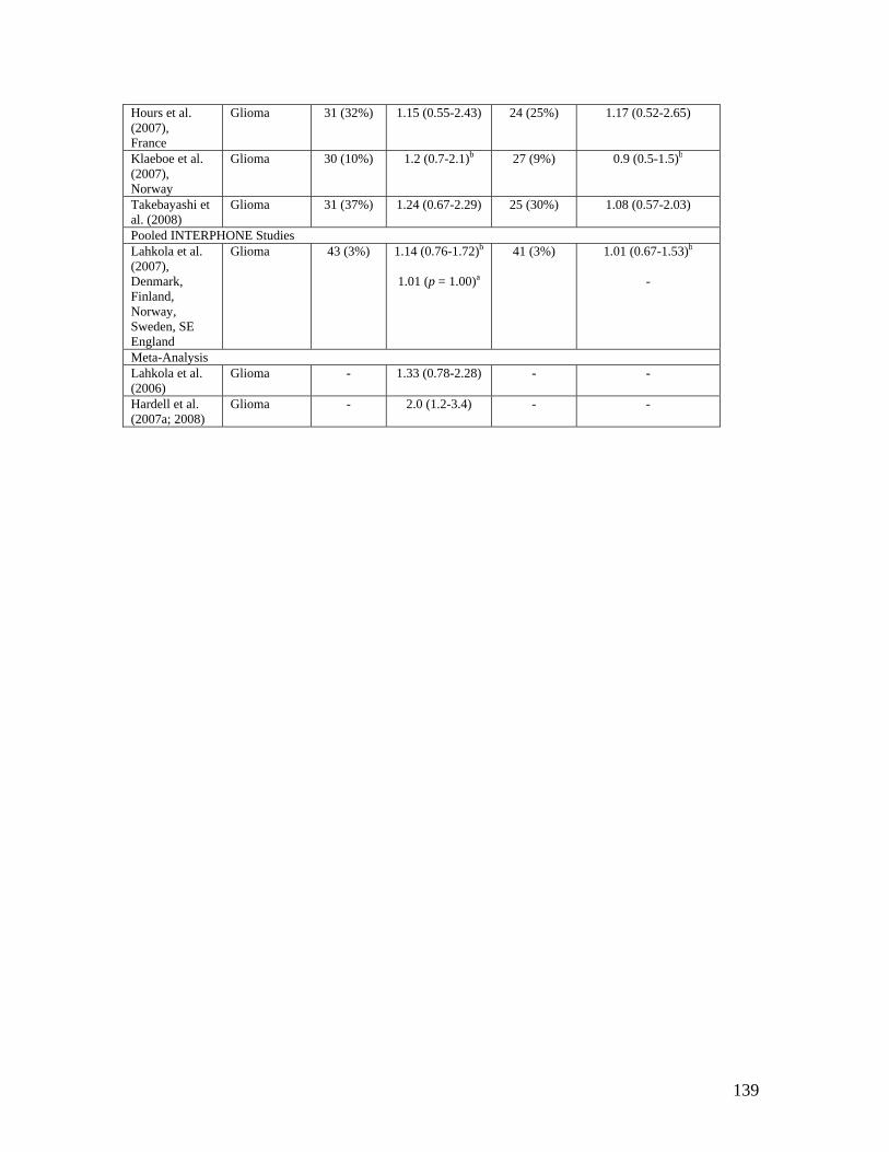
139
Hours et al. (2007), France
Glioma 31 (32%) 1.15 (0.55-2.43) 24 (25%) 1.17 (0.52-2.65)
Klaeboe et al. (2007), Norway
Glioma
30 (10%) 1.2 (0.7-2.1)b 27 (9%) 0.9 (0.5-1.5)b
Takebayashi et al. (2008)
Glioma 31 (37%) 1.24 (0.67-2.29) 25 (30%) 1.08 (0.57-2.03)
Pooled INTERPHONE Studies Lahkola et al. (2007), Denmark, Finland, Norway, Sweden, SE England
Glioma 43 (3%) 1.14 (0.76-1.72)b
1.01 (p = 1.00)a
41 (3%) 1.01 (0.67-1.53)b
-
Meta-Analysis Lahkola et al. (2006)
Glioma - 1.33 (0.78-2.28) - -
Hardell et al. (2007a; 2008)
Glioma - 2.0 (1.2-3.4) - -
140
Table 6c. Relative risk estimates for glioma associated with handheld cellular telephone use according to type of phone used26 Reference, Country
Endpoint Analog Digital
n cases (%)
OR (95% CI) n cases (%)
OR (95% CI)
Population-Based Case-Control Studies Auvinen et al. (2002), Finland
Glioma 11 (6%) 2.0 (1.0-4.0) 7 (4%) 1.4 (0.6-3.4)
Hardell et al. (2002b), Sweden
Malignant brain tumour
43 (7%) 1.17 (0.75-1.81) 12 (2%) 1.71 (0.67-4.34)
Hardell et al. (2006a), Sweden
Malignant brain tumour
48 (15%) 3.5 (2.0-6.4) 19 (6%) 3.6 (1.7-7.5)
Hardell et al. (2006c), Sweden
Malignant brain tumour
82 (9%) 2.4 (1.6-3.4) 19 (2%) 2.8 (1.4-5.7)
INTERPHONE Lonn et al. (2005a), Sweden
Glioma 25 (7%) 0.8 (0.5-1.5) 83 (22%) 0.8 (0.6-1.2)
Hepworth et al. (2006), UK
Glioma 10 (1%) 1.20 (0.48-3.04) 378 (39%) 0.96 (0.79-1.16)
Klaeboe et al. (2007), Norway
Glioma
10 (3%) 0.7 (0.4-1.2)
24 (8%) 0.7 (0.4-1.3)
Takebayashi et al. (2008), Japan
Glioma 6 (7%) 0.83 (0.23-3.00) 50 (60%) 1.29 (0.66-2.53)
Pooled INTERPHONE Studies Lahkola et al. (2007), Denmark, Finland, Norway, Sweden, SE England
Glioma 16 (1%) 0.92 (0.48-1.77) 198 (13%) 0.83 (0.67-1.04)
26 ORs for longest duration of use as defined in Table 6a are presented with the exception of Auvinen et al. (2002) where results for digital use are for use of 1-2 years; results for studies by Hardell et al. (2002b) are for > 6 years latency and Hardell et al. (2006a; 2006c) are for >10 year latency; Lonn et al. (2005a) where results are presented for >=10 years for analog phones and >= 5 years for digital phones; Hepworth et al. (2006) where results are presented for regular digital use only; Takebayashi et al. (2008) overall results presented; Lahkola et al. (2007) results for digital phones are for 5-9 years of use. Note all results presented in studies of Hardell et al. consider use of hands-free devices. Results from Takebayashi et al. (2008) are based on self-reported cellular telephone use.
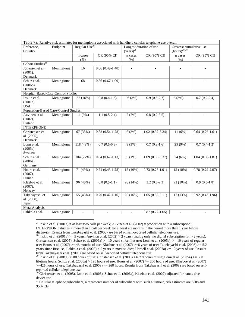
141
Table 7a. Relative risk estimates for meningioma associated with handheld cellular telephone use overall. Reference, Country
Endpoint Regular Use27 Longest duration of use (years)28
Greatest cumulative use (hours)29,30
n cases (%)
OR (95% CI) n cases (%)
OR (95% CI) n cases (%)
OR (95% CI)
Cohort Studies31 Johansen et al. (2001), Denmark
Meningioma 16 0.86 (0.49-1.40) - - - -
Schuz et al. (2006b), Denmark
Meningioma 68 0.86 (0.67-1.09) - - - -
Hospital-Based Case-Control Studies Inskip et al. (2001a), USA
Meningioma 32 (16%)
0.8 (0.4-1.3) 6 (3%) 0.9 (0.3-2.7) 6 (3%)
0.7 (0.2-2.4)
Population-Based Case-Control Studies Auvinen et al. (2002), Finland
Meningioma 11 (9%) 1.1 (0.5-2.4)
2 (2%) 0.8 (0.2-3.5) - -
INTERPHONE Christensen et al. (2005), Denmark
Meningioma 67 (38%) 0.83 (0.54-1.28) 6 (3%) 1.02 (0.32-3.24) 11 (6%) 0.64 (0.26-1.61)
Lonn et al. (2005a), Sweden
Meningioma 118 (43%)
0.7 (0.5-0.9)
8 (3%) 0.7 (0.3-1.6)
25 (9%) 0.7 (0.4-1.2)
Schuz et al. (2006a), Germany
Meningioma 104 (27%) 0.84 (0.62-1.13) 5 (1%) 1.09 (0.35-3.37) 24 (6%) 1.04 (0.60-1.81)
Hours et al. (2007), France
Meningioma 71 (49%) 0.74 (0.43-1.28) 15 (10%) 0.73 (0.28-1.91) 15 (10%) 0.78 (0.29-2.07)
Klaeboe et al. (2007), Norway
Meningioma 96 (46%)
0.8 (0.5-1.1)
28 (14%) 1.2 (0.6-2.2) 21 (10%) 0.9 (0.5-1.8)
Takebayashi et al. (2008), Japan
Meningioma 55 (43%) 0.70 (0.42-1.16) 20 (16%) 1.05 (0.52-2.11) 17 (13%) 0.92 (0.43-1.96)
Meta-Analysis Lahkola et al. Meningioma - - - 0.87 (0.72-1.05) - -
27 Inskip et al. (2001a) = at least two calls per week; Auvinen et al. (2002) = proportion with a subscription; INTERPHONE studies = more than 1 call per week for at least six months in the period more than 1 year before diagnosis. Results from Takebayashi et al. (2008) are based on self-reported cellular telephone use. 28 Inskip et al. (2001a) >= 5 years; Auvinen et al. (2002) > 2 years (analog only, no digital subscription for > 2 years); Christensen et al. (2005), Schuz et al. (2006a) >= 10 years since first use; Lonn et al. (2005a), >= 10 years of regular use; Hours et al. (2007) >= 46 months of use; Klaeboe et al. (2007) >=6 years of use; Takebayashi et al. (2008) >= 5.2 years since first use; Lahkola et al. (2006) > 5 years in most studies; Hardell et al. (2007a) >= 10 years of use. Results from Takebayashi et al. (2008) are based on self-reported cellular telephone use. 29 Inskip et al. (2001a) >500 hours of use; Christensen et al. (2005) >467.9 hours of use; Lonn et al. (2005a) >= 500 lifetime hours; Schuz et al. (2006a) > 195 hours of use; Hours et al. (2007) >= 260 hours of use; Klaeboe et al. (2007) >=425 hours of use; Takebayashi et al. (2008) >= 260 hours. Results from Takebayashi et al. (2008) are based on self-reported cellular telephone use. 30 Christensen et al. (2005), Lonn et al. (2005), Schuz et al. (2006a), Klaeboe et al. (2007) adjusted for hands-free device use 31 Cellular telephone subscribers, n represents number of subscribers with such a tumour, risk estimates are SIRs and 95% CIs
142
(2006) Hardell et al. (2007a ; 2008)
Meningioma - - - 1.3 (0.9-1.8) - -
Kan et al. (2008)
Meningioma - 0.64 (0.56-0.74) - - - -
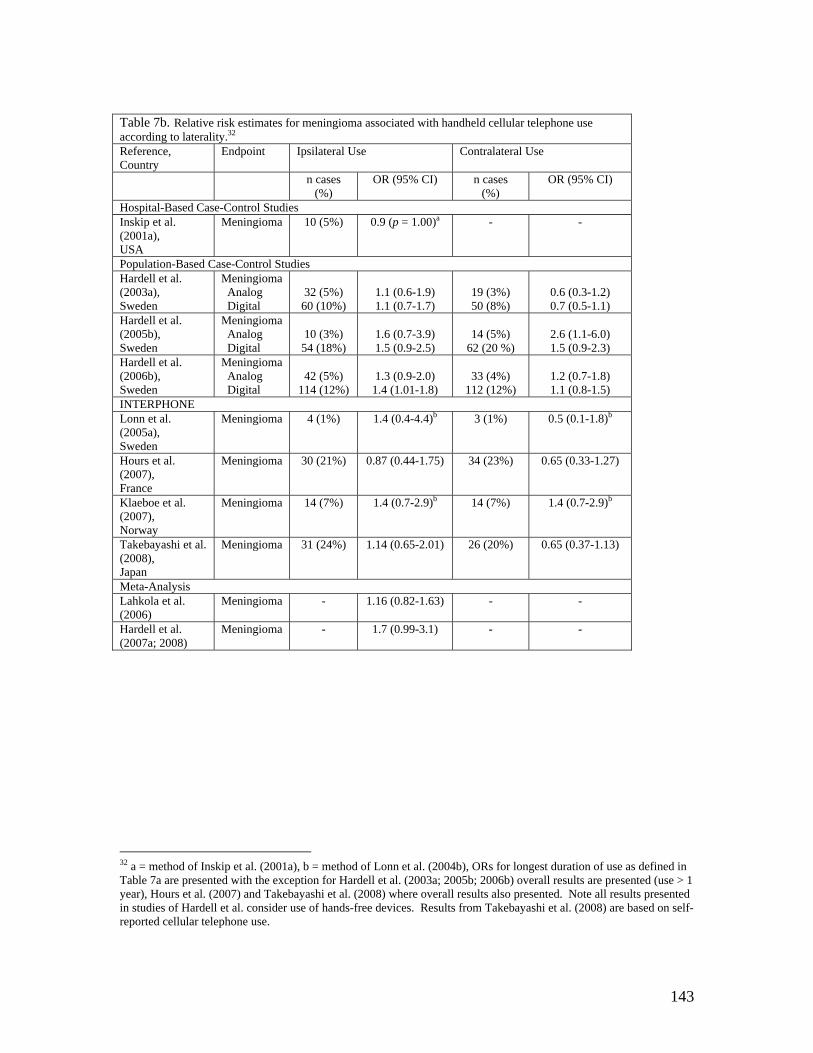
143
Table 7b. Relative risk estimates for meningioma associated with handheld cellular telephone use according to laterality.32 Reference, Country
Endpoint Ipsilateral Use Contralateral Use
n cases (%)
OR (95% CI) n cases (%)
OR (95% CI)
Hospital-Based Case-Control Studies Inskip et al. (2001a), USA
Meningioma 10 (5%) 0.9 (p = 1.00)a - -
Population-Based Case-Control Studies Hardell et al. (2003a), Sweden
Meningioma Analog Digital
32 (5%)
60 (10%)
1.1 (0.6-1.9) 1.1 (0.7-1.7)
19 (3%) 50 (8%)
0.6 (0.3-1.2) 0.7 (0.5-1.1)
Hardell et al. (2005b), Sweden
Meningioma Analog Digital
10 (3%)
54 (18%)
1.6 (0.7-3.9) 1.5 (0.9-2.5)
14 (5%)
62 (20 %)
2.6 (1.1-6.0) 1.5 (0.9-2.3)
Hardell et al. (2006b), Sweden
Meningioma Analog Digital
42 (5%)
114 (12%)
1.3 (0.9-2.0)
1.4 (1.01-1.8)
33 (4%)
112 (12%)
1.2 (0.7-1.8) 1.1 (0.8-1.5)
INTERPHONE Lonn et al. (2005a), Sweden
Meningioma 4 (1%) 1.4 (0.4-4.4)b 3 (1%) 0.5 (0.1-1.8)b
Hours et al. (2007), France
Meningioma 30 (21%) 0.87 (0.44-1.75) 34 (23%) 0.65 (0.33-1.27)
Klaeboe et al. (2007), Norway
Meningioma 14 (7%) 1.4 (0.7-2.9)b 14 (7%) 1.4 (0.7-2.9)b
Takebayashi et al. (2008), Japan
Meningioma 31 (24%) 1.14 (0.65-2.01) 26 (20%) 0.65 (0.37-1.13)
Meta-Analysis Lahkola et al. (2006)
Meningioma - 1.16 (0.82-1.63) - -
Hardell et al. (2007a; 2008)
Meningioma - 1.7 (0.99-3.1) - -
32 a = method of Inskip et al. (2001a), b = method of Lonn et al. (2004b), ORs for longest duration of use as defined in Table 7a are presented with the exception for Hardell et al. (2003a; 2005b; 2006b) overall results are presented (use > 1 year), Hours et al. (2007) and Takebayashi et al. (2008) where overall results also presented. Note all results presented in studies of Hardell et al. consider use of hands-free devices. Results from Takebayashi et al. (2008) are based on self-reported cellular telephone use.
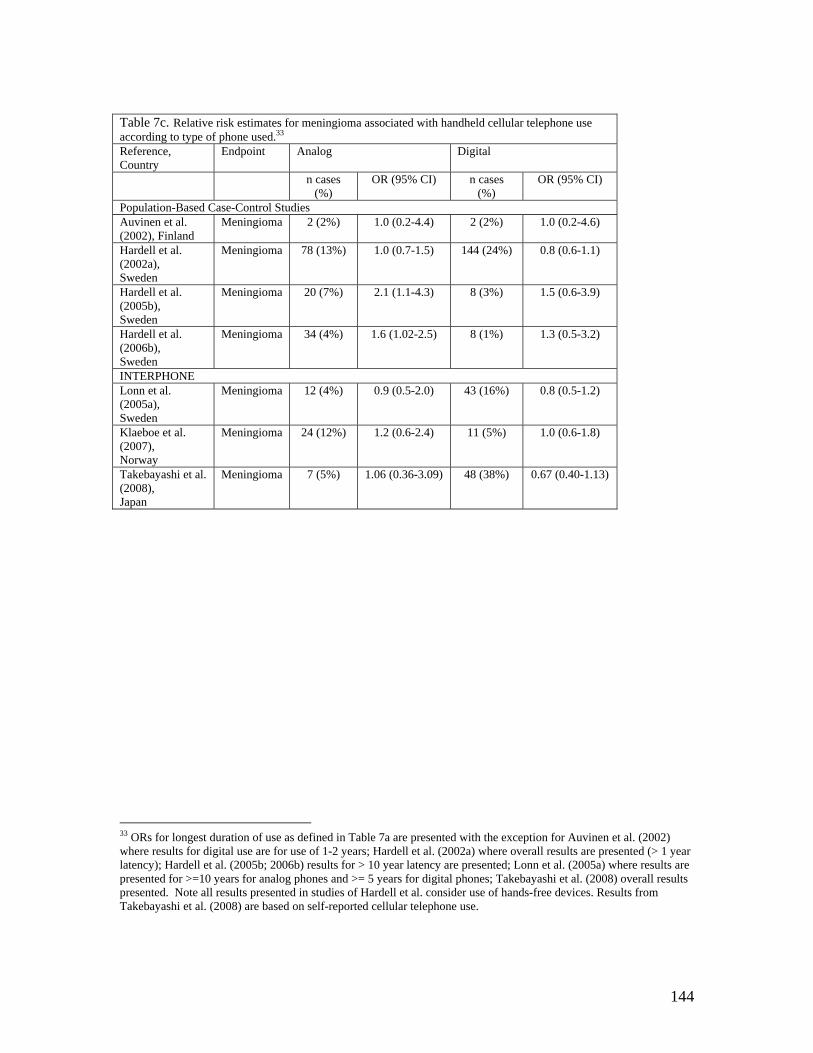
144
Table 7c. Relative risk estimates for meningioma associated with handheld cellular telephone use according to type of phone used.33 Reference, Country
Endpoint Analog Digital
n cases (%)
OR (95% CI) n cases (%)
OR (95% CI)
Population-Based Case-Control Studies Auvinen et al. (2002), Finland
Meningioma 2 (2%) 1.0 (0.2-4.4) 2 (2%) 1.0 (0.2-4.6)
Hardell et al. (2002a), Sweden
Meningioma 78 (13%) 1.0 (0.7-1.5) 144 (24%) 0.8 (0.6-1.1)
Hardell et al. (2005b), Sweden
Meningioma 20 (7%) 2.1 (1.1-4.3) 8 (3%) 1.5 (0.6-3.9)
Hardell et al. (2006b), Sweden
Meningioma 34 (4%) 1.6 (1.02-2.5) 8 (1%) 1.3 (0.5-3.2)
INTERPHONE Lonn et al. (2005a), Sweden
Meningioma 12 (4%) 0.9 (0.5-2.0) 43 (16%) 0.8 (0.5-1.2)
Klaeboe et al. (2007), Norway
Meningioma 24 (12%) 1.2 (0.6-2.4) 11 (5%) 1.0 (0.6-1.8)
Takebayashi et al. (2008), Japan
Meningioma 7 (5%) 1.06 (0.36-3.09) 48 (38%) 0.67 (0.40-1.13)
33 ORs for longest duration of use as defined in Table 7a are presented with the exception for Auvinen et al. (2002) where results for digital use are for use of 1-2 years; Hardell et al. (2002a) where overall results are presented (> 1 year latency); Hardell et al. (2005b; 2006b) results for > 10 year latency are presented; Lonn et al. (2005a) where results are presented for >=10 years for analog phones and >= 5 years for digital phones; Takebayashi et al. (2008) overall results presented. Note all results presented in studies of Hardell et al. consider use of hands-free devices. Results from Takebayashi et al. (2008) are based on self-reported cellular telephone use.
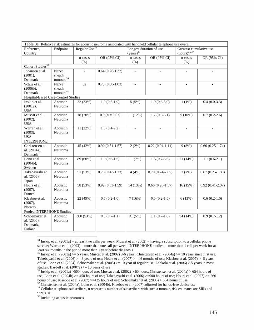
145
Table 8a. Relative risk estimates for acoustic neuroma associated with handheld cellular telephone use overall. Reference, Country
Endpoint Regular Use34 Longest duration of use (years)35
Greatest cumulative use (hours)36,37
n cases (%)
OR (95% CI) n cases (%)
OR (95% CI) n cases (%)
OR (95% CI)
Cohort Studies38 Johansen et al. (2001), Denmark
Nerve sheath tumours39
7 0.64 (0.26-1.32) - - - -
Schuz et al. (2006b), Denmark
Nerve sheath tumours41
32 0.73 (0.50-1.03) - - - -
Hospital-Based Case-Control Studies Inskip et al. (2001a), USA
Acoustic Neuroma
22 (23%) 1.0 (0.5-1.9) 5 (5%) 1.9 (0.6-5.9) 1 (1%) 0.4 (0.0-3.3)
Muscat et al. (2002), USA
Acoustic Neuroma
18 (20%) 0.9 (p = 0.07) 11 (12%) 1.7 (0.5-5.1) 9 (10%) 0.7 (0.2-2.6)
Warren et al. (2003), USA
Acoustin Neuroma
11 (22%) 1.0 (0.4-2.2) - - - -
INTERPHONE Christensen et al. (2004a), Denmark
Acoustic Neuroma
45 (42%) 0.90 (0.51-1.57)
2 (2%) 0.22 (0.04-1.11) 9 (8%) 0.66 (0.25-1.74)
Lonn et al. (2004b), Sweden
Acoustic Neuroma
89 (60%) 1.0 (0.6-1.5)
11 (7%) 1.6 (0.7-3.6) 21 (14%) 1.1 (0.6-2.1)
Takebayashi et al. (2006), Japan
Acoustic Neuroma
51 (53%) 0.73 (0.43-1.23) 4 (4%) 0.79 (0.24-2.65) 7 (7%) 0.67 (0.25-1.83)
Hours et al. (2007), France
Acoustic Neuroma
58 (53%) 0.92 (0.53-1.59) 14 (13%) 0.66 (0.28-1.57) 16 (15%) 0.92 (0.41-2.07)
Klaeboe et al. (2007), Norway
Acoustic Neuroma
22 (49%) 0.5 (0.2-1.0) 7 (16%) 0.5 (0.2-1.5) 6 (13%) 0.6 (0.2-1.6)
Pooled INTERPHONE Studies Schoemaker et al. (2005), Denmark, Finland,
Acoustic Neuroma
360 (53%) 0.9 (0.7-1.1)
31 (5%) 1.1 (0.7-1.8) 94 (14%) 0.9 (0.7-1.2)
34 Inskip et al. (2001a) = at least two calls per week; Muscat et al. (2002) = having a subscription to a cellular phone service; Warren et al. (2003) = more than one call per week; INTERPHONE studies = more than 1 call per week for at least six months in the period more than 1 year before diagnosis 35 Inskip et al. (2001a) >= 5 years; Muscat et al. (2002) 3-6 years; Christensen et al. (2004a) >= 10 years since first use; Takebayashi et al. (2006) >= 8 years of use; Hours et al. (2007) >= 46 months of use; Klaeboe et al. (2007) >=6 years of use; Lonn et al. (2004), Schoemaker et al. (2005) >= 10 year of regular use; Lahkola et al. (2006) > 5 years in most studies; Hardell et al. (2007a) >= 10 years of use 36 Inskip et al. (2001a) >500 hours of use; Muscat et al. (2002) > 60 hours; Christensen et al. (2004a) > 654 hours of use; Lonn et al. (2004b) >= 450 hours of use; Takebayashi et al. (2006) >=900 hours of use; Hours et al. (2007) >= 260 hours of use; Klaeboe et al. (2007) >=425 hours of use; Schoemaker et al. (2005) > 534 hours of use 37 Christensen et al. (2004a), Lonn et al. (2004b), Klaeboe et al. (2007) adjusted for hands-free device use 38 Cellular telephone subscribers, n represents number of subscribers with such a tumour, risk estimates are SIRs and 95% CIs 39 including acoustic neuromas
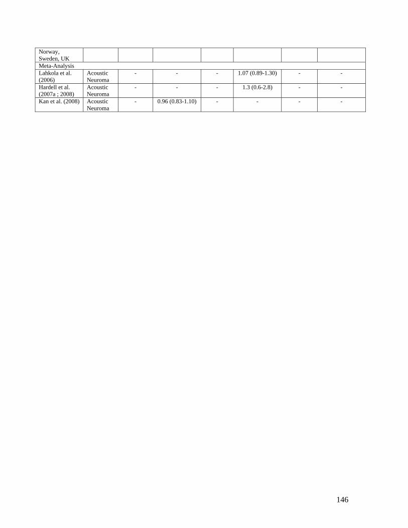
146
Norway, Sweden, UK Meta-Analysis Lahkola et al. (2006)
Acoustic Neuroma
- - - 1.07 (0.89-1.30) - -
Hardell et al. (2007a ; 2008)
Acoustic Neuroma
- - - 1.3 (0.6-2.8) - -
Kan et al. (2008) Acoustic Neuroma
- 0.96 (0.83-1.10) - - - -
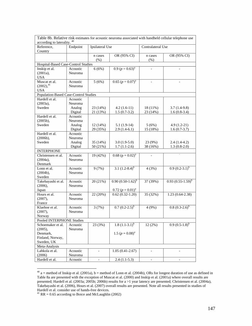
147
Table 8b. Relative risk estimates for acoustic neuroma associated with handheld cellular telephone use according to laterality. 40 Reference, Country
Endpoint Ipsilateral Use Contralateral Use
n cases (%)
OR (95% CI) n cases (%)
OR (95% CI)
Hospital-Based Case-Control Studies Inskip et al. (2001a), USA
Acoustic Neuroma
6 (6%) 0.9 (p = 0.63)a - -
Muscat et al. (2002),41 USA
Acoustic Neuroma
5 (6%) 0.65 (p = 0.07)a - -
Population-Based Case-Control Studies Hardell et al. (2003a), Sweden
Acoustic Neuroma Analog Digital
23 (14%) 21 (13%)
4.2 (1.6-11) 1.5 (0.7-3.2)
18 (11%) 23 (14%)
3.7 (1.4-9.8) 1.6 (0.8-3.4)
Hardell et al. (2005b), Sweden
Acoustic Neuroma Analog Digital
12 (14%) 29 (35%)
5.1 (1.9-14) 2.9 (1.4-6.1)
5 (6%) 15 (18%)
4.9 (1.2-21) 1.6 (0.7-3.7)
Hardell et al. (2006b), Sweden
Acoustic Neuroma Analog Digital
35 (14%) 50 (21%)
3.0 (1.9-5.0) 1.7 (1.1-2.6)
23 (9%) 38 (16%)
2.4 (1.4-4.2) 1.3 (0.8-2.0)
INTERPHONE Christensen et al. (2004a), Denmark
Acoustic Neuroma
19 (42%) 0.68 (p = 0.02)a - -
Lonn et al. (2004b), Sweden
Acoustic Neuroma
9 (7%) 3.1 (1.2-8.4)b 4 (3%) 0.9 (0.2-3.1)b
Takebayashi et al. (2006), Japan
Acoustic Neuroma
20 (21%) 0.90 (0.50-1.62)b
0.72 (p = 0.01)a
37 (39%) 0.93 (0.55-1.59)b
Hours et al. (2007), France
Acoustic Neuroma
22 (20%) 0.62 (0.32-1.20) 35 (32%) 1.23 (0.64-2.38)
Klaeboe et al. (2007), Norway
Acoustic Neuroma
3 (7%) 0.7 (0.2-2.5)b 4 (9%) 0.8 (0.3-2.6)b
Pooled INTERPHONE Studies Schoemaker et al. (2005), Denmark, Finland, Norway, Sweden, UK
Acoustic Neuroma
23 (3%) 1.8 (1.1-3.1)b
1.5 (p = 0.08)a
12 (2%) 0.9 (0.5-1.8)b
Meta-Analysis Lahkola et al. (2006)
Acoustic Neuroma
- 1.05 (0.41-2.67) - -
Hardell et al. Acoustic - 2.4 (1.1-5.3) - -
40 a = method of Inskip et al. (2001a), b = method of Lonn et al. (2004b), ORs for longest duration of use as defined in Table 8a are presented with the exception of Muscat et al. (2000) and Inskip et al. (2001a) where overall results are presented; Hardell et al. (2003a; 2005b; 2006b) results for a >1 year latency are presented; Christensen et al. (2004a), Takebayashi et al. (2006), Hours et al. (2007) overall results are presented. Note all results presented in studies of Hardell et al. consider use of hands-free devices. 41 RR = 0.65 according to Boice and McLaughlin (2002)

148
(2007a; 2008) Neuroma
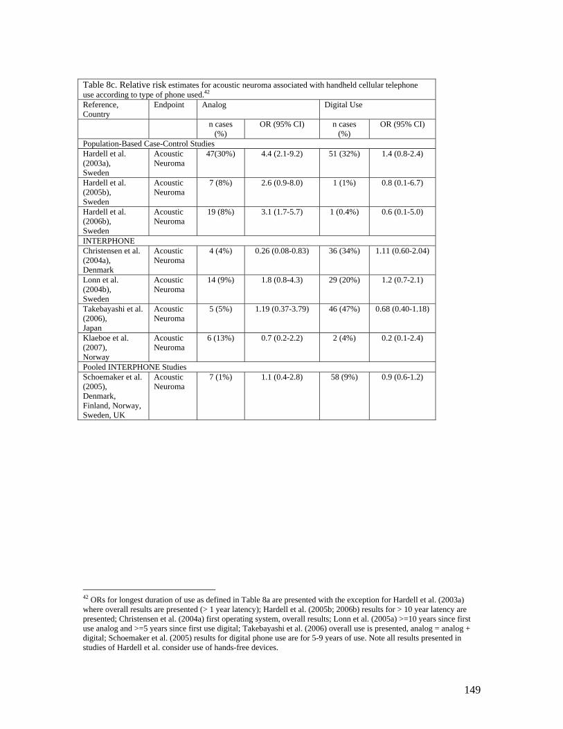
149
Table 8c. Relative risk estimates for acoustic neuroma associated with handheld cellular telephone use according to type of phone used.42 Reference, Country
Endpoint Analog Digital Use
n cases (%)
OR (95% CI) n cases (%)
OR (95% CI)
Population-Based Case-Control Studies Hardell et al. (2003a), Sweden
Acoustic Neuroma
47(30%) 4.4 (2.1-9.2) 51 (32%) 1.4 (0.8-2.4)
Hardell et al. (2005b), Sweden
Acoustic Neuroma
7 (8%) 2.6 (0.9-8.0) 1 (1%) 0.8 (0.1-6.7)
Hardell et al. (2006b), Sweden
Acoustic Neuroma
19 (8%) 3.1 (1.7-5.7) 1 (0.4%) 0.6 (0.1-5.0)
INTERPHONE Christensen et al. (2004a), Denmark
Acoustic Neuroma
4 (4%) 0.26 (0.08-0.83) 36 (34%) 1.11 (0.60-2.04)
Lonn et al. (2004b), Sweden
Acoustic Neuroma
14 (9%) 1.8 (0.8-4.3) 29 (20%) 1.2 (0.7-2.1)
Takebayashi et al. (2006), Japan
Acoustic Neuroma
5 (5%) 1.19 (0.37-3.79) 46 (47%) 0.68 (0.40-1.18)
Klaeboe et al. (2007), Norway
Acoustic Neuroma
6 (13%) 0.7 (0.2-2.2) 2 (4%) 0.2 (0.1-2.4)
Pooled INTERPHONE Studies Schoemaker et al. (2005), Denmark, Finland, Norway, Sweden, UK
Acoustic Neuroma
7 (1%) 1.1 (0.4-2.8) 58 (9%) 0.9 (0.6-1.2)
42 ORs for longest duration of use as defined in Table 8a are presented with the exception for Hardell et al. (2003a) where overall results are presented (> 1 year latency); Hardell et al. (2005b; 2006b) results for > 10 year latency are presented; Christensen et al. (2004a) first operating system, overall results; Lonn et al. (2005a) >=10 years since first use analog and >=5 years since first use digital; Takebayashi et al. (2006) overall use is presented, analog = analog + digital; Schoemaker et al. (2005) results for digital phone use are for 5-9 years of use. Note all results presented in studies of Hardell et al. consider use of hands-free devices.
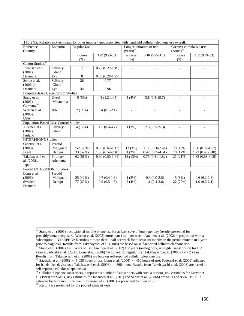
150
Table 9a. Relative risk estimates for other tumour types associated with handheld cellular telephone use overall. Reference, Country
Endpoint Regular Use43 Longest duration of use (years)44
Greatest cumulative use (hours)45
n cases (%)
OR (95% CI) n cases (%)
OR (95% CI) n cases (%)
OR (95% CI)
Cohort Studies46 Johansen et al. (2001), Denmark
Salivary Gland Eye
7 8
0.72 (0.29-1.49)
0.65 (0.28-1.27)
- - - -
Schuz et al. (2006b), Denmark
Salivary Gland Eye
26
44
0.77
0.96
- - - -
Hospital-Based Case-Control Studies Stang et al. (2001), Germany47
Uveal Melanoma
6 (5%) 4.2 (1.2-14.5) 5 (4%) 3.8 (0.8-19.7) - -
Warren et al. (2003), USA
IFN 2 (11%) 0.4 (0.1-2.1) - - - -
Population-Based Case-Control Studies Auvinen et al. (2002), Finland
Salivary Gland
4 (12%) 1.3 (0.4-4.7) 1 (3%) 2.3 (0.2-25.3) - -
INTERPHONE Studies Sadetzki et al. (2008), Israel
Parotid Malignant Benign
252 (63%) 33 (57%)
0.85 (0.64-1.12) 1.06 (0.54-2.10)
12 (3%) 1 (2%)
1.11 (0.50-2.44) 0.47 (0.05-4.51)
73 (18%) 10 (17%)
1.08 (0.72-1.62) 1.22 (0.43-3.48)
Takebayashi et al. (2008), Japan
Pituitary Adenoma
62 (61%) 0.90 (0.50-1.61) 13 (13%) 0.75 (0.31-1.82) 21 (21%) 1.33 (0.58-3.09)
Pooled INTERPHONE Studies Lonn et al. (2006), Sweden, Denmark
Parotid Malignant Benign
25 (42%) 77 (69%)
0.7 (0.4-1.3) 0.9 (0.5-1.5)
1 (2%) 5 (4%)
0.3 (0.0-2.5) 1.1 (0.4-3.6)
5 (8%)
22 (20%)
0.6 (0.2-1.8) 1.0 (0.5-2.1)
43 Stang et al. (2001) occupational mobile phone use for at least several hours per day (results presented for probably/certain exposure; Warren et al. (2003) more than 1 call per week; Auvinen et al. (2002) = proportion with a subscription; INTERPHONE studies = more than 1 call per week for at least six months in the period more than 1 year prior to diagnosis. Results from Takebayashi et al. (2008) are based on self-reported cellular telephone use. 44 Stang et al. (2001) >= 3 years of use; Auvinen et al. (2002) > 2 years (analog only, no digital subscription for > 2 years); Sadetzki et al. (2008), Lonn et al. (2006) >= 10 year of regular use; Takebayashi et al. (2008) >= 7.2 years. Results from Takebayashi et al. (2008) are base on self-reported cellular telephone use. 45 Sadetzki et al. (2008) >= 1,035 hours of use; Lonn et al. (2006) >= 450 hours of use; Sadetzki et al. (2008) adjusted for hands-free device use; Takebayashi et al. (2008) >= 560 hours. Results from Takebayashi et al. (2008) are based on self-reported cellular telephone use. 46 Cellular telephone subscribers, n represents number of subscribers with such a tumour, risk estimates for Dreyer et al. (1999) are SMRs, risk estimates for Johansen et al. (2001) and Schuz et al. (2006b) are SIRs and 95% CIs. SIR estimate for tumours of the eye in Johansen et al. (2001) is presented for men only 47 Results are presented for the pooled analysis only
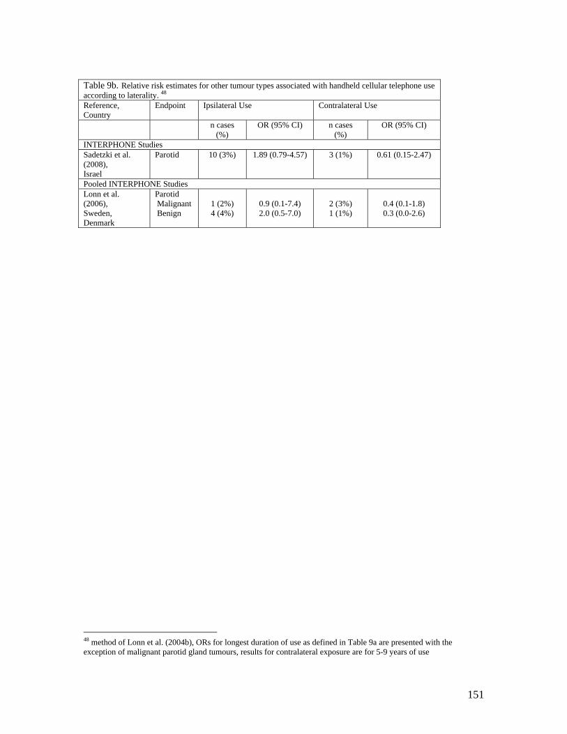
151
Table 9b. Relative risk estimates for other tumour types associated with handheld cellular telephone use according to laterality. 48 Reference, Country
Endpoint Ipsilateral Use Contralateral Use
n cases (%)
OR (95% CI) n cases (%)
OR (95% CI)
INTERPHONE Studies Sadetzki et al. (2008), Israel
Parotid
10 (3%)
1.89 (0.79-4.57) 3 (1%) 0.61 (0.15-2.47)
Pooled INTERPHONE Studies Lonn et al. (2006), Sweden, Denmark
Parotid Malignant Benign
1 (2%)
4 (4%)
0.9 (0.1-7.4)
2.0 (0.5-7.0)
2 (3%) 1 (1%)
0.4 (0.1-1.8)
0.3 (0.0-2.6)
48 method of Lonn et al. (2004b), ORs for longest duration of use as defined in Table 9a are presented with the exception of malignant parotid gland tumours, results for contralateral exposure are for 5-9 years of use
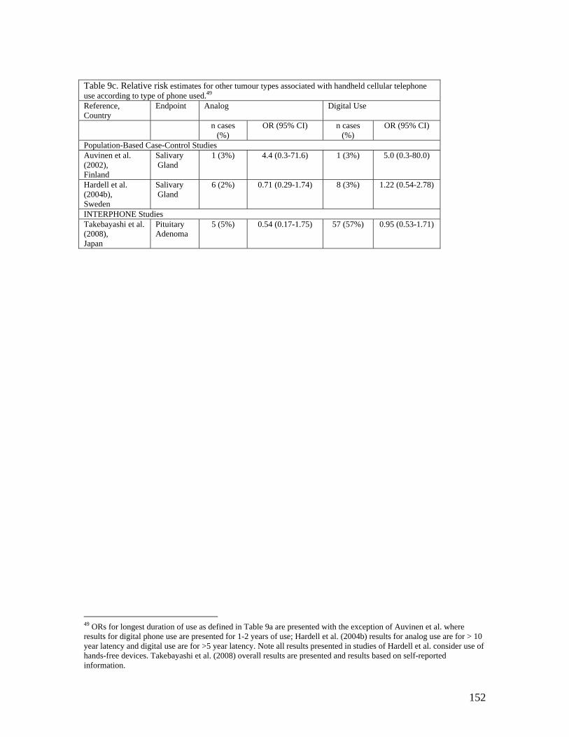
152
Table 9c. Relative risk estimates for other tumour types associated with handheld cellular telephone use according to type of phone used.49 Reference, Country
Endpoint Analog Digital Use
n cases (%)
OR (95% CI) n cases (%)
OR (95% CI)
Population-Based Case-Control Studies Auvinen et al. (2002), Finland
Salivary Gland
1 (3%) 4.4 (0.3-71.6) 1 (3%) 5.0 (0.3-80.0)
Hardell et al. (2004b), Sweden
Salivary Gland
6 (2%)
0.71 (0.29-1.74) 8 (3%) 1.22 (0.54-2.78)
INTERPHONE Studies Takebayashi et al. (2008), Japan
Pituitary Adenoma
5 (5%) 0.54 (0.17-1.75) 57 (57%) 0.95 (0.53-1.71)
49 ORs for longest duration of use as defined in Table 9a are presented with the exception of Auvinen et al. where results for digital phone use are presented for 1-2 years of use; Hardell et al. (2004b) results for analog use are for > 10 year latency and digital use are for >5 year latency. Note all results presented in studies of Hardell et al. consider use of hands-free devices. Takebayashi et al. (2008) overall results are presented and results based on self-reported information.
153
APPENDIX 1 – Description of other electronic resources used to identify potential
epidemiologic studies for inclusion in the review
154
The McLaughlin Centre for Population Health Risk Assessment at the University
of Ottawa coordinates the website www.rfcom.ca. As of June 30, 2007, it included 750
references. Human studies form a subsection of the database, and there are 133
epidemiological studies listed.
The WHO International EMF Project (www.who.int/peh/) was launched in 1996
due to concerns related to possible health effects from regular daily exposure to EMF.
Funding is provided by contributions from WHO member states and non-governmental
organizations approved by the WHO. The project assesses the health and environmental
effects of exposure to electromagnetic fields including radiofrequency fields (>10 MHz-
300GHz), which includes the range of cellular telephones. The website contains a
database of completed and ongoing research projects on the effects of EMF on biological
systems throughout the world. It contains information on studies conducted in humans
(epidemiological and laboratory provocation), animals (in vivo), and in cultured cells and
artificial systems (in vitro). It also contains engineering studies that characterize and
quantify EMF exposure in these systems, and theoretical studies that consider feasible
mechanisms for EMF energy interaction. The database contains studies spanning the non-
ionizing part of the electromagnetic spectrum. It is divided into two sections, a Project
Database and Citation List.
The Project Database contains completed studies as well as recently initiated
projects and follow-on work that is ongoing and not yet published. It is searchable on a
variety of categories (e.g. frequency range and sub-range, study type and sub-type,
funding agency, investigator name) and each entry includes a condensed summary
description of a project from a given laboratory or group. As of June 30, 2007, there
155
were 1,274 studies in the Database in the radiofrequency range. In the "Epidemiology"
subtype, mention is made of six INTERPHONE studies that are ongoing, and have not
yet published results - Australia, Canada, France, Israel, Italy, and New Zealand. These,
and the summary paper of all participating countries, are expected to publish results in
the near future. Two other studies are also discussed. One, by Elliot et al., is an ongoing
large cohort study in England and Europe. Another, in five European countries, by
Blettner et al., is stalled because of lack of funding. An initial feasibility study had
shown a poor response rate to a questionnaire survey.
The Citation List provides the complete reference and is searchable by a more
limited number of categories (e.g. frequency range and sub-range, study type and sub-
type, investigator name, reference key words, and date of publication). As of June 30,
2007, there were 2,963 studies in the radiofrequency range. In the "Epidemiology"
subtype, there were 331 studies. Search of these studies revealed one review paper, and
five letters that related to our study question.
Finally, a study chart function has been provided to view a specific study
category(s) in terms of number of ongoing projects, projects reported but not published,
and published studies. On June 30, 2007, there were 309 ongoing projects listed, 118
reported but not published, and 2,166 published. The categories are: 1) engineering and
physics, 2) epidemiology, 3) human/provocation, 4) in vitro, 5) in vivo, 6) literature
review, letter, book chapter, and report, 7) plant studies, and 8) social sciences. Each
study chart entry is further linked to the Project Database and Citation List for additional
information.
156
The Research Centre for Bioelectromagnetic Interaction at the University
Hospital of Aachen University (www.femu.rwth-aachen.de/) conducts
interdisciplinary research on the interaction of electromagnetic fields. The site
maintains a database on the biological effects of low and high frequency fields.
Access to a database of publication titles is available on-line. Table A1 summarizes
the other websites that were also examined. No new references were found.
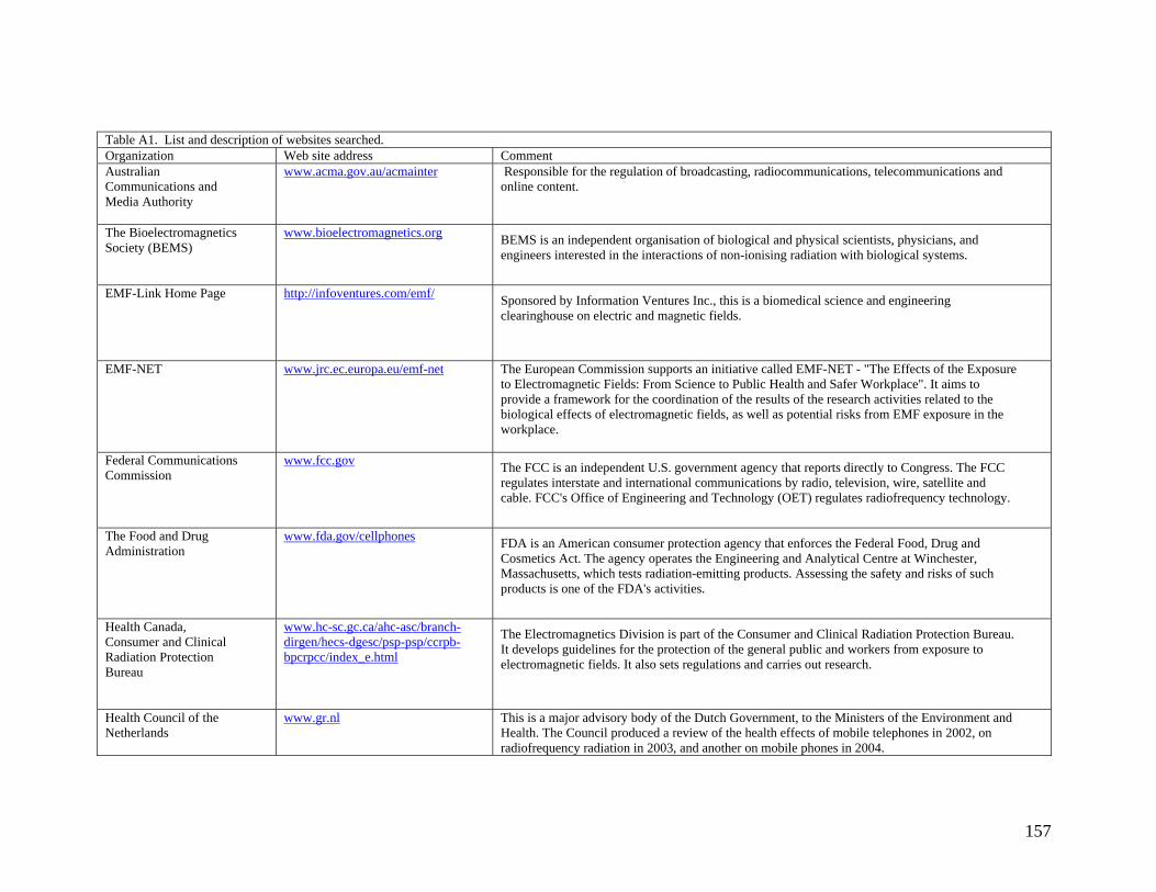
157
Table A1. List and description of websites searched. Organization Web site address Comment Australian Communications and Media Authority
www.acma.gov.au/acmainter
Responsible for the regulation of broadcasting, radiocommunications, telecommunications and online content.
The Bioelectromagnetics Society (BEMS)
www.bioelectromagnetics.org BEMS is an independent organisation of biological and physical scientists, physicians, and engineers interested in the interactions of non-ionising radiation with biological systems.
EMF-Link Home Page
http://infoventures.com/emf/ Sponsored by Information Ventures Inc., this is a biomedical science and engineering clearinghouse on electric and magnetic fields.
EMF-NET
www.jrc.ec.europa.eu/emf-net The European Commission supports an initiative called EMF-NET - "The Effects of the Exposure to Electromagnetic Fields: From Science to Public Health and Safer Workplace". It aims to provide a framework for the coordination of the results of the research activities related to the biological effects of electromagnetic fields, as well as potential risks from EMF exposure in the workplace.
Federal Communications Commission
www.fcc.gov The FCC is an independent U.S. government agency that reports directly to Congress. The FCC regulates interstate and international communications by radio, television, wire, satellite and cable. FCC's Office of Engineering and Technology (OET) regulates radiofrequency technology.
The Food and Drug Administration
www.fda.gov/cellphones FDA is an American consumer protection agency that enforces the Federal Food, Drug and Cosmetics Act. The agency operates the Engineering and Analytical Centre at Winchester, Massachusetts, which tests radiation-emitting products. Assessing the safety and risks of such products is one of the FDA's activities.
Health Canada, Consumer and Clinical Radiation Protection Bureau
www.hc-sc.gc.ca/ahc-asc/branch-dirgen/hecs-dgesc/psp-psp/ccrpb-bpcrpcc/index_e.html
The Electromagnetics Division is part of the Consumer and Clinical Radiation Protection Bureau. It develops guidelines for the protection of the general public and workers from exposure to electromagnetic fields. It also sets regulations and carries out research.
Health Council of the Netherlands
www.gr.nl This is a major advisory body of the Dutch Government, to the Ministers of the Environment and Health. The Council produced a review of the health effects of mobile telephones in 2002, on radiofrequency radiation in 2003, and another on mobile phones in 2004.
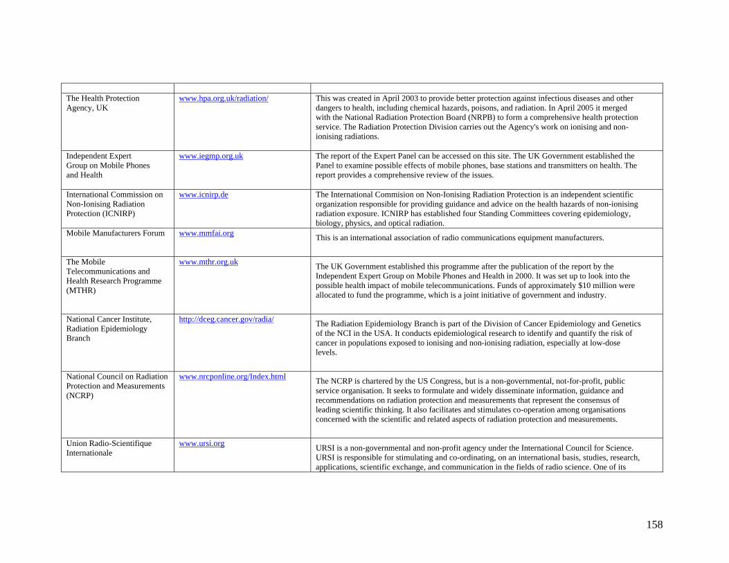
158
The Health Protection Agency, UK
www.hpa.org.uk/radiation/ This was created in April 2003 to provide better protection against infectious diseases and other dangers to health, including chemical hazards, poisons, and radiation. In April 2005 it merged with the National Radiation Protection Board (NRPB) to form a comprehensive health protection service. The Radiation Protection Division carries out the Agency's work on ionising and non-ionising radiations.
Independent Expert Group on Mobile Phones and Health
www.iegmp.org.uk The report of the Expert Panel can be accessed on this site. The UK Government established the Panel to examine possible effects of mobile phones, base stations and transmitters on health. The report provides a comprehensive review of the issues.
International Commission on Non-Ionising Radiation Protection (ICNIRP)
www.icnirp.de
The International Commision on Non-Ionising Radiation Protection is an independent scientific organization responsible for providing guidance and advice on the health hazards of non-ionising radiation exposure. ICNIRP has established four Standing Committees covering epidemiology, biology, physics, and optical radiation.
Mobile Manufacturers Forum www.mmfai.org This is an international association of radio communications equipment manufacturers.
The Mobile Telecommunications and Health Research Programme (MTHR)
www.mthr.org.uk The UK Government established this programme after the publication of the report by the Independent Expert Group on Mobile Phones and Health in 2000. It was set up to look into the possible health impact of mobile telecommunications. Funds of approximately $10 million were allocated to fund the programme, which is a joint initiative of government and industry.
National Cancer Institute, Radiation Epidemiology Branch
http://dceg.cancer.gov/radia/ The Radiation Epidemiology Branch is part of the Division of Cancer Epidemiology and Genetics of the NCI in the USA. It conducts epidemiological research to identify and quantify the risk of cancer in populations exposed to ionising and non-ionising radiation, especially at low-dose levels.
National Council on Radiation Protection and Measurements (NCRP)
www.nrcponline.org/Index.html The NCRP is chartered by the US Congress, but is a non-governmental, not-for-profit, public service organisation. It seeks to formulate and widely disseminate information, guidance and recommendations on radiation protection and measurements that represent the consensus of leading scientific thinking. It also facilitates and stimulates co-operation among organisations concerned with the scientific and related aspects of radiation protection and measurements.
Union Radio-Scientifique Internationale
www.ursi.org URSI is a non-governmental and non-profit agency under the International Council for Science. URSI is responsible for stimulating and co-ordinating, on an international basis, studies, research, applications, scientific exchange, and communication in the fields of radio science. One of its
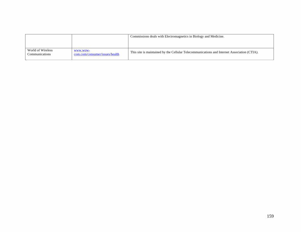
159
Commissions deals with Electromagnetics in Biology and Medicine.
World of Wireless Communications
www.wow-com.com/consumer/issues/health This site is maintained by the Cellular Telecommunications and Internet Association (CTIA).