Embed Size (px)
Citation preview
Med Oral Patol Oral Cir Bucal. 2012 Jul 1;17 (4):e716-20. Biocompatibility of Epiphany sealer
e716
Journal section: EndodonticsPublication Types: Research
Epiphany root canal sealer prepared with resinous solvent is irritating to rat subcutaneous tissues
Élcio Daleffe 1, José-Estevam Vieira-Ozório 1, Manoel-Damião Sousa-Neto 2, Danyel-Elias da-Cruz-Perez 3
1 DDS, MSc. DDS, MSc. School of Dentistry, University of Ribeirao Preto (UNAERP), Ribeirao Preto, Sao Paulo, Brazil2 DDS, PhD. Department of Restorative Dentistry, School of Dentistry of Ribeirao Preto, University of Sao Paulo (USP), Ribei-DDS, PhD. Department of Restorative Dentistry, School of Dentistry of Ribeirao Preto, University of Sao Paulo (USP), Ribei-Department of Restorative Dentistry, School of Dentistry of Ribeirao Preto, University of Sao Paulo (USP), Ribei-rao Preto, Sao Paulo, Brazil 3 DDS, PhD. Department of Clinical and Preventive Dentistry, Oral Pathology Unit, Federal University of Pernambuco, Recife, Pernambuco, Brazil
Correspondence:Universidade Federal de PernambucoDepartamento Clínica e Odontologia Preventiva, Área de Patologia OralAv. Prof. Moraes Rego, 1235. Cidade UniversitáriaCEP: 50670-901 Recife/PE, [email protected]
Received: 05/07/2011Accepted:16/09/2011
AbstractObjective: This study assessed the biocompatibility of the Epiphany endodontic sealer prepared with resinous solvent of Epiphany system (Thinning resin) in rat subcutaneous tissues. Study Design: Polyethylene tubes were filled with the sealer and 4 groups were established: GI, Epiphany prepared with 1 drop of resinous solvent (RS); GII, Epiphany prepared with 1 drop of RS and photoactivated; GIII, Epiph-any associated with self-etch primer and prepared with 1 drop of RS; GIV, Epiphany associated with self-etch primer, prepared with 1 drop of RS and photoactivated. The filled tubes were implanted into 4 different regions of the dorsum of 20 adult male rats. Results: After 7, 14 and 21 days, all groups presented a moderate to severe chronic inflammation, necrosis and foreign-body giant cells. At 42 days, although the intensity of chronic inflammatory reaction decreased, the other features still were observed. Conclusion: The Epiphany sealer prepared with the RS was irritating to rat subcutaneous tissues.
Key words: Biocompatibility, Epiphany, methacrylate resin sealer, resinous solvent, root canal sealer.
Daleffe É, Vieira-Ozório JE, Sousa-Neto MD, da-Cruz-Perez DE. Epiphany root canal sealer prepared with resinous solvent is irritating to rat subcuta-neous tissues. Med Oral Patol Oral Cir Bucal. 2012 Jul 1;17 (4):e716-20. http://www.medicinaoral.com/medoralfree01/v17i4/medoralv17i4p716.pdf
Article Number: 17788 http://www.medicinaoral.com/© Medicina Oral S. L. C.I.F. B 96689336 - pISSN 1698-4447 - eISSN: 1698-6946eMail: [email protected] Indexed in:
Science Citation Index ExpandedJournal Citation ReportsIndex Medicus, MEDLINE, PubMedScopus, Embase and Emcare Indice Médico Español
doi:10.4317/medoral.17788http://dx.doi.org/doi:10.4317/medoral.17788
Med Oral Patol Oral Cir Bucal. 2012 Jul 1;17 (4):e716-20. Biocompatibility of Epiphany sealer
e717
IntroductionThe biocompatibility of root canal sealers is an essential property for ensuring their good performance and suc-cessful endodontic treatment (1-3). In vivo and in vitro studies are important to determine the genotoxicity, cytotoxicity, irritant potential and the biological com-patibility of these materials (1-7), mainly that recently introduced to the market.The Resilon-Epiphany canal filling system is composed by Resilon, a synthetic polycaprolactone polymer, based on a polyester-based polymer that contains dimethacr-ylates, which can bond to methacrylate-based resin seal-ers, such as Epiphany, a resin-based dual-cure root ca-nal sealer. This system also uses a self-etch primer that contains sulfonic acid terminated functional monomer, HEMA, water, and polymerization initiator. One of the main characteristics of this system is the formation of a resin monoblock that adheres to the dentin of the radic-ular canal through intratubular tags (8-10). Owing these features, some studies found greater sealing capacity of this system than other materials, being associated with lower index of apical periodontitis (8,11). This canal filling system is also followed by a resinous solvent (Epiphany Thinning Resin, Pentron Clinical Technologies, LLC, Wallingford, CT, USA), which is an aqueous solution composed by ethoxylated bisphe-nol-A-dimethacrylate (EBPADMA) resins with photo-initiators, amines, stabilizers and pigments (12). The manufacturer recommends the use of 1 to 2 drops of this solution in order to adjust the sealer viscosity, when necessary. Rached-Junior et al. (13) observed that the resinous solvent increases the bond strength of Epipha-ny sealer to dentin walls when photoactivated. According to the manufacturer, the Epiphany sealer is biocompatible and non-cytotoxic. In vivo studies confirmed the good biological properties of the sealer (3,14), even when associated with the self-etch primer that accompanies it (5). However, in vitro tests revealed that the photoactivated and fresh sealer presents mod-erate and severe cytotoxicity, respectively (4,15). The self-etch primer also showed cytotoxicity (4). Moreo-ver, Epiphany showed higher cytotoxicity than other
conventional sealers (16) and its citotoxicity depends on the concentration and contact time with the culture cells (17). Regarding to the solvent resinous that also accompanies the system, there is only one study that shows moderate cytotoxicity of the solution when in contact with HeLa cells (4). To the best of our knowledge, there are not pre-vious in vivo studies that evaluated the biocompatibility of the resinous solvent. Thus, the aim of this study was to evaluate the biocompatibility of the Epiphany root canal sealer prepared with resinous solvent (Thinning resin) in rat subcutaneous connective tissues. Material and MethodsThis study was previously evaluated and approved by the Animal Research Ethics Committee of the Univer-sity of Ribeirao Preto, Sao Paulo, Brazil and the ethical concepts for use of laboratory animals were observed in all phases of the experiment. The material assessed was the Epiphany root canal sealer (Pentron®-Clinical Tech-nologies, L.L.C. Wallingford CT., USA), which was as-sociated or not with self-etch primer and prepared or not with the resinous solvent (Epiphany Thinning Resin). According to the preparation of the tested sealer, the following 4 experimental groups were established: GI: Epiphany prepared with 1 drop of resinous solvent; GII: Epiphany prepared with 1 drop of resinous solvent and photoactivated; GIII: Epiphany associated with self-etch primer and prepared with 1 drop of resinous sol-vent; GIV: Epiphany associated with self-etch primer, prepared with 1 drop of resinous solvent and photoacti-vated (Table 1). The lateral wall of the tubes was used as negative control. For each group, it was used 1.5 cm of the sealer obtained from the mixture of the 2 pastes for 1 drop of the resinous solvent, which was released from the flask positioned perpendicular to the glass plate surface.For this study, 20 male adult rats (Rattus novergicus, Albinus Wistar), weighing between 200 g and 250 g were used. They were kept in an acclimatized room and received a balanced diet and water ad libitum. The
GROUPS CONTENTS SPECIMENS
(n per analyzed time)
I Epiphany prepared with 1 drop of resinous solvent 5
IIEpiphany prepared with 1 drop of resinous solvent and photoactivated
5
IIIEpiphany associated with self-etch primer and prepared with 1 drop of resinous solvent
5
IVEpiphany associated with self-etch primer, prepared with 1 drop of resinous solvent and photoactivated
5
Table 1. Experimental groups and their respective contents.
Med Oral Patol Oral Cir Bucal. 2012 Jul 1;17 (4):e716-20. Biocompatibility of Epiphany sealer
e718
animals were anesthetized by intramuscular injection of ketamine chlorhydrate (0.1 mg/ml) associated with xy-lazine (0.05 mg/ml), followed by shaving of dorsal fur, disinfection, incision and divulsion of the subcutaneous tissue to insert the test material. To implant the root ca-nal sealer into the rat subcutaneous tissues, sterilized polyethylene tubes 1.2 mm in diameter (0.8 mm internal diameter) and 10 mm long were used. To prevent the sealer from escaping, one of the extremities of the tubes was close by heat. The Epiphany root canal sealer was manipulated in accordance with the manufacturer’s rec-ommendation for clinical use, in a dark room, because it is a dual-cure sealer. In Groups III and IV, a brush was used to coat the internal surface of the polyethyl-ene tubes with the primer supplied with the sealer. The manipulated sealer was carefully put into polyethylene tubes with aid of a paper cone compatible with the diam-eter of tubes, ensuring that there were no empty spaces and that the sealer did not overflow. Next, the sealer in the tubes was light activated (groups II and IV) and im-mediately implanted in the subcutaneous tissue in the rat dorsa. The surgical wounds were correctly sutured.Each animal received four tubes with material, two be-ing in the scapular region and two in the pelvic region (one on the right side and other on the left), with each tube representing a distinct group. After 7, 14, 21 and 42 days, 5 rats were killed by anesthetic overdose and the tubes removed for histological analysis. Thus, there were 5 samples for each experimental group in all the analyzed periods. The specimens were fixed in 10% buffered formalin solution for 24 hours and processed for conventional histological examination.The connective tissue adjacent to open extremity of each tube was examined in histological cuts (5 µm thick) stained by the hematoxylin and eosin technique, to evaluate the presence or absence of neutrophils, macrophages, lymphocytes, plasma cells, giant foreign body cells, dispersed material and necrotic tissue, clas-
sifying each criteria according to quantity in negative (-), slight (+), moderate (++) and intense (+++). Depend-ing on these features, except the presence of dispersed material, the inflammatory reaction of the connective tissue was classified as none to slight (grade I), mo-derate (grade II), and severe (grade III), according to Campos-Pinto et al. (5).
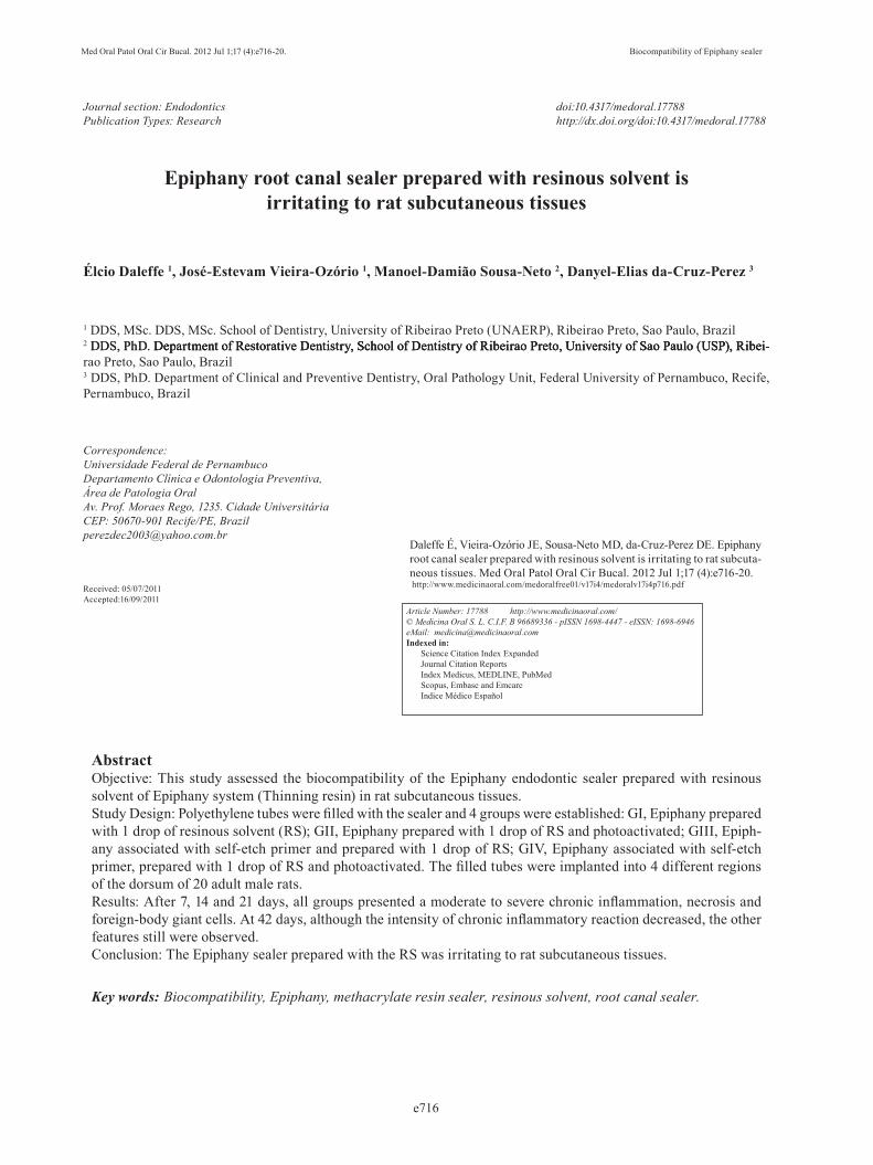
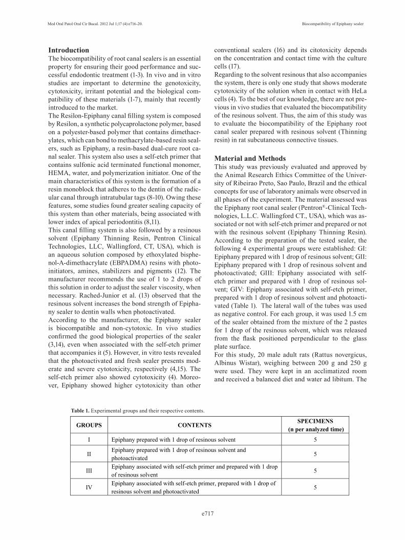
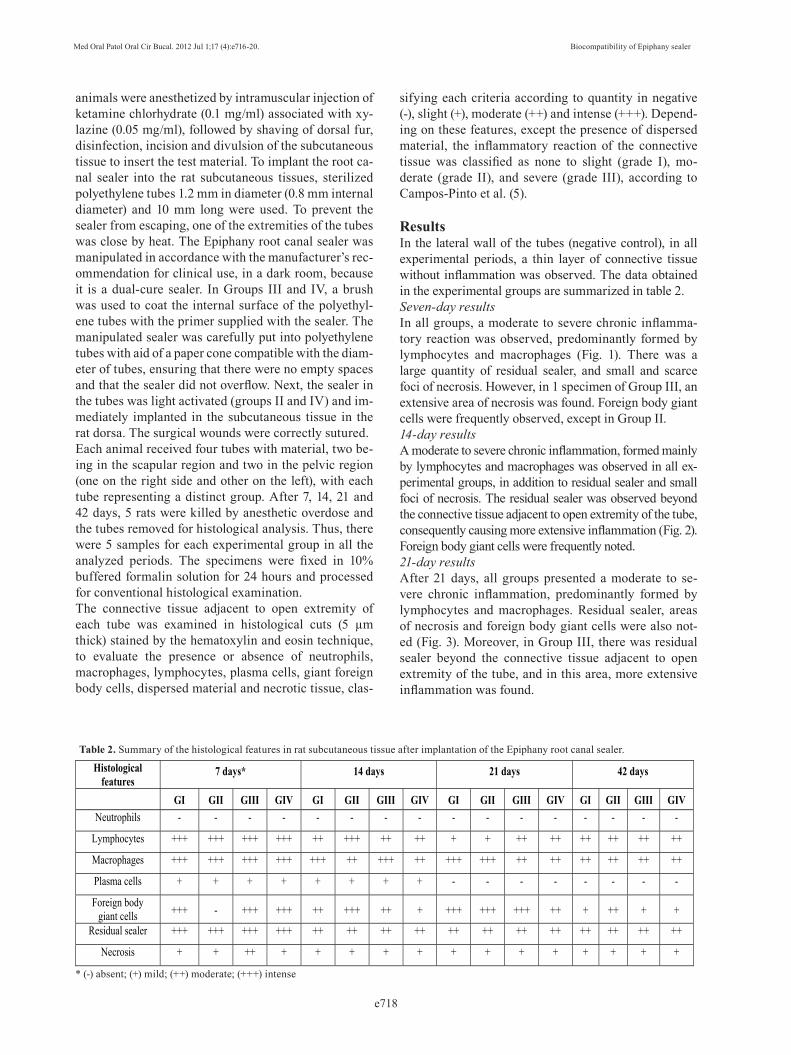
ResultsIn the lateral wall of the tubes (negative control), in all experimental periods, a thin layer of connective tissue without inflammation was observed. The data obtained in the experimental groups are summarized in table 2.Seven-day results In all groups, a moderate to severe chronic inflamma-tory reaction was observed, predominantly formed by lymphocytes and macrophages (Fig. 1). There was a large quantity of residual sealer, and small and scarce foci of necrosis. However, in 1 specimen of Group III, an extensive area of necrosis was found. Foreign body giant cells were frequently observed, except in Group II.14-day resultsA moderate to severe chronic inflammation, formed mainly by lymphocytes and macrophages was observed in all ex-perimental groups, in addition to residual sealer and small foci of necrosis. The residual sealer was observed beyond the connective tissue adjacent to open extremity of the tube, consequently causing more extensive inflammation (Fig. 2). Foreign body giant cells were frequently noted.21-day resultsAfter 21 days, all groups presented a moderate to se-vere chronic inflammation, predominantly formed by lymphocytes and macrophages. Residual sealer, areas of necrosis and foreign body giant cells were also not-ed (Fig. 3). Moreover, in Group III, there was residual sealer beyond the connective tissue adjacent to open extremity of the tube, and in this area, more extensive inflammation was found.
Histological features
7 days* 14 days 21 days 42 days
GI GII GIII GIV GI GII GIII GIV GI GII GIII GIV GI GII GIII GIV Neutrophils - - - - - - - - - - - - - - - -
Lymphocytes +++ +++ +++ +++ ++ +++ ++ ++ + + ++ ++ ++ ++ ++ ++
Macrophages +++ +++ +++ +++ +++ ++ +++ ++ +++ +++ ++ ++ ++ ++ ++ ++
Plasma cells + + + + + + + + - - - - - - - -
Foreign body giant cells +++ - +++ +++ ++ +++ ++ + +++ +++ +++ ++ + ++ + +
Residual sealer +++ +++ +++ +++ ++ ++ ++ ++ ++ ++ ++ ++ ++ ++ ++ ++
Necrosis + + ++ + + + + + + + + + + + + +
Table 2. Summary of the histological features in rat subcutaneous tissue after implantation of the Epiphany root canal sealer.
* (-) absent; (+) mild; (++) moderate; (+++) intense
Med Oral Patol Oral Cir Bucal. 2012 Jul 1;17 (4):e716-20. Biocompatibility of Epiphany sealer
e719
42-day resultsAt 42 days, moderate chronic inflammation was ob-served in all groups, predominantly composed of lym-phocytes and macrophages. However, in 1 specimen of Groups I and III, there is a mild inflammatory reaction and in 1 sample of Group IV, a severe inflammation was noted. In all specimens, the connective tissue also presented foci of necrosis, residual sealer and a foreign body granulomatous inflammatory reaction. As had occurred in other groups from previous experimental periods, in Group III there was residual sealer beyond the connective tissue adjacent to open extremity of the tube.
DiscussionAs the manufacturer postulate, some authors also demonstrated that the Epiphany sealer presents low ir-ritability to subcutaneous and bone tissues of animals (3,5,14,18). However, in vitro studies revealed moder-ate cytotoxicity of this sealer (4,15,17). Moreover, when the cells were exposed to the fresh Epiphany sealer, a greater cytotoxicity was observed (15). With regard to resinous solvent, Merdad et al. (4) found moderate cy-totoxicity of this solution when in contact with HeLa cells.In the present study, after 7, 14 and 21 days, all groups presented connective tissue with moderate to severe chronic inflammatory reaction, necrosis, foreign body giant cells and abundant residual sealer. At 42 days, al-though there was a slight decrease in the intensity of inflammation (moderate), necrosis, foreign body giant cells and residual sealer still were observed. As the resinous solvent contains similar components to those of the Epiphany sealer, such as ethoxylated bisphenol-A-dimethacrylate (EBPADMA) resins with photo-ini-tiators, amines and stabilizers (12) the sealer prepared with the resinous solvent could eventually increase the concentration of these components and consequently cause irritation and aggression to the tissues. Although Merdad et al. (4) observed a moderate cytotoxicity of the self-etch primer that accompanies the Epiphany system, this substance presented low irritability to subcutane-ous tissues of rats when it was used associated with the Epiphany sealer without the resinous solvent (5). There-fore, the tissue irritation observed in the current study is apparently related to the use of the resinous solvent.Another hypothesis, perhaps the most probable, is based on the fact that the addition of the resinous solvent in-creased the sealer flow, providing a greater surface of contact with the connective tissue and consequently causing more extensive inflammatory reaction. Rached-Junior et al. (13) observed that the addition of the resin-ous solvent provided alterations in the Epiphany sealer structure, resulting in monomers of smaller size, which probably increases the sealer flow. In addition, in the
Fig. 1. Group III (Epiphany associated with self-etch primer and pre-pared with 1 drop of resinous solvent), 7 days – Severe chronic in-flammatory reaction, residual sealer (white arrow) and foreign body giant cells (yellow arrow) (hematoxylin-eosin, original magnifica-tion, x100).
Fig. 2. Group IV (Epiphany associated with self-etch primer, prepared with 1 drop of resinous solvent and photoactivated), 14 days – Large quantity of residual sealer beyond the connective tissue adjacent to open extremity of the tube, foci of necrosis (arrows) and se-vere inflammation (hematoxylin-eosin, original mag-nification, x100).
Fig. 3. Group I (Epiphany prepared with 1 drop of resinous solvent), 21 days – Large quantity of residual sealer (yellow arrow), with foreign body giant cells (black arrow) (hema-toxylin-eosin, original magnification, x100).
Med Oral Patol Oral Cir Bucal. 2012 Jul 1;17 (4):e716-20. Biocompatibility of Epiphany sealer
e720
present study, residual sealer was observed beyond the connective tissue adjacent to open extremity of the tube in several specimens, suggesting higher flow of the sealer when prepared with the resinous solvent.A foreign body granulomatous inflammatory reaction associated with abundant residual sealer was frequently observed after all the periods of time analyzed. Con-sidering clinical practice, this is an important finding because, if there were overfilling of Epiphany sealer prepared with the resinous solvent during endodontic therapy, this combination could eventually lead to for-eign body granulomatous inflammation in the periapi-cal tissues and favor the persistence of periapical peri-odontitis (19). However, multinucleated giant cells was rarely seen when the Epiphany SE sealer (a nonetching version of Epiphany) was used (7). Although the addition of the resinous solvent to the Epiphany sealer was irritating to subcutaneous tissues in animals, further in vivo and in vitro studies are nec-essary in order to obtain more information about this canal filling system and to compare the results found in different studies.
References1. Hauman CH, Love RM. Biocompatibility of dental materials used in contemporary endodontic therapy: a review. Part 2. Root-canal-filling materials. Int Endod J. 2003;36:147-60.2. Bernath M, Szabo J. Tissue reaction initiated by different sealers. Int Endod J. 2003;36:256-61.3. Sousa CJ, Montes CR, Pascon EA, Loyola AM, Versiani MA. Comparison of the intraosseous biocompatibility of AH Plus, En-doREZ, and Epiphany root canal sealers. J Endod. 2006;32:656-62.4. Merdad K, Pascon AE, Kulkarni G, Santerre P, Friedman S. Short-term cytotoxicity assessment of the epiphany resin-percha obturat-ing system by indirect and direct contact millipore filter assays. J Endod. 2007;33:24-7.5. Campos-Pinto MM, de Oliveira DA, Versiani MA, Silva-Sousa YT, de Sousa-Neto MD, da Cruz Perez DE. Assessment of the biocompat-ibility of Epiphany root canal sealer in rat subcutaneous tissues. Oral Surg Oral Med Oral Pathol Oral Radiol Endod. 2008;105:e77-81.6. Brzovic V, Miletic I, Zeljezic D, Mladinic M, Kasuba V, Ram-ic S, et al. In vitro genotoxicity of root canal sealers. Int Endod J. 2009;42:253-63.7. Yamanaka Y, Shigetani Y, Yoshiba K, Yoshiba N, Okiji T. Immu-nohistochemical analysis of subcutaneous tissue reactions to meth-acrylate resin-based root canal sealers. Int Endod J. 2011;44:669-75.8. Shipper G, Orstavik D, Teixeira FB, Trope M. An evaluation of mi-crobial leakage in roots filled with a thermoplastic synthetic polymer-based root canal filling material (Resilon). J Endod. 2004;30:342-7.9. Ezzie E, Fleury A, Solomon E, Spears R, He J. Efficacy of retreat-ment techniques for a resin-based root canal obturation material. J Endod. 2006;32:341-4.10. Versiani MA, Carvalho-Junior JR, Padilha MI, Lacey S, Pas-con EA, Sousa-Neto MD. A comparative study of physicochemical properties of AH Plus and Epiphany root canal sealants. Int Endod J. 2006;39:464-71.11. Shipper G, Teixeira FB, Arnold RR, Trope M. Periapical inflam-mation after coronal microbial inoculation of dog roots filled with gutta-percha or resilon. J Endod. 2005;31:91-6.12. Skrtic D, Antonucci JM. Dental composites based on amorphous calcium phosphate-resin composition/physicochemical properties study. J Biomater Appl. 2007;21:375-93.
13. Rached-Junior FJ, Souza-Gabriel AE, Alfredo E, Miranda CE, Silva-Sousa YT, Sousa-Neto MD. Bond strenght of Epiphany sealer prepared with resinous solvent. J Endod. 2009;35:251-5.14. Onay EO, Ungor M, Ozdemir BH. In vivo evaluation of the bio-compatibility of a new resin-based obturation system. Oral Surg Oral Med Oral Pathol Oral Radiol Endod. 2007;104:60-6.15. Lodienè G, Morisbak E, Bruzell E, Orstavik D. Toxicity evalua-tion of root canal sealers in vitro. Int Endod J. 2008;41:72-7.16. Key JE, Rahemtulla FG, Eleazer PD. Cytotoxicity of a new root canal filling material on human gingival fibroblasts. J Endod. 2006;32:756-8.17. Heitman EP, Joyce AP, McPberson JC, Roberts S, Cbuang A. An in vitro evaluation of the growth of human periodontal ligament fi-broblasts after exposure to a methacrylate-based endodontic sealer. J Endod. 2008;34:186-9.18. Silveira CM, Pinto SC, Zedebski RA, Santos FA, Pilatti GL. Bio-compatibility of four root canal sealers: a histopathological evaluation in rat subcutaneous connective tissue. Braz Dent J. 2011;22:21-7.19. Nair PN. Pathogenesis of apical periodontitis and the causes of endodontic failures. Crit Rev Oral Biol Med. 2004;15:348-81.