Embed Size (px)
Citation preview
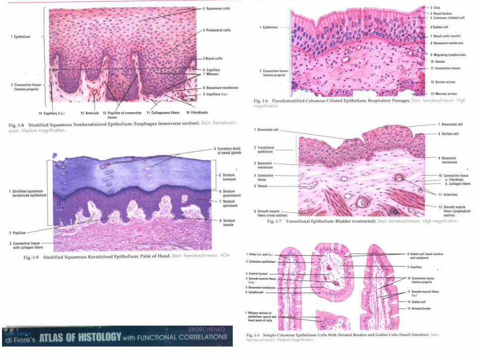
Epithelium and Junctions
VIBS 443/602
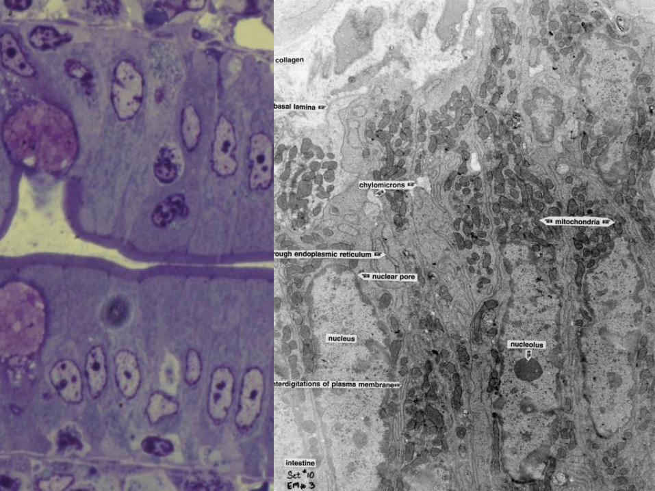
EM 3: region of basal lamina and
connective tissue beneath intestinal
epithelial absorptive cells
1. Plasma membrane
2. Basal lamina
3. Connective tissue

EM 17; capillary in the lamina
propria of the intestine
(duodenum); 13,500x
1. Smooth muscle cell
2. Capillary
3. Endothelial cell
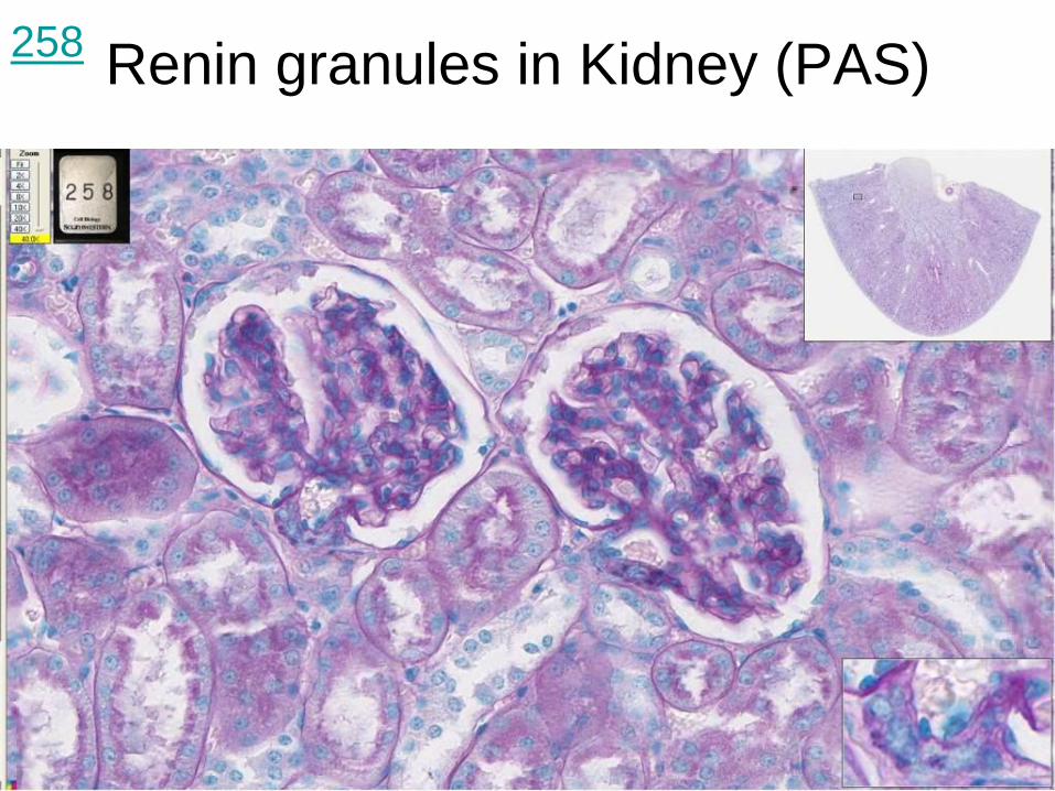
Renin granules in Kidney (PAS)
258

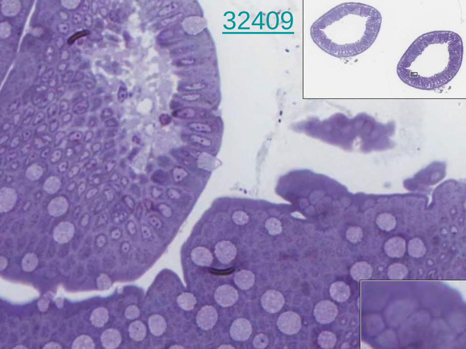
Ileum, monkey (PAS)
249
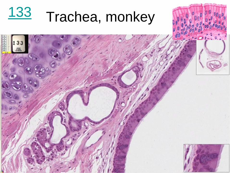
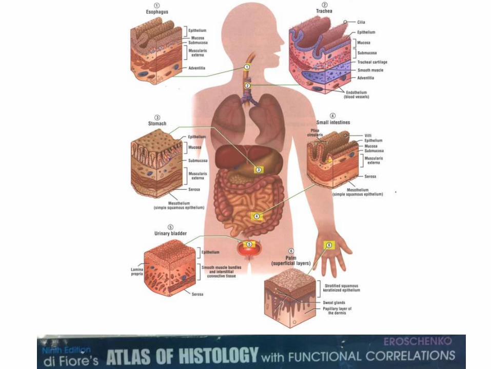
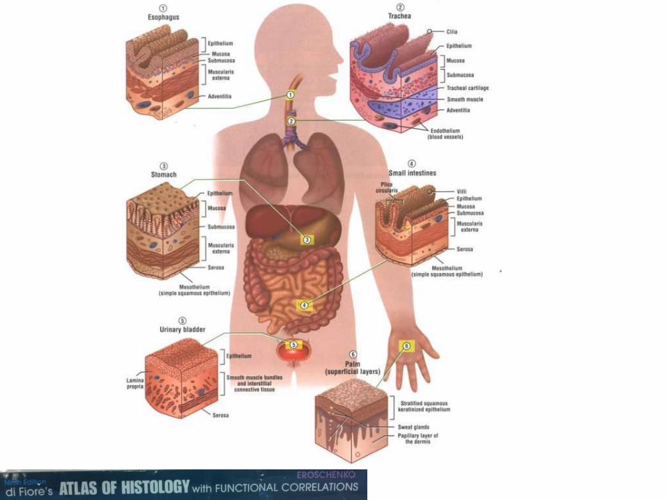
Trachea, monkey 133

Esophagus and trachea, monkey –
glands in trachea
242

Larynx (Gallego's stain) 429
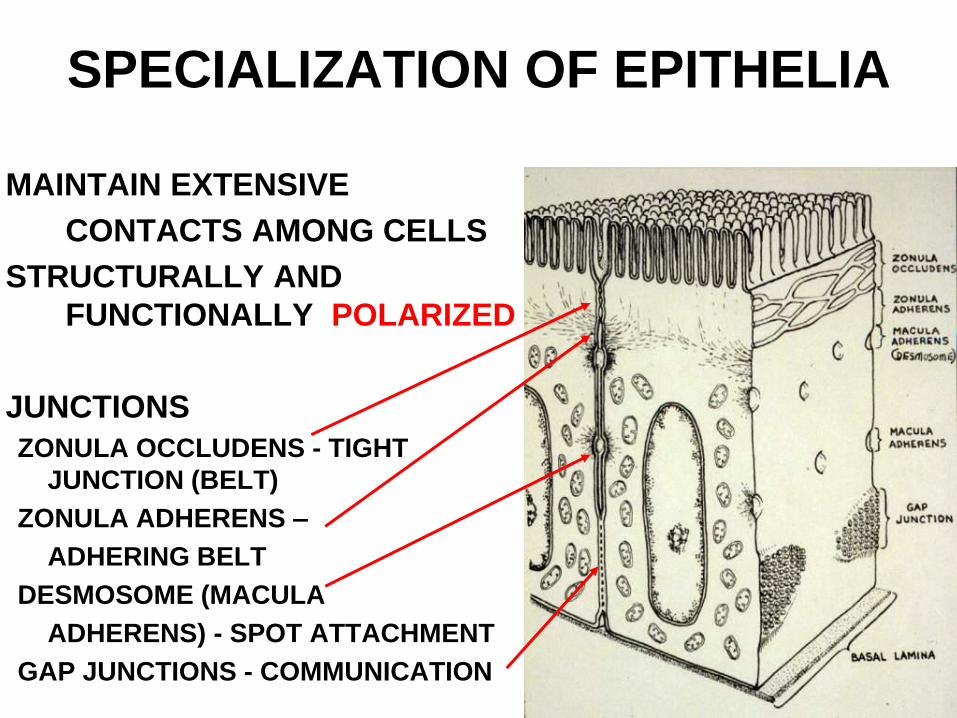
SPECIALIZATION OF EPITHELIA
MAINTAIN EXTENSIVE
CONTACTS AMONG CELLS
STRUCTURALLY AND
FUNCTIONALLY POLARIZED
JUNCTIONS
ZONULA OCCLUDENS - TIGHT
JUNCTION (BELT)
ZONULA ADHERENS –
ADHERING BELT
DESMOSOME (MACULA
ADHERENS) - SPOT ATTACHMENT
GAP JUNCTIONS - COMMUNICATION
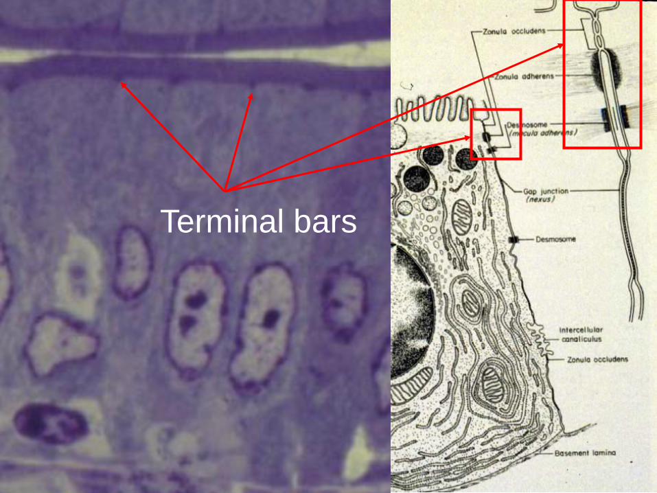
Terminal bars

EM 6b: Region showing zonula
occludens, zonula adherents,
and macula adherents junctions
between cells.
1. Zonula occludens
2. Zonula adherens
3. Desmosome EM 6b
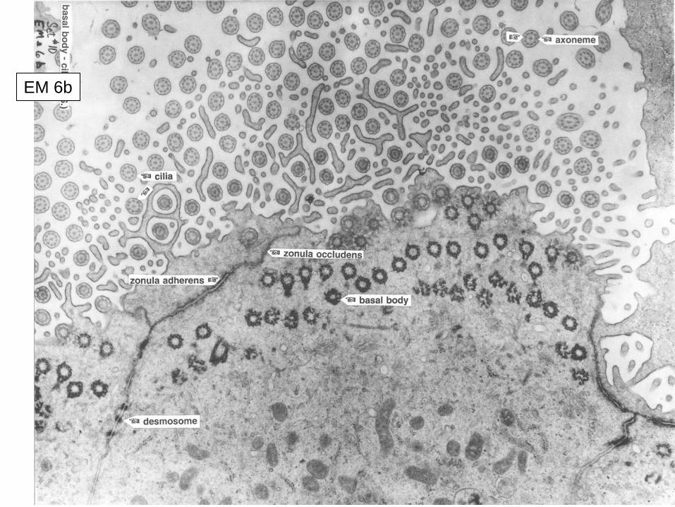
EM 6b
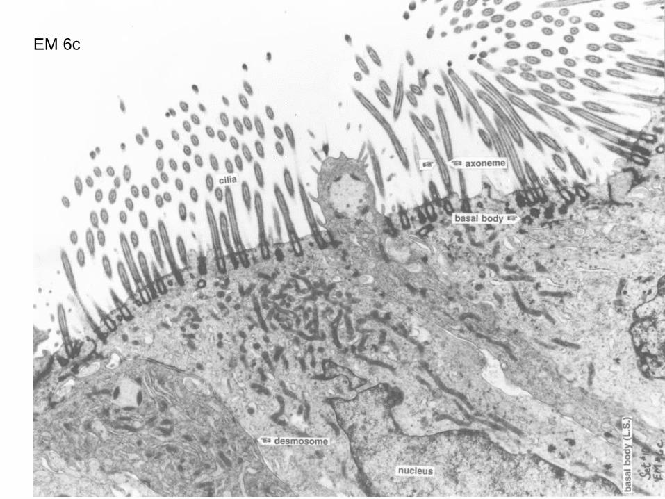
EM 6c
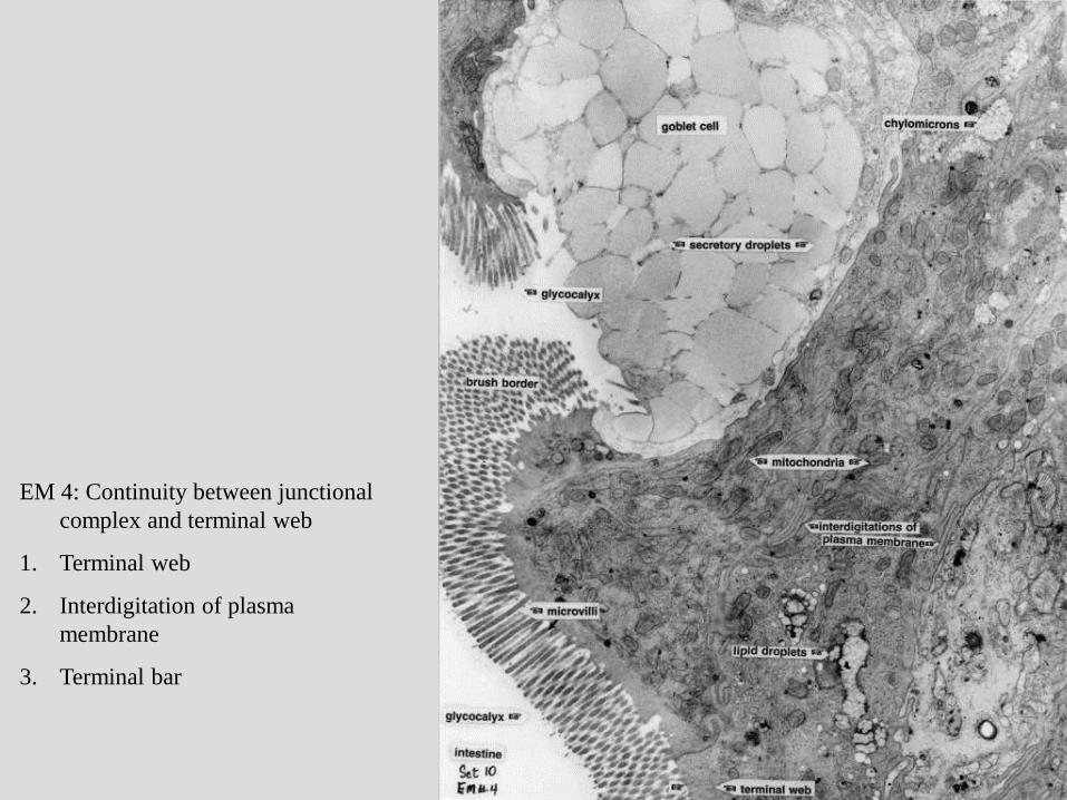
EM 4: Continuity between junctional
complex and terminal web
1. Terminal web
2. Interdigitation of plasma
membrane
3. Terminal bar
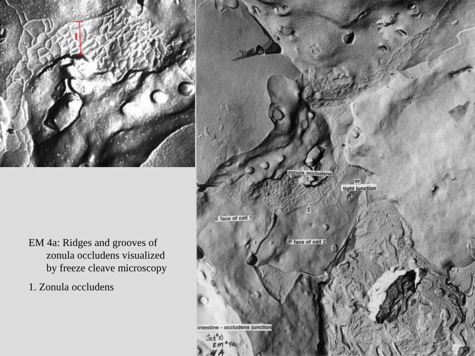
EM 4a: Ridges and grooves of
zonula occludens visualized
by freeze cleave microscopy
1. Zonula occludens
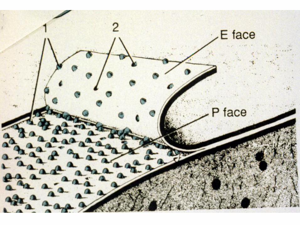

Inside the cell Outside the cell
P = Protoplasmic side E = Ectoplasmic side

EM 4a
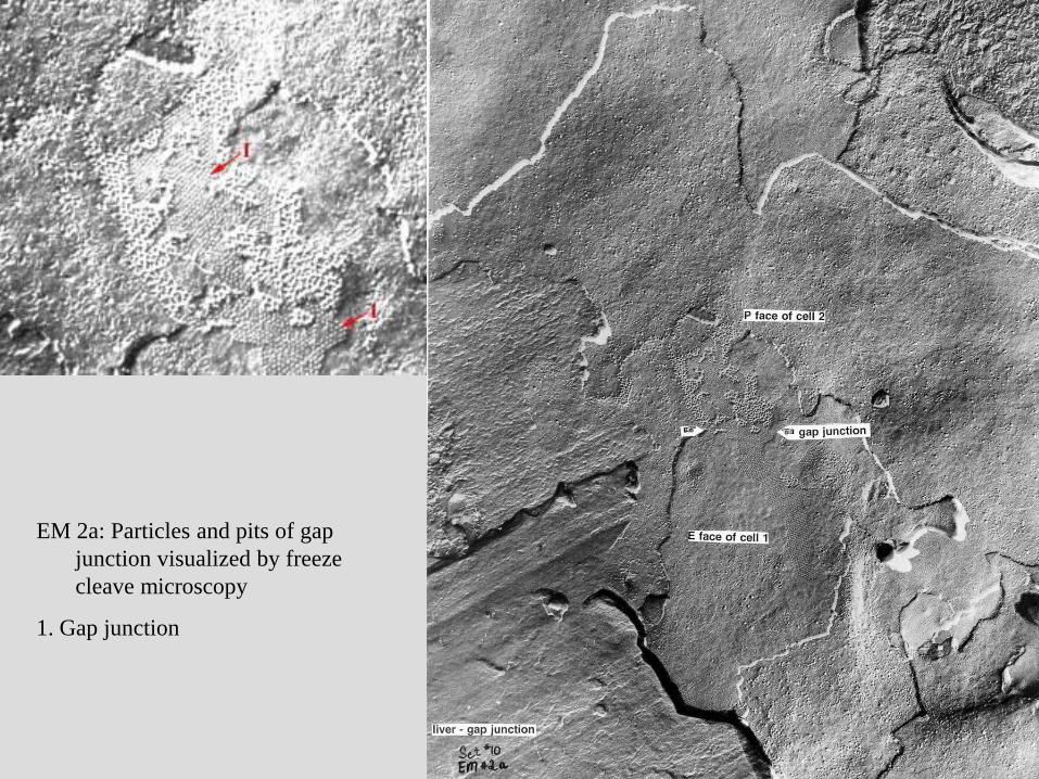
EM 2a: Particles and pits of gap
junction visualized by freeze
cleave microscopy
1. Gap junction

EM 2a
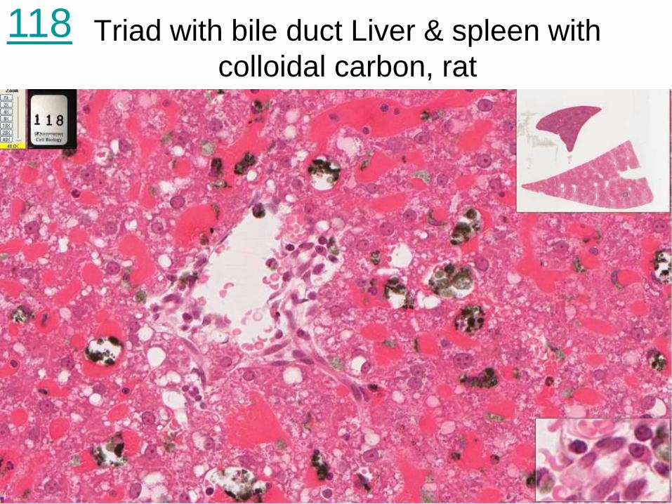
Triad with bile duct Liver & spleen with
colloidal carbon, rat
118
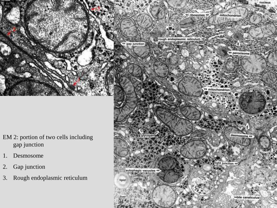
EM 2: portion of two cells including
gap junction
1. Desmosome
2. Gap junction
3. Rough endoplasmic reticulum
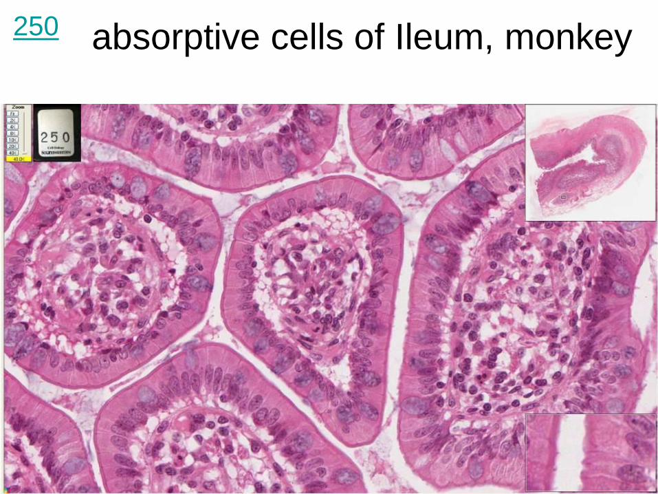
absorptive cells of Ileum, monkey
250
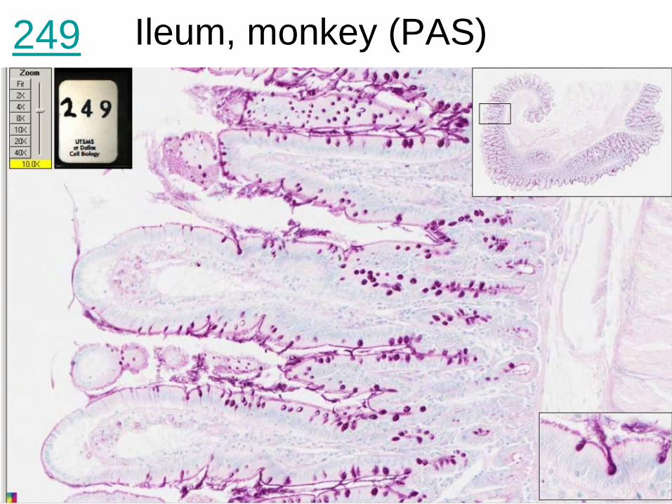
Ileum, monkey (PAS)
249
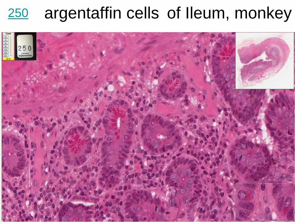
argentaffin cells of Ileum, monkey
250
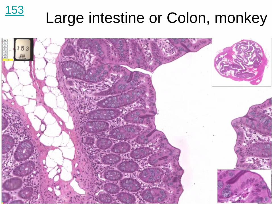
Large intestine or Colon, monkey 153
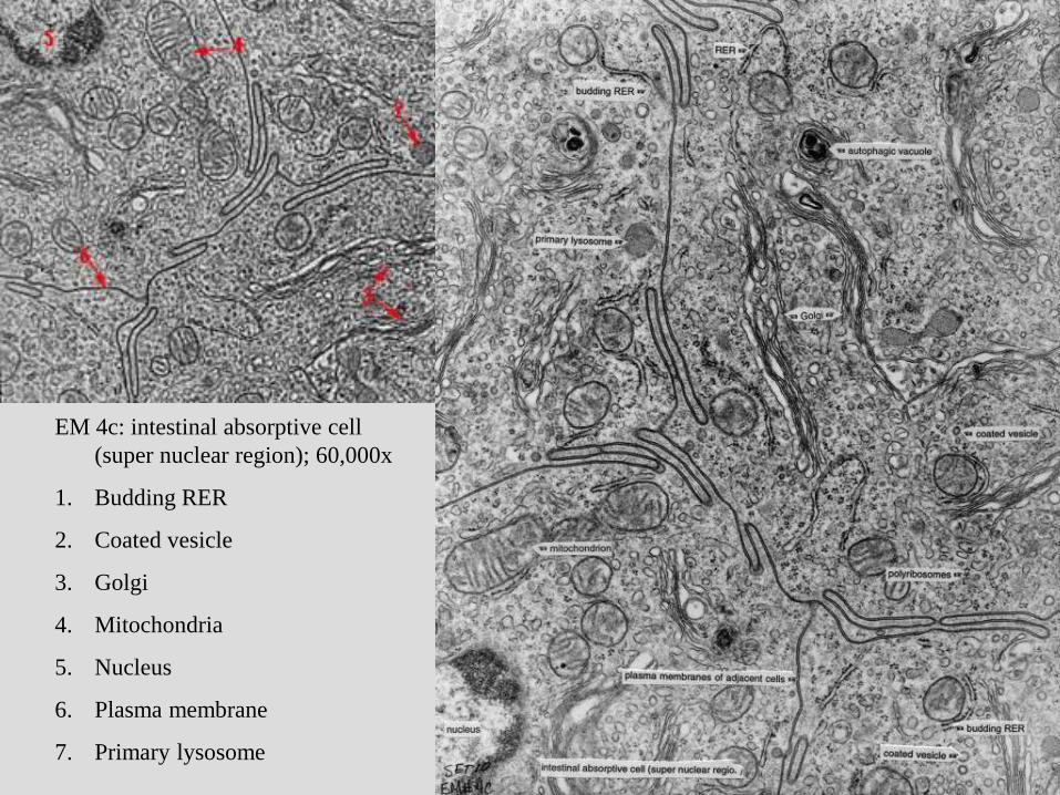
EM 4c: intestinal absorptive cell
(super nuclear region); 60,000x
1. Budding RER
2. Coated vesicle
3. Golgi
4. Mitochondria
5. Nucleus
6. Plasma membrane
7. Primary lysosome
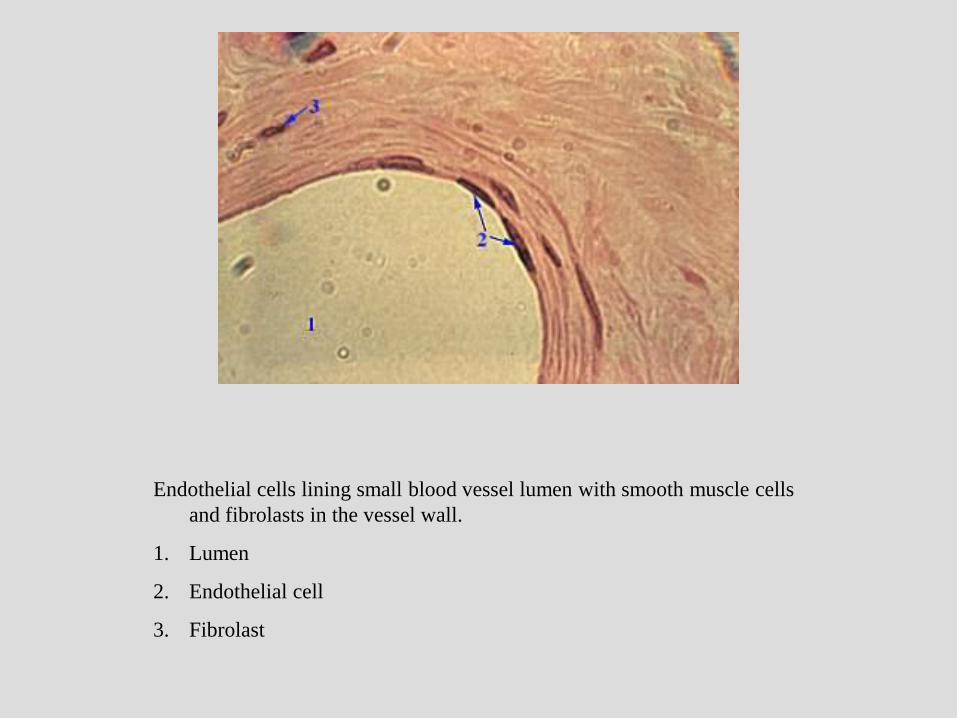
Endothelial cells lining small blood vessel lumen with smooth muscle cells
and fibrolasts in the vessel wall.
1. Lumen
2. Endothelial cell
3. Fibrolast
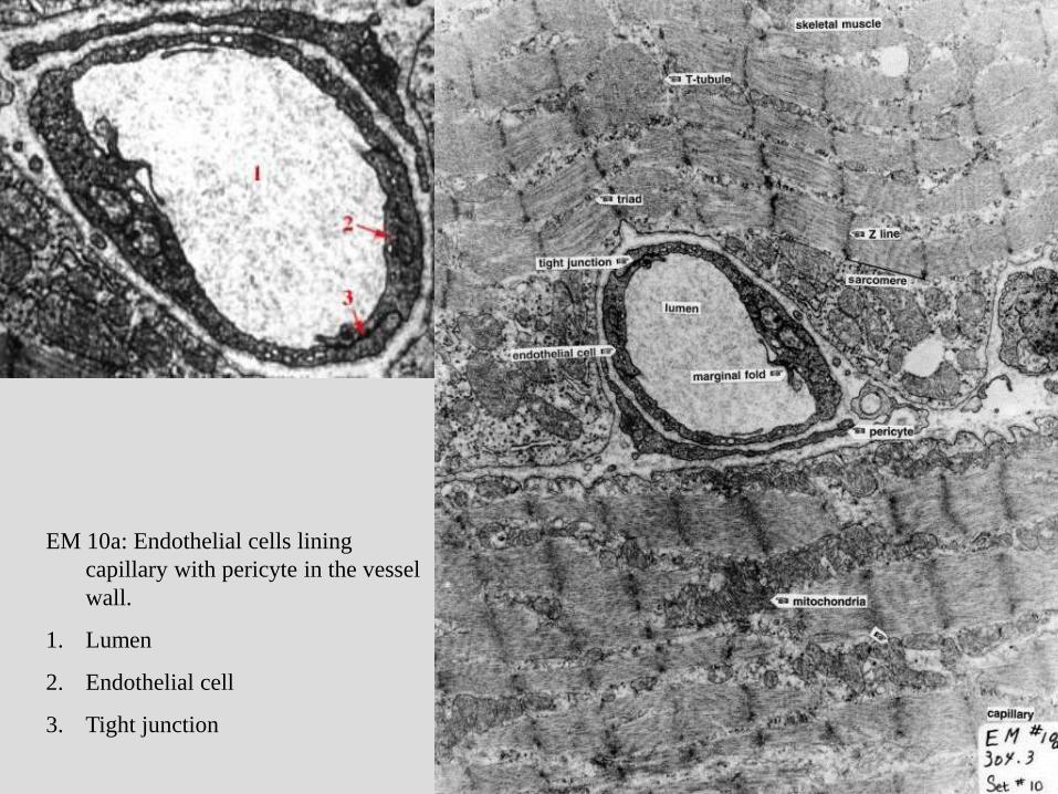
EM 10a: Endothelial cells lining
capillary with pericyte in the vessel
wall.
1. Lumen
2. Endothelial cell
3. Tight junction
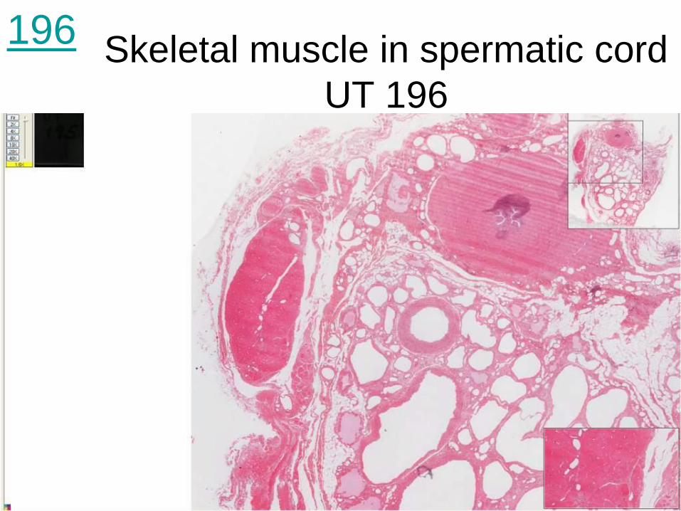
Skeletal muscle in spermatic cord
UT 196
196
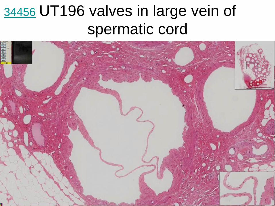
UT196 valves in large vein of
spermatic cord
34456
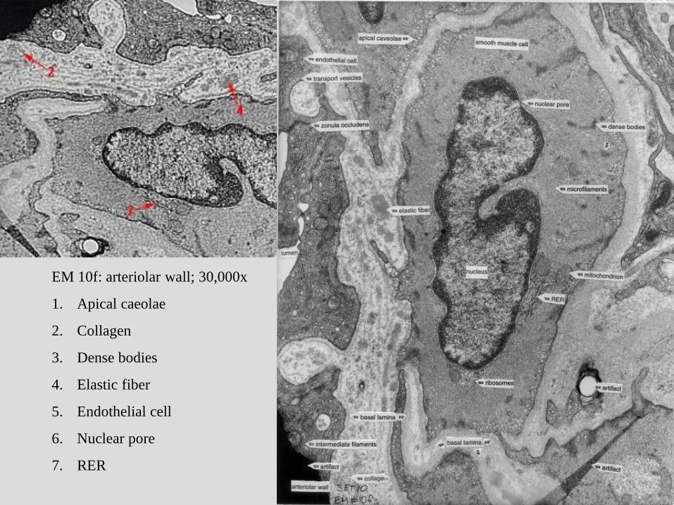
EM 10f: arteriolar wall; 30,000x
1. Apical caeolae
2. Collagen
3. Dense bodies
4. Elastic fiber
5. Endothelial cell
6. Nuclear pore
7. RER
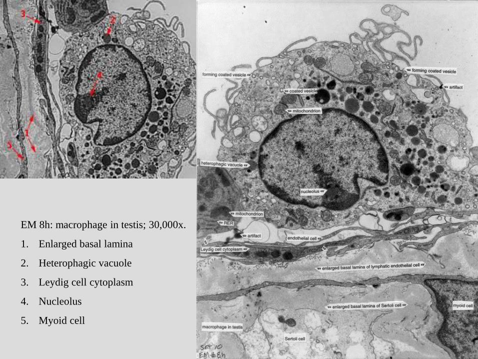
EM 8h: macrophage in testis; 30,000x.
1. Enlarged basal lamina
2. Heterophagic vacuole
3. Leydig cell cytoplasm
4. Nucleolus
5. Myoid cell
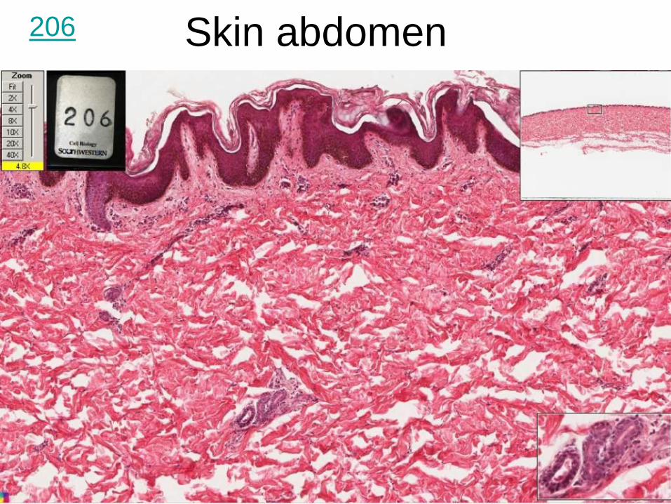
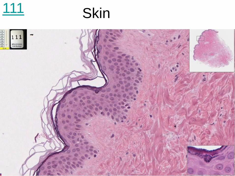
Skin abdomen 206
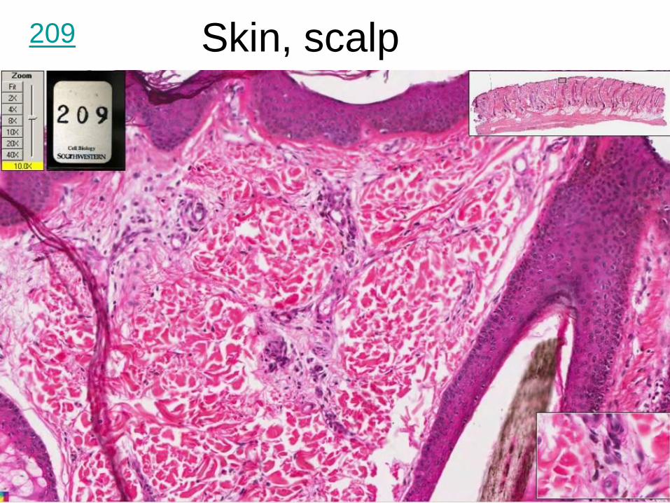
Skin, scalp 209

Skin hand monkey 109
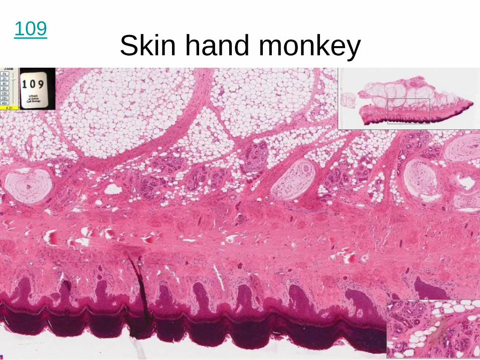
Skin hand monkey 109
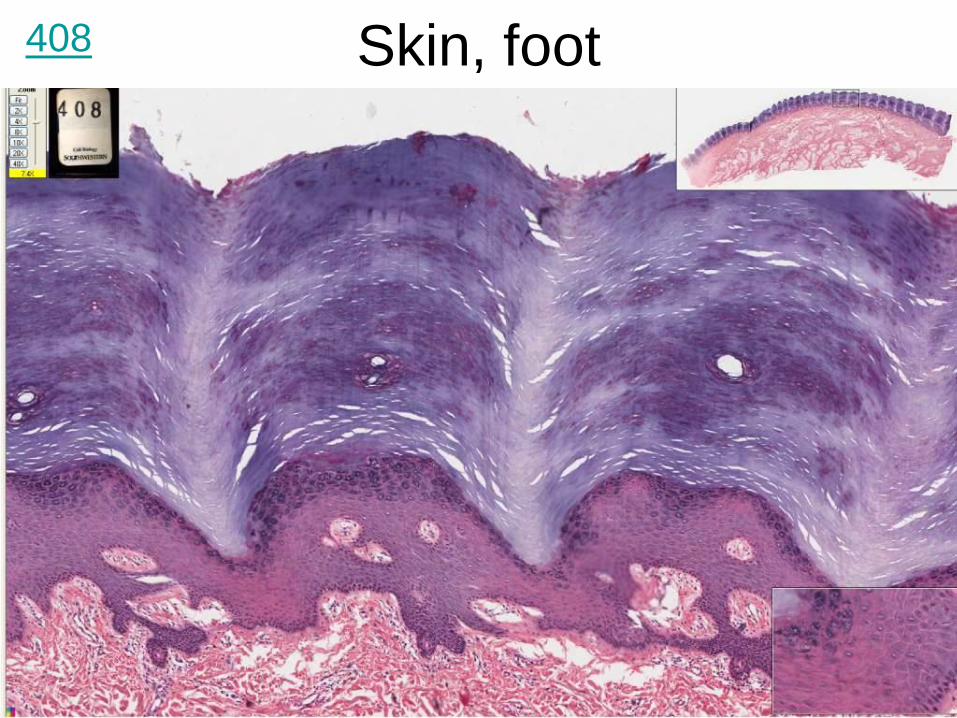
Skin, foot 408
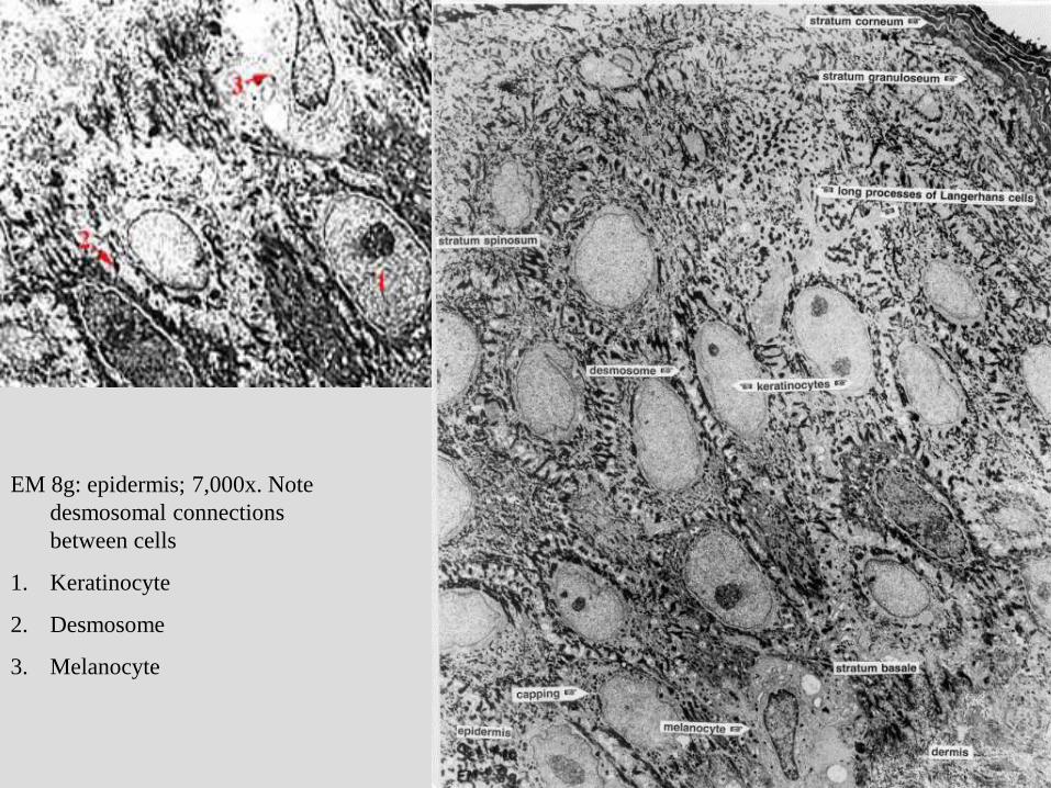
EM 8g: epidermis; 7,000x. Note
desmosomal connections
between cells
1. Keratinocyte
2. Desmosome
3. Melanocyte
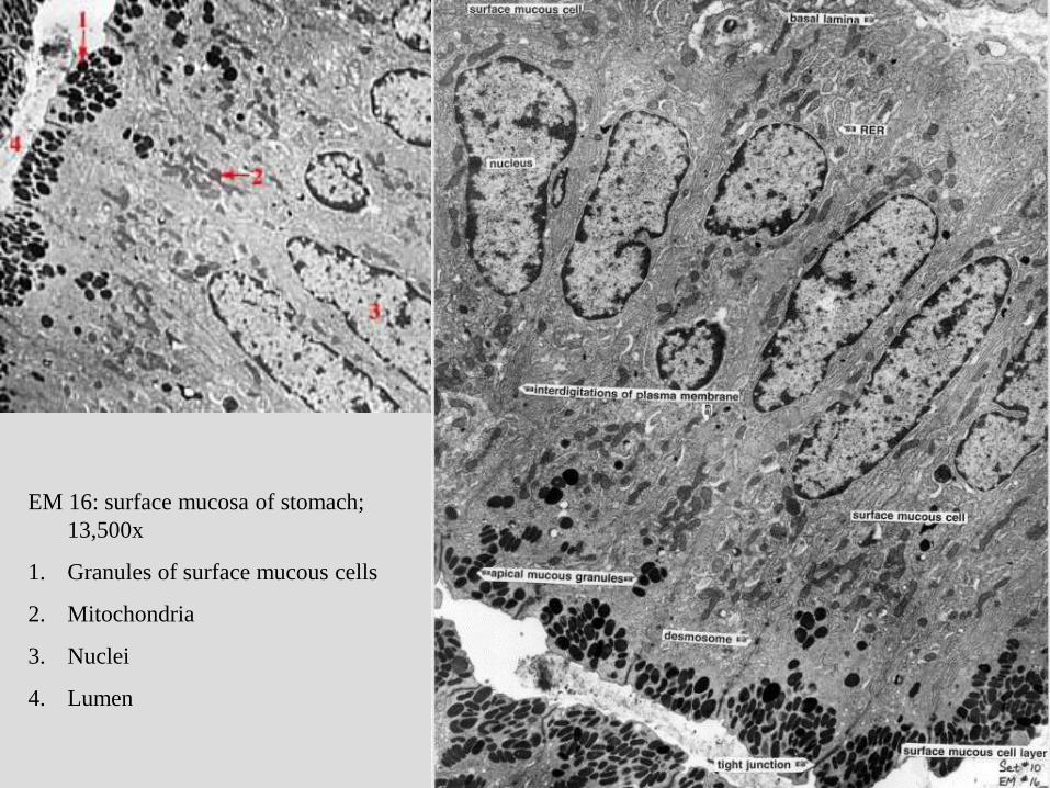
EM 16: surface mucosa of stomach;
13,500x
1. Granules of surface mucous cells
2. Mitochondria
3. Nuclei
4. Lumen
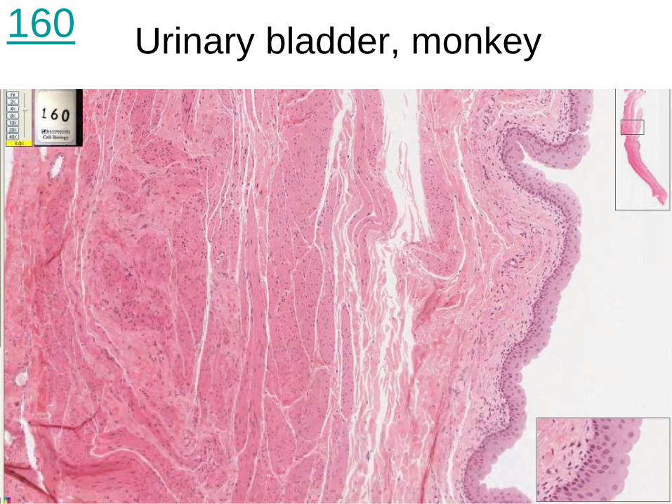
Urinary bladder, monkey
160
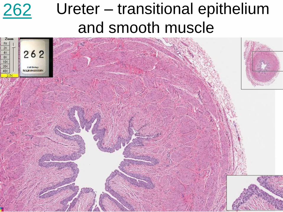
Ureter – transitional epithelium
and smooth muscle
262
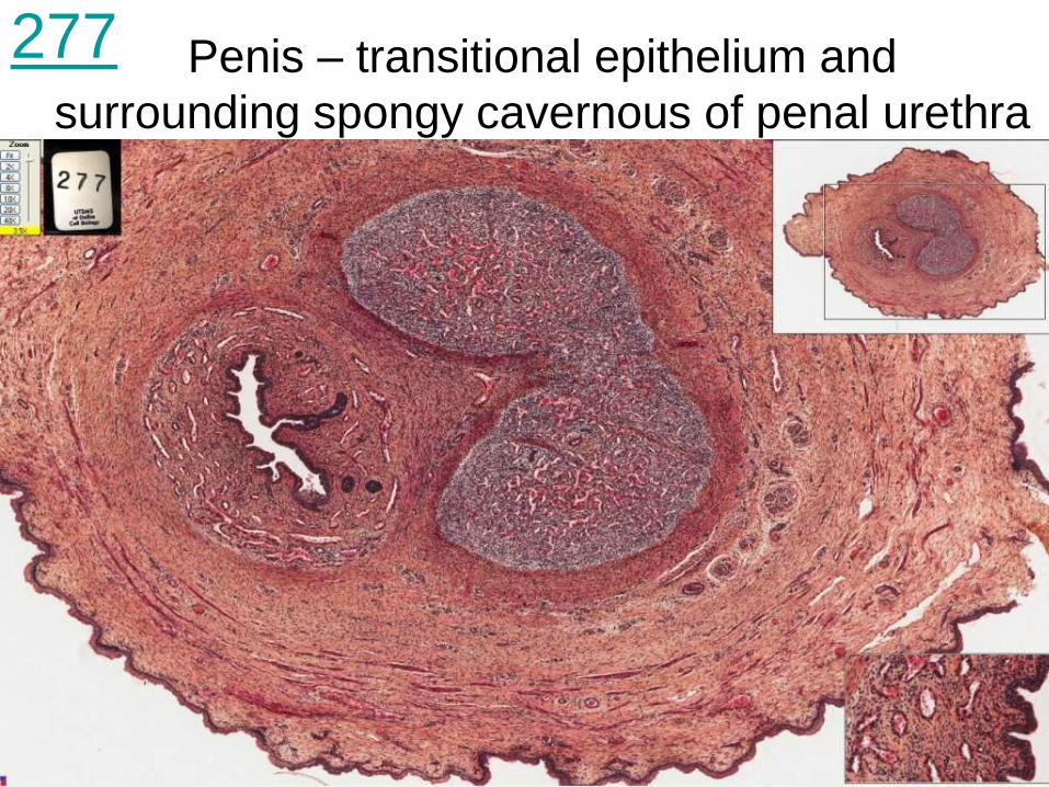
Penis – transitional epithelium and
surrounding spongy cavernous of penal urethra
277
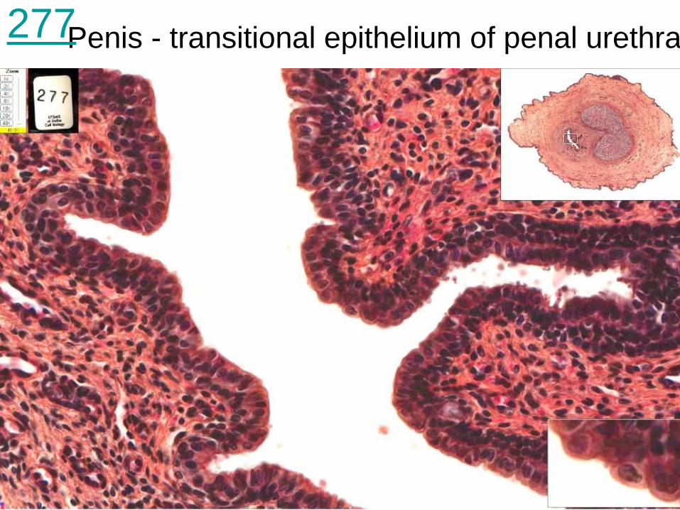
Penis - transitional epithelium of penal urethra 277