Embed Size (px)
Citation preview

Flinders Biomedical Engineering
A joint department of Flinders Medical Centre and Flinders University of South Australia, Faculty of Health Sciences
Orientation pages created and compiled by FBE: mailto:[email protected]
Equipment Orientation
An introduction to typical hospital equipment:
A straight forward description of typical hospital equipment that Bio-meds will come across as they go about their tasks. Good for "just-been-born" Bio-meds, it may also be useful for those who have English as a second language. The contents of these pages were derived from teaching material created and compiled by FBE Anaesthesia Machines Aspirators Blood Pressure Monitors Blood Warmers Capnographs Cardiotocographs Cold Light Sources Defibrillators Dental Chairs Electrocardiograph Electroencephalographs Electromyographs Endoscope Electro-Surgical Units Haemodialysis Humidifiers Intra Aortic Balloon Pumps Infant Incubator Infusion Pumps Syringe Infusion Pumps Volumetric Insufflator Laryngoscopes Laser Medical Flowmeter Medical Regulator Microscopes Non Invasive Blood Pressure Ophthalmoscopes Otoscopes Patient Monitor Phototherapy Pulse Oximeters Radiant Warmers Ripple Mattress Short Wave Treadmills Ultrasound Ventilators X-ray Machines Comments or feedback about these pages: [email protected] Reprinted by the CMIA with permission of FME. While the material is believed correct neither FME or CMIA can guarantee its accuracy.
http://som.flinders.edu.au/FUSA/BME/FBEHomePage.htm

Flinders Biomedical Engineering
A joint department of Flinders Medical Centre and Flinders University of South Australia, Faculty of Health Sciences
Anaesthesia Machines – Basics What does "Anaesthesia" mean?
an = without aesthesia = sensation
What does it do? The anaesthesia machine mixes the correct concentrations of gas and drugs to be inhaled and exhaled by the patient so they lose consciousness. They are held unconscious and have no sensation while being operated on, then regain consciousness after the operation.
Physiology Anaesthetic gas affects the nervous system, resulting in a numbing of the nerve pathways. The patient becomes unconscious, unaware of what is happening, has no pain, is immobile (may need breathing support) and free from memory while under the influence of the anaesthetic agent.
How it works Air, Oxygen, and Nitrous Oxide are supplied from wall outlets or cylinders. The pressures are reduced by pressure regulators then pass through an O2 failure alarm. The flow of gas is controlled by flowmeters. The gas is mixed and passed over a vaporiser containing the anaesthetic agent. An Anti-hypoxic device ensures that the flowmeters deliver a minimum of 25% O2 to the agent vaporiser. The gas then goes through an over pressure relief valve and back flow valve, then to the common gas outlet (CGO), ready to be given to the patient. There is an emergency O2 flush valve connected to give 100% O2 if required. The gas then enters a patient circuit and ventilator. Expired gas can be stripped of CO2 in a soda-lime filter and recirculated, or scavenged out of the room.
Units of measurement
Flow: L/min, Pressure: kPa and cmH2O
Typical values Dependant on patient size, rate and strength of breathing, circulation and solubility of the anaesthetic. Pictures of equipment
Datex Ohmeda ADU
http://www.datex-ohmeda.com/products/anelifesupport.htm Ulco
http://www.ulco.com.au/AN_WORK.asp
Back
Orientation pages created and compiled by FBE: mailto:[email protected]

Flinders Biomedical Engineering
A joint department of Flinders Medical Centre and Flinders University of South Australia, Faculty of Health Sciences
Aspirators – Basics Abbreviation / other names
Suction unit
What does it do? Removes unwanted body fluids and irrigation fluids from an open wound, surgical site or any other area where excess fluid has gathered.
How it works Aspirators generally consist of an electric motor driving a pump that generates a negative pressure. A suction regulator controls the pressure to the required level. The fluid is sucked through a tube into a large jar (disposable in many cases) for examination or disposal. The amount of suction (negative pressure) is normally low to avoid tissue and organ damage. Suction may also be used from wall outlets and portable medical gas cylinders. However, a regulator must be used to reduce pressure.
Units of measurement
Negative Pressure: mmHg or kPa
Typical values 0 to -760 mmHg or 0 to -100 kPa
Picture of equipment
Clements Aspirator
Back
Orientation pages created and compiled by FBE: mailto:[email protected]

Flinders Biomedical Engineering
A joint department of Flinders Medical Centre and Flinders University of South Australia, Faculty of Health Sciences
Blood Pressure Monitors– Basics What does it do? It measures the pressure in the veins or arteries.
Physiology When the heart beats, it pumps blood to the body through the arteries. The blood then returns to the heart
through the veins. The amount the heart contracts, the volume of the blood and the diameter of the blood vessels all determine the pressure inside the vessels.
How it works A narrow fluid filled tube is inserted into a blood vessel. The end of the tube is connected to a pressure sensor. The pressure in the vessel pressurises the fluid in the tube and the pressure sensor measures this. The pressure sensor sends an electrical signal to the monitor, which displays the pressure on a screen. An invasive blood pressure machine is also used to measure other pressures inside the body such as intracranial pressure (the fluid around the brain), intrapleural (between the lung tissue and the ribs) and gastro-intestinal pressures (inside the stomach or intestines)
Units of measurement
mmHg
Typical values
Venous: mmHg Systolic: 120 mmHg Diastolic: 80 mmHg Mean arterial: 100 mmHg
Picture of equipment
HP/Philips Merlin monitor showing invasive blood pressure display and input module
Back
Orientation pages created and compiled by FBE: mailto:[email protected]

Flinders Biomedical Engineering
A joint department of Flinders Medical Centre and Flinders University of South Australia, Faculty of Health Sciences
Blood Warmers – Basics What does it do? Blood is stored at 2 C° and needs to be warmed up during blood transfusion to between 37 C°
and 41 C°. This prevents hypothermia (body temperature too low)
Physiology
blood loss the patient can suffer from anaemia (reduced number of blood cells) and low blood pressure (from the loss of blood volume) which can be life threatening. Blood transfusions are also often used for treatments of blood related diseases like leukaemia. The blood is warmed up to body temperature before infusing it into the patient. The blood must not be heated above 43 C° or the blood cells will die (haemolysis).
How it works There are different types of blood warmers. Some blood warmers have a water bath where the water is heated up to a set temperature e.g. 40 C°. A blood bag is put in to this bath to warm up. The other types (e.g. the Prisma Prismatherm) has a coiled blood line around its body. While the blood is flowing through the coiled blood line it heats the blood to the set temperature. All blood warmers must have an overheat cutout thermostat to prevent the blood from overheating above 43 C°.
Units of measurement Temperature = C°
Typical values Operation temperature 37 C° – 41 C° Over-temperature alarm/cut-out 42 C° - 43 C°
Picture of equipment
Back
Orientation pages created and compiled by FBE: mailto:[email protected]
Flinders Biomedical Engineering
A joint department of Flinders Medical Centre and Flinders University of South Australia, Faculty of Health Sciences
Capnographs – Basics Abbreviation / other names
What does "Capnograph" mean?
End Tidal CO2 (ETCO2), Expired CO2 Monitor
Capno = Carbon dioxide Graph = Graphic display read out or recording.
What does it do? A Capnograph measures the amount of CO2 in the expired air from the lungs of a patient.
Physiology Cells of our body require oxygen, which is given to them via the blood. O2 is given to cells, and when used the waste gas, CO2, produced within the cells is exhausted into the blood. The Blood then transports it to the capillaries in the lungs and the CO2 is then transferred into the air in the lung. As the lungs exhale the CO2 is exhausted to atmosphere.
How it works There are two types of Capnographs, Main stream or Side stream. Main stream. Capnographs have a CO2 sensor that attaches to a disposable airway adaptor inserted between the endotracheal tube in the patient and the ventilator circuit (after the wye piece). The sensor is made up of a light source that sends infra red light through the adaptor, the sample of gas and a filter chopper wheel to the photo detector. Infra red is absorbed by CO2 in the exhaled gas. The more CO2 gas the more the infra red is absorbed and less reaches the photo detector. This signal is sent to the Capnograph and processed to give a graphic reading on the screen. The Side stream Capnograph takes a sample of gas from the airway (from the same place as the Main stream sensor) through a small tube into the Capnograph and all of the measurement and signal processing takes place inside the Side stream Capnograph. (same method as Main stream Capnograph)
Units of measurement %, mmHg partial pressure.
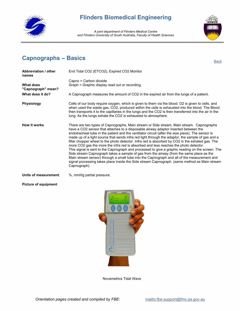
Picture of equipment
Novametrics Tidal Wave
Back
Orientation pages created and compiled by FBE: mailto:[email protected]

Flinders Biomedical Engineering
A joint department of Flinders Medical Centre and Flinders University of South Australia, Faculty of Health Sciences
Cardiotocographs - Basics Abbreviation / other names What does "Cardiotocograph" mean?
What does “Tocodynamometer” mean?
CTG = Cardiotocograph , “Toco” or Tocodynamometer Fetal Monitors
Cardio = heart Toco = contractions (of uterus during labour) Graph = machine to record Cardiotocograph = machine to record the heart rate (fetal heart) and contractions of uterus during labour
Toco = contractions (of uterus during labour) Dyna = force Meter = device to measure Tocodynamometer = device to measure the force of contractions of the uterus during labour CTG = Cardiotocograph , “Toco” or Tocodynamometer
What does it do? Cardiotocograph can monitor the fetus’ heart rate, the mother’s heart rate, the mother’s contractions and the pressure inside the uterus during labour.
Physiology During labour the fetus can become stressed. The heart rate of the fetus is monitored throughout labour so stress can be detected early. The contractions are monitored also so that the midwife and mother know when the contraction is occurring and also to check for fetal distress during the contraction.
How it works The Cardiotocograph has a disc-shaped ultrasound transducer, which is strapped onto the mother’s abdomen over the area of the uterus. The transducer has several crystals (usually about 7), which emit ultrasound waves into the uterus and also detect the waves that are reflected back. The ultrasound waves are reflected from different surfaces in the uterus and detected by the transducer. The fetal heart moves every time it beats and this beating movement is detected by the changing ultrasound waves being reflected back to the transducer. From this the cardiotocograph can calculate the fetal heart rate. A second ultrasound transducer can also be used at the same time so that twins can be monitored. The second transducer has a different frequency so the two signals can be separated. The Cardiotocograph also has a different type of transducer to monitor the uterine contractions (the toco transducer). This transducer is also disc-shaped and is strapped around the uterus. The patient’s abdominal muscles depress the transducer’s central section during a contraction. A strain gauge behind the moving part measures the movement. The size of the contraction is proportional to the amount of change in the strain gauge. The Cardiotocograph can also monitor fetal heart rate and contractions invasively. Fetal heart rate can be measured by using a special ECG electrode on the scalp of the baby. The mother’s ECG can also be recorded by a maternal ECG leads. (See Electrocardiograph for explanation). The pressure inside the uterus can be monitored invasively by a Intra Uterine Pressure (IUP) transducer (see invasive blood pressure for explanation)
Units of measurement Fetal heart rate (ultrasound/FECG): BPM (beats per minute) Contractions: % (relative measure of magnitude of contractions) IUP: mmHg
Back
Orientation pages created and compiled by FBE: mailto:[email protected]

Flinders Biomedical Engineering
A joint department of Flinders Medical Centre and Flinders University of South Australia, Faculty of Health Sciences
Cardiotocographs – Basics - Continued Picture of equipment
HP ECG & Tocograph
Orientation pages created and compiled by FBE
Back
Orientation pages created and compiled by FBE: mailto:[email protected]

Flinders Biomedical Engineering
A joint department of Flinders Medical Centre and Flinders University of South Australia, Faculty of Health Sciences
Cold Light Sources - Basics Abbreviation / other names
Light source
What does it do? High intensity light used in conjunction with endoscopes, rigid scopes, headlamps and operating microscopes etc.
Physiology Cold light sources are normally used to illuminate a specific work area during surgery or medical examinations. It is important that the light is as near in colour to natural light so doctors are able to detect abnormal colourations of the skin and tissue. It is also important that the light does not heat the tissue.
How it works The high intensity light source consists of a power supply, cooling fan, brightness controls, lamp life indicator and a spare lamp to enable procedures to be finished if the primary lamp fails. Filters are also employed to reduce the transmission of excessive heat to the operating site. This is why it is called ‘Cold Light’. The two main types of lamps used are xenon arc and halogen. Some xenon lamps have a useable life of only 500 hours. The light is channelled to the required area via a flexible fibre optic light guide, made from tightly bundled glass fibres. This concentrates the light and allows the light to be easily directed to where it is required.
Picture of equipment
Olympus CLK3 Cold Light Source
Back
Orientation pages created and compiled by FBE: mailto:[email protected]

Flinders Biomedical Engineering
A joint department of Flinders Medical Centre and Flinders University of South Australia, Faculty of Health Sciences
Defibrillators - Basics What does "Defibrillator" mean?
What does it do?
De = to undo, or reverse Fibrillate = uncoordinated contractions of the heart It stops fibrillation of the heart, and restores the normal coordinated contractions.
Physiology The cells inside the heart muscle conduct electrical current. Some cells in the heart (pacemaker cells) charge by themselves regularly, and they cause the other cells to charge. A current flows the heart muscle in a particular way. Every time this happens the heart muscle contracts and pumps the blood - 'heart beat". This coordinated electrical pattern is essential to cause the heart to fill and empty with every beat pushing blood around the body. When this electrical pattern becomes uncoordinated (fibrillation), the heart muscle contractions are not coordinated and no blood is pumped. This will lead to decreased blood pressure, lack of oxygen, heart muscle damage, and untimley death. The defibrillator is used to reverse this fibrillation and restore the coordinated contractions. It can also be used if the heart has stopped beating to start it again. (e.g. after an electric shock)
How it works The defibrillator uses the mains power or a large internal battery to charge a large capacitor to between 5 and 400 Joules. Two metal "paddles" (electrodes) are connected from the defibrillator, and placed on either side of the patient's chest. The energy stored in the capacitor is then discharged from one paddle to the other through the patient's chest, which shocks the heart into beating rythmically again Defibrillators also have built in ECG monitors, and ECG recorders to continuously display and document ECG waveforms.
Units of measurement Joules Picture of equipment
Philips (formerly Agilent) Zoll - Defib
Back
Orientation pages created and compiled by FBE: mailto:[email protected]
Flinders Biomedical Engineering
A joint department of Flinders Medical Centre and Flinders University of South Australia, Faculty of Health Sciences
Dental Chairs - Basics What does it do? Dental chairs provide a platform from which a dentist can safely and comfortably perform oral
surgery and examination with all the necessary instruments and functions close at hand. How it works Typical dental chairs are hydraulically operated so they can be raised, lowered and also laid down
to a horizontal position. Incorporated into the chair are High and low speed dental drills, air and water sprays (syringes) to wash debris from the area being worked on and also a suction system to remove water and debris. High speed dental drills are usually air driven turbines, whereas low speed drills need more torque and are generally electric motor driven. Dental drills also have water sprays to cool the area, which is being cut away. Water that is used for irrigation is normally distilled or at least filtered to avoid unnecessary contamination. A water and amalgam separator
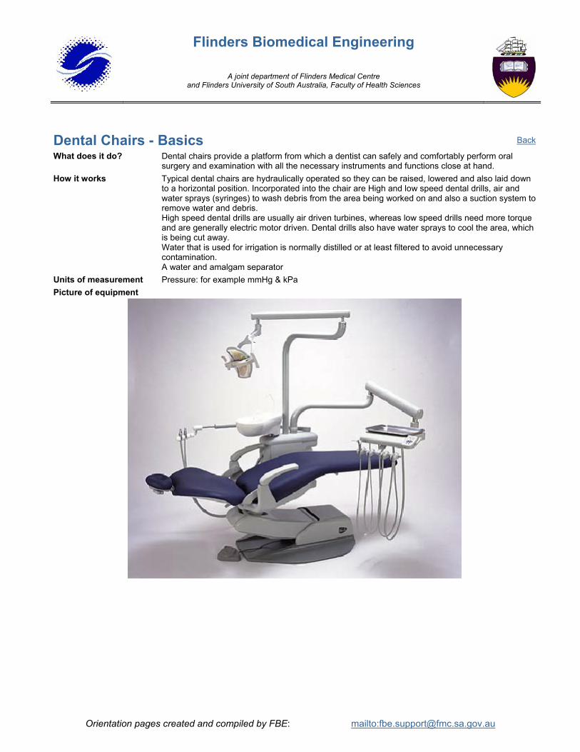
Units of measurement Pressure: for example mmHg & kPa Picture of equipment
Back
Orientation pages created and compiled by FBE: mailto:[email protected]

Flinders Biomedical Engineering
A joint department of Flinders Medical Centre and Flinders University of South Australia, Faculty of Health Sciences
Electrocardiographs - Basics Abbreviation / other names
What does "Electrocardiograph" mean?
ECG Electrocardiogram (In America it is sometimes called EKG)
Electro = electrical activity Cardio = heart Graph = machine to record
What does it do? Cardiotocograph can monitor the fetus’ heart rate, the mother’s heart rate, the mother’s contractions and the pressure inside the uterus during labour.
Physiology The cells inside the heart muscle conduct electrical current. Some cells in the heart (the pace-maker cells) charge by themselves regularly and they cause the other cells to charge. A current flows through the heart muscle in a particular way. Every time this happens the heart muscle contracts and pumps the blood – “heart beat”.
How it works The tissues in the human body are electrical conductors and when the electrical current flows in the heart we can measure it in the surface of the body. ECG electrodes are made of silver-silverchloride and they are stuck to the skin together with ECG gel. The electrical signals from the heart are picked up by these electrodes and measured and recorded by the ECG machine.
Units of measurement Measurement of amplitude in mV Typical values Adults: 50 – 90 BPM
Neonates: 120 – 180 BPM The repetitive electrical signal produced by the Heart and measured at the patient electrodes is approx. 1 mV in amplitude.
Picture of equipment
HP ECG & Tocograph
Back
Orientation pages created and compiled by FBE: mailto:[email protected]
Flinders Biomedical Engineering
A joint department of Flinders Medical Centre and Flinders University of South Australia, Faculty of Health Sciences
Electroencephalographs - Basics Abbreviation / other names
What does "Electroencephalograph" mean?
EEG
Electro = electrical activity Encephalo = brain Graph = recording
What does it do? It records the electrical activity in the brain. Physiology The cells in the brain transmit messages to each other by tiny electrical currents. How it works These electrical potentials in the brain can be measured on the surface of the head as tiny voltages.
EEG electrodes made of gold, silver or platinum are placed on the head. Electrode gel is used between the electrode and the skin to transmit the signal from the skin to the electrode. The EEG will display the signal on the screen as a series of many lines representing activity in different areas of the brain. The EEG may also filter the signals into different types such as a, b, d etc.
Units of measurement Amplitude is measured in mV Frequency is measured in Hz
Typical values The signals will be analysed by a Neurologist. They look for bursts of activity, patterns, lack of signals. There is no simple typical value.
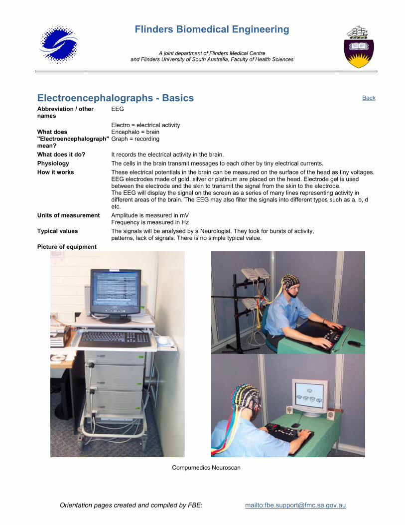
Picture of equipment
Compumedics Neuroscan
Back
Orientation pages created and compiled by FBE: mailto:[email protected]

Flinders Biomedical Engineering
A joint department of Flinders Medical Centre and Flinders University of South Australia, Faculty of Health Sciences
Electromyographs - Basics Abbreviation / other names
What does "Electromyograph" mean?
E.M.G.
Electro = electrical activity Myo = muscle Graph = recording
What does it do? Records the electrical activity of a muscle by means of electrodes placed near, or inserted into muscle fibres.
Physiology The technique is used for diagnosing various nerve disorders and assessing the progress in recovery from some form of paralysis. It is also able to estimate the density of muscle fibres belonging to a single muscle activity.
How it works Electromyography tests nerves the same way an electrician would test a wire. Since nerves operate by conducting electrical impulses, the easiest way to see if they are damaged is to send a current through a nerve from one end to see if it reaches the other. If the current reaches the other point at full strength, there is no damage to the nerve. If the impulse is reduced, or does not come through at all, there may be damage to the nerve.
Units of measurement Microamperes - milliampere Typical values Many variables depending on the site of the EMG. Picture of equipment
Compumedics Neuroscan
Back
Orientation pages created and compiled by FBE: mailto:[email protected]

Flinders Biomedical Engineering
A joint department of Flinders Medical Centre and Flinders University of South Australia, Faculty of Health Sciences
Endoscope - Basics Abbreviation / other names
What does "Endoscope" mean?
Colonoscopes, Duodenoscopy, Gastroscope, Bronchoscope, Rhinoscope
Endo = inside Scope = device to look or see Endoscope = a device to look inside
What does it do? An endoscope is used to examine and take biopsies inside passages in the body (eg. inside lining of the upper and lower digestive system, bronchus etc). The endoscopes are named and are designed to be used in specific areas, for example the bronchoscope is used only to go into the bronchus, while the gastroscope is designed to examine and take biopsies of the stomach.
Physiology Endoscopy can be helpful in the evaluation or diagnosis of various medical problems including intestinal, stomach, abdomen, duodenum and bronchus bleeding, ulcers, and tumours. Biopsies can be taken for examination.
How it works The endoscope is a long, thin, flexible tube with a tiny video camera or eyepiece and light on the end. By adjusting the various controls on the endoscope, the user can safely guide the instrument to carefully examine the inside lining of the upper or lower digestive system, nasal, bronchus etc. The endoscopes are designed with an air and water channel, while the larger endoscopes also have a biopsy and suction channel. The air and water is used to keep the viewing area clean. A fiberoptic light channel illuminates the viewing area. The distal end of the scope can be angled in almost all directions for examining the surroundings
Picture of equipment
Back
Orientation pages created and compiled by FBE: mailto:[email protected]

Flinders Biomedical Engineering
A joint department of Flinders Medical Centre and Flinders University of South Australia, Faculty of Health Sciences
Electrosurgical units - Basics Abbreviation / other names
What does "Diathermy" mean?
ESU Diathermy
Dia = through Thermo = temperature heat
What does it do? The ESU cuts tissue and/or stops the bleeding by coagulating the blood (haemostasis)
Physiology During surgery the tissue and blood vessels are cut which causes bleeding. To prevent too much blood loss and to keep the operating field free of excess blood, electro surgical units are used. Different modes of operation can be set. “Cut” is for cutting tissue, “Blend” is a mixture of cutting and coagulation. This mode is used to cut and the same time reduces bleeding (haemostatic). “Coagulation” is used for maximum Haemostasis, “Desiccation” is used for destroying tissue. The monopolar mode (single electrode) is used for cutting and coagulation, the bipolar mode (forceps like electrode) is used mainly for desiccation (destroying tissue)
How it works An electric current with a frequency of about 500kHz is used to cut and coagulate tissue. This process involves applying an RF (Radio Frequency) spark between a probe and the tissue. The electric current through the tissue heats up the tissue and evaporates the water in the cell destroying it. This achieves special surgical effects namely cutting, coagulation and desiccation. The voltage on the electrode is between 1000 – 10,000 V p-p.
Units of measurement Watts
Typical values Bipolar Cut: 50 Watts Coag: 8 Watts Monopolar Cut: 150 – 300 Watts Coag: 40 – 80 Watts
Picture of equipment
Valleylab Force 2 http://www.valleylabeducation.org/pages/vliceesself.html
Back
Orientation pages created and compiled by FBE: mailto:[email protected]

Flinders Biomedical Engineering
A joint department of Flinders Medical Centre and Flinders University of South Australia, Faculty of Health Sciences
Haeomodialysis units - Basics Abbreviation / other names
What does "Haemodialysis" mean?
Renal Machine, Dialysis Machines
Haemo = blood Dialysis = removal of waste products from the blood Dialyser = an artificial kidney
What does it do? Decontaminate the blood Maintain liquid balance Maintain electrolyte balance
Physiology Replacing the function of the failed kidneys
How it works Solutions are mixed with reverse osmosis(RO) water to create dialysate (cleansing fluid). The dialysate is heated to 37C, de-aerated and passed through the dialyser (artificial kidney), where the blood is cleaned by osmosis, diffusion, and ultra-filtration. Anti clotting agents are used to prevent dialyser blockages. Waste fluid is pumped to drain.
Units of measurement Temperature: C, Conductivity: milli mohs, Pressure: mm/Hg, Flow: ml/min Typical values Temperature 37C, Conductivity 14 Ms, Venous Pressure 100-200mm/Hg,
Dialysate flow 500 ml/min, Blood flow 200-300 ml/min Picture of equipment
Fresenius
Back
Orientation pages created and compiled by FBE: mailto:[email protected]
Flinders Biomedical Engineering
A joint department of Flinders Medical Centre and Flinders University of South Australia, Faculty of Health Sciences
Humidifiers - Basics What does "Humidifier" mean?
Humid = moist Humidifier = a device to make air moist
What does it do? Decontaminate the blood Maintain liquid balance Maintain electrolyte balance
Physiology The gas from a bottle or hospital gas supply is very dry and cool. If a patient on a ventilator inhales this cool dry air, the tissue in the airway passages and lung tissues can dry out and be damaged. When the airway tissue becomes dry the mucous becomes thick and difficult to remove by suction. The humidifier humidifies the air to prevent all of the above. The air is also warmed to prevent temperature loss in the patient. The ideal gas to enter a patient is at a temperature of 37 C°, and moisture levels in the gas of 44mg/L (100% humidity at 37 C°)
How it works A humidifier is a controlled heater block with a chamber of water on top. The heater plate heats the water in the chamber and causes some water to evaporate. The gas in the ventilator patient circuit passes over the water chamber and is warmed to the desired temperature and humidified. A heater wire is also inserted along the tubing going to the patient to prevent the gas from cooling along the length of the tube. The controller also includes various alarms of high or low temperature in the airway or water chamber.
Units of measurement Degrees Celsius. C°, water content mg/L, %Relative Humidity
Typical values 37 deg C, 44mg/L, 100%Relative Humidity
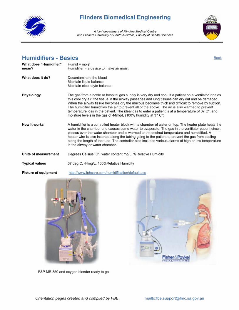
Picture of equipment http://www.fphcare.com/humidification/default.asp
F&P MR 850 and oxygen blender ready to go
Back
Orientation pages created and compiled by FBE: mailto:[email protected]

Flinders Biomedical Engineering
A joint department of Flinders Medical Centre and Flinders University of South Australia, Faculty of Health Sciences
Intra Aortic Balloon Pumps - Basics What does "Intra Aortic" mean?
Intra = Between, Inside Aorta = The major artery from the heart to the body. Intra-aortic = inside the Aorta - next to the heart
What does it do? Helps the heart recover by reducing the amount of work it has to do. Physiology If the blood supply to the heart muscle becomes reduced by a blood clot, the heart's ability to do its
normal work is reduced because there is less oxygen being carried to the muscle. The heart muscle in the area affected by the blocked supply works on, depleting oxygen. The region begins to die. If the normal body load the heart "sees" is reduced by a Balloon Pump, the work and oxygen demand is reduced, while the heart muscle blood supply is improved. The muscle can now survive and heal.
How it works A catheter or "balloon" is inserted into the femoral artery, and using an ultrasonic detecting device, is slid into position below the aortic arch. It is then connected to the Balloon Pump. The Balloon pump uses helium gas to drive the catheter rather than air because it lets the balloon inflate and deflate much more rapidly. The inflation and deflation cycles are timed electronically from an arterial pressure wave or an ECG signal. As the left ventricle begins to contract and pushes the blood out through the semilunar aortic valve, the flow is obstructed by the body resistance, requiring appropriate work from the heart muscle. If the Balloon filling the aortic arch is suddenly deflated at this time, it "sucks" out the contents of the ventricle, and the heart chamber can empty with little effort. When the left ventricle is empty, the body's natural back-pressure closes the Aortic semilunar valve. Suddenly inflating the Balloon now pushes the blood around the body and the coronary arteries. The balloon remains inflated until the ventricle is ready to contract again. As the heart heals, the assistance the IABP gives can be gradually decreased by reducing the volume of inflation, and assisting only every second or third heart beat. IABP assistance is typically required for 5 days.
Units of measurement Pressure Volts
Typical values 70mmHg Picture of equipment Datascope 98XT http://www.datascope.com/
Back
Orientation pages created and compiled by FBE: mailto:[email protected]

Flinders Biomedical Engineering
A joint department of Flinders Medical Centre and Flinders University of South Australia, Faculty of Health Sciences
Infant Incubators- Basics Abbreviation / other names
What does "Incubator" mean
Humidi crib (not a correct name) Incubate = to keep warm Incubator = a device to keep warm
What does it do? An infant incubator is used mainly to keep a baby’s core temperature stable at 37 degrees Celsius. Most incubators also humidify the air and can add extra oxygen.
Physiology The core temperature of the human body needs to be kept at a constant temperature of 37 degrees Celsius. If the temperature goes too high or too low, then the organs can be damaged and illness or death can result. Premature babies (babies born before they are due to be born) have undeveloped nervous systems and also lack the energy to regulate their own temperature, so their temperature needs to be maintained by an incubator. We can only give small babies a small amount of food for growing. We want them to use all of their energy for growth rather than wasting it on keeping warm, so sometimes we use the incubator to help them grow faster.
How it works The mattress where the baby lies is completely enclosed by a clear plastic canopy. The temperature in the incubator is increased by a heater element below the mattress. A motor driven fan near the heater draws in fresh air through a filter and blows it past the heater, warming the air. The air is directed up through slots into the area above the mattress and circulated around. The air temperature is monitored by temperature sensors and is adjusted by controlling the current to the heater. The incubator can also monitor the baby’s skin temperature by using a skin temperature probe, which is stuck onto the skin. The user can either set the incubator to control the temperature of the air or to control the temperature of the baby’s skin (servo control mode). Supplementary oxygen can be taken in by an oxygen inlet connection where it is mixed with the fresh air through the filter. The humidity can be increased by the use of water baths (passive humidification) or by dripping water on a heated element (active humidification). The baby is cared for through special access doors or arm ports.
Units of measurement Temperature: degrees Celsius Total gas intake: L/min Relative Humidity % Oxygen concentration: %
Typical values Air Temperature: 32 to 38 C° Baby skin temperature: 34 to 36 C° Total gas intake: 35 L/min Relative humidity: 50-100%
Picture of equipment
Drager Caelo
Back
Orientation pages created and compiled by FBE: mailto:[email protected]

Flinders Biomedical Engineering
A joint department of Flinders Medical Centre and Flinders University of South Australia, Faculty of Health Sciences
Infusion Pumps - Syringe - Basics Abbreviation / other names Syringe Pump
What does it do? It is used to pump fluids (drugs, liquid food, glucose, saline) into the body.
How it works A plastic syringe containing fluid is placed into the syringe holder. A tube (or “giving set”) is
connected to the patient’s vein by a needle, cannula or into the patient’s stomach via a tube. When the flow rate is set, the pump pushes the syringe plunger so that the fluid flows. The speed that the plunger is pushed depends on both the diameter of the syringe and the flow rate set.
When Syringe Infusion Pumps are in use, Flow Rate, Volume and Pressure of Fluid delivery are constantly measured and the operator alerted by alarms if an error exists in these and other parameters. Over or Under Infusion of certain drugs can be very hazardous to the patient. Plastic Syringes made by different manufacturers are not exactly the same. Syringe Pumps are labelled or programmed to specify which brand of Syringe to use. Significant errors in flow rate and volume delivered will be introduced if the wrong Syringe is used. Syringe Infusion Pumps do not allow the pressure of the liquid infused to go too high as damage to the vein will result. High pressures will result in an Occlusion Alarm.
Units of measurement mL/h Typical values 0 – 250 mL/h Picture of equipment
Grasbey PCA (Patient Controlled Analgesia)
Back
Orientation pages created and compiled by FBE: mailto:[email protected]

Flinders Biomedical Engineering
A joint department of Flinders Medical Centre and Flinders University of South Australia, Faculty of Health Sciences
Infusion Pumps - Volumetric - Basics Abbreviation / other names
Peristaltic Pump
What does it do? It is used to pump fluids (drugs, liquid food, glucose, saline) into the body.
How it works A bag of fluid is hung on a pole or hook above the pump and is connected to a tube. The tube is filled with fluid and is placed in the pump against a series of little fingers or rollers. As the fingers move or as the rollers turn the fluid is pushed along toward the patient. The user sets the required flow rate and volume on the pump, then the rollers or fingers move at the speed required for the flow rate set. Once the required volume has been delivered an alarm is activated. The tube passes through an air-in-line detector and will alarm and stop the flow of fluid if it detects an air bubble. Volumetric Infusion Pumps monitor pressure and do not allow the pressure of the liquid infused into the patient to go too high as damage to the vein will result. High pressures will result in an Occlusion Alarm
Units of measurement mL/h Typical values 0 – 250 mL/h
Picture of equipment
IMED Gemini
Lifecare
Back
Orientation pages created and compiled by FBE: mailto:[email protected]

Flinders Biomedical Engineering
A joint department of Flinders Medical Centre and Flinders University of South Australia, Faculty of Health Sciences
Insufflators - Basics What does "Insufflator" mean?
Inflate= to fill with air
What does it do? An insufflator inflates the abdominal cavities (like a balloon), removes gas and smoke during laparoscopic (keyhole) surgery.
Physiology Laparoscopic or keyhole surgery is used increasingly for exploratory surgery, removing the gallbladder, hernia repair and obesity reduction surgery. Three holes are punctured about a few centimetres apart in a circle with a trocar. The holes are used for the laparoscope, the forceps, light, insufflator inlet and suction. This type of surgery has many advantages. It is much less invasive than normal surgery therefore reducing blood loss, days in hospital, permanent scaring, recovery time and the patient only needs a local anaesthetic.
How it works CO2 gas is used to pressurise abdominal cavities in a very controlled way. CO2 high pressure gas is regulated down from bottle pressure to a maximum of 25 mmHg. The user sets the outlet pressure and flow of gas. During surgery gas will leak out through the keyholes. The insufflator must keep the pressure inside the cavity constant. The unit has many safety features to prevent the abdominal pressure rising more than 5 mmHg above the set value. The fail safe over pressure valve prevents the abdominal pressure rising above 35 mmHg. Some units are equipped with a suction outlet.
Units of measurement Pressure: mmHg Flow: L/min
Typical values Safety valve pressure: 35 mmHg Operating pressure: 3 mmHg – 25 mmHg Flow rate: .5 – 35 L/min Excessive: 5 mmHg above set pressure Suction: Max 40 L/min
Picture of equipment http://www.wisap.de/gyn/hyst-insuff/1142-hyst-data.html
WISAP Hystero-Insufflator
Back
Orientation pages created and compiled by FBE: mailto:[email protected]

Flinders Biomedical Engineering
A joint department of Flinders Medical Centre and Flinders University of South Australia, Faculty of Health Sciences
Laryngoscopes - Basics What does "Laryngoscope" mean?
Laryngo = larynx
Scope = to view
What does it do? It is used to view the larynx.
Physiology The larynx is the area in your airway between the mouth and the trachea. It is otherwise known as the vocal cords. When a patient requires artificial ventilation, a tube (“endotracheal tube”) is inserted into the trachea via the mouth or nose. When the tube is being inserted the doctor uses a laryngoscope to gently lift the tongue to see where the tube is going to ensure the tube goes in the trachea (path to the lungs) and not the oesophagus (path to the stomach)
How it works The laryngoscope consists of a handle and a blade. There are batteries in the handle, which are sometimes rechargeable. There is a small light bulb either in the handle or the blade. The laryngoscopes with the bulb in the handle have a fiberoptic path from the bulb to the end of the blade. When the blade is pushed into position the light comes on. The blade is inserted into the mouth and is shaped so that the user can see past the blade into the larynx. There are different sized blades for Adults, Children and Neo Nates. Often these different blades come as a set with one handle.
Picture of equipment
Laryngoscope
Back
Orientation pages created and compiled by FBE: mailto:[email protected]

Flinders Biomedical Engineering
A joint department of Flinders Medical Centre and Flinders University of South Australia, Faculty of Health Sciences
Lasers - Basics What does "LASER" mean?
LASER is an acronym Light Amplification by Stimulated Emission of Radiation
What does it do? Lasers are used in hospitals for surgical procedures such as, cutting tissue, controlling bleeding (gastric ulcers), ophthalmology (repairing detached retinas) and oncology (destroying tumours).
Physiology Lasers are able to produce a range of responses in living tissue, such as various optical interactions, thermal, photo ablative (separation of molecular bonds), electromechanical (thunder like shock wave), photochemical (like skin tanning in the sunlight) and biostimulation (e.g. improving circulation by cellular stimulation). The laser interaction with tissue can be reflected, scattered (change in lights direction), absorbed (the light energy is transferred into the tissue) and transmitted (no loss of energy while going through tissue). This interaction depends on the wavelength, power density and pulse duration of the laser light.
How it works Four main components of a laser are: 1. The actual substance or active medium in which the laser action takes place, this can be a solid, liquid, or gas, which determines the type of laser (eg. Helium-Neon, Carbon Dioxide, Nd:YAG, Ruby) 2. The optical cavity contains the substance or active medium and has mirrors at each end to amplify the light. One of the mirrors is only a partial mirror and it is through this that the laser beam emerges. 3. External energy source in the form of intense light or voltage is delivered to the active medium to excite the atoms that give off light when stimulated, triggering the laser process. 4. Laser delivery system includes a form of control (e.g. power, exposure time), delivery path (e.g. fibre optics, mirrors and tube, beam spot size) and method of aiming the beam (with another low power, visible safe laser) to the desired surgical site.
The surgical laser will emit a concentrated, single wavelength beam, in phase and in the same direction aimed at tissue to provide the desired effect.
Units of measurement Power: Watts
Typical values Wave lengths: Argon 490-510nm, Helium-neon, 630nm, Nd-YAG 1064nm, CO2 10600 nm
Picture of equipment Alcon - Laser
Back
Orientation pages created and compiled by FBE: mailto:[email protected]

Flinders Biomedical Engineering
A joint department of Flinders Medical Centre and Flinders University of South Australia, Faculty of Health Sciences
Medical Flowmeters - Basics What does it do? Flowmeters measure and control the flow of medical gases such as oxygen, air and nitrous oxide. Physiology Medical gases such as oxygen, breathing air and nitrous oxide are used in anaesthetic machines,
ventilators, or directly to the patient by means of a breathing mask. Oxygen is used for patients with breathing problems to increase the level of oxygen into their blood. A mixture of nitrous oxide with oxygen (entonox) is used for pain relief or relaxing muscles. Medical air and oxygen are used on ventilators. Oxygen, nitrous oxide and medical air are used on anaesthetic machines. Flow meters are used in all of these applications
How it works Flowmeters consist of a tapered glass tube containing a which floats on the stream of moving gas. As the gas flow rate increases, the ball is carried further up the tube, indicating the flow rate. Flowmeters are specifically constructed for each gas, since the flow rate depends on both the viscosity and density of the gas. Only the correct tube and ball can be used to repair flowmeters Some Flow Meters have a dial type gauge to measure flow. Again only the correct gauge can be used to repair the flowmeter.
Flow meter valves The needle valve is the most common means of controlling gas flow rate. As the valve is opened, the orifice around the needle becomes larger and flow increases. When closing, the valve must not be over-tightened - this will drill out the orifice and cause it to leak
Units of measurement Flow : l/min Typical values 0-15 l/min Picture of equipment
Medical Air Flowmeter
Back
Orientation pages created and compiled by FBE: mailto:[email protected]

Flinders Biomedical Engineering
A joint department of Flinders Medical Centre and Flinders University of South Australia, Faculty of Health Sciences
Medical Regulators - Basics What does "Regulate" mean?
Regulate = Control and maintain
What does it do? A regulator reduces high pressure gas down to a lower useable pressure (normally about 400 kPa) and keeps the pressure constant.
Physiology Medical gases such as oxygen, breathing air and nitrous oxide are used in anaesthetic machines, ventilators, or directly to the patient by means of a breathing mask. Oxygen is used for patients with breathing problems to increase the level of oxygen into their blood. A mixture of nitrous oxide with oxygen (entonox) is used for pain relief or relaxing muscles. Medical air and oxygen are used on ventilators. Oxygen, nitrous oxide and medical air are used on anaesthetic machines. Regulators are used in all of these applications..
How it works As gas flows out of the low-pressure chamber, the drop in pressure reduces the force generated by the diaphragm against the spring , allowing the valve to open and admit gas from the high-pressure chamber. The output pressure may be adjusted by a screw that alters the force applied by the spring. Most medical regulators produce a higher output as the supply pressure drops. Regulators can be built tohave a double stage pressure reduction to improve regulation.
Warning: Never lubricate oxygen regulators with oil or grease. Always use oxygen compatible lubricant, pressure gauges and
oxygen compatible thread tape. Oil/grease when mixed together with high pressure oxygen is very explosive.
Units of measurement
Pressure: kPa
Typical values Hospital use 415 kPa or 60 PSI Picture of equipment
Nitrous Oxide Regulator
Back
Orientation pages created and compiled by FBE: mailto:[email protected]

Flinders Biomedical Engineering
A joint department of Flinders Medical Centre and Flinders University of South Australia, Faculty of Health Sciences
Microscopes - Basics What does "Microscope" mean?
Micro = small Scope = optical instrument Operating = surgical procedure Microscope = an optical instrument for magnifying images of small objects
What does it do? During surgical procedures it enables small parts of the body to be optically enlarged to enable fine surgical inspections and procedures to be performed with optimum precision.
Physiology Operating microscopes are used in many different applications. The main areas are neurosurgery, ophthalmology, plastic surgery, gynaecology, ENT or any area where image enlargement is required and the instrument is accessible.
How it works The three main components are the 1) Microscope body, 2) Light source, 3) Microscope stand.
The microscope body is made up of the eyepieces, binocular tube, beam splitter, magnification changer, fine focus control, objective lens and dovetail connection to the microscope stand. It is the body that performs all the optical magnification of small objects to enable a clearer, larger view of the operating site. The beam splitter enables a second viewer and a camera to be installed to go to a monitor.
The light source is most often in the form of a cold light source mounted on the stand with fiberoptics directing the light to the operating site highlighting the object often from 2 different directions to reduce shadows.
The microscope stand is a series of counterbalanced swivelling joints that can be smoothly moved to the optimum position for viewing then easily locked to give a solid platform from which the body can be mounted to reduce vibration while microscope is in use.
Picture of equipment
Operating Microscope
Back
Orientation pages created and compiled by FBE: mailto:[email protected]

Flinders Biomedical Engineering
A joint department of Flinders Medical Centre and Flinders University of South Australia, Faculty of Health Sciences
Non-invasive Blood Pressure Machines - Basics Abbreviation / other names
What does “Non-Invasive Blood Pressure Monitor” mean?
N.I.B.P
Non = not Invasive = inside the body Non-Invasive = not inside the body Blood pressure = the pressure of the blood in the veins / arteries
What does it do? It measures the pressure of the blood in the arteries Some NIBP Machines also display Pulse (Heart) Rate.
Physiology When the heart beats it pumps blood to the body through the arteries. The pumping of the heart, the volume of the blood and the diameter of the blood vessels all determine the pressure inside the vessels. When the heart contracts the pressure increases to the “systolic” pressure and in between heart beats the pressure falls to the “diastolic” pressure. The average pressure (mean pressure) is calculated.
How it works A cuff is placed around the arm or leg and is inflated with air until the blood flow in the limb is stopped. The monitor then lets the air out of the cuff, slowly decreasing the pressure. When the pressure in the cuff decreases to the systolic pressure the blood starts to spurt through the artery causing oscillations in the cuff pressure. This noise is picked up by a microphone or pressure transducer. When the pressure is reduced to the diastolic pressure the oscillation noise stops. The systolic and diastolic pressures are recorded as the pressures in the cuff where the oscillations start and finish. The Blood spurting through the artery (between the Systolic and Diastolic Pressures) coincides with a Heart ‘beat’ The time interval between 4 - 6 Beats is measured and Pulse Rate is then calculated.
Units of measurement mmHg Typical values Systolic: 120 mmHg
Diastolic: 80 mmHg Mean arterial: 100 mmHg
Picture of equipment
Criticare NIBP & SpO2
Back
Orientation pages created and compiled by FBE: mailto:[email protected]

Flinders Biomedical Engineering
A joint department of Flinders Medical Centre and Flinders University of South Australia, Faculty of Health Sciences
Ophtalmoscopes - Basics What does "Ophthalmoscope" mean?
Ophthalmo = eyes
Scope = to view What does it do? It is used to view inside the eye Physiology When light is shone through the pupil of the eye the inside of the eye can be viewed. By looking in the
inside of the eye, an ophthamologist (eye doctor) can diagnose problems such as cataracts, blindness and damage to the retina.
How it works The ophthalmoscope consists of a handle and an optical attachment with a light bulb and a lens. There are batteries in the handle, which are sometimes rechargeable. The lens in the attachment focuses the light in different ways depending on its shape. The light is shone through the pupil so the doctor can see inside.
Picture of equipment
Ophthalmoscopes
Back
Orientation pages created and compiled by FBE: mailto:[email protected]

Flinders Biomedical Engineering
A joint department of Flinders Medical Centre and Flinders University of South Australia, Faculty of Health Sciences
Otoscopes - Basics What does "Otoscope" mean?
"Otoscope" mean? Oto = ears
Scope = to view What does it do? It is used to view inside the ear canal How it works The otoscope consists of a handle with a light bulb and a cone shaped
attachment to insert into the ear. There are batteries in the handle, which are sometimes rechargeable. The attachment is inserted into the ear and is shaped so that the user can see into the ear canal.
What does "Otoscope" mean?
"Otoscope" mean? Oto = ears Scope = to view
Picture of equipment
Otoscopes
Back
Orientation pages created and compiled by FBE: mailto:[email protected]

Flinders Biomedical Engineering
A joint department of Flinders Medical Centre and Flinders University of South Australia, Faculty of Health Sciences
Patient Monitors - Basics Abbreviation / other name
Bedside monitor, Physiological Monitoring System, Vital signs monitor Monitor = to keep a check and display.
What does it do? Patient Monitors gather vital medical information from the patient, display it on a screen and alerts medical staff of any undesirable health condition.
Physiology Patient monitors record many parameters from the patient by using different methods. The parameters can include ECG, IBP, NIBP, temperature, SpO2, cardiac output, heart rate, respiration rate, respiratory gases, EEG and transcutaneous blood gasses.
How it works The monitor gathers patient data using various methods for the different parameters (see sections on ECG, IBP, NIBP, temperature, SpO2, cardiac output, heart rate, respiration rate, respiratory gases, EEG and transcutaneous blood gasses) The inputs are then amplified and processed and are displayed as a waveform and/or a numerical value. Alarm limits can then be adjusted to desired levels to alert the medical staff of undesirable health conditions.
Units of measurement
Heart rate: beats per minute (BPM), Blood pressures: mmHg, Temperature: degrees Celsius (C°), SPO2: %, Cardiac Output: litres per minute (L/min), Respiratory Rate: breaths per min (BPM), Respiratory gases: % or partial pressure mmHg
Typical values ADULT: Heart Rate: 50-90 BPM, Blood pressure: 120/80 (100) mmHg, Temperature: 37oC, SPO2: 95-100%, Cardiac Output: 6 L/min, Respiratory Rate: 12 BPM, Expired gases: Nitrogen 79%, Oxygen 17%, Carbon dioxide 4% Neonates: Heart Rate: 120-180 BPM, Blood pressure: ??? Temperature: 37oC, SPO2: 95-98%, Cardiac Output: ?? mL/min, Respiratory Rate: 40-80 BPM, Expired gases: Nitrogen 79%, Oxygen 17%, Carbon dioxide 4%.
Picture of equipment GE Marquette - Dash
Back
Orientation pages created and compiled by FBE: mailto:[email protected]

Flinders Biomedical Engineering
A joint department of Flinders Medical Centre and Flinders University of South Australia, Faculty of Health Sciences
Phototherapy Devices - Basics What does "Phototherapy" mean?
Photo = light Phototherapy = Therapy using light
What does it do? The phototherapy light shines light onto the baby’s skin. The light must be the correct wavelength (colour) and the correct intensity (brightness). It is used for treating a condition called Jaundice or Hyperbilirubinemia.
Physiology When red blood cells die and are broken down, a chemical called “bilirubin” is produced. Normally the bilirubin is processed by the liver and excreted from the body by the kidneys in the urine. The baby’s liver sometimes cannot process the bilirubin quickly enough and it begins to build up in the blood. Bilirubin is deposited in the skin, whites of the eyes, and mucous membranes (for example the inside of the mouth). When this occurs, the baby appears yellow and is said to be “Jaundiced”. Usually Jaundice disappears in 1-2 weeks and does not require special treatment. Some bilirubin in the blood is normal but when the concentration rises too high it is dangerous hyperbilirubinemia. An excessive level of bilirubin can lead to serious neurological damage such as brain damage and hearing loss.
How it works During phototherapy the baby’s skin is exposed to blue light (420 – 500nm). The bilirubin deposited in the skin is “photoisomerised” (changed shape by the light) and becomes water soluble. This is a similar change that occurs normally in the liver. The photoisomerised bilirubin then dissolves back into the blood where it is excreted from the body in urine. The untreated bilirubin in the blood then deposits in the skin and the process continues until all or most of the bilirubin is removed. This happens over a long period of time, usually several days. The effectiveness of the phototherapy depends on: - the intensity of the therapeutic light - the wavelength (colour) of the light - the surface area of skin exposed
Some phototherapy lights use white light instead of pure blue. White light contains all the colours but it is only the blue wavelengths that treat the Jaundice.
Units of measurement mW/cm2 Typical values Wavelength = 420-500 nm (with the most important wavelength of 470 nm)
Intensity = 8 uW/cm2/nm to 25 uW/cm2/nm or 0.65 mW/cm2 to 2 mW/cm2 (with a blue filter of 80nm bandwidth)
Picture of equipment
Ohio Radiant Warmer with Phototherapy
Back
Orientation pages created and compiled by FBE: mailto:[email protected]

Flinders Biomedical Engineering
A joint department of Flinders Medical Centre and Flinders University of South Australia, Faculty of Health Sciences
Pulse Oximeters - Basics What does “Pulse Oximeter” mean?
Pulse = the changes in arterial blood with every heart beat Oxi = oxygen Meter = measurement Pulse oximeter = device to measure the rate and the amount of oxygen in the beating arterial blood
What does it do? The pulse oximeter measures the amount of oxygen in the arterial blood. It measures the amount of oxygen as a percentage of haemoglobin molecules that are oxygenated versus the total amount of haemoglobin molecules. Most Pulse Oximeters also display Pulse (Heart) Rate.
Physiology Haemoglobin is a protein in red blood cells. When oxygen enters the blood it is picked up by the haemoglobin and carried around the body attached to the haemoglobin. Haemoglobin with oxygen attached to it is called Oxyhaemoglobin.
How it works A pulse oximeter probe consists of: - 2 low power LEDs: Infra Red 940 nm & Red 660 nm - One photodetector The infrared and red light is shone alternatively through some tissue (finger, foot, toe, earlobe or nose). As the light is passed through the tissue some of it is absorbed. The amount of light absorbed changes every time the heart beats as the blood pulses past the sensor. The light is absorbed differently by haemoglobin and oxyhaemoglobin. The light intensity of the infrared and red light is measured by the photodetector after it has passed through the finger. The pulse oximeter calculates the percentage of haemoglobin which it oxygenated. SpO2 = Oxyhaemoglobin x 100 % Total Haemoglobin The time interval between 4 – 8 Heart Beats is measured and from that a Pulse (or Heart) Rate is calculated and displayed.
Units of measurement SpO2: %, Pulse rate: Beats Per Minute Typical values Adults 95 – 100 % 50 – 90 BPM
Neonates 90 – 98 % 120 – 180 BPM Picture of equipment
Datex TuffSat Pulse Oximeter
Back
Orientation pages created and compiled by FBE: mailto:[email protected]

Flinders Biomedical Engineering
A joint department of Flinders Medical Centre and Flinders University of South Australia, Faculty of Health Sciences
Radiant Warmers - Basics Abbreviation / other names
Open incubator (for infants) Patient warmer (for adults)
What does it do? A radiant warmer is used to keep the patient’s core temperature stable at 37C°. There are different radiant warmers used for infants and adults.
Physiology The core temperature of the human body needs to be kept at a constant temperature of 37 degrees Celsius. Premature infants need to use as little energy as possible in keeping warm or cool, using it for growth instead. If the temperature goes too high or too low, then the organs can be damaged and illness or death can result.
The radiant warmer is used in a number of different situations: to warm a baby directly after birth, to regulate a baby’s temperature during long term care in hospital, to keep the patient warm during or after surgery, to keep a patient warm when they are minimally covered (because they are having a procedure or need to be accessible)
Read the physiology section on “Infant Incubators” for a more detailed explanation for infant thermoregulation.
How it works The patient lies on a bed with the skin exposed. The radiant warmer element is positioned above the patient. The warmer element emits infrared radiation, which is absorbed by the patient’s skin and warms the patient. The air around the patient does not need to be warm because the radiant energy is absorbed directly by the skin.
Units of measurement Degrees Celsius Typical values 34 – 37C° skin temperature
36 – 37C° for core temperature Picture of equipment http://www.fphcare.com/neonatal/warming.asp
F&P infant Warmer
Back
Orientation pages created and compiled by FBE: mailto:[email protected]

Flinders Biomedical Engineering
A joint department of Flinders Medical Centre and Flinders University of South Australia, Faculty of Health Sciences
Ripple Mattress - Basics Abbreviation / other names
What does “Alternating” mean?
Alternating pressure pad
Alternating = To occur by turns.
What does it do? Prevention, treatment and management of pressure sores.
Physiology When patients are lying in bed for long periods of time there are points on their body that push onto the mattress harder than other points. Due to the pressure on these sites the blood flow in the tissue is reduced which results in the tissue dying. This is known as a pressure sore. Pressure sores are very painful and they take a long time to heal. To prevent and treat pressure sores a ripple mattress is used beneath the patient to relieve the pressure on the tissue and allow blood flow in the tissue.
How it works A ripple mattress consists of an alternating pressure pump and a special air-mattress. The mattress consists of groups of cells which can be pressurised and depressurised in turn. The pump pushes air into the cells at different times so that different mattress cells are pressurised alternately. This allows the patient to be in contact with the mattress only when that cell is pressurised. Blood can flow to the tissues near the unpressurised cells. So in time all cells receive blood.
Units of measurement Pressure: mmHg Typical values 20-80mmHg Picture of equipment
Mattress and Control Unit
Back
Orientation pages created and compiled by FBE: mailto:[email protected]
Flinders Biomedical Engineering
A joint department of Flinders Medical Centre and Flinders University of South Australia, Faculty of Health Sciences
Shortwave Physiotherapy Machines - Basics Abbreviation / other names What does "Diathermy" mean?
Short Wave Diathermy Dia = through Thermo = temperature heat
What does it do? The use of short wave radio frequency for heat therapy
Physiology Radio Frequency (RF) is used to penetrate deep into the body tissue to stimulate blood flow and to heat the treated area. The heating effect on parts of the body that have been exposed to RF may last up to 90 min with a temperature increase of 1 to 2 C°. Treated tissue can have increased circulation without stressing the tissue from movement or exercise. The applications of short wave therapy are for rheumatic disorders for joints and muscles, inflammatory disorders of the respiratory organs, the kidneys and urinary tracts and all disorders resulting from poor circulation.
How it works Short-wave diathermy is an electrical field that oscillates at varying frequencies and different wavelengths and is applied to a patient by capacitor field treatment or coil field treatment. The oscillating fields produce distortion of molecules, rotation of dipoles and vibration of ions. The movement of the molecules and ions generates heat within the tissues. The RF field can be continuous or pulsed depending on the application. A timer is often installed for length of treatment. With capacitor field treatment, the treatment site is placed between the plates of a capacitor and becomes the dielectric. The RF energy is diffused through the part of the body located between the plate electrodes. Heat is developed in the entire diffused area. With coil field treatment the generated RF flows through a coil in an insulated housing that is applied to the body’s treatment site. An RF magnetic eddy field arises around the coil that is transferred into heat at the treatment site that decreases in intensity, the deeper the field penetrates into the site
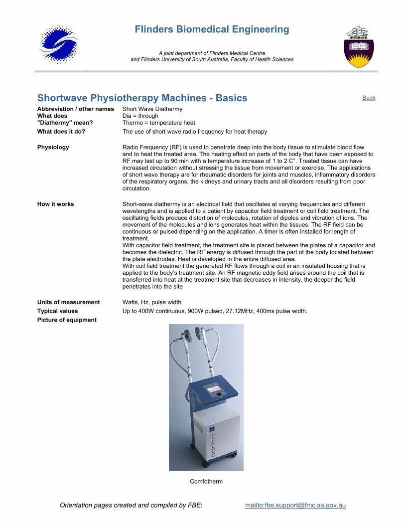
Units of measurement Watts, Hz, pulse width Typical values Up to 400W continuous, 900W pulsed, 27.12MHz, 400ms pulse width. Picture of equipment
Comfotherm
Back
Orientation pages created and compiled by FBE: mailto:[email protected]

Flinders Biomedical Engineering
A joint department of Flinders Medical Centre and Flinders University of South Australia, Faculty of Health Sciences
Treadmills - Basics What does it do? A treadmill is a motor driven moving platform able to adjusted in speed and also
elevation. Physiology Treadmills are used for stress testing where the patient is exercised beyond their normal limit
and the cardio vascular system is closely monitored. The aim of this test is to reveal any abnormalities in the heart’s function under stress.
Treadmills are also used in Physiotherapy as a form of exercise for rehabilitation after injury or surgery.
How it works The Treadmill consists of a wide belt, which can be driven at various speeds by means of an electric motor.
The inclination (angle) of the platform can also be adjusted by motor driven feet at the control end so that the patient is walking up hill.
The control panel allows for adjustments in speed, elevation, duration and calculates time distance walked energy used. It may also have a heart rate monitor fitted.
If the treadmill is used for stress testing additional monitoring, such as an ECG machine, is utilised, maximising patient safety and data collection.
Typical values Speed = 1.5 to 16 km/h Elevation = 0 to 10%
Picture of equipment
Treadmill
Back
Orientation pages created and compiled by FBE: mailto:[email protected]

Flinders Biomedical Engineering
A joint department of Flinders Medical Centre and Flinders University of South Australia, Faculty of Health Sciences
Ultrasound - Basics What does "Ultrasound" mean?
Ultra = High or extreme Ultrasound = sound waves above the frequency detectable by human ears
Physiology Ultrasound or ultrasonography is a medical imaging technique that uses high frequency sound waves and their echoes. It is used to form an image of organs or tissue inside the body non-invasively. The technique is similar to the echolocation used by bats, whales and dolphins.
Ultrasound is also used as a physiotherapy treatment where the sound waves are used to warm muscle tissue deep inside the body.
How it works The ultrasound machine transmits high-frequency (1 to 10 MHz) sound pulses into the body using a probe. The probe has special crystals that generate the sound waves and also detect sound waves reflected.
When the sound waves hit a boundary between tissues (e.g. between fluid and soft tissue, soft tissue and bone) some of the sound waves are reflected back to the probe and the rest travel on further until they reach another boundary and are reflected back.
The reflected waves are detected by the probe and relayed to the machine.
The machine calculates the distance from the probe to the tissue or organ (boundaries) using the speed of sound in tissue (1,540 m/s) and the time of each echo's return (usually in the order of millionths of a second).
The machine displays the distances and intensities of the echoes on the screen and forms a two dimensional image like the one shown below.
Picture of equipment
Ultrasound Image
Ultrasound
Back
Orientation pages created and compiled by FBE: mailto:[email protected]
Flinders Biomedical Engineering
A joint department of Flinders Medical Centre and Flinders University of South Australia, Faculty of Health Sciences
Ventilator - Basics What does "Ventilator" mean?
Vent = an opening allowing gas to pass into or out of. Ventilator = a device that assists the passing of gas into and out of the lungs
Physiology All living cells in the body use oxygen (O2) and produce carbon dioxide (CO2). Oxygen is delivered to the cells and carbon dioxide is transferred away from the cells via the circulating blood. The oxygen consumed by the cells needs to be replaced and the carbon dioxide needs to be removed from the blood. This occurs in the lungs: oxygen in the air inhaled into the lugs is transferred into the blood and carbon dioxide is transferred from the blood into the lungs and is expired. Inspired air contains 79% Nitrogen, 20.96% Oxygen, 0.04% Carbon Dioxide. Expired air contains 79% Nitrogen, 17% Oxygen, 4% Carbon Dioxide.
How it works A ventilator mixes oxygen and air to required levels and then delivers it to the patient via special tubing called a “breathing circuit”. The gas from the ventilator is humidified and heated in the breathing circuit before it goes to the patient. The ventilator increases the pressure in the breathing circuit so that the air is pushed into the lungs for inspiration. The ventilator reduces the pressure so that the air in the lungs can be expired and the expired air is vented to atmosphere. Numerous ventilator parameters are adjustable and are determined by the doctor prior to attachment and during operation on the patient, some of these include:
• Mode of ventilation - CMV (controlled mandatory ventilation), SIMV (synchronised intermittent mandatory ventilation), CPAP (constant positive airway pressure).
• Tidal Volume – the volume of air inspired with each breath • Respiratory rate – how many breaths per minute (BPM) • O2 concentration • Flow rate • Airway pressure • Minute volume – the volume inspired and expired in one minute
All of the above are monitored and have various alarm adjustable limits Units of measurement Pressure: cmH20, mbar. Volume: millilitres. Oxygen: %. Flow: litre/min. Typical values Adults: tidal volume, 600 - 1200ml, flow 30-50 litre/min, resp rate 12 BPM
Neonates: tidal volume = 100-200ml, flow = 6-10 L/min resp rate = 60 BPM Picture of equipment
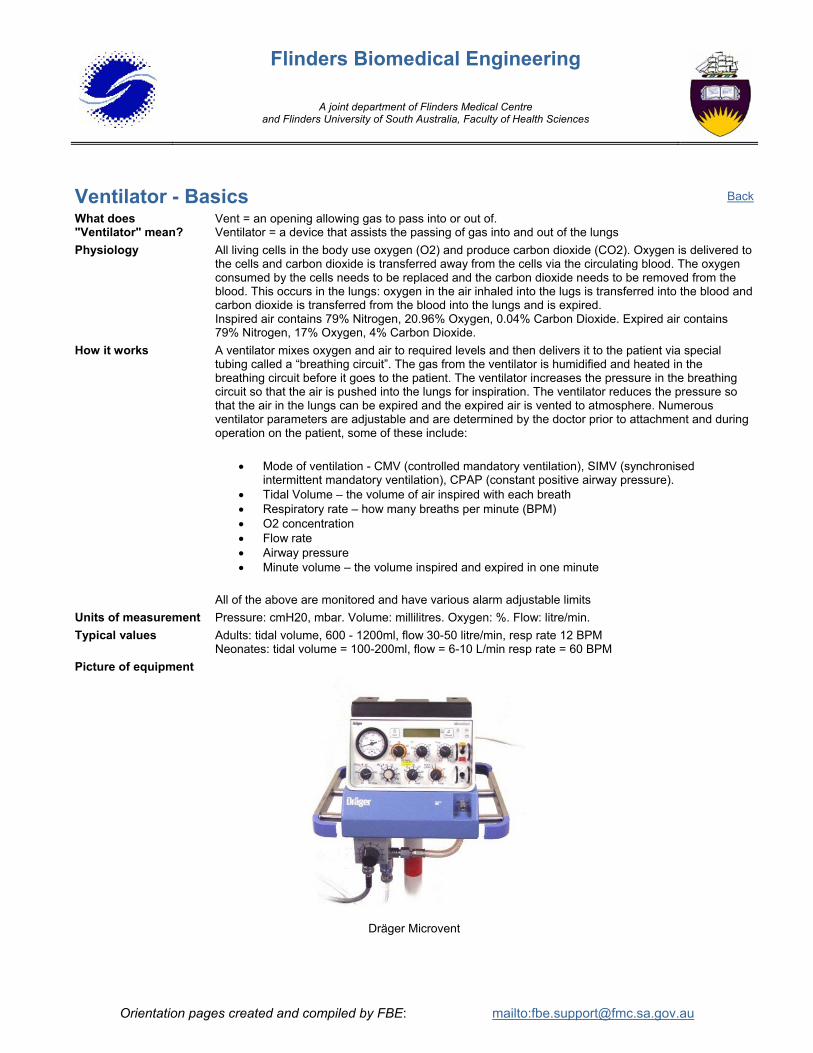
Dräger Microvent
Back
Orientation pages created and compiled by FBE: mailto:[email protected]

Flinders Biomedical Engineering
A joint department of Flinders Medical Centre and Flinders University of South Australia, Faculty of Health Sciences
Orientation pages created and compiled by FBE: mailto:[email protected]
X-Ray Machines - Basics What does "X-ray" mean?
Mobile X-Ray or Fixed X-Ray (General Screening, Angiography, Fluoroscopy, Mammography and CT Scanners)
In an X-Ray tube an electron gun fires high energy electrons at a target. This produces photons of electromagnetic interference called X-Rays
What does it do? X-Rays are directed through the body onto a film resulting in a ‘picture’ (called an X-Ray) of the internal parts of that part of the body
Physiology The Human body has different densities of tissue, Hard (eg Bones and tendons) and Soft (eg Skin, Muscle and Internal Organs). The X-Ray is used to form an image of the dense tissue in the body.
How it works The X-Ray tube is placed on one side of the patient and a film is placed on the other side. As the X-Rays pass through the body they are absorbed more by the hard tissue than the soft tissue. The X-Rays that are not absorbed travel through the body and hit the X-Ray film. The X-Ray film is exposed less in the areas where the X-Rays have been absorbed by the hard tissue and when the film is developed these areas look lighter. This gives a very good picture of the Bones and Tendons in that part of the body.
Units of measurement X-Rays are measured photographically. There are two common sets of units, Rads and Sieverts (typically milliSieverts, mS)
X-Ray Machine Outputs are measured with test equipment in kilovolts (kV) and milliamp seconds (mAs) Higher values of kV and mAs produce more X-Rays than lower values
Typical values The output required of an X-Ray Machine depends on the thickness of the Body part being X-Rayed. Higher power is needed for thicker parts (eg a Chest X-Ray requires more power than an Arm)
Outputs vary from 70 – 130 kV and 20 – 95 mAs Picture of equipment
Mobile X-Ray Fixed X-Ray
Back