Embed Size (px)
Citation preview
VESICULOBULLOUS DISEASES
OF THE NEWBORN
N Engl J Med 357;13
September 2007

A. Infectious Neonatal Vesiculopustular Dermatoses
1. HSV
Devastating consequences if not detected and treated
Infection in utero transplacental or ascending infection from the maternal genital tract
History of recent HSV infection in the mother, but often no
3 forms: mucocutaneous infection, disseminated infection, and infection of the central nervous system
Begin as solitary, grouped, or diffuse erythematousmacules; within 24 to 48 h vesiculopustules,crusts,erosions
Oral involvement is common
Skin lesions typically appear days after onset of systemic manifestations : lethargy, fever, and poor muscle tone
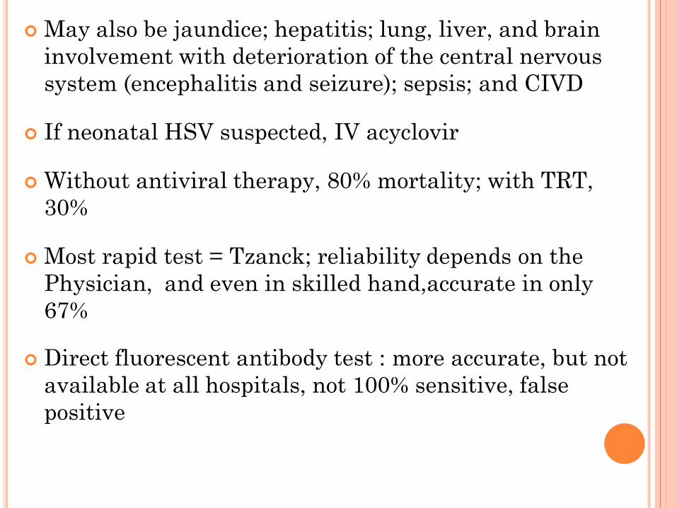
May also be jaundice; hepatitis; lung, liver, and brain
involvement with deterioration of the central nervous
system (encephalitis and seizure); sepsis; and CIVD
If neonatal HSV suspected, IV acyclovir
Without antiviral therapy, 80% mortality; with TRT,
30%
Most rapid test = Tzanck; reliability depends on the
Physician, and even in skilled hand,accurate in only
67%
Direct fluorescent antibody test : more accurate, but not
available at all hospitals, not 100% sensitive, false
positive
Viral culture of skin, conjunctiva, cerebrospinal
fluid, or urine specimens remains the gold
standard
Examination of cerebrospinal fluid samples may
show pleocytosis, elevated protein levels, and the
presence of red cells
MRI may show encephalitis within 3 days after
birth, whereas findings on CT Scan may not be
abnormal until after 5 days
2. Varicella
Occur when mother has infection in the last 3
weeks of pregnancy
Appears in infant 9 to 15 days after the maternal
rash develops, administration of varicella–zoster
Ig may prolong incubation period to 28 days
Cutaneous manifestations: pink macules that
develop into papules and ―teardrop‖ vesicles,
sparse or numerous, necrotic and hemorrhagic in
severe cases
Systemic findings: pneumonitis, respiratory
distress, hepatitis, and encephalitis
3. Cytomegalovirus
One of the most common infections of the neonate
5 to 10% of infected neonates have symptoms
Vesicles are rare and present only at birth
When vesicles are present at birth,
cytomegalovirus infection must be considered
Viral cultures of blood and urine have the highest
sensitivity when performed within 1 week after
infection
PCR for CMV DNA in plasma : very sensitive
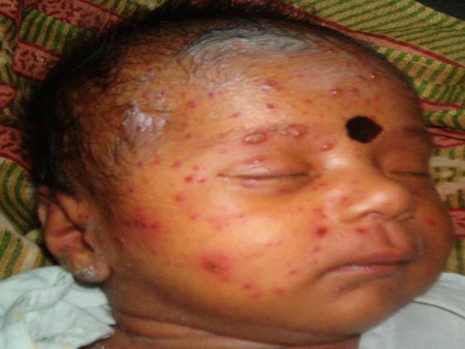
4. Candida
Most common fungal infection of the neonate
Congenital candidiasis usually manifests on the first day of life as result of exposure in utero or during delivery or within 1 week if acquired during delivery
Risk factors: foreign body in the uterus (such as an intrauterine device or a cervical suture), a maternal history of vaginal candidiasis, and premature delivery
Most cases are mild and confined to the skin
Generalized eruption of red macules, pustules, vesicles, and vesiculopustules on the first day of life
Spores and pseudohyphae , KOH preparation of skin scrapings
Repeated blood, cerebrospinal fluid, and urine cultures if
suspected disseminated infection, because initial cultures
often negative
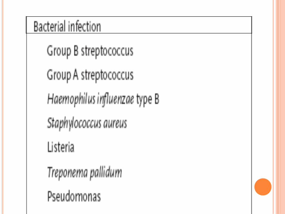
5. Bacterial Infection
Symptoms of bacterial sepsis — including lethargy,
jaundice, purpura, fever, and shock —typical
Group B streptococcus = most common cause of
bacterial sepsis in newborn
Skin : vesicles, bullae, crusts, and erosions, but often
systemic manifestations: bacteremia, meningitis,
pneumonia
Impetigo neonatorum : infection with Staphylococcus
aureus (phage group II, type 55, with 71 subtypes);
large, flaccid bullae and moist, sometimes golden-
crusted erosions from impetiginization
Typically, mild eruptions but may be severe and
life-threatening, with osteomyelitis, pneumonia, and
sepsis.
Bullae usually appear in the second week of life.
Staphylococcal scalded skin syndrome begins with a
scarlatiniform eruption that rapidly progresses to bullae
and desquamation of large portions of skin, resembling
toxic epidermolytic necrosis, with a characteristic golden
crusting around the mouth and nose.
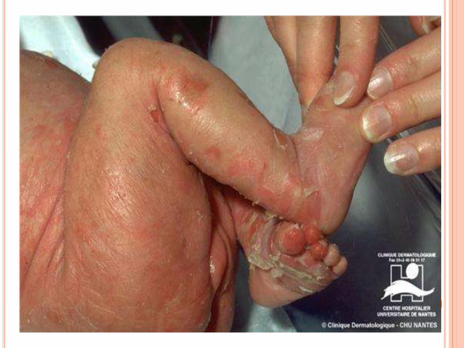
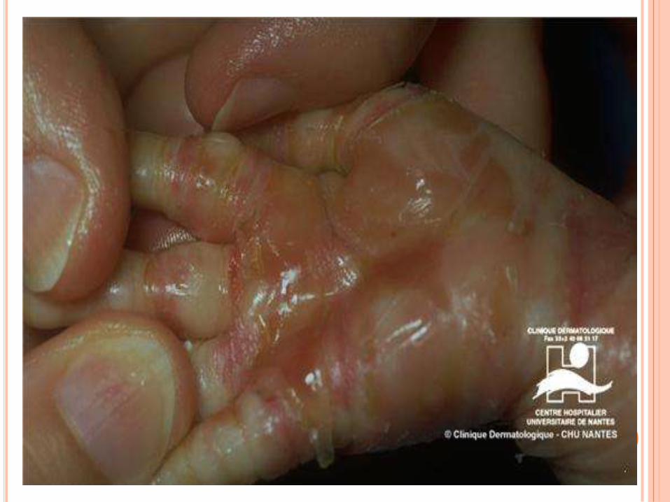
Congenital Syphilis
• Infected mothers, transplacental transmission
• 40% of infected newborns have skin findings at
delivery: papulosquamous eruptions, condylomata
lata, and desquamation; more rarely, vesicles and
bullae specific to the newborn
• Hemorrhagic bullae on palms and soles
• Serologic tests in the mother during pregnancy and
in the neonate, as well as a VDRL test of a
cerebrospinal fluid specimen
Imperative to treat with AB for bacterial
and viral infections until conclusively ruled
out
Ideally, cultures performed before the broad
AB coverage with IV ampicillin, gentamicin,
and acyclovir
Infants who have vesiculopustular rashes
and who appear ill should be tested for
Candida, viral, and bacterial infections
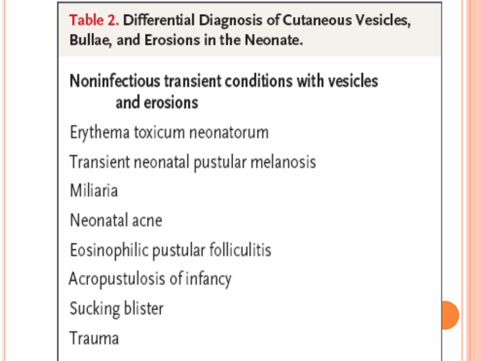
B. Noninfectious Conditions in the Neonate
with Vesicles and Erosions
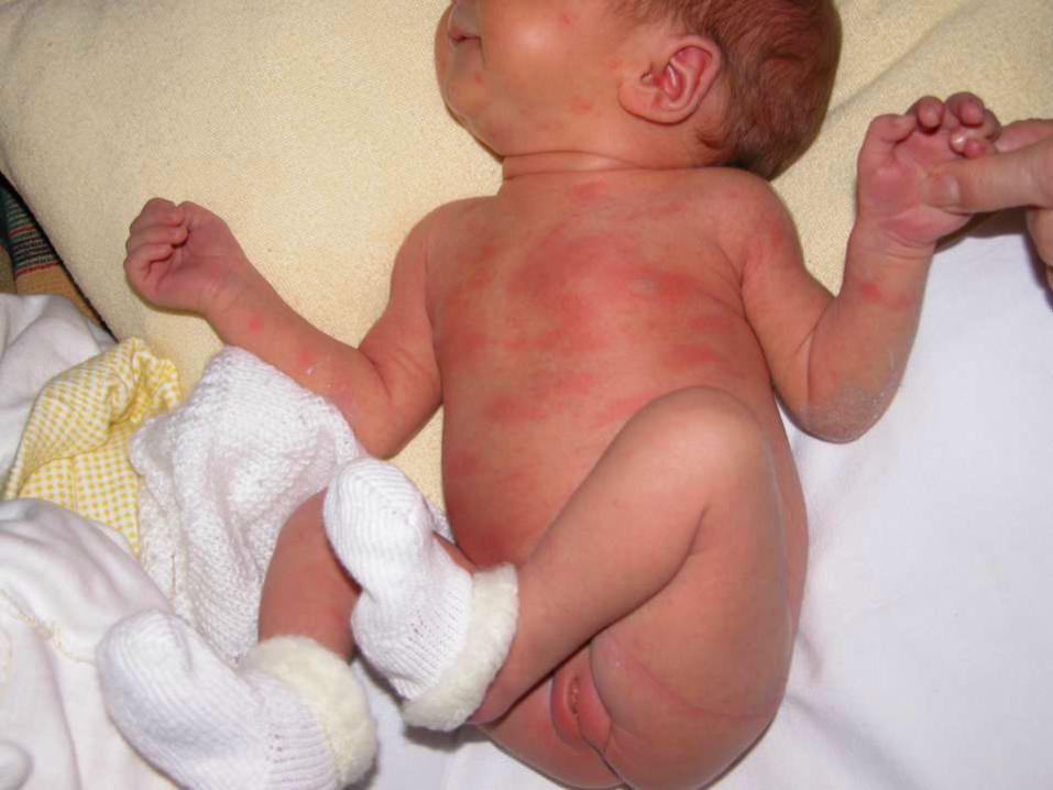
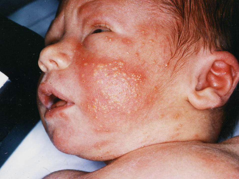
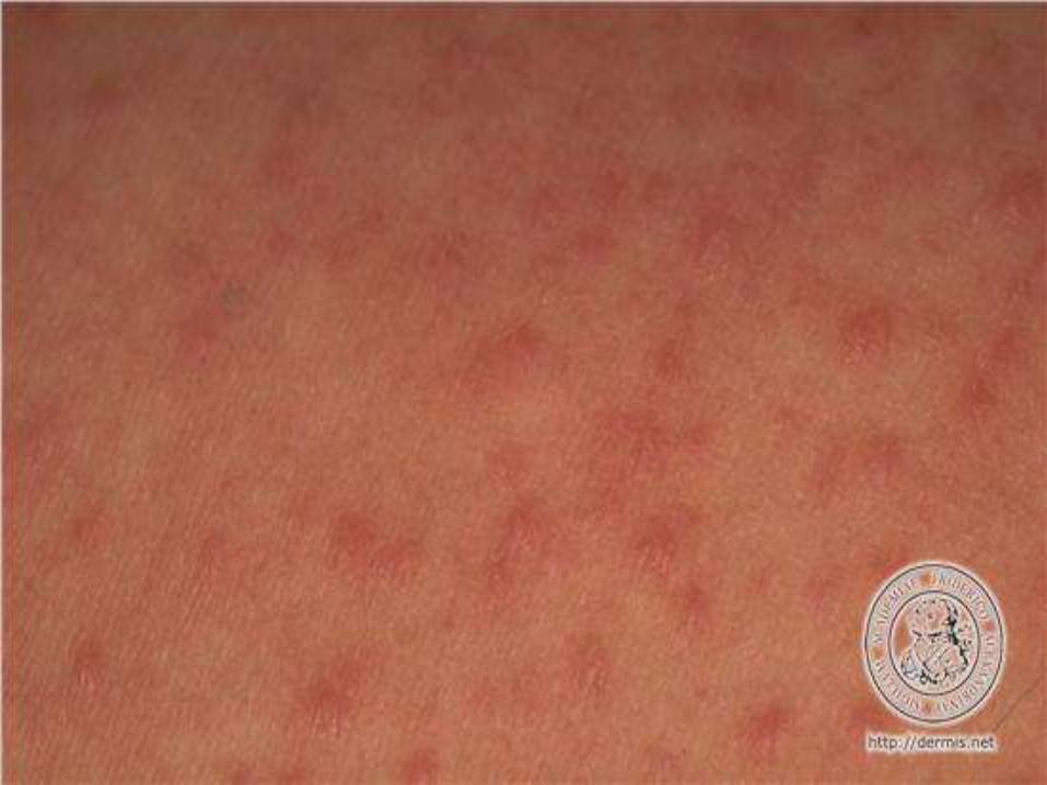
1. Erythema toxicum neonatorum:
• Common in healthy infants
• First week of life
• Erythematous macules, wheals, papules, and
pustules that wax and wane, with new lesions
persisting for several days
• Begins on the face and migrates to the trunk
• No involvement of palms or soles
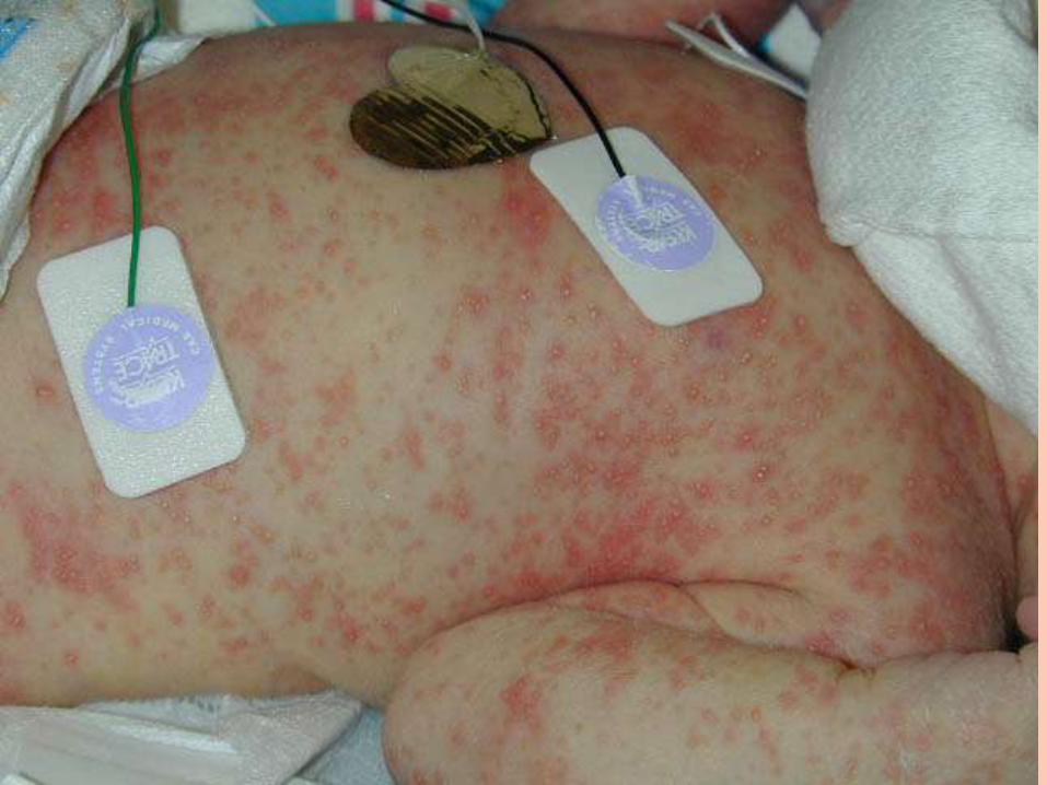
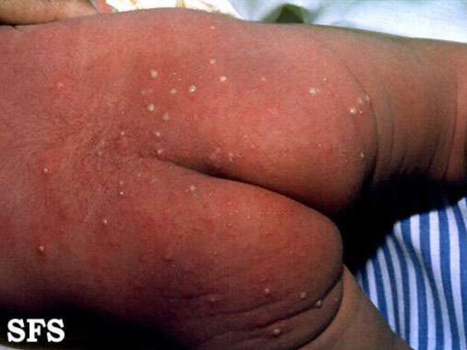
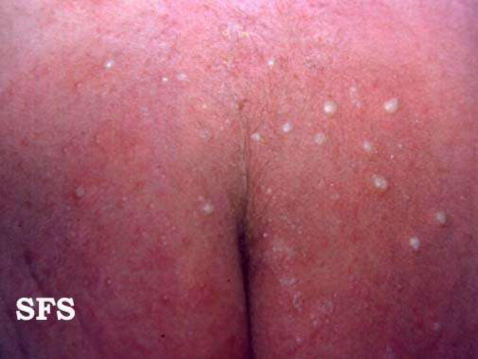
2. Transient neonatal pustular melanosis :
• Superficial pustules with no erythema
• Rupture easily, scaling
• Hyperpigmented macules with or without scale
• Anywhere on the skin, including the palms and
soles
• Resolution within 3 to 4 weeks
• No treatment indicated

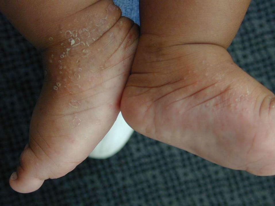
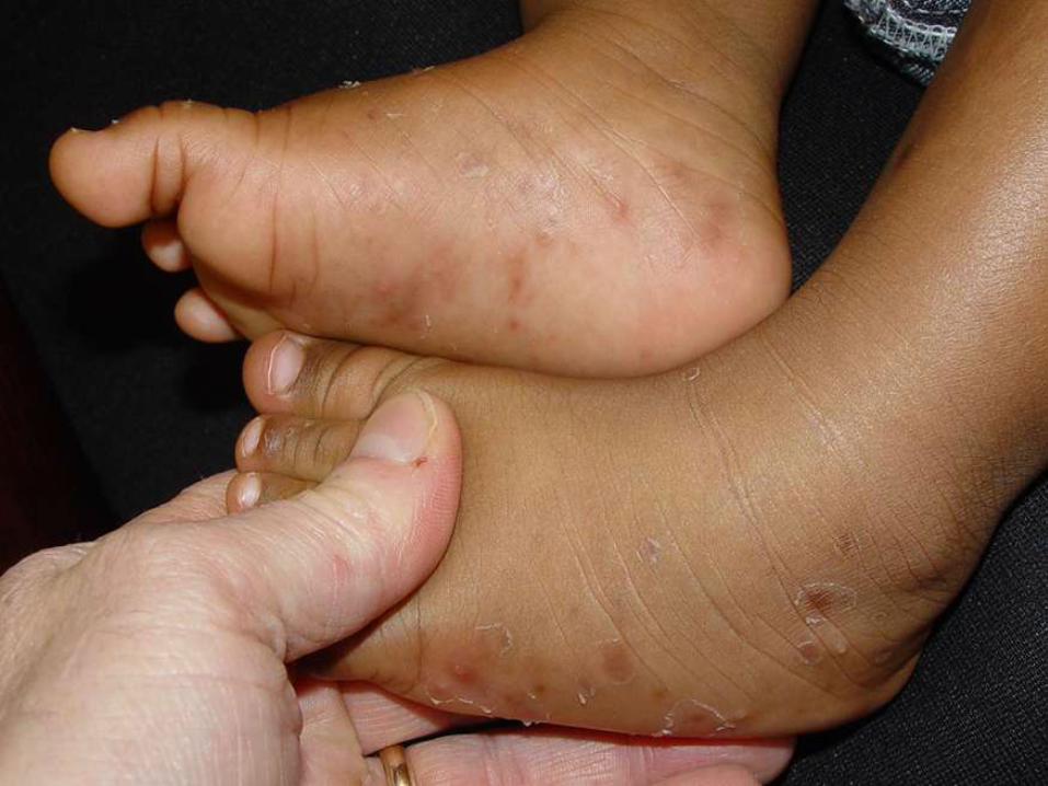
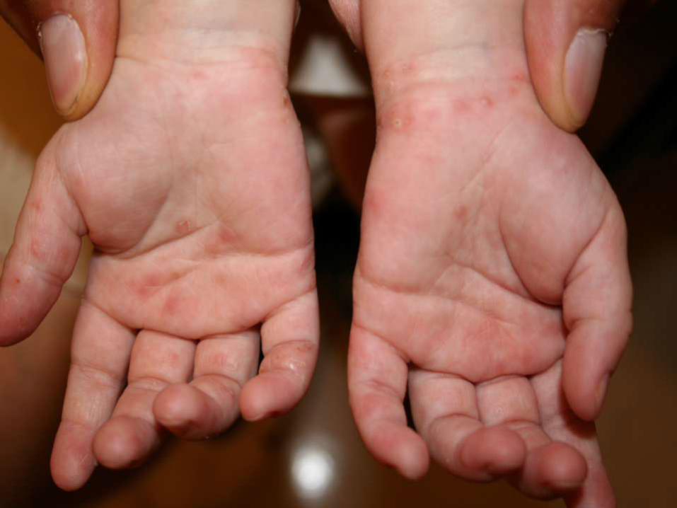
3. Acropustulosis of infancy :
• Extremely pruritic
• Vesiculopustular eruption on the hands and feet
present at birth or within the first few weeks of life
• Appears every 2 to 4 weeks, persisting for 5 to 10
days during each episode
• Usually resolves in 1 to 2 years and is responsive to
topical CS
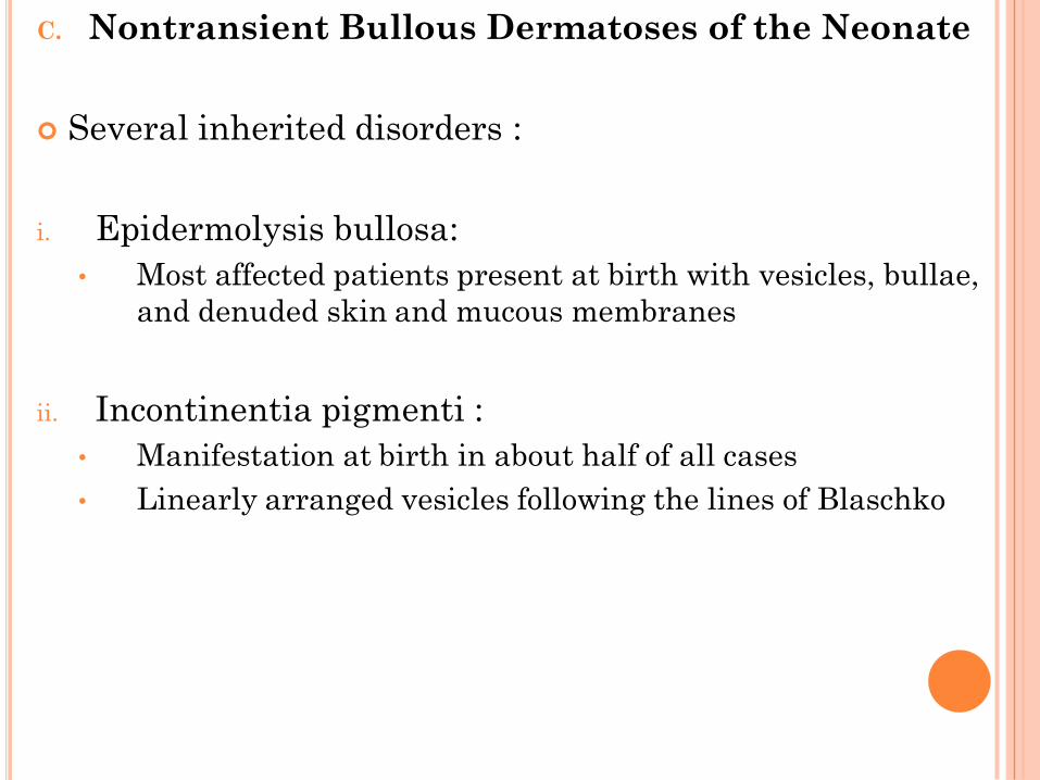
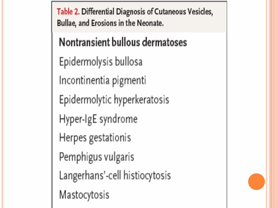
C. Nontransient Bullous Dermatoses of the Neonate
Several inherited disorders :
i. Epidermolysis bullosa:
• Most affected patients present at birth with vesicles, bullae,
and denuded skin and mucous membranes
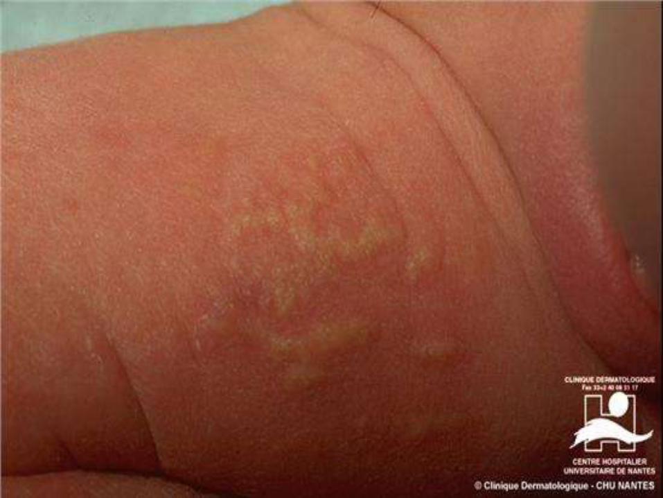
ii. Incontinentia pigmenti :
• Manifestation at birth in about half of all cases
• Linearly arranged vesicles following the lines of Blaschko
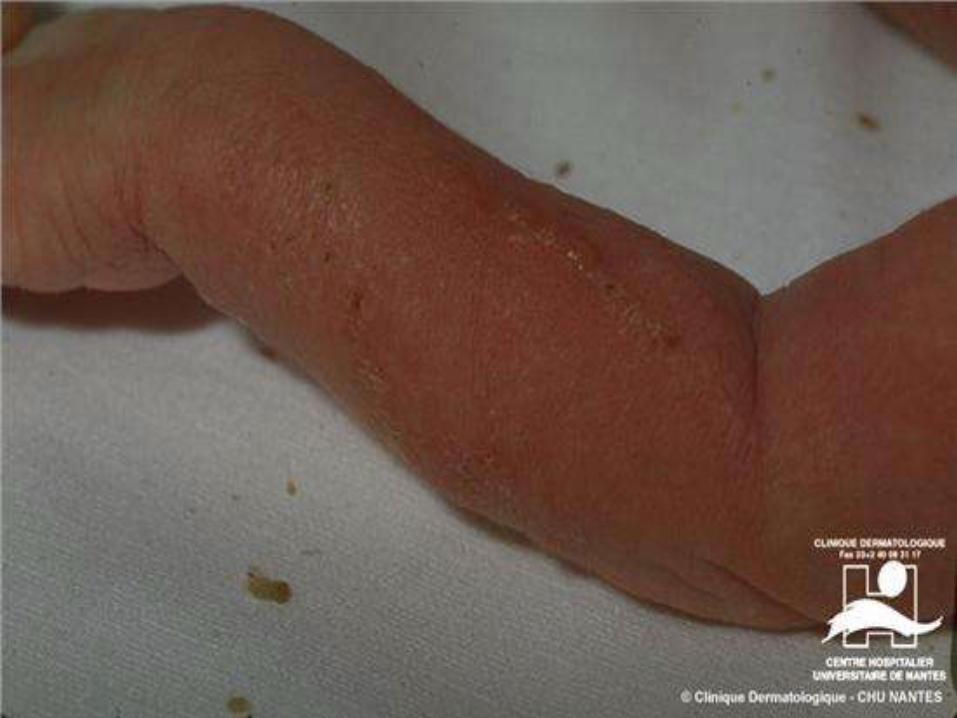
iii. Epidermolytic hyperkeratosis :
• Very rare, AD disorder
• Defects in the genes encoding keratins 1 and 10
• Widespread bullae, erythroderma, and
desquamation at birth
iv. The hyper-IgE syndrome (Job’s syndrome):
• Solitary or numerous vesicles tense, with
surrounding erythema
• Head and shoulders

Two autoimmune diseases of the mother can affect
neonates:
1. Herpes gestationis
2. Pemphigus vulgaris: maternal autoantibodies can
pass transplacentally to the newborn
Langerhans’-Cell Histiocytosis
• Congenital form = clonal proliferative disorder of
Langerhans’ cells (= APC derived from bone marrow)
• Single organ or multiorgan-system involvement at birth
• Hashimoto–Pritzker’s disease, or congenital self-healing
reticulohistiocytosis = one end of the spectrum of single-
organ–system LH, high likelihood of complete, sp.
resolution
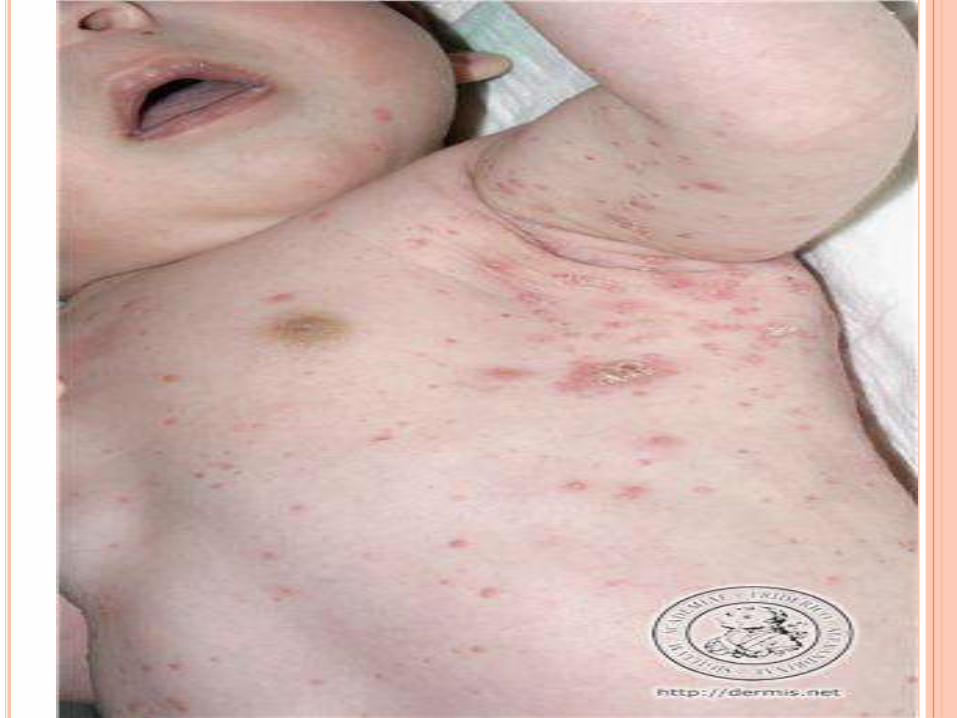
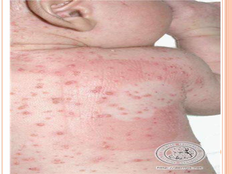
• Skin lesions : solitary or multiple vesicles, bullae, erosions,
papules, nodules, crusts, petechiae, milia, and atrophy.
Erosions have more poorly defined borders than those of
neonatal herpes
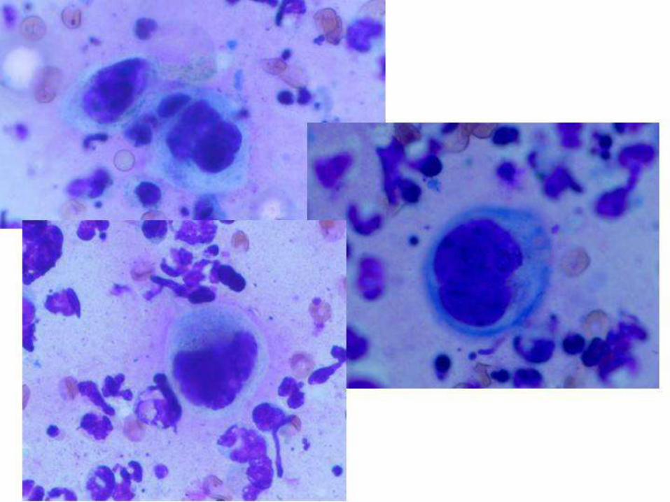
• Tzanck preparation: characteristic reniform nuclei and
abundant cytoplasm
• Skin biopsy for a definitive diagnosis

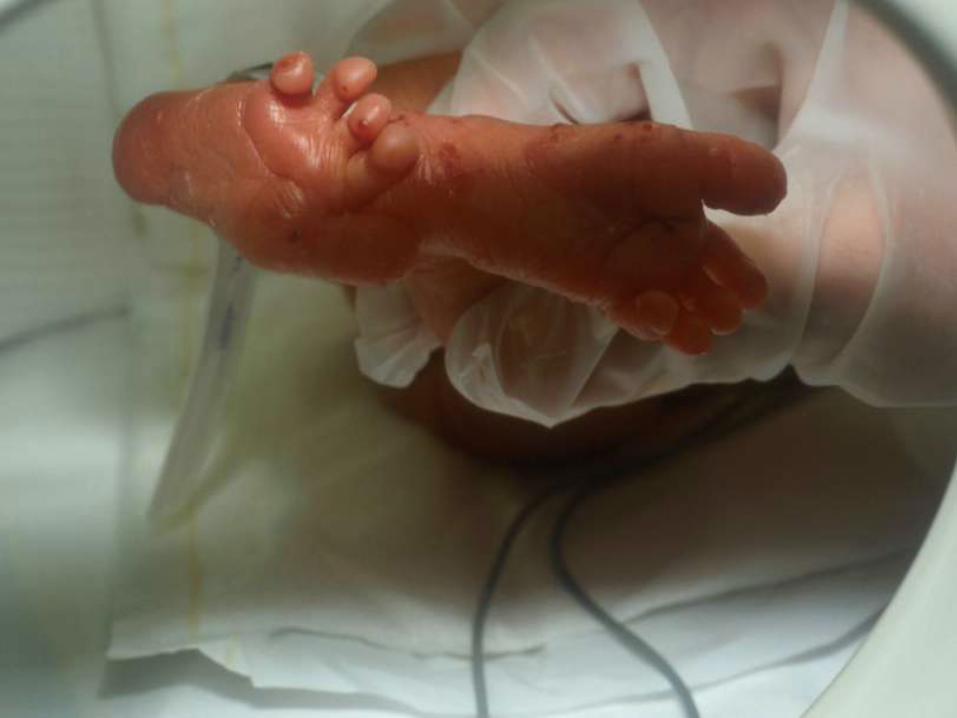
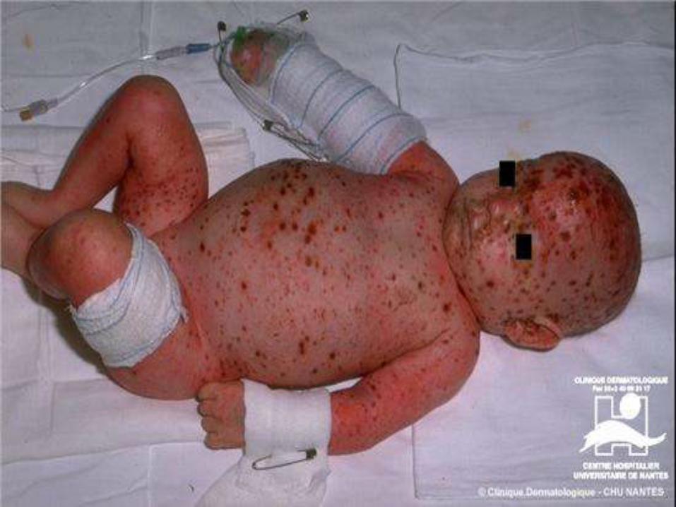
CLINICAL CASE
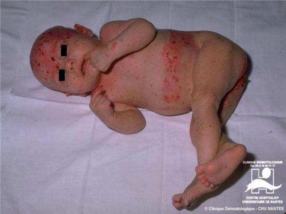
Newborn girl
Weight = 3105 g, spontaneous vaginal delivery at 40 weeks’ gestation
Healthy 27-year old mother after an uncomplicated pregnancy
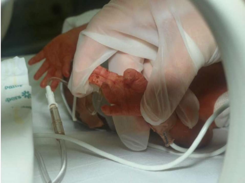
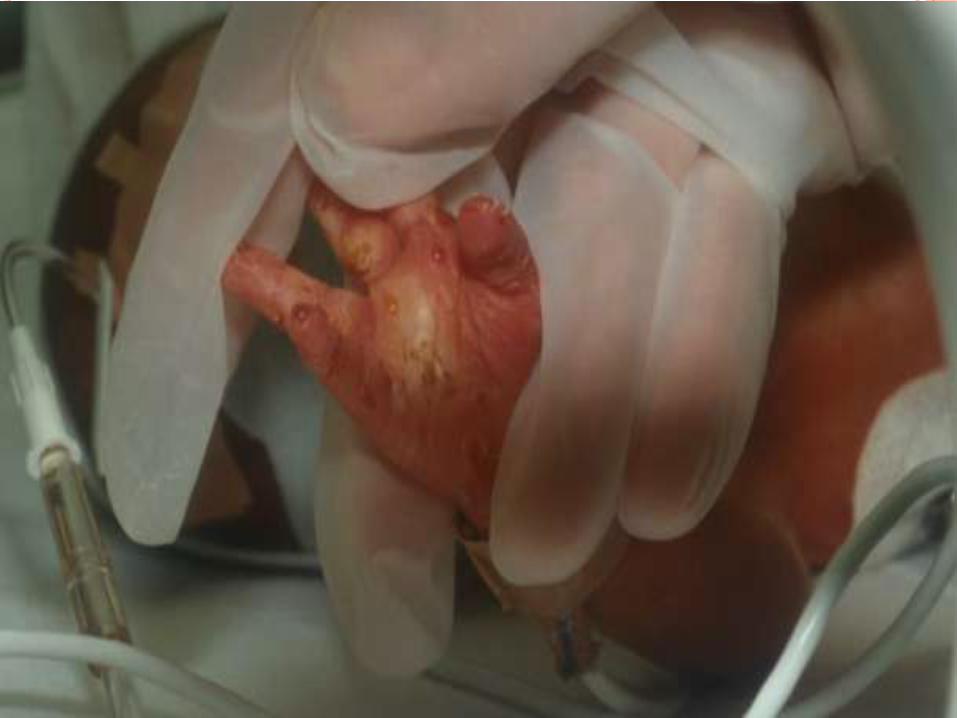
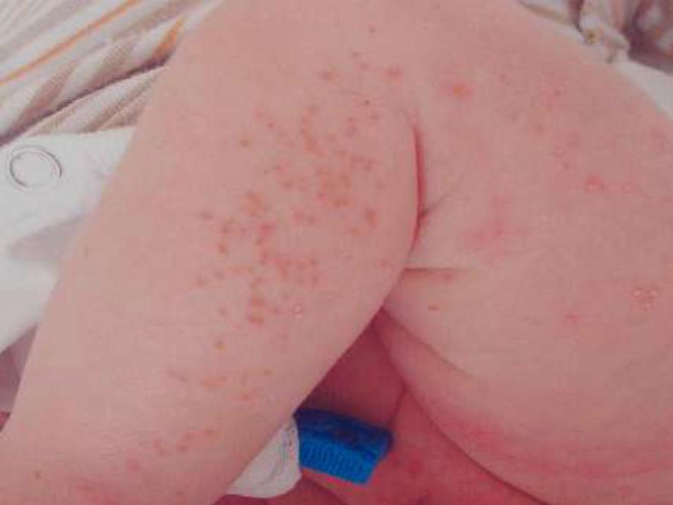
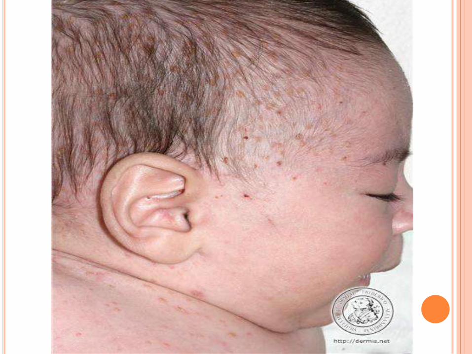
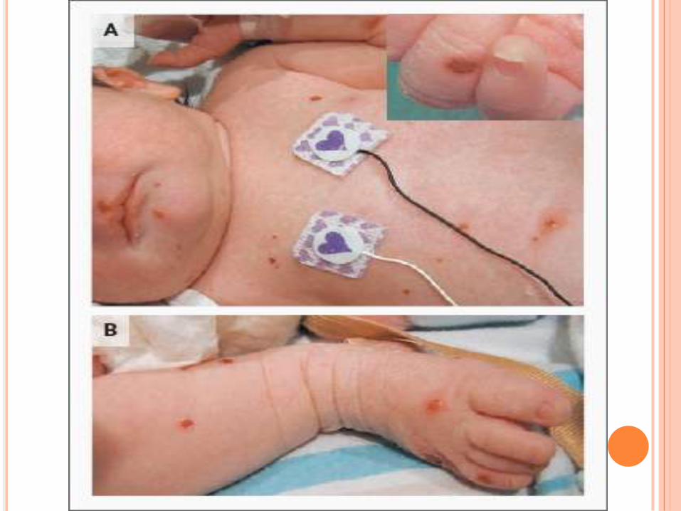
Numerous pustules on the face, trunk, arms, and legs
Mother: no exposures to infectious diseases, genital infections, or rashes during the pregnancy ;
Tests for antibodies to rubella and HSV were positive
Tests for antibodies to syphilis and hepatitis B were negative
Rectovaginal culture : positive for group B streptococcus
Mother had varicella infection in childhood
No medications and no illicit drugs, alcohol, or tobacco
Rupture of membranes 2 hours before vaginal delivery, and AB administered IV during delivery
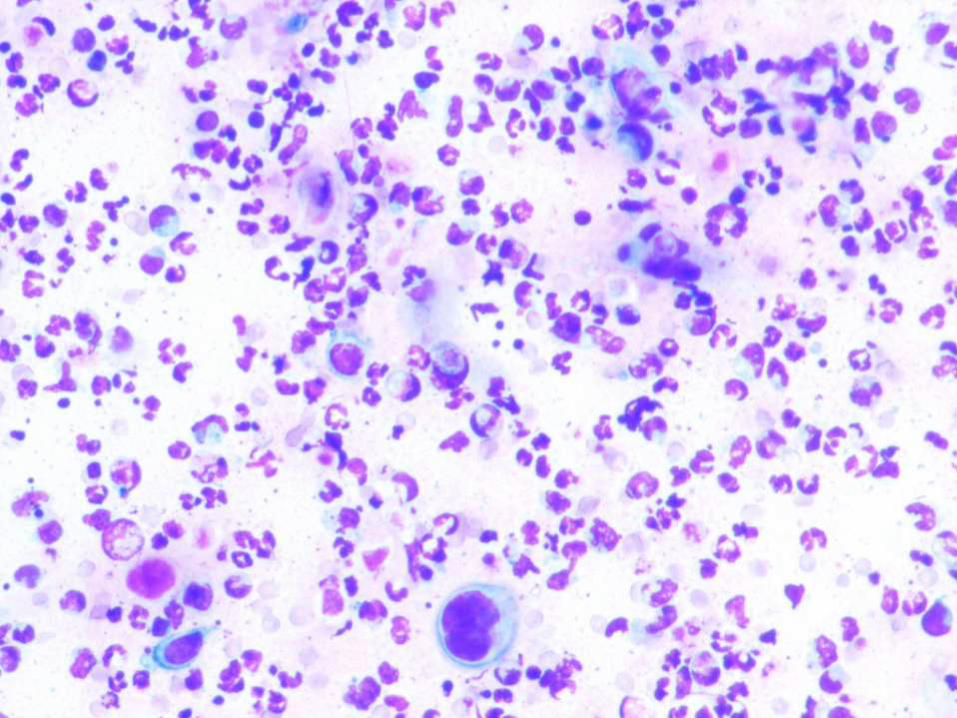
Tzanck smear : positive for multinucleated giant cells
Lumbar puncture attempted three times but unsuccessful
IV ampicillin, gentamicin, and acyclovir were administered
On examination: temperature = 36.8°C, pulse = 135 beats per minute, blood pressure = 74/58 mm Hg, SaO2 = 99%; 56 breaths per minute
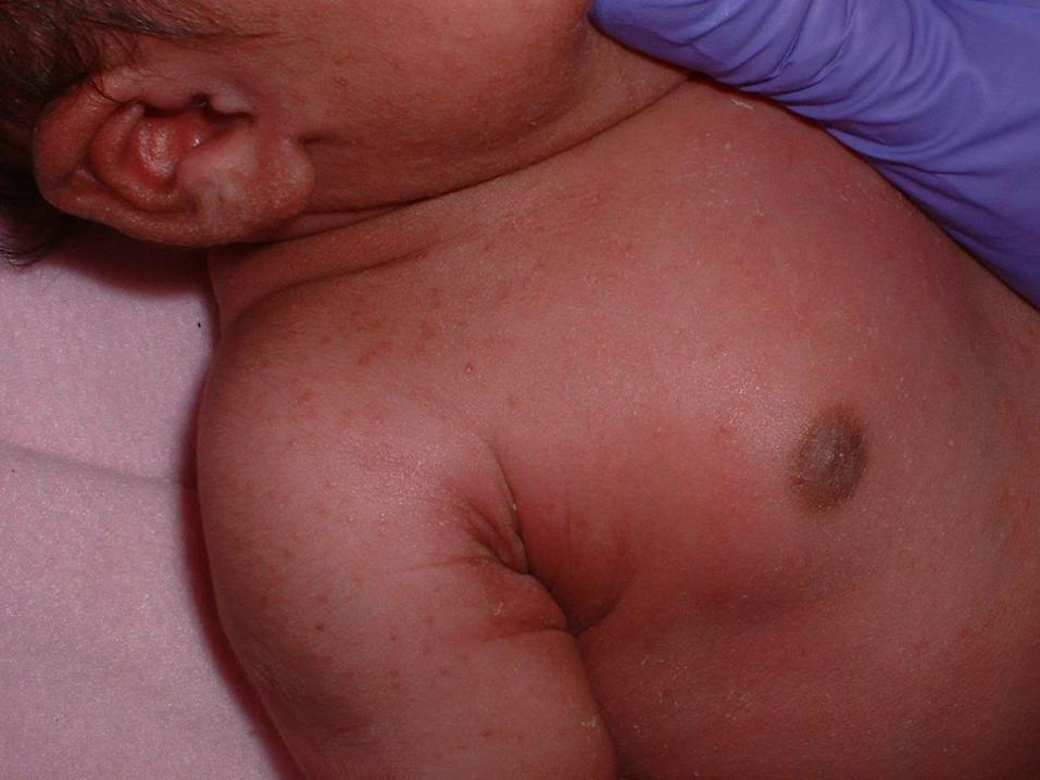
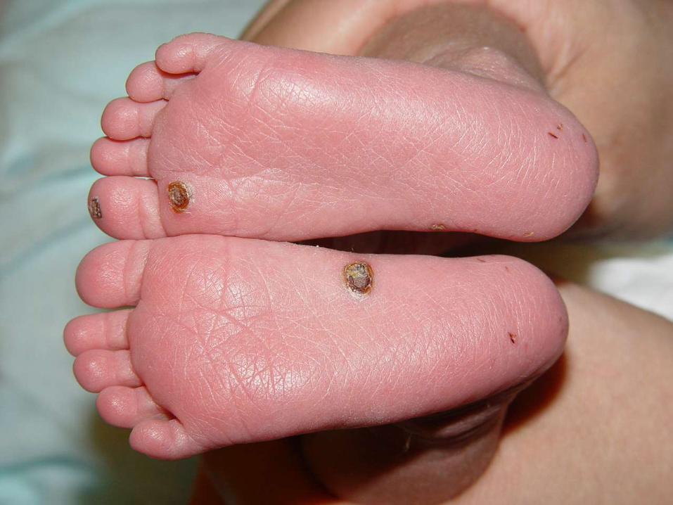
50 cutaneous erosions and papules, 3 to 7 mm in diameter with serosanguineous crust, surrounding pink erythema; over the scalp, face, trunk, arms, and legs
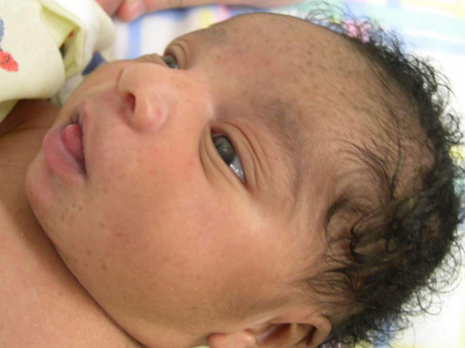
No involvement of groin area or oral, conjunctival, or vaginal mucosae
No dysmorphic features
Mucous membranes were pink and moist, palate intact
Neck was supple
Auscultation of the heart and lungs : nl
No enlarged lymph nodes in the cervical, axillary, or inguinal regions
Hepatomegaly?
Genitalia normal
Neurologic examination normal
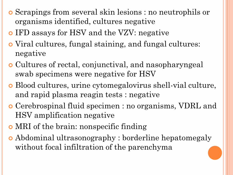
Scrapings from several skin lesions : no neutrophils or
organisms identified, cultures negative
IFD assays for HSV and the VZV: negative
Viral cultures, fungal staining, and fungal cultures:
negative
Cultures of rectal, conjunctival, and nasopharyngeal
swab specimens were negative for HSV
Blood cultures, urine cytomegalovirus shell-vial culture,
and rapid plasma reagin tests : negative
Cerebrospinal fluid specimen : no organisms, VDRL and
HSV amplification negative
MRI of the brain: nonspecific finding
Abdominal ultrasonography : borderline hepatomegaly
without focal infiltration of the parenchyma
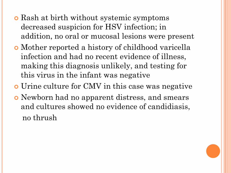
Rash at birth without systemic symptoms
decreased suspicion for HSV infection; in
addition, no oral or mucosal lesions were present
Mother reported a history of childhood varicella
infection and had no recent evidence of illness,
making this diagnosis unlikely, and testing for
this virus in the infant was negative
Urine culture for CMV in this case was negative
Newborn had no apparent distress, and smears
and cultures showed no evidence of candidiasis,
no thrush
Well-appearing infant → bacterial sepsis
unlikely, and appropriate bacterial cultures of
specimens from the lesion, blood, urine, and
cerebrospinal fluid were negative
Although likelihood of a serious bacterial
infection or sepsis was low in this infant, it was
imperative to treat her with AB for bacterial and
viral infections until they were conclusively ruled
out. Then investigation for noninfectious causes
of vesiculopustular eruptions
None of the benign transient dermatoses
resembled the presentation of this newborn
Epidermolysis bullosa, Incontinentia pigmenti,
Epidermolytic hyperkeratosis, hyper-IgE
syndrome: none of these disorders appeared
likely in this patient with no family history
Herpes gestationis, pemphigus vulgaris: the
mother had no history of either of these disorders
Biopsy of a lesion on the flank
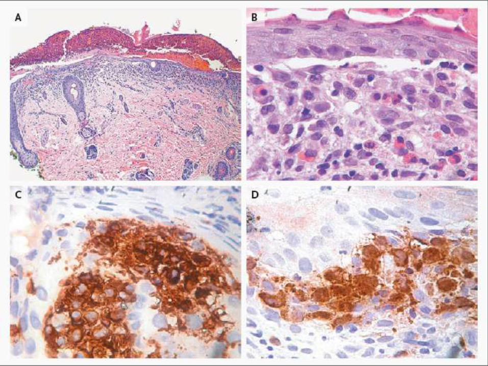
Epidermal ulceration and crust formation
Infiltrate of medium-sized to large mononuclear cells with
indented or grooved nuclei, typical of Langerhans’ cells
Clusters of cells present in papillae and scattered in the
upper dermis
Eosinophils also present
Immunohistochemical analysis : (+) for CD1a and S-100
Histologic differential diagnosis : dendritic-cell sarcoma
(indeterminate-cell histiocytosis), xanthogranuloma,
xanthoma disseminatum, benign cephalic histiocytosis,
and occasionally mycosis fungoides (in cases of MF with
numerous eo and histiocytes
Characteristic morphologic features of Langerhans’ cells
and, most important, positive staining for CD1
The diagnosis:
Cutaneous Langerhans’-cell histiocytosis
No histologic or immunophenotypic features
permit distinction between the two clinical forms
of Langerhans’-cell histiocytosis involving the
skin
Differential diagnosis in this case :
single-organ–system cutaneous Langerhans’-cell
histiocytosis, also known as cutaneous self-
healing Langerhans’-cell histiocytosis, and
multisystem Langerhans’-cell histiocytosis, also
known as Letterer–Siwe disease
Prognosis and treatment vary markedly depending on
involvement of other organs
Prognosis of truly isolated cutaneous single-organ–
system Langerhans’-cell histiocytosis is excellent, and
observation without treatment is justified
However, multiorgan-system Langerhans’-cell
histiocytosis, particularly in neonates, with involvement
of the liver, spleen, lung, the hematopoietic system, or a
combination of these is a potentially fatal disease
In addition, congenital cutaneous single-organ–system
Langerhans’- cell histiocytosis not infrequently evolves
into multiorgan-system Langerhans’-cell histiocytosis.
5-year survival rate of neonates with single-
organ–system Langerhans’- cell histiocytosis
(most of whom did not receive treatment) was
94%, whereas in multiorgan-system Langerhans’-
cell histiocytosis(all of whom were treated) 57%
Evaluation for the presence of disease in
locations other than the skin.
A skeletal survey, CT and MRI of the abdomen
confirmed that the size of the liver was at the
upper limit of normal and that there was no
splenomegaly or intrasplenic lesions
All skin lesions resolved by 1 month of age
Median time to regression in retrospective studies of
only small numbers of patients is about 4 months.
However, relapse with subsequent dissemination to
multiorgan-system Langerhans’- cell histiocytosis has
been described
Isolated lesions of the pituitary, including diabetes
insipidus — a classic manifestation of Langerhans’-cell
histiocytosis — and other neurodegenerative lesions
detected in such patients many years after the initial
diagnosis
Followed closely during the first year of life and will
continue long-term follow- up at longer intervals.
At 1 year of age, no evidence of either cutaneous or
systemic disease