Embed Size (px)
Citation preview
1
NEONATAL SURGICAL EMERGENCIES
Liz Graf-Brennen, RNC, CNSSwedish Medical Center
Esophageal Atresia & Tracheo-Esophageal Fistula
� Esophagus dead-ends in a blind pouch.� TE fistula is an abnormal passage between
the esophagus and trachea.� Occur together in 85% of cases.� Results from failure of trachea and
esophagus to divide at 34-36 days of gestation.
� Not always obvious on prenatal US
Esophageal Atresia & Tracheo-Esophageal Fistula
Esophageal Atresia & Tracheo-Esophageal Fistula
� Incidence: 1 in 4500� Frequently, history of polyhydramnios
� Identification� Cannot swallow saliva with atresia� Choking or cyanosis with feeding� Gastric Tube will not pass to stomach� May develop gastric distension if there is a
fistula, as air cannot get out of stomach
Air in stomach indicates TE fistula. No air means isolated esophageal atresia.
Tube meets resistance at T2-3. Do NOT force!
Esophageal Atresia & TEF:Management� Elevate head of bed 30°- prone may
help avoid aspiration� NPO� IV fluids� Feeding tube or Replogle to low suction
in pouch� Assess for associated anomalies (30-
70%)
2
VACTERR (or VACTERL)
V: VertebralA: Anorectal
C: CardiacT: TrachealE: Esophageal
R: RenalR: Radial (or L: Limb)
TEF/Esophageal Atresia: Repair
� Primary repair often possible� Repair is delayed if there are coexisting
problems or baby is too small.� Staged repair when the gap between distal
and proximal esophagus too large. Gastrostomy placed and repair done in 6-8 weeks.
� Prognosis excellent if no other anomalies. Over 90% survival.
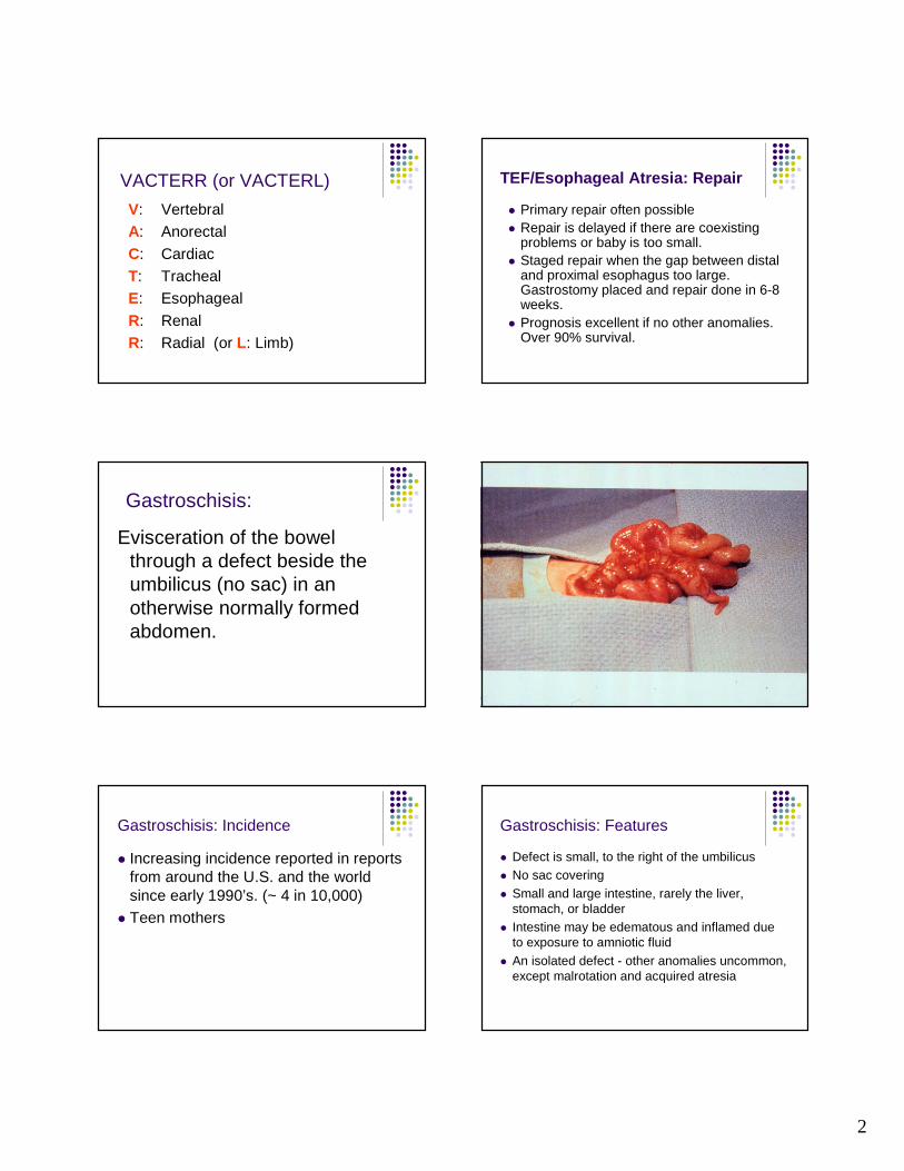
Gastroschisis:
Evisceration of the bowel through a defect beside the umbilicus (no sac) in an otherwise normally formed abdomen.
Gastroschisis: Incidence
� Increasing incidence reported in reports from around the U.S. and the world since early 1990’s. (~ 4 in 10,000)
� Teen mothers
Gastroschisis: Features
� Defect is small, to the right of the umbilicus� No sac covering� Small and large intestine, rarely the liver,
stomach, or bladder� Intestine may be edematous and inflamed due
to exposure to amniotic fluid� An isolated defect - other anomalies uncommon,
except malrotation and acquired atresia
3
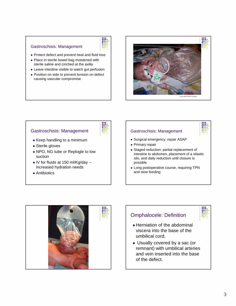
Gastroschisis: Management
� Protect defect and prevent heat and fluid loss� Place in sterile bowel bag moistened with
sterile saline and cinched at the axilla� Leave intestine visible to watch gut perfusion� Position on side to prevent tension on defect
causing vascular compromise
Photo with permission of parents
Gastroschisis: Management
� Keep handling to a minimum� Sterile gloves� NPO, NG tube or Replogle to low
suction� IV for fluids at 150 ml/Kg/day --
increased hydration needs
� Antibiotics
Gastroschisis: Management
� Surgical emergency: repair ASAP� Primary repair� Staged reduction: partial replacement of
intestine to abdomen, placement of a silastic silo, and daily reduction until closure is possible
� Long postoperative course, requiring TPN and slow feeding
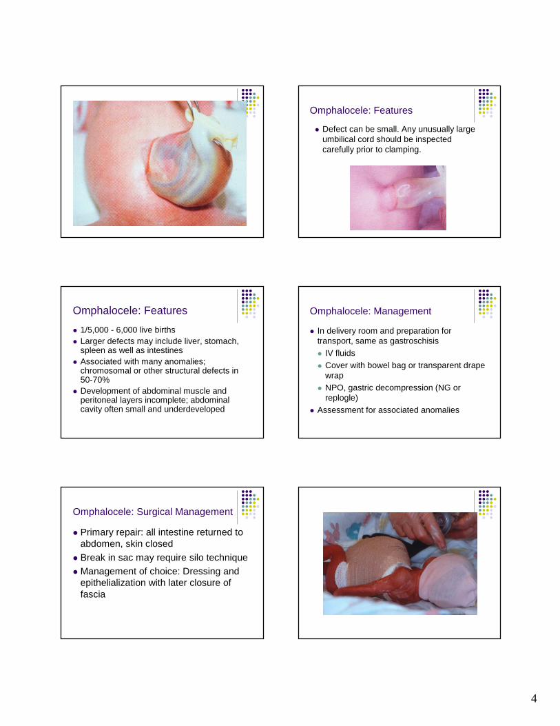
Omphalocele: Definition
� Herniation of the abdominal viscera into the base of the umbilical cord.
� Usually covered by a sac (or remnant) with umbilical arteries and vein inserted into the base of the defect.
4
Omphalocele: Features
� Defect can be small. Any unusually large umbilical cord should be inspected carefully prior to clamping.
Omphalocele: Features
� 1/5,000 - 6,000 live births� Larger defects may include liver, stomach,
spleen as well as intestines� Associated with many anomalies;
chromosomal or other structural defects in 50-70%
� Development of abdominal muscle and peritoneal layers incomplete; abdominal cavity often small and underdeveloped
Omphalocele: Management
� In delivery room and preparation for transport, same as gastroschisis� IV fluids� Cover with bowel bag or transparent drape
wrap� NPO, gastric decompression (NG or
replogle)� Assessment for associated anomalies
Omphalocele: Surgical Management
� Primary repair: all intestine returned to abdomen, skin closed
� Break in sac may require silo technique� Management of choice: Dressing and
epithelialization with later closure of fascia
5
Bowel Obstruction
� Blockage of the GI tract may be mechanical (anatomical), acquired mechanical, or functional
Bowel Obstruction: Causes
� Atresias (1 in 2,500-5,000 births)
� Hirschprungs Disease (1 in 5,000 births)
� Meconium ileus/plug (? Cystic Fibrosis)� Hernia� Malrotation/volvulus� Necrotizing enterocolitis (more common in
preemies, but can occur in term infants)
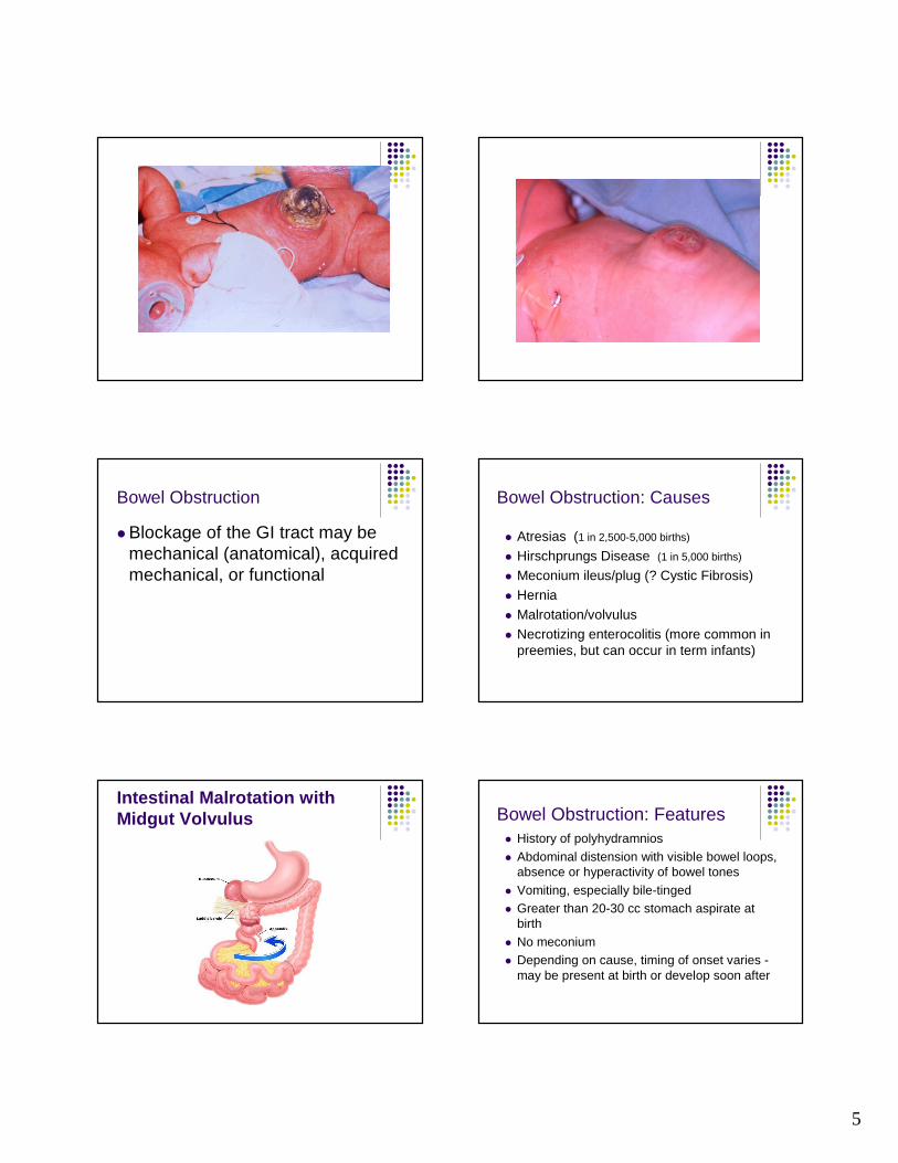
Intestinal Malrotation with Midgut Volvulus Bowel Obstruction: Features
� History of polyhydramnios� Abdominal distension with visible bowel loops,
absence or hyperactivity of bowel tones� Vomiting, especially bile-tinged� Greater than 20-30 cc stomach aspirate at
birth� No meconium� Depending on cause, timing of onset varies -
may be present at birth or develop soon after
6
Bowel Obstruction: Management
� NPO with NG or Replogle to low suction, or aspirate every 20-30 minutes
� IV for fluid, glucose, electrolyte management
� Measure and track abdominal girth� Abdominal films may help diagnose if air is
absent from the distal bowel.� Good surgical outcomes
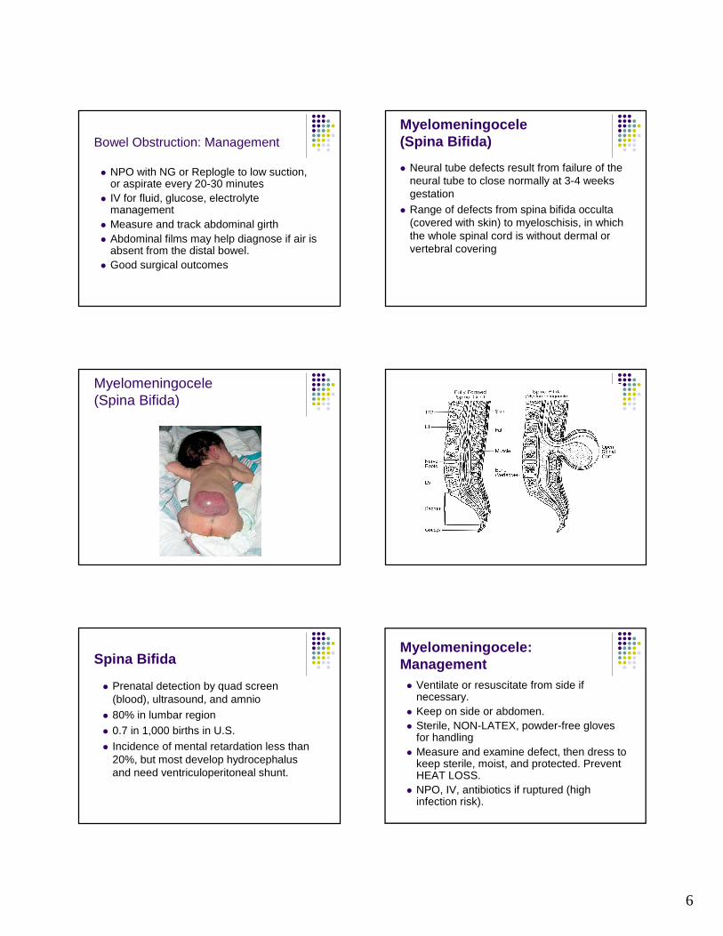
Myelomeningocele (Spina Bifida)
� Neural tube defects result from failure of the neural tube to close normally at 3-4 weeks gestation
� Range of defects from spina bifida occulta (covered with skin) to myeloschisis, in which the whole spinal cord is without dermal or vertebral covering
Myelomeningocele (Spina Bifida)
Spina Bifida
� Prenatal detection by quad screen (blood), ultrasound, and amnio
� 80% in lumbar region� 0.7 in 1,000 births in U.S. � Incidence of mental retardation less than
20%, but most develop hydrocephalus and need ventriculoperitoneal shunt.
Myelomeningocele: Management � Ventilate or resuscitate from side if
necessary. � Keep on side or abdomen.� Sterile, NON-LATEX, powder-free gloves
for handling� Measure and examine defect, then dress to
keep sterile, moist, and protected. Prevent HEAT LOSS.
� NPO, IV, antibiotics if ruptured (high infection risk).
7
Secure feeding tube to back of Telfa dressing and a ttach syringe of sterile saline.
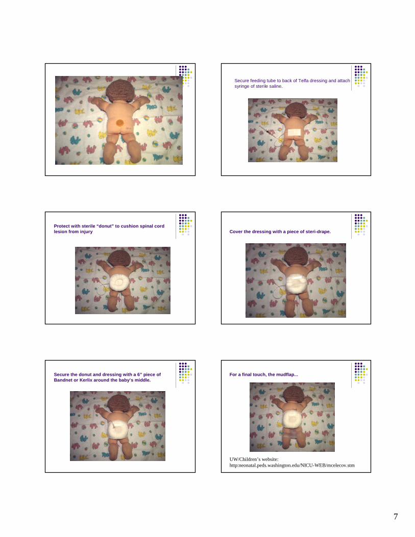
Protect with sterile “donut” to cushion spinal cord lesion from injury Cover the dressing with a piece of steri-drape.
Secure the donut and dressing with a 6” piece of Bandnet or Kerlix around the baby’s middle.
For a final touch, the mudflap...
UW/Children’s website: http:neonatal.peds.washington.edu/NICU-WEB/mcelecov.stm
8
Diaphragmatic Hernia� Herniation of abdominal organs into the
thoracic cavity through a defect in the diaphragm due to early failure of the closure of the diaphram. This usually results in hypoplasia of the lung.
Diaphragmatic Hernia: Features Diaphragmatic Hernia: Features
� Incidence: 1 in 4000; 90% on left� Detectable on prenatal ultrasound� 50% associated with other anomalies: neural
tube defects, heart defects, intestinal malrotation
� May be mild and asymptomatic or severe and life-threatening
Diaphragmatic Hernia: Features
� Respiratory distress at birth or soon after� Cyanosis, decreased breath sounds on
one side of chest� Muffled or displaced heart sounds on
(usually) right side of chest� Bowel tones in chest� Diagnosis confirmed by X-Ray
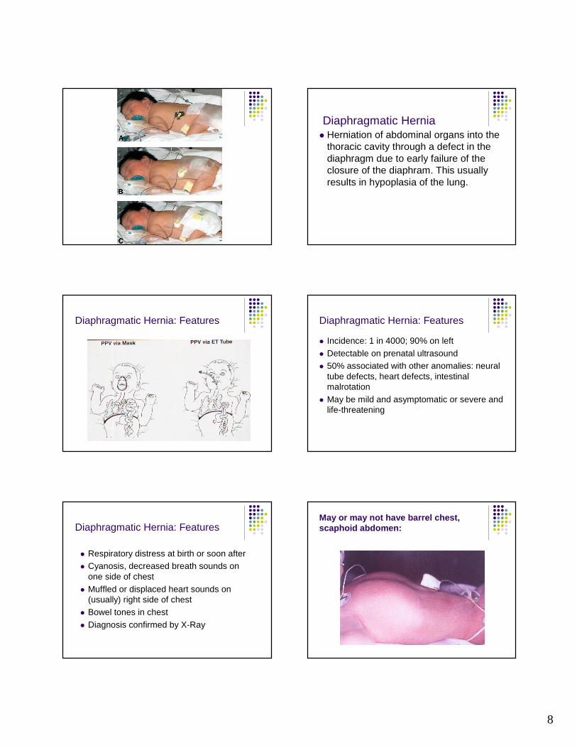
May or may not have barrel chest, scaphoid abdomen:
9
Diaphragmatic Hernia: Management
� Avoid bag-mask ventilation! Early intubation recommended.
� Decompress stomach with OG or Replogle.� Elevate head of bed� NPO, IV for fluid and electrolyte management� Antibiotics if risk of sepsis
Diaphragmatic Hernia: Management
� Surgical repair usually deferred until pulmonary hypertension is controlled.
� May have lung hypoplasia and need high frequency ventilation, nitric oxide or ECMO
� Primary closure usually possible� Patch or muscle flap may be used to close
defect.
Diaphragmatic Hernia: Management
� Survival depends on preoperative status. 40-60% survival if severe symptoms appear within the first 6 - 8 hours of life.
� Depends on severity of defect and any other associated anomalies.
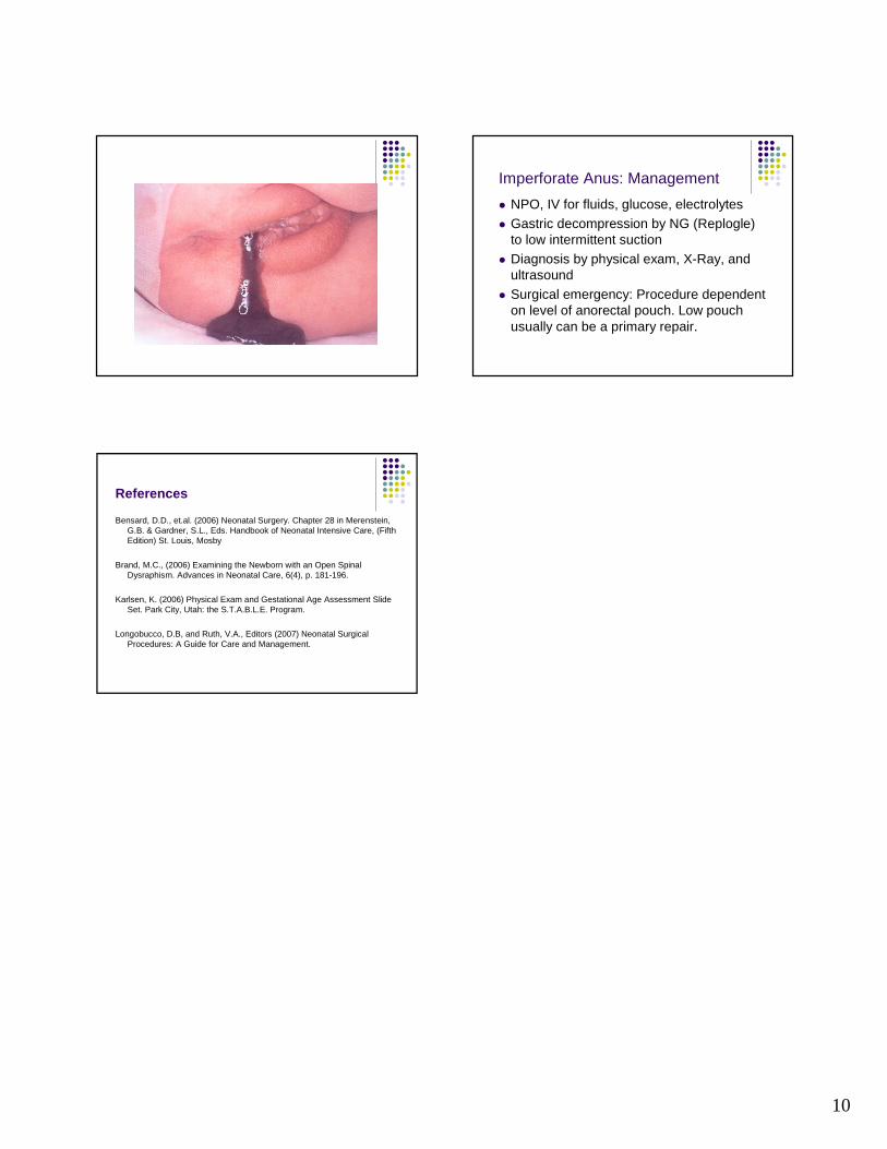
Imperforate Anus
� Failure of differentiation of urogenital sinus and cloaca during embryological development. May be high or low in colon.
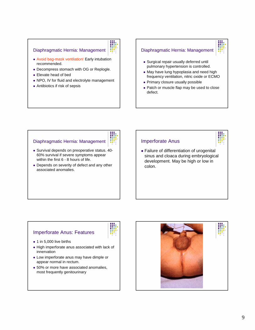
Imperforate Anus: Features
� 1 in 5,000 live births� High imperforate anus associated with lack of
innervation� Low imperforate anus may have dimple or
appear normal in rectum. � 50% or more have associated anomalies,
most frequently genitourinary
10
Imperforate Anus: Management
� NPO, IV for fluids, glucose, electrolytes� Gastric decompression by NG (Replogle)
to low intermittent suction� Diagnosis by physical exam, X-Ray, and
ultrasound� Surgical emergency: Procedure dependent
on level of anorectal pouch. Low pouch usually can be a primary repair.
References
Bensard, D.D., et.al. (2006) Neonatal Surgery. Chap ter 28 in Merenstein, G.B. & Gardner, S.L., Eds. Handbook of Neonatal Int ensive Care, (Fifth Edition) St. Louis, Mosby
Brand, M.C., (2006) Examining the Newborn with an O pen Spinal Dysraphism. Advances in Neonatal Care, 6(4), p. 181 -196.
Karlsen, K. (2006) Physical Exam and Gestational Ag e Assessment Slide Set. Park City, Utah: the S.T.A.B.L.E. Program.
Longobucco, D.B, and Ruth, V.A., Editors (2007) Neo natal Surgical Procedures: A Guide for Care and Management.