Embed Size (px)
Citation preview
Journal of Case Reports and Images in Otolaryngology, Vol. 2, 2021.
J Case Rep Images Otolaryngol 2021;2:100004Z18AM2021. www.ijcriotolaryngology.com
Mahmud et al. 1
CASE REPORT OPEN ACCESS
Esophageal denture impaction: A report of four cases
Ahmad Mahmud, Mohammed Bello Fufore, Babangida Sabo Miya
ABSTRACT
Esophageal denture impactions are common occurrence in the developing world mostly due to inappropriate use or improper fixture of these dentures. Incidence of these impactions appears to be alarming especially in places where private dental services manned by unqualified staff are proliferating. Four cases of esophageal denture impaction were managed over a period of six months in our institution. Three of the patients had successful surgery and were discharged home. One of the patients had diagnostic dilemma and declined further management. To check the rising cases of denture impaction, patients should be advised to use permanent dentures and patronize only specialists for dental fitting so as to minimize complications.
Keywords: Denture impaction, Dysphagia, Esophago-scopy, Foreign body
How to cite this article
Mahmud A, Fufore MB, Miya BS. Esophageal denture impaction: A report of four cases. J Case Rep Images Otolaryngol 2020;1:100004Z18AM2021.
Article ID: 100004Z18AM2021
*********
Ahmad Mahmud1, MBBS, MSc, FWACS, Mohammed Bello Fufore1, MBBS, MPH, FWACS, Babangida Sabo Miya1, MBBSAffiliation: 1Department of Ear Nose & Throat, Federal Med-ical Centre, Yola, Lamido Zubairu Way, PMB 2017, Yola, Adamawa State, Nigeria.Corresponding Author: Dr Ahmad Mahmud, Department of Ear Nose & Throat, Federal Medical Centre, Yola, Lamido Zubairu Way, PMB 2017, Yola, Adamawa State, Nigeria; Email: [email protected]
Received: 06 February 2021Accepted: 02 August 2021Published: 06 September 2021
CASE SERIES PEER REVIEWED | OPEN ACCESS
doi: 10.5348/100004Z18AM2020CS
INTRODUCTION
Tooth has remarkable function in articulation of speech and also plays role in maintaining the normal contour and shape of the face [1]. It is because of this aesthetic and articulation function that people often resort to wearing dentures for one reason or the other once they lose their teeth; this has culminated to the rising cases of esophageal foreign body (denture impaction). Esophageal denture impaction is common in Nigeria, and the patients are often aware of the diagnosis even before seeking for medical help [2]. In a single center survey done in Northwestern Nigeria, denture was found to be the second most common aero-digestive foreign body [3]. Impacted denture is usually removed by rigid esophagoscopy which may sometimes be very challenging or even impossible warranting open surgery such as cervical esophagotomy [2–5].
We present a series of four cases of esophageal denture impaction, presented and managed in a Tertiary Centre in the Northeastern Nigeria over a period of six months. All the three patients who had surgery underwent rigid esophagoscopy and foreign body removal under General Anesthesia. They had guided oral intubation.
CASE SERIES
Case 1A 62-year-old woman presented with one day history
of dysphagia following accidental swallowing of her denture. There was associated pooling of saliva in the oral cavity. On examination there was positive pointing sign on the neck. X-ray soft tissue neck shows opacity around the esophageal inlet corresponding to C5–C6 vertebrae (Figure 1A). The patient optimized and planned for emergency esophagoscopy, which she had successfully. The denture was visualized at 18 cm from the upper incisor and was removed with minimal difficulty (Figure 1B). The patient was placed on nil per oral for 24 hours. Had post-operative intravenous antibiotic and intravenous fluid. No any complications observed, she was discharged home after 72 hours.
Journal of Case Reports and Images in Otolaryngology, Vol. 2, 2021.
J Case Rep Images Otolaryngol 2021;2:100004Z18AM2021. www.ijcriotolaryngology.com
Mahmud et al. 2
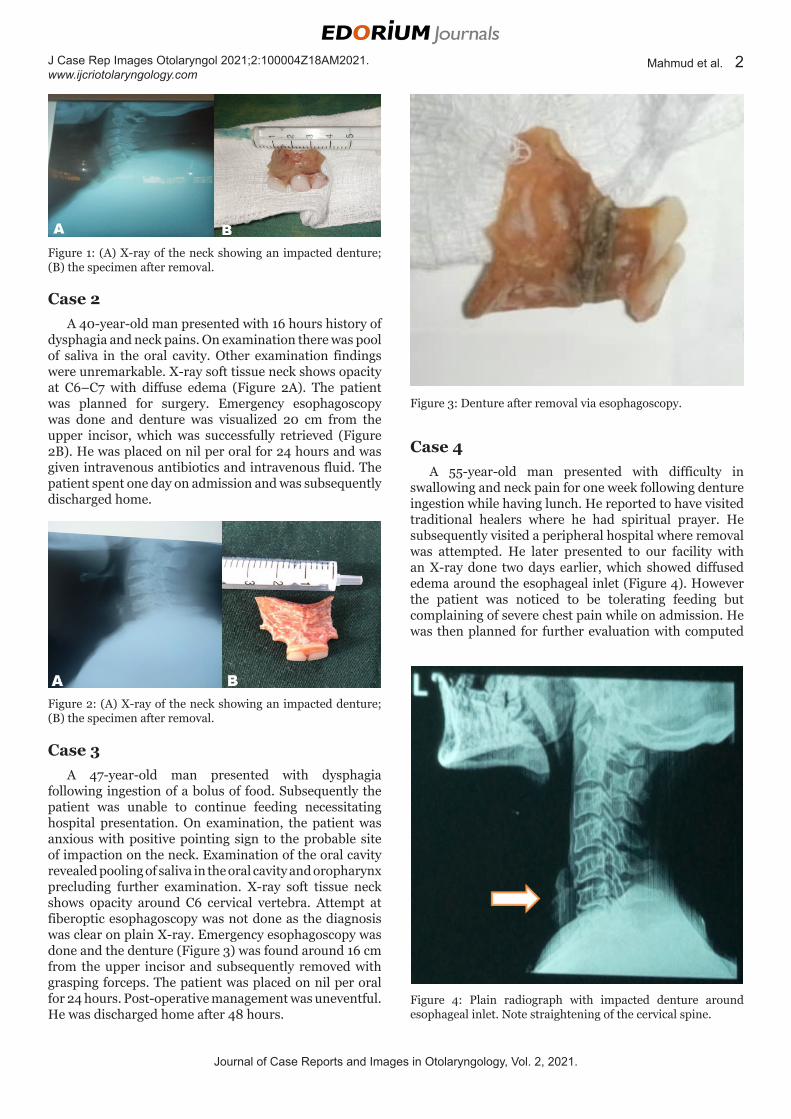
Case 2A 40-year-old man presented with 16 hours history of
dysphagia and neck pains. On examination there was pool of saliva in the oral cavity. Other examination findings were unremarkable. X-ray soft tissue neck shows opacity at C6–C7 with diffuse edema (Figure 2A). The patient was planned for surgery. Emergency esophagoscopy was done and denture was visualized 20 cm from the upper incisor, which was successfully retrieved (Figure 2B). He was placed on nil per oral for 24 hours and was given intravenous antibiotics and intravenous fluid. The patient spent one day on admission and was subsequently discharged home.
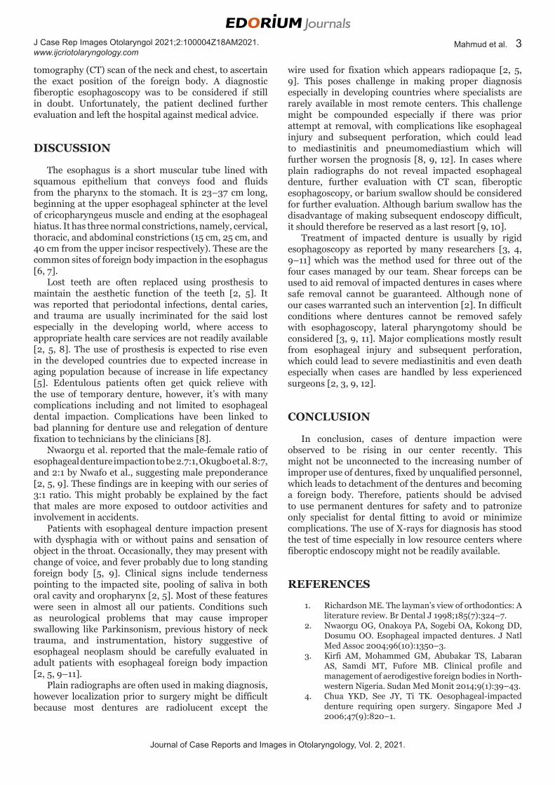
Case 4A 55-year-old man presented with difficulty in
swallowing and neck pain for one week following denture ingestion while having lunch. He reported to have visited traditional healers where he had spiritual prayer. He subsequently visited a peripheral hospital where removal was attempted. He later presented to our facility with an X-ray done two days earlier, which showed diffused edema around the esophageal inlet (Figure 4). However the patient was noticed to be tolerating feeding but complaining of severe chest pain while on admission. He was then planned for further evaluation with computed
Figure 1: (A) X-ray of the neck showing an impacted denture; (B) the specimen after removal.
Figure 2: (A) X-ray of the neck showing an impacted denture; (B) the specimen after removal.
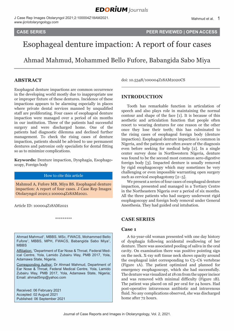
Case 3A 47-year-old man presented with dysphagia
following ingestion of a bolus of food. Subsequently the patient was unable to continue feeding necessitating hospital presentation. On examination, the patient was anxious with positive pointing sign to the probable site of impaction on the neck. Examination of the oral cavity revealed pooling of saliva in the oral cavity and oropharynx precluding further examination. X-ray soft tissue neck shows opacity around C6 cervical vertebra. Attempt at fiberoptic esophagoscopy was not done as the diagnosis was clear on plain X-ray. Emergency esophagoscopy was done and the denture (Figure 3) was found around 16 cm from the upper incisor and subsequently removed with grasping forceps. The patient was placed on nil per oral for 24 hours. Post-operative management was uneventful. He was discharged home after 48 hours.
Figure 3: Denture after removal via esophagoscopy.
Figure 4: Plain radiograph with impacted denture around esophageal inlet. Note straightening of the cervical spine.
Journal of Case Reports and Images in Otolaryngology, Vol. 2, 2021.
J Case Rep Images Otolaryngol 2021;2:100004Z18AM2021. www.ijcriotolaryngology.com
Mahmud et al. 3
tomography (CT) scan of the neck and chest, to ascertain the exact position of the foreign body. A diagnostic fiberoptic esophagoscopy was to be considered if still in doubt. Unfortunately, the patient declined further evaluation and left the hospital against medical advice.
DISCUSSION
The esophagus is a short muscular tube lined with squamous epithelium that conveys food and fluids from the pharynx to the stomach. It is 23–37 cm long, beginning at the upper esophageal sphincter at the level of cricopharyngeus muscle and ending at the esophageal hiatus. It has three normal constrictions, namely, cervical, thoracic, and abdominal constrictions (15 cm, 25 cm, and 40 cm from the upper incisor respectively). These are the common sites of foreign body impaction in the esophagus [6, 7].
Lost teeth are often replaced using prosthesis to maintain the aesthetic function of the teeth [2, 5]. It was reported that periodontal infections, dental caries, and trauma are usually incriminated for the said lost
especially in the developing world, where access to appropriate health care services are not readily available [2, 5, 8]. The use of prosthesis is expected to rise even in the developed countries due to expected increase in aging population because of increase in life expectancy [5]. Edentulous patients often get quick relieve with the use of temporary denture, however, it’s with many complications including and not limited to esophageal dental impaction. Complications have been linked to bad planning for denture use and relegation of denture fixation to technicians by the clinicians [8].
Nwaorgu et al. reported that the male-female ratio of esophageal denture impaction to be 2.7:1, Okugbo et al. 8:7, and 2:1 by Nwafo et al., suggesting male preponderance [2, 5, 9]. These findings are in keeping with our series of 3:1 ratio. This might probably be explained by the fact that males are more exposed to outdoor activities and involvement in accidents.
Patients with esophageal denture impaction present with dysphagia with or without pains and sensation of object in the throat. Occasionally, they may present with change of voice, and fever probably due to long standing foreign body [5, 9]. Clinical signs include tenderness pointing to the impacted site, pooling of saliva in both oral cavity and oropharynx [2, 5]. Most of these features were seen in almost all our patients. Conditions such as neurological problems that may cause improper swallowing like Parkinsonism, previous history of neck trauma, and instrumentation, history suggestive of esophageal neoplasm should be carefully evaluated in adult patients with esophageal foreign body impaction [2, 5, 9–11].
Plain radiographs are often used in making diagnosis, however localization prior to surgery might be difficult because most dentures are radiolucent except the
wire used for fixation which appears radiopaque [2, 5, 9]. This poses challenge in making proper diagnosis especially in developing countries where specialists are rarely available in most remote centers. This challenge might be compounded especially if there was prior attempt at removal, with complications like esophageal injury and subsequent perforation, which could lead to mediastinitis and pneumomediastium which will further worsen the prognosis [8, 9, 12]. In cases where plain radiographs do not reveal impacted esophageal denture, further evaluation with CT scan, fiberoptic esophagoscopy, or barium swallow should be considered for further evaluation. Although barium swallow has the disadvantage of making subsequent endoscopy difficult, it should therefore be reserved as a last resort [9, 10].
Treatment of impacted denture is usually by rigid esophagoscopy as reported by many researchers [3, 4, 9–11] which was the method used for three out of the four cases managed by our team. Shear forceps can be used to aid removal of impacted dentures in cases where safe removal cannot be guaranteed. Although none of our cases warranted such an intervention [2]. In difficult conditions where dentures cannot be removed safely with esophagoscopy, lateral pharyngotomy should be considered [3, 9, 11]. Major complications mostly result from esophageal injury and subsequent perforation, which could lead to severe mediastinitis and even death especially when cases are handled by less experienced surgeons [2, 3, 9, 12].
CONCLUSION
In conclusion, cases of denture impaction were observed to be rising in our center recently. This might not be unconnected to the increasing number of improper use of dentures, fixed by unqualified personnel, which leads to detachment of the dentures and becoming a foreign body. Therefore, patients should be advised to use permanent dentures for safety and to patronize only specialist for dental fitting to avoid or minimize complications. The use of X-rays for diagnosis has stood the test of time especially in low resource centers where fiberoptic endoscopy might not be readily available.
REFERENCES
1. Richardson ME. The layman’s view of orthodontics: A literature review. Br Dental J 1998;185(7):324–7.
2. Nwaorgu OG, Onakoya PA, Sogebi OA, Kokong DD, Dosumu OO. Esophageal impacted dentures. J Natl Med Assoc 2004;96(10):1350–3.
3. Kirfi AM, Mohammed GM, Abubakar TS, Labaran AS, Samdi MT, Fufore MB. Clinical profile and management of aerodigestive foreign bodies in North-western Nigeria. Sudan Med Monit 2014;9(1):39–43.
4. Chua YKD, See JY, Ti TK. Oesophageal-impacted denture requiring open surgery. Singapore Med J 2006;47(9):820–1.
Journal of Case Reports and Images in Otolaryngology, Vol. 2, 2021.
J Case Rep Images Otolaryngol 2021;2:100004Z18AM2021. www.ijcriotolaryngology.com
Mahmud et al. 4
5. Okugbo SU, Onyeagwara NC. Oesophageal impacted dentures at the University of Benin Teaching Hospital, Benin City, Nigeria. J West Afr Coll Surg 2012;2(2):102–11.
6. Oezcelik A, DeMeester SR. General anatomy of the esophagus. Thorac Surg Clin 2011;21(2):289–97.
7. Moore KL, Dally FA, Agur AMR. Esophagus. In: Moore KL, editor. Clinically Oriented Anatomy. 7ed. Philadelphia: Lippincott Williams & Wilkins; 2014. p. 229–30.
8. Bilhan H, Erdogan O, Ergin S, Celik M, Ates G, Geckili O. Complication rates and patient satisfaction with removable dentures. J Adv Prosthodont 2012;4(2):109–15.
9. Nwafo DC, Anyanwu CH, Egbue MO. Impacted esophageal foreign bodies of dental origin. Ann Otol Rhinol Laryngol 1980;89(2 Pt 1):129–31.
10. Hashmi S, Walter J, Smith W, Latis S. Swallowed partial dentures. J R Soc Med 2004;97(2):72–5.
11. Orji FT, Akpeh JO, Okolugbo NE. Management of esophageal foreign bodies: Experience in a developing country. World J Surg 2012;36(5):1083–8.
12. Nageris B, Feinmesser R. Dentures in the oesophagus complicated by pneumomediastinum. Ear Nose Throat J 1990;69(11):737–8.
*********
AcknowledgmentAll staff of ENT Department of Federal Medical Centre, Yola.
Author ContributionsAhmad Mahmud – Conception of the work, Design of the work, Acquisition of data, Analysis of data, Interpretation of data, Drafting the work, Revising the work critically for important intellectual content, Final approval of the version to be published, Agree to be accountable for all aspects of the work in ensuring that questions related to the accuracy or integrity of any part of the work are appropriately investigated and resolved
Mohammed Bello Fufore – Conception of the work, Design of the work, Acquisition of data, Analysis of data,
Interpretation of data, Drafting the work, Revising the work critically for important intellectual content, Final approval of the version to be published, Agree to be accountable for all aspects of the work in ensuring that questions related to the accuracy or integrity of any part of the work are appropriately investigated and resolved
Babangida Sabo Miya – Conception of the work, Design of the work, Acquisition of data, Analysis of data, Interpretation of data, Drafting the work, Revising the work critically for important intellectual content, Final approval of the version to be published, Agree to be accountable for all aspects of the work in ensuring that questions related to the accuracy or integrity of any part of the work are appropriately investigated and resolved
Guarantor of SubmissionThe corresponding author is the guarantor of submission.
Source of SupportNone.
Consent StatementWritten informed consent was obtained from the patient for publication of this article.
Conflict of InterestAuthors declare no conflict of interest.
Data AvailabilityAll relevant data are within the paper and its Supporting Information files.
Copyright© 2021 Ahmad Mahmud et al. This article is distributed under the terms of Creative Commons Attribution License which permits unrestricted use, distribution and reproduction in any medium provided the original author(s) and original publisher are properly credited. Please see the copyright policy on the journal website for more information.
Access full text article onother devices
Access PDF of article onother devices