Embed Size (px)
Citation preview

[CANCER RESEARCH 36, 3978-3984, November 1976]
SUMMARY
A human colon carcinoma cell line, Co-uS, has beenestablished in vitro from solid xenognafts maintained innude mice and subcultuned for 95 passages. Co-uS cellsgrow in vitro as tightly packed, epithelial-like colonies, havea doubling time of about 36 hn, have a relatively low platingefficiency in agar, and release significant amounts of cancinoembryonic antigen to the culture medium. Their epithehal nature has been confirmed by ultnastnuctunal examination. The injection of Co-i 15 cells into nude mice reinducedthe formation of solid tumor masses that could be retransplanted and showed a morphology comparable to that ofthe original xenognaft.
INTRODUCTION
Cell lines of human colon carcinomas are difficult toestablish. Indeed, only a few of them have been reported inthe literature (3, 7, 17). One of the major difficulties toovercome in establishing cell lines of human colonectalcarcinomas is the bacterial contamination present in thesurgical specimens. Furthermore, when primary cultures ofcolon carcinomas could be obtained, the neoplastic cellswere usually overshadowed by fibroblast-like cells in a fewmonths. Yet, cultures of gastrointestinal tumors representuseful experimental material for a variety of studies. Inparticular, human tumor cells have been shown to possesstumor-associated on -specific antigens, and cell culturesmay be a convenient source of malignant material for antigenic studies. Indeed, CEA3 (6) has been demonstrated incultures of gastrointestinal neoplasms (2, 7, 17). We wish toreport here the establishment and preliminary chanactenization of an epithelial tumor cell line, referred to as Co-115and derived from a human colon carcinoma previouslygrafted and maintained by serial transplantation into nudemice. The human tumor growing in the nude mouse wasfound to be supported by a stroma of munine origin, andcontamination by human fibroblasts was therefore minimalor absent. Isolation of tumor material from nude mice hasthe advantage of providing sterile material and of allowing
I Supported in part by Grants 3.50075 and 0841 .4200 from the Swiss
National Foundation for Scientific Research.2 To whom requests for reprint should be addressed, at Ludwig Institute
for Cancer Research, 1066 Epalinges s/Lausanne, Switzerland.3 The abbreviations used are: CEA, carcinoembryonic antigen; FCS, fetal
calf serum.Received May 17, 1976; accepted August 9, 1976.
the elimination of muminefibroblasts present in the primaryculture.
MATERIALS AND METHODS
Tumor Tissue. The primary tumor was obtained shortlyafter surgery from a 77-year-old Caucasian female with acarcinoma of the ascending colon. Histological examination revealed a poorly differentiated adenocarcinoma (seeFig. 1A) that invaded the entire thickness of the bowel walland metastasized to the regional lymph nodes (Duke's C;Duke's classification, which refers to the pathological extent of the neoplasm). Rare gland-like structures were present, with little if any mucin production.
Heterotransplantation. The tumor fragments wereminced in culture medium and inoculated s.c. with a trocarinto the flanks of the nude mice. Six-week-old nude mice,backcross mated with BALB/c and bred in our colony underconventional conditions, were used in these experiments.For serial passage of the transplants, the tumor noduleswere dissected and freed from necrotic tissue before reinoculation. Serial samples were taken for examination bylight and electron microscopy.
Cell Cultures. Cell cultures from nodules of different passages were prepared by mincing the tissue with a scalpelinto fragments of approximately 2 mm in diameter. Thefragments were then suspended in the culture medium andseeded in Falcon plastic flasks (25 sq cm). In addition, a finecell suspension was prepared by digestion of tumor fragments with 0.25% trypsin for 20 mm at room temperature.About i0@ tumor cells obtained by this procedure wereplaced in tissue culture flasks of the same size. Dulbecco'smodified Eagle's medium supplemented with 20% heatinactivated FCS (Gibco Bio-Cult Ltd. , Scotland) containing200 j@gstreptomycin per ml and 200 IU penicillin G per mlwas used to initiate the cultures. All cultures were incubatedat 37°in 5% CO2 in air. Subsequently, the cultures wereserially transferred by treatment with 0.05% trypsin and0.05% EDTA and maintained in the same medium containing 10% FCS.
Co-uS cells were viably frozen in medium plus 7.5%dimethyl sulfoxide and were maintained at —80°at differentpassages over the past 2 years.
Growth Curves and Plating Efficiency. Cultures of human colon carcinoma Co-uS cells were collected after 20and 50 passages for the determination of growth curves.For each passage studied, semiconfluent cultures were
3978 CANCER RESEARCH VOL. 36
Establishment of a Cell Line (Co-115) from a Human ColonCarcinoma Transplanted into Nude Mice1
S. Carrel,2 B. Sordat, and C. Merenda
Unit of Human Cancer Immunology, Lausanne Branch, Ludwig Institute for Cancer Research (S. C.); Department of Immunology, Swiss Institute forExperimental Cancer Research (B. S.); and Department of Biochemistry, University of Lausanne (C. MI, 1011 Lausanne Switzerland
Research. on September 5, 2018. © 1976 American Association for Cancercancerres.aacrjournals.org Downloaded from

Co!on Carcinoma Ce!! Line
tnypsinized (trypsin:EDTA), and the cells were washed twiceand resuspended in medium with 5% FCS for counting.Three plastic flasks (25 sq cm) were plated with 10 cells andcounts were carried out at 48-hr intervals up to Day 10.Trypsinized cells were counted in a hemocytometem andtheir numbers were averaged for each time interval.
The plating efficiency of Co-i15 was determined in softagar according to the method of MacPhenson and Montagnier (12). Cells were suspended in growth medium containing 0.3% agan (Difco Laboratories, Detroit, Mich.) and wereseeded into 60- x 15-mm Petni dishes containing a base of 3ml of 0.5% agan in growth medium. Tumor cells wereseeded in duplicate dishes at concentrations of i0@, i0@,and 10@cells. Colony formation in agan was scored after 3weeks of culture, and colonies with 30 or more cells werecounted.
Reinoculation of Cultured Tumor Cells into Nude Mice.Cultured Co-i 15 cells (30 x 106)were injected s.c. into nudemice as previously done for other cell lines (5, 8, 9, i3, iS).The tumors that developed were used in part for histologicalexamination and in part for transplantation into other mice.By this technique, several serial passages were carried out.
Cytogenetic Analysis. Confluent monolayers of culturesof Co-uS cells were treated with colchicine at a final concentration of 0.08 mg/mI in the medium. After 4 hr, thecultures were dispersed at 37°with 0.25% trypsin and thecells were centrifuged at 800 rpm for 5 mm. The cells werethen resuspended in a hypotonic solution (1% sodium citrate in distilled water) for 20 mm at 37°.Cells were thenpelleted and fixed in ethanol:acetic acid (3:1) at room temperatune for 30 mm. The cell suspension was dropped on achilled wet slide (distilled water), air dried, and stained with2% Giemsa for 20 mm.
CEA Assays. CEA was identified in 0.6 M perchlomicacidextracts of Co-i 15 tumor heterografts by means of the madioimmunoassay described by Thompson et a!. (16) andmodified by Mach et a!. (ii). CEA in the sena of tumorbeaning nude mice, as well as in supennatants of confluentcultures, was determined by means of a modification of thedouble-antibody radioimmunoassay described by Egan etal. (1). Indirect fluorescent staining of CEA was performedon cryostat sections of a tumor transplant obtained after the2nd transfer and on monolayems of Co-uS cells grown oncovenslips.
Blood Group Antigens. The persistence of blood groupsubstance(s) on Co-ii5 cell membranes was tested by anindirect immunofluonescence technique using human donor sera antiblood groups A, B, and 0.
Microscopy. Tumor fragments for light microscopic examination were fixed with 2.5% cacodylate-buffemed glutaraldehyde, embedded in butoxyethanol glycolmethacrylate(Sorvall, Newton, Conn.), sectioned at 2 @.tm,and stainedwith Giemsa and periodic acid-Schiff on Alcyan blue at pH2.6 reagents.
For electron microscopy, the tissues were fixed with 2.5%glutanaldehyde buffered with 0.i M cacodylate, pH 7.3,postfixed in i% osmium tetroxide, and embedded in Analdite. Ultnathin sections were stained with umanylacetate andlead citrate.
Confluent cultures of Co-i i Scells (95th passage) growing
on a Falcon plastic flask surface were fixed in situ with theuse of glutanaldehyde and osmium, as above. The cells weredehydrated and embedded in the flask. Ultnathin sectionswere performed parallel and perpendicular to the plasticsurface.
RESULTS
Heterotransplantation. Tumor nodules (s.c.) developedin 3 of 3 mice originally grafted and reached a diameter of 5to 8 mm within 2 to 3 weeks. Up to the present time, 50successive transplantations have been carried out. Thegrowth rate of the tumors increased during the 1st 4 passages and then remained constant, yielding approximately3 to 4g of tumor in 4 weeks.
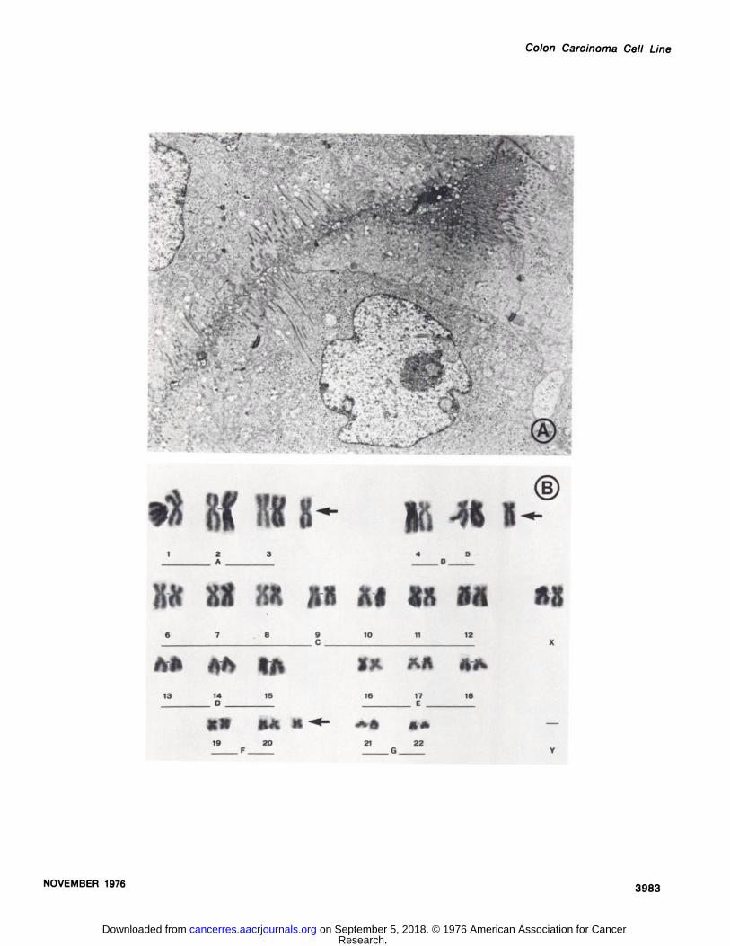
Microscopic examination revealed that all tumors growing in nude mice were composed of carcinoma cells featuring essentially the same degree of differentiation as theoriginal tumor (Fig. iB). These cells were arranged inmasses supported by a vasculanized stroma of munine originwhich appeared less abundant than that of the originaltumor (B. Sondat, unpublished results, i974).@Necrotic fociwere frequently observed in the center of larger nodules. Atthe periphery, the tumor nodules appeared to be well delimited but not encapsulated. At the electron microscopiclevel, the tumor transplants consisted mostly of poorly diffementiated cells with lange nuclei and distinct nucleoli.Their cytoplasm was rich in polysomes and contained numerous mitochondnia but mareprofiles of ergastoplasm andof Golgi complexes. The intercellular spaces presented frequent intendigitationsand micnovilli,as illustratedin Fig.2A. Cell junctions between the tumor cells were mainly ofthe desmosome type. The fine structure of these neoplasticcells did not change in subsequent passages. No basallamina was ever recognized at the periphery of the tumornodule, although some amorphous electron-dense materialwas occasionally juxtaposed with the tumor cells. No virusparticles have been seen in association with the tumor cells.
Cell Cultures. Primary cultures were established moreeasily from later than from earlier transplants. These cultunes were characterized by colonies of cells featuring anepithelial pattern of growth after 2 to 5 days of explantation.The epithelial colonies consisted of relatively slow-growingcells showing pleomomphism and some vacuolization.These colonies increased in size for 3 to 4 weeks. As theygrew larger, piling up of cells occurred in their center.Fibnoblasts of munine origin were present in variable numbers. When tumor cells from early passages were used,every attempt to subculture these slow-growing epithelialcells was unsuccessful, the only remaining cells being ofmouse origin. In contrast, when tumor cells from the 10th to15th passages in the mouse were used, the colonies consisted of more rapidly growing epithelial cells which couldbe easily subcultuned. Cell line Co-uS was established inOctober i974 from such colonies that demonstrated astrong adherence to the plastic surface and grew in tightly
4 The murine origin of the stroma within transplanted tumors has been
demonstrated by indirect immunofluorescence with antispecies antisera oncryostat sections of colorectal xenografts, including Co-uS tumor.
NOVEMBER 1976 3979
Research. on September 5, 2018. © 1976 American Association for Cancercancerres.aacrjournals.org Downloaded from

S. Carrel et a!.
packed, epithelial-like formations (Fig. iC). The tumor cellswere separated from the mouse fibroblasts by repeatedshort periods of trypsinization (3 times at 2-day intervals),during which most of the fibroblast-like cells could be removed by gentle shaking. This procedure proved to be verysuccessful in yielding almost pure colonies of tumor cellswhich grew to confluence. The remaining fibroblasts werelost during successive subcultures. In addition, cytotoxicitytests performed on 51Cr-Iabeled Co-uS cells in the presence of antimouse antiserum dilutions plus complement didnot reveal a significant radioactive release. These resultsstrongly indicate the disappearance and elimination of munine cells in the Co-115 cultures. Moreover, the tumor cellshad essentially the same morphological appearance as didthe primary cultures and showed some mucin production.
The fine structure of the cultured cells was remarkablysimilar to that of the tumor cells described for the solidtransplants. Sections parallel to the surface of the monolayer showed that the cells were in close contact with eachother and presented numerous cell-to-cell junctions of thedesmosome type. The intercellular spaces were often focally dilated, forming “intercellularlumens―containing numerous microvilli (Fig. 3A). Desmosomes were sometimespresent at both sides of these spaces. Rare lumens entirelysurrounded by cytoplasm were occasionally seen; it couldnot be established, however, whether they were entirelyintracellular or whether they represented extensions of theextracellular spaces. The microvilli at the surface of thecells were generally numerous and displayed a core of finefilaments extendihg into the cytoplasm in the form of deeprootlets. Filaments were present in all cells, the larger ones(80 to 120 A in diameter) arranged in bundles sometimesconverging toward desmosomes, and the finer ones (40 to80 A) forming a felt-like meshwork. Small vesicles werepresent along the plasma membrane of most cells. In someof them, membrane-bound granules containing materialreminiscent of mucin were also observed (Fig. 38).
Growth Curves and Plating Efficiency. Co-i 15 cell cultunes first showed an initial growth lag of about 5 days, thenan exponential phase of growth between Days 5 and 10 afterplating. The mean population doubling time was about 56hr when tested between Passages 8 and 15 and was about36 hr after the 26th passage. The mitotic index, counted oncells 7 days after the 80th in vitro passage, varied between0.9 and 1.2%. The plating efficiency was determined withthe same cell preparations that were used to establish thegrowth curves. The colonies were counted 3 weeks afterplating . The absolute plating efficiency of Co-i 15 cell line atPassage 34 was 3%. This result was relatively low, cornpared with another colon carcinoma line, HT-29 (initiallycultured by Dr. J. Fogh of the Sloan-Kettering Institute forCancer Research), which exhibited, under similar cultureconditions, a plating efficiency of 13%.
Tumorigenicity in Vivo. Co-i15 cells (30 x 106) were injected s.c. into 3 nude mice, all of which developed a tumorafter a latency period of 3 to 4 weeks. These tumors wereeasily retransplantable into nude mice where they reproduced a morphology comparable to that of the solid tnansplants (see Fig. 1D).
Blood Group Antigens. Among the different human sera
tested against the ABO blood group substances, only theanti-A serum gave a distinct positive fluorescence, whichconfirmed the donor's known blood group.
CEA Assays. Co-i 15 cells produced significant quantitiesof CEA in vitro, as well as in hetenotransplants. Culturemedium of confluent monolayers contained as much as 300ng CEA in 10 ml medium after 5 days of culture.
CEA was also demonstrated in the circulating blood ofnude mice bearing large tumors. In these instances, serumconcentrations of CEA reached values up to 118 ng/mI.Extracts of Co-i 15 heterografts gave 1.5 to 6.3 @gCEA per gof wet tumor tissue.
Demonstration of CEA by indirect fluorescence on cryostat sections of Co-uS tumor nodule at the 2nd transferrevealed a localization in the intercellular spaces while, onCo-i 15 monolayens grown on covenslips, CEA was shown tobe localized on the membrane of the tumor cells.
Cytogenetic Analysis. Karyotypicanalysiswas performedon Co-115 cells from the 50th passage. Among 8 metaphases that could be analyzed, 7 exhibited a near-diploidmode of 49 chromosomes and 1 had 46 chromosomes. The3 additional chromosomes of the 7 abnormal karyognamswere classified as an extra A, 1 related to the B group, andan extra E or F chromosome. No marker chromosomes forHeLa cells were found in this material. A karyognam, nepresented in Fig. 2B, illustrates these preliminary results.
DISCUSSION
The present report describes the establishment of cell lineCo-i15 from a human colonic adenocarcinoma. This line,which was obtained from solid heterotransplants maintamed in nude mice, grows in culture as tightly packedcolonies of epithelial-like cells adherent to the plastic sunface of the flask. Ultrastructunal examination of the culturedcells confirms the epithelial nature of the Co-uS cell line.Furthermore, the morphology of microvilli and the appearance of secretory material are very comparable to those ofintestinal epithelial cells. The mouse fibroblasts presentinitially could be removed from the cultures after severalpassages in vitro. The tumors induced in nude mice by s.c.injection of Co-115 cells grown in vitro for 8 months werehistologically comparable to the original malignant tumor,although with a less abundant stroma. Similarly, Goldenbenget a!. (7) succeeded in establishing a mucin-producing human colon carcinoma line, GW-39, from a solid xenograftgrowing in the cheek pouch of adult golden hamsters. Inthis case, fibroblast-like cells admixed with the tumor cellsdied off after about 3 months of culture. Moreover, 2 penmanent cell lines (END-i and END-2) from poorly differentiatedendometnial carcinomas maintained in the nude mousecould also be established in vitro in this laboratory (13). Thisreport raises the question of the importance of the nudemouse as an intermediate host for the establishment oflong-term human neoplastic cell lines. Previous attempts inthis laboratory to establish cell lines from colon carcinomastaken directly from surgical specimens have been unsuccessful, and a recent report (14) suggests that, if 5 to 10% ofthe various human tumors initially placed in culture pro
3980 CANCERRESEARCHVOL. 36
Research. on September 5, 2018. © 1976 American Association for Cancercancerres.aacrjournals.org Downloaded from

Co!on Carcinoma Cell Line
vided an epithelial type of growth, less than 1% may surviveas continuous cell lines. Our experience suggests that theestablishment of line Co-i 15 was facilitated by previouspassages of the tumor cells through nude mice. This isfurther supported by the fact that several other humantumor cell lines were established in this laboratory fromhetenotnansplanted human tumors (1 renal cell carcinoma, 4malignant melanomas, and 1 moderately differentiated coIon carcinoma). The synthesis of CEA and its release in theculture medium by human tumor cells have been previouslyreported by several authors. Egan and Todd (2) showed,with colon carcinoma line HT-29, that CEA could be detected in the medium after 5 days of culture and that itsconcentration increased up to 2 months after initial transfer. CEA has also been demonstrated in the supernatantfrom a lysate of COLO 16, a human squamous carcinomaline (14), and in the culture fluid of Me180, a cervical carcinoma cell line (2). In addition, Tompkinsetal. (17) reportedthe establishment of 2 cell strains derived from colorectalneoplasms HCT-8 and HRT-i8, both of which synthesizedCEA in vitro . The Co-i 15 cells retained the ability to produceCEA in vivo (10) and also in vitro in amounts comparable tothose of the HCT-8 and HRT-18 cell strains. Therefore, thisline may be useful in further investigations of the possibleantigenic differences of CEA molecules produced by thesevarious strains. Moreover, it also retained the capacity toproduce another human oncofetal antigen, referred to asBOFA in a recent publication (4). As a source of malignanttarget material, it may also be used in in vitro studies ofhumonal and cell-mediated immunity in patients with coloncarcinomas.
ACKNOWLEDGMENTS
we are grateful for the excellent technical assistance of J. Bamat, L. Kolly,and S. Salvi in these studies. We thank Dr. M. Jotterand-Bellomo, Department of Medical Genetics, University Hospital of Lausanne, for performingthe karyotypic analysis, as well as Professor C. Bron, Institute of Biochemistry, University of Lausanne, for his gift of antispecies antisera. We also thankProfessor L. Ozzello, Professor K. T. Brunner, and Professor J.-P. Mach forsuggestions and advice in the preparation of the manuscript.
Fig . 1. A , histology of the original Co-i 15 tumor specimen featuring poorly differentiated carcinoma cells arranged in cords and supported by an abundantstroma. No mucin production in this field. Periodic acid-Schiff, x 500. B, Co-i 15 heterotransplant after 37 passages in the nude mouse. Cords of carcinomacells are separated by finely vascularized connective tissue trabeculae. Numerous mitotic figures are present, especially in the vicinity of intratumoral vessels.Giemsa, x 500. C, Co-115 carcinoma cells after 80 passages in vitro. Epithelial-type colony. The cells are irregular in shape with basophilic cytoplasm contaming vacuoles and large clear nuclei showing several nucleoli. Giemsa, x 500. D, Co-i15 cells established in vitro and injected into the nude mouse.Cytological and stromal characteristics are comparable to those in B. Giemsa, x 500.
Fig. 2. A, Co-115 heterotransplant after 23 passages in the nude mouse. Note the presence of interdigitations and microvillous projections in the intercellular spaces. Desmosomes are frequent between the tumor cells, x 14,000. B, karyogram of Co-1i5 cells after 50 passages in vitro. Representativemetaphase with 49 chromosomes. The 3 additional chromosomes (arrows) have been classified as extra A and B and the 3rd one is related to the E or Fgroups.
Fig. 3. A, Co-ii5 cultured cells; an intercellular space showing a focal dilation (intercellular lumen). Notice desmosome and numerous microvillous projectionswith deeplypenetratingrootlets.Numeroussuperficialvesiclesarealsovisible, x 24,000.B, cytoplasmicportionsof Co-i 15cellsjoined byadesmosome. The appearance of vacuoles, lined by double membrane and containing dense material, is suggestive of secretory granules. x 42,500.
NOVEMBER1976 3981
REFERENCES
1. Egan, M. L., Lautenschleger, J. F., Coligan, J. E., and Todd, C. W.Radloimmune Assay of Carcinoembryonic Antigen. Immunochemistry.9: 289-299, 1972.
2. Egan,M. L., andTodd,C. W.CarcinoembryonicAntigen:SynthesisbyaContinuous Line of Adenocarcinoma Cells. J. NatI. Cancer Inst. , 49:887-889, 1972.
3. Fogh,J., andTrempe,G.NewHumanTumorCell Lines.In: J. Fogh(ed),Human Tumor Cells in Vitro, pp. 115-151 . New York: Plenum Press,1975.
4. Fritsche, R., and Mach, J.-P. Identification of a New Oncofoetal AntigenAssociated with Several Types of Human Carcinomas. Nature, 258: 734-737, 1975.
5. Giovanella, B. C., Yim, S. 0., Stehlin, J. S., and Williams, L. J. Development of Invasive Tumors in the “Nude―Mouse after Injection of CulturedHuman Melanoma Cells. J. NatI. Cancer Inst. , 48: 1531-1533, 1972.
6. Gold, P., and Freedman,S. 0. Specific CarcinoembryonicAntigensofthe HumanDigestiveSystem.J. Exptl. Med., 122:467-481, 1965.
7. Goldenberg, D. M., Pavia, A. A., Hansen, H. J., and vandevoorde, J. P.Synthesis of Carcinoembryonic Antigen in Vitro. Nature New Biol. , 239:189-190, 1972.
8. Kameya, T., Shimosato, Y., Tumuraya, M., Ohsawa, N., and Nomura, T.Human Gastric Choriocarcinoma Serially Transplated in Nude Mice. J.NatI. Cancer Inst., 56: 325—332,1976.
9. Kuga,N., Yoshida,K., Seido,T., Oboshi,S., Koide,T., Shimosato,Y.,and Nomura, T. Heterotransplantation of Cultured Human Cancer Cellsand Human Cancer Tissues into Nude Mice. Gann, 66: 547-560, 1975.
10. Mach, J.-P., Carrel, S.. Merenda, @.,Sordat, B., and Cerottini, J.-C. Invivo Localization of Radiolabelled Antibodies to Carcinoembryonic Antigen in Human Colon Carcinoma Grafted into Nude Mice. Nature,248:704-706, 1974.
11. Mach, J.-P. and Pusztaszeri, 0. Carcinoembryonic Antigen (CEA) in Nondigestive Cancerous and Normal Tissue. Immunochemistry, 10: 197-204,1973.
12. MacPherson, I., and Montagnier, L. Agar suspension Culture for theSelective Assay of Cells Transformed by Polyoma Virus. Virology, 23:291-294, 1964.
13. Merenda, C., Sordat, B., Mach, J.-P., and Carrel, S. Human EndometrialCarcinomas Serially Transplanted in Nude Mice and Established in Continuous Cell Lines. Intern. J. Cancer, 16: 559-570, 1975.
14. Moore, G. E., Merrick, S. B., Woods, L. K., and Arabasz, N. M. A HumanSquamous Cell Carcinoma Cell Line. Cancer Res., 35: 2684-2688, 1975.
15. Ozzello, L., Sordat, B., Merenda, C., Carrel, 5., Hurlimann, J., andMach, J.-P. Transplantation of a Human Mammary Carcinoma Cell Line(BT2O) into Nude Mice. J. NatI. Cancer Inst., 52: 1669-1672, 1974.
16. Thompson, P. M., Krupey, J., Freedman, S. 0., and Gold, P. The Radioimmunoassay of Circulating Carcinoembryonic Antigen of the HumanDigestive System. Proc. NatI. Acad. Sci. U. 5.. 64: 161-167, 1969.
17. Tompkins, W. A. F., Watrach, A. M., Schmale, J. D., Schultz, R. M., andHarris, J. A. Cultural and Antigenic Properties of Newly Established CellStrains Derived from Adenocarcinomas of the Human Colon and Rectum. J. NatI. Cancer Inst., 52: 1101-1110, 1974.
Research. on September 5, 2018. © 1976 American Association for Cancercancerres.aacrjournals.org Downloaded from
— — .. .— -.
S. Carrel et a!.
©
3982
CANCER RESEARCH VOL. 36
,
@i.
. a.
‘ @/
Research. on September 5, 2018. © 1976 American Association for Cancercancerres.aacrjournals.org Downloaded from
Colon Carcinoma Cell Line
‘@ _@f@ ...,@-@ @.. ‘1@
@@ ;: .m@S@f @“4'/
-..-@ ‘@‘. :-:‘..@ 1'@ ‘ •.\ . ..::‘ ‘-“.@ @.-. .@
,.? 3 ‘@ “
... . - &]‘ ‘@ . .@ ..‘ . .. . ,. S':'@@@
‘ ‘@1 @_ :@ \““ — ::.@:r @/
. ,.. - ::@..@ @.@ C:@?.
,) I @‘@ Ec@@.-.. :@lsI@
@@:‘@- -. \ .@ [email protected] ‘@‘:@•‘
‘@:@@ \@:@ . •-‘@@-‘i@Y@-:â€â€¢@ .-, . @[email protected] ‘?*.)@_t .@ ... \\ ,@ ‘@@@
‘ Chj& , \ uf@—,.---P ,@ @@0
4. @,-@ @t / If ..@ ‘
p. ‘@.@ . •@ .4' . . . . ... :@@@@ - . .@@ .. .@‘@ @:. @,
. . . . . .- . . . ‘@ ., . , . ,. , f. ,).\‘r@ -‘:- . . . . . . .@ •: .@@@@ ,‘ “(, 7. - .
@ .@@ :@‘@ • .@@ ‘?@
. .@ .
.... @. . .@ ,/“ .
II *1 a-o is ii
6 7 8 9 10 11 12_____________ C _____________
‘a 1*
13 14 15 16 17 18____ 0 ____ ____ E____
a. M@ @- d*f• $.@
19 20 21 22F,....,.,.. G,.. V
NOVEMBER1976 3983
Research. on September 5, 2018. © 1976 American Association for Cancercancerres.aacrjournals.org Downloaded from

S. Carre! et a!.
@,‘ @‘4@ @,,. ‘@r@@
@ r ) ‘@?@@::.á'@@@ ‘:.,..@
.@ ‘. S
. . ,.@@
@ @, ‘6.@ . @. @•‘
S.@ f@@@ @#y. .‘
@@ @•,@ .
. . . .-@: :@.;L@ @•.. @.@. .
.. .. . :@ . . “@, #- . . .,.. .4i@ - ‘@.@@@
.,, * @A •@ @r—―
@:‘@@
.@ ?:@@ @J@ #@-. •‘@@ C,.
- 5,. -5 . ;. .....-“ .@ ‘a. .@@ .@ .@ -.†. . @) ‘ @. . ;@ . @-:@@ • •@ , I ‘P.:'.@. @.@@ .@
,...‘ •: ‘.
‘@ .@, @.... . . ‘.
@@@ •@ .
:@ ‘@;Aø@*?,@.*@
*@L'@L'.@
,@
:@@ “@
-$
4. \@
- .@ @.
.@‘)i@2@@
@ 4±@
@“ •rk@ @7@ 4,:@ @@“:‘@ @‘.
./@@ !. : •-: , ‘ -i―@ ,.@..
-@@ .*@
@@ I@ ‘@ .@
@k@ 9;@
CANCER RESEARCH VOL. 36
‘. @..
@@@@@@ @z
@.•1@•f. . t@ ,
‘_$ @: “@@
@ .@, .
@, , .@
- @.@@.f @4•'@
:@p...- @. :
,. .@ @.@ ‘ @.
@.@
‘, .@ .. . -- :@‘ ..@.@ .@@ .
@@ .@@@ @. @4
,@ .- .‘,-@
@ ( @;
@ ?@ ‘-@—‘@i!,@;
,@t
3984
Research. on September 5, 2018. © 1976 American Association for Cancercancerres.aacrjournals.org Downloaded from
1976;36:3978-3984. Cancer Res S. Carrel, B. Sordat and C. Merenda Carcinoma Transplanted into Nude MiceEstablishment of a Cell Line (Co-115) from a Human Colon
Updated version
http://cancerres.aacrjournals.org/content/36/11_Part_1/3978
Access the most recent version of this article at:
E-mail alerts related to this article or journal.Sign up to receive free email-alerts
Subscriptions
Reprints and
To order reprints of this article or to subscribe to the journal, contact the AACR Publications
Permissions
Rightslink site. Click on "Request Permissions" which will take you to the Copyright Clearance Center's (CCC)
.http://cancerres.aacrjournals.org/content/36/11_Part_1/3978To request permission to re-use all or part of this article, use this link
Research. on September 5, 2018. © 1976 American Association for Cancercancerres.aacrjournals.org Downloaded from
![DifferentialExpressionofTransformingGrowthFactor- ...cancerres.aacrjournals.org/content/45/11_Part_1/5413.full.pdf^i^'-rf''?^ [CANCERRESEARCH45,5413-5416,November1985] DifferentialExpressionofTransformingGrowthFactor-«duringPrenatal](https://img.pdfslide.net/doc/110x75/5b0658237f8b9a5c308cd438/differentialexpressionoftransforminggrowthfactor-i-rf-cancerresearch455413-5416november1985.jpg)