Embed Size (px)
Citation preview
RESEARCH Open Access
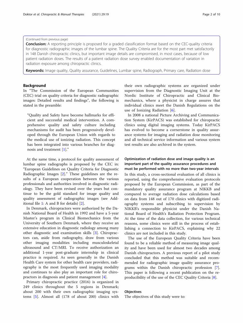
European guidelines on radiographic imagequality in chiropractic practice – proposalof a cross-sectional graded classificationreporting principleKlaus Doktor1,2,3* , Maria Lind Vilholm2, Aldis Hardardóttir4, Henrik Wulff Christensen3 and Jens Lauritsen5
Abstract
Background: The Commission of the European Communities (CEC) has published: European Guidelines on QualityCriteria for Diagnostic Radiographic Images. These guidelines are considered a gold standard, recommended foruse in quality assurance protocols.The objectives of this study: 1) Propose a graded classification format for Danish chiropractic clinics according tothe CEC-quality criteria for diagnostic radiographic images of the lumbar spine. 2) Propose a reporting principle forquality of radiographic images. 3) Document variation in radiation exposure among clinics.
Methods: This is a cross-sectional study of image quality based on random sampling from 148 chiropractic clinics.Clinics were included if using: 1) Digital radiography and 2) The chiropractic picture and archiving system (KirPACS)at the Nordic Institute of Chiropractic and Clinical Biomechanics (NIKKB) in Denmark. A sample of 296 lumbar spineseries were randomly collected from KirPACS (January 2018). Two independent observers reviewed 50 lumbar spineseries twice with a 4-week interval, testing intra- and inter-observer reproducibility. The same observers thenreviewed the remaining 246 radiographic studies. All studies were evaluated using the CEC Quality Criteria. Patientradiation dose values were retrieved from KirPACS (First quarter of 2020).
Results: A reporting and classification principle of diagnostic image quality was used in 148 chiropractic clinics.Compliance with the 22 CEC Quality Criteria had proportions ranging from 0.72–0.96 for 18 criteria, while 4 criteriaspecifying detail and definition ranged between 0.20–0.66. The proposed rating system (A to E) revealed: 18 Aclinics, 28 B clinics, 32 C clinics, 25 D clinics and 45 E clinics (A = highest quality; E = lowest quality). The patientradiation reference dose in Denmark is 7 mGy for the AP/PA lumbar spine. Very few clinics exceed the referencedose value, approximately 50% of clinics were below 5mGy.
(Continued on next page)
© The Author(s). 2021 Open Access This article is licensed under a Creative Commons Attribution 4.0 International License,which permits use, sharing, adaptation, distribution and reproduction in any medium or format, as long as you giveappropriate credit to the original author(s) and the source, provide a link to the Creative Commons licence, and indicate ifchanges were made. The images or other third party material in this article are included in the article's Creative Commonslicence, unless indicated otherwise in a credit line to the material. If material is not included in the article's Creative Commonslicence and your intended use is not permitted by statutory regulation or exceeds the permitted use, you will need to obtainpermission directly from the copyright holder. To view a copy of this licence, visit http://creativecommons.org/licenses/by/4.0/.The Creative Commons Public Domain Dedication waiver (http://creativecommons.org/publicdomain/zero/1.0/) applies to thedata made available in this article, unless otherwise stated in a credit line to the data.
* Correspondence: [email protected] Unit for Clinical Biomechanics, University of Southern Denmark,Campusvej 55, 5250 Odense M, Denmark2Private Chiropractic Practice, Back Center Midwestern Jutland, Dalgas Allé2A, 7400 Herning, DenmarkFull list of author information is available at the end of the article
Doktor et al. Chiropractic & Manual Therapies (2021) 29:19 https://doi.org/10.1186/s12998-021-00375-4
(Continued from previous page)
Conclusion: A reporting principle is proposed for a graded classification format based on the CEC-quality criteriafor diagnostic radiographic images of the lumbar spine. The Quality Criteria are for the most part met satisfactorilyin 148 Danish chiropractic clinics, but important image details are compromised, in most cases, because of lowpatient radiation doses. The results of a patient radiation dose survey enabled documentation of variation inradiation exposure among chiropractic clinics.
Keywords: Image quality, Quality assurance, Guidelines, Lumbar spine, Radiograph, Primary care, Radiation dose
BackgroundIn “The Commission of the European Communities(CEC) trial on quality criteria for diagnostic radiographicimages: Detailed results and findings”, the following isstated in the preamble:
“Quality and Safety have become hallmarks for effi-cient and successful medical intervention. A com-prehensive quality and safety culture includingmechanisms for audit has been progressively devel-oped through the European Union with regards tothe medical use of ionizing radiation. This concepthas been integrated into various branches for diag-nosis and treatment [1].”
At the same time, a protocol for quality assessment oflumbar spine radiographs is proposed by the CEC in:“European Guidelines on Quality Criteria for DiagnosticRadiographic Images [2].” These guidelines are the re-sults of a European cooperation between the variousprofessionals and authorities involved in diagnostic radi-ology. They have been revised over the years but con-tinue to be the gold standard for image quality andquality assessment of radiographic images (see Add-itional file 1: A and B for details) [2].In Denmark, chiropractors were authorized by the Da-
nish National Board of Health in 1992 and have a 5-yearMaster’s program in Clinical Biomechanics from theUniversity of Southern Denmark, where they receive anextensive education in diagnostic radiology among manyother diagnostic and examination skills [3]. Chiroprac-tors can, aside from radiography, draw from variousother imaging modalities including musculoskeletalultrasound and CT/MRI. To receive authorization anadditional 1-year post-graduate internship in clinicalpractice is required. As seen generally in the DanishHealth Care system for other health care providers, radi-ography is the most frequently used imaging modalityand continues to also play an important role for chiro-practors in diagnosis and patient management [4].Primary chiropractic practice (2016) is organized in
249 clinics throughout the 5 regions in Denmark;about 200 with their own radiographic imaging sys-tems [5]. Almost all (178 of about 200) clinics with
their own radiographic systems are organized undersupervision from the Diagnostic Imaging Unit at theNordic Institute of Chiropractic and Clinical Bio-mechanics, where a physicist in charge assures thatindividual clinics meet the Danish Regulations on theuse of Ionizing Radiation [6].In 2008 a national Picture Archiving and Communica-
tion System (KirPACS) was established for chiropracticclinics using digital imaging systems. Today KirPACShas evolved to become a cornerstone in quality assur-ance systems for imaging and radiation dose monitoringand all technical service information and various systemtest results are also archived in the system.
Optimization of radiation dose and image quality is animportant part of the quality assurance procedures andmust be performed with no more than two-year intervals
In this study, a cross-sectional evaluation of all clinics isreported, using the comprehensive evaluation protocolsproposed by the European Commission, as part of themandatory quality assurance program at NIKKB andcompared to average radiation dose calculations basedon data from 148 out of 170 clinics with digitized radi-ography systems and subscribing to supervision byNIKKB’s responsible physicist under the Danish Na-tional Board of Health’s Radiation Protection Program.At the time of the data collection, for various technicalreasons, some clinics were still in the process of estab-lishing a connection to KirPACS, explaining why 22clinics are not included in this study.The use of the European Quality Criteria have been
found to be a reliable method of measuring image qual-ity and have been used for almost two decades amongDanish chiropractors. A previous report of a pilot studyconcluded that this method was suitable and recom-mended for radiographic image quality assurance pro-grams within the Danish chiropractic profession [7].This paper is following a recent publication on the re-producibility of the use of the CEC Quality Criteria [8].
ObjectivesThe objectives of this study were to:
Doktor et al. Chiropractic & Manual Therapies (2021) 29:19 Page 2 of 10
1) Propose a reporting principle for individual clinicsin relation to quality of radiographic imaging.
2) Propose a graded classification format based on theCEC-quality criteria for diagnostic radiographic im-ages of the lumbar spine.
3) Document variation in radiation exposure amongchiropractic clinics in Denmark.
DesignThis is a double-blinded cross-sectional study of radio-graphic image quality based on random sampling.
Materials and methodsData collectionThe inter- and intra-observer reproducibility study in-cluded 50 radiographic studies of the lumbar spine andhas been reported in full detail in a separate paper [8].The assessment of the diagnostic quality of lumbar
spine radiographs in chiropractic practice in Denmark,included clinics: 1) Using digitalized radiographic
imaging systems and, 2) Storing studies in KirPACS atNIKKB in Odense, Denmark. The study was initiatedin January 2018 and was completed in the first quarterof 2020.The project was initiated by anonymizing and numbering
all studies. The study reviewers or clinicians obtaining the ra-diographs were blinded to the identity of patients/clinics andpotential participation in the quality assurance procedure.After randomly retrieving 2 studies per clinic from 148clinics in KirPACS, the studies were analyzed using theimage viewer Osirix version 5.7.1.for Mac and a digitized for-mat of the CEC: Quality Criteria for Diagnostic RadiographicImages. The results were tabulated directly into a softwaremodule made in Epidata Entry Client and Epidata Manager(version 2.0.7.22r547) [9]. Acceptance tested, high resolution(2 million pixel) diagnostic monitors (BARCO MDNC-2121)were used for the image evaluation process [10]. Two ob-servers, licensed chiropractors, with 2 years of clinical experi-ence handled the readings of all the samples. The twoobservers were blinded to patient and diagnostic information
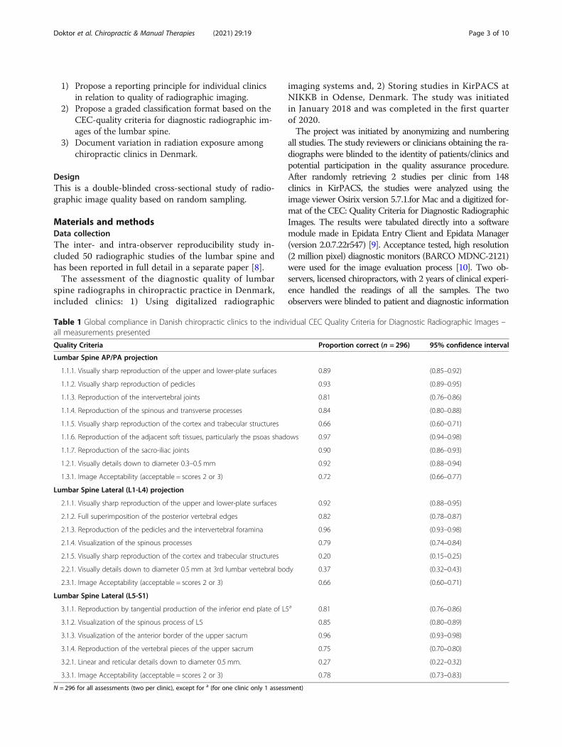
Table 1 Global compliance in Danish chiropractic clinics to the individual CEC Quality Criteria for Diagnostic Radiographic Images –all measurements presented
Quality Criteria Proportion correct (n = 296) 95% confidence interval
Lumbar Spine AP/PA projection
1.1.1. Visually sharp reproduction of the upper and lower-plate surfaces 0.89 (0.85–0.92)
1.1.2. Visually sharp reproduction of pedicles 0.93 (0.89–0.95)
1.1.3. Reproduction of the intervertebral joints 0.81 (0.76–0.86)
1.1.4. Reproduction of the spinous and transverse processes 0.84 (0.80–0.88)
1.1.5. Visually sharp reproduction of the cortex and trabecular structures 0.66 (0.60–0.71)
1.1.6. Reproduction of the adjacent soft tissues, particularly the psoas shadows 0.97 (0.94–0.98)
1.1.7. Reproduction of the sacro-iliac joints 0.90 (0.86–0.93)
1.2.1. Visually details down to diameter 0.3–0.5 mm 0.92 (0.88–0.94)
1.3.1. Image Acceptability (acceptable = scores 2 or 3) 0.72 (0.66–0.77)
Lumbar Spine Lateral (L1-L4) projection
2.1.1. Visually sharp reproduction of the upper and lower-plate surfaces 0.92 (0.88–0.95)
2.1.2. Full superimposition of the posterior vertebral edges 0.82 (0.78–0.87)
2.1.3. Reproduction of the pedicles and the intervertebral foramina 0.96 (0.93–0.98)
2.1.4. Visualization of the spinous processes 0.79 (0.74–0.84)
2.1.5. Visually sharp reproduction of the cortex and trabecular structures 0.20 (0.15–0.25)
2.2.1. Visually details down to diameter 0.5 mm at 3rd lumbar vertebral body 0.37 (0.32–0.43)
2.3.1. Image Acceptability (acceptable = scores 2 or 3) 0.66 (0.60–0.71)
Lumbar Spine Lateral (L5-S1)
3.1.1. Reproduction by tangential production of the inferior end plate of L5a 0.81 (0.76–0.86)
3.1.2. Visualization of the spinous process of L5 0.85 (0.80–0.89)
3.1.3. Visualization of the anterior border of the upper sacrum 0.96 (0.93–0.98)
3.1.4. Reproduction of the vertebral pieces of the upper sacrum 0.75 (0.70–0.80)
3.2.1. Linear and reticular details down to diameter 0.5 mm. 0.27 (0.22–0.32)
3.3.1. Image Acceptability (acceptable = scores 2 or 3) 0.78 (0.73–0.83)
N = 296 for all assessments (two per clinic), except for a (for one clinic only 1 assessment)
Doktor et al. Chiropractic & Manual Therapies (2021) 29:19 Page 3 of 10

and did not have access to previous readings, and effortswere made to minimize confounding factors, such as visibleclinic identification or modality manufacture information,during the readings. The observers were given 4weeks to fin-ish their evaluations and could log on and off to access theimages any time they wished. After the initial evaluations of50 studies for the reproducibility study (reported separately),the remaining studies were divided in two portions, one foreach observer. Observers could evaluate in any order theywished within the timeframe.
Statistics and proposed quality definition groupsAll analysis was performed using STATA 15 for Win-dows, Stata Corporation, USA [11] and Microsoft Excel2010, Microsoft Office Package, Microsoft Corporation,USA [12]. Statistical analysis was carried out at eithermeasurement level, clinic level or lumbar projectionlevel (AP/PA; L1-L4; L5/S1) as indicated in the tablesand graphs.Quality in this study was defined according to the
CEC Quality Criteria as: Ok = Sum of correct (accept-able) measurements; error = Sum of incorrect (not ac-ceptable) measurements; diff. = Difference betweenmeasurements within a given clinic (variation).All clinics were ranked according to counts of: “error”
(sum of not acceptable in both measurements), “Ok”(count of correct in both measurements) and “stability”
(number of differences in assessment of the two mea-surements) and classified in 5 percentile groups (A: 0–10, B: 11–40, C: 41–59; D: 60–89, E: 90+) with poorquality as (errors = highest; ok = lowest; diff = highest) foreach projection.All clinics were then classified on the combination of
the percentile groups for all projections and measure-ments, as follows: Overall A: (only A grading), B: (B or Agrading), C: (No D grading), D: (maximum one E grad-ing), E: (two or more E). Since probit plots showed thattotals for ok, error and diff were all reasonably Gaussiandistributed we also applied an alternative according to(A: < mean-2 SD; B <mean-1 SD; C: mean +/− 1 SD; D:> mean + 1 SD; E: >mean + 2 SD) based on I Chart graphvalues (EpiData Analysis, www.epidata.dk) for allmeasurements.
ResultsThese are the results of a study of radiographic imagequality based on “European Guidelines on Quality Cri-teria for Diagnostic Radiographic Images” (EUR 16260).The study was performed as part of the quality assur-ance program for 148 chiropractic clinics in Denmarkusing computerized radiography or direct radiography(CR or DR systems) in their primary care practice. Atotal of 296 lumbar spine studies were retrieved from
Table 2 Sum scores divided in: correct (compliance with quality criteria), errors (no compliance with quality criteria) and stability(same quality in both radiographic series per clinic) in assessments for all clinics. Mean (95% CI) and presented in percentile (p)groups with a proposed grading: A-E
Mean 95% conf. interval Min p10 p25 Median p75 p90 Max
Correctd
Proposed scoringaE D C B A
All projections 33.56 (32.69 34.43) 14 25 31 34 37 40 43
AP/PA 15.26 (14.88 15.65) 7 12 14 16 17 18 18
L1-L4 9.45 (9.11 9.78) 3 7 8 9 11 12 14
L5/S1 8.85 (8.50 9.21) 2 6 8 9 10 12 12
Errorc
Proposed scoringaA B C D E
All projections 10.42 (9.55 11.29) 1 4 7 10 13 19 30
AP/PA 2.72 (2.34 3.10) 0 0 1 2 4 6 11
L1-L4 4.55 (4.22 4.89) 0 2 3 5 6 7 11
L5/S1 3.14 (2.79 3.50) 0 0 2 3 4 6 10
Stabilityb
Proposed scoringaE D C B A
All projections 5.20 (4.72 5.68) 0 2 3 5 7 9 14
AP/PA 1.96 (1.70 2.21) 0 0 1 2 3 4 6
L1-L4 1.76 (1.56 1.95) 0 0 1 2 2 3 5
L5/S1 1.49 (1.28 1.69) 0 0 0 1 2 3 5aProposed cut point in grading A-E. Only used for the “all projections” group. A = highest gradebStability in quality measured as whether the two assessed radiographic series had the same scoring (ok versus error)cSum of errors in all assessments. d Sum of correct assessments. Classified in grades according to mean +/− SD criteria, see Materials and methods section
Doktor et al. Chiropractic & Manual Therapies (2021) 29:19 Page 4 of 10
KirPACS, analyzed and scored according to the pro-posed image criteria.
Compliance with the CEC diagnostic image quality criteriaIn Table 1, the global results for 148 Danish chiropracticclinics is presented for Lumbar Spine projections (AP/PA, Lateral L1-L4 and Lateral L5/S1), as a percentage ofthe total sample size fulfilling the individual CEC Diag-nostic Quality Criteria in this study.
Proposed grading system for individual clinicsIn Table 2 the results for individual clinics are presentedas a percentage of the fulfilled CEC Diagnostic QualityCriteria for all projections and divided into individualprojections: Lumbar AP/PA, Lateral L1-L4 and LateralL5/S1 projections. This allows for a ranking of clinics bypercentiles from maximum to minimum. Comparison ofclinics is crucial in pinpointing potential problem areasregarding the imaging quality. The maximal achievablescores for all projections are 44 points (AP/PA: 9 vari-ables, Lateral L1-L4: 7 variables and, Lateral L5/S1: 6
variables = 22 variables total per series). The combinedscore for both series is: All projections 22 × 2 = 44 (AP/PA = 18, Lateral L1-L4 = 14 and Lateral L5/S1 = 12).The proposed grading makes it possible to combine all
grades and present a final ranking of clinics based on a3-letter classification/grading system, as can be seenbelow in Table 3.
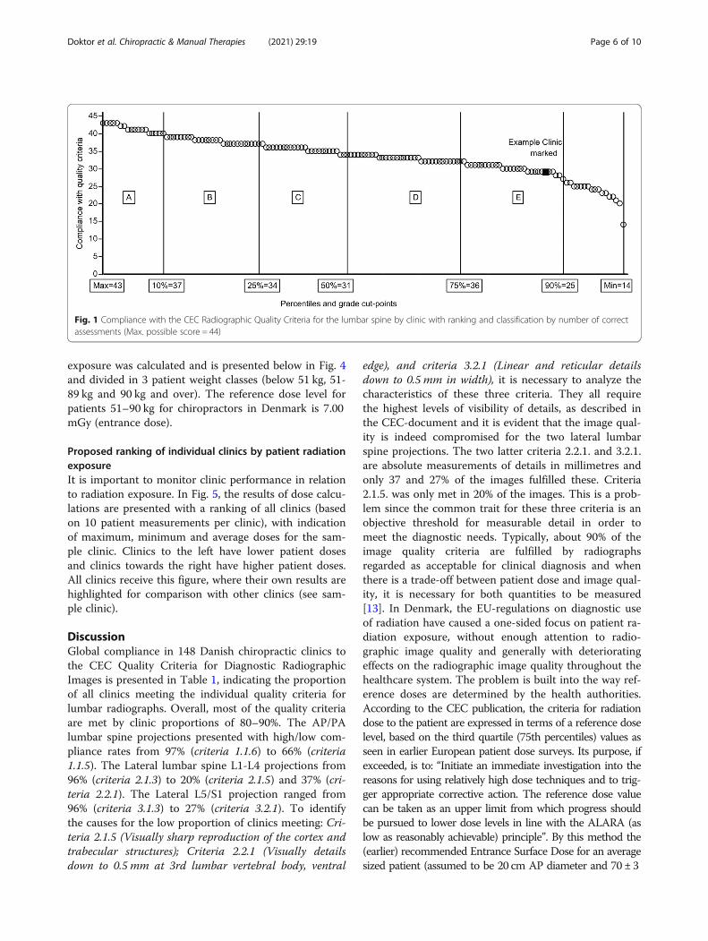
Proposed reporting principle for individual clinics inrelation to image quality in overviewOne of our objectives was to propose a reportingprinciple for individual clinics to present individual re-sults in a simple and clear format, making it possible tocompare the results with the rest of the group, as can beseen in Fig. 1 below.
Variation in radiation exposure among clinics withelectronic image storage (KirPACS)Our last objective was to document the variation in radi-ation exposure among clinics connected to the KirPACS.Based on reporting to the responsible physicist, radiation
Table 3 Median and range of errors and correct for all assessments ranked according to overall classification of clinics
Gradea NumberofClinics
OK assessments (out of possible 44) Errors (out of possible 44)
median range median range
AAA 7 43 (41–43) 1 (1–3)
AAB 5 40 (40–42) 4 (2–4)
AAC 6 41 (40–41) 3 (3–4)
BBA 5 37 (37–39) 7 (5–7)
BBB 2 38 (38–38) 6 (6–6)
BBC 10 38 (37–39) 6 (5–7)
BBD 9 38 (37–39) 6 (5–7)
BBE 2 37 (37–37) 7 (7–7)
CCB 10 36 (34–36) 8 (8–10)
CCC 9 35 (35–36) 9 (8–9)
CCD 10 34 (34–35) 10 (9–10)
CCE 3 34 (34–34) 10 (10–10)
DCE 1 33 (33–33) 10 (10–10)
DDC 6 32 (32–33) 12 (11–12)
DDD 10 33 (32–33) 12 (11–12)
DDE 8 33 (32–33) 11 (11–12)
EDD 3 31 (31–31) 13 (13–13)
EDE 7 31 (31–31) 13 (12–13)
EEB 1 24 (24–24) 20 (20–20)
EEC 2 28 (26–30) 16 (14–18)
EED 9 25 (20–29) 19 (15–24)
EEE 23 28 (14–30) 16 (14–30)aGrade nomination: First letter = Grading according to Ok. Second letter = Grading according to error. Third letter = Grading according to difference between thetwo assessments. Grades are defined according to ranking of all clinics. OK: Best to have a high number of OK = Grade AAA, and worst to have a low number ofOK = Grade EEE. A: top 10% of the entire group; B: 11–25%; C: 26–50%; D: 51–75%; E: bottom 76–100%. Error + differences: Best to have a low number of Errorsand/or Differences between x-ray series (homogenizes). Scale now reversed. Grade A: bottom 10% etc.
Doktor et al. Chiropractic & Manual Therapies (2021) 29:19 Page 5 of 10
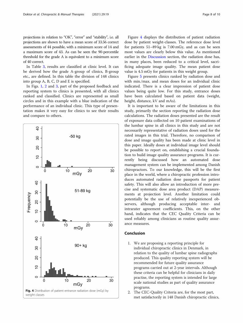
exposure was calculated and is presented below in Fig. 4and divided in 3 patient weight classes (below 51 kg, 51-89 kg and 90 kg and over). The reference dose level forpatients 51–90 kg for chiropractors in Denmark is 7.00mGy (entrance dose).
Proposed ranking of individual clinics by patient radiationexposureIt is important to monitor clinic performance in relationto radiation exposure. In Fig. 5, the results of dose calcu-lations are presented with a ranking of all clinics (basedon 10 patient measurements per clinic), with indicationof maximum, minimum and average doses for the sam-ple clinic. Clinics to the left have lower patient dosesand clinics towards the right have higher patient doses.All clinics receive this figure, where their own results arehighlighted for comparison with other clinics (see sam-ple clinic).
DiscussionGlobal compliance in 148 Danish chiropractic clinics tothe CEC Quality Criteria for Diagnostic RadiographicImages is presented in Table 1, indicating the proportionof all clinics meeting the individual quality criteria forlumbar radiographs. Overall, most of the quality criteriaare met by clinic proportions of 80–90%. The AP/PAlumbar spine projections presented with high/low com-pliance rates from 97% (criteria 1.1.6) to 66% (criteria1.1.5). The Lateral lumbar spine L1-L4 projections from96% (criteria 2.1.3) to 20% (criteria 2.1.5) and 37% (cri-teria 2.2.1). The Lateral L5/S1 projection ranged from96% (criteria 3.1.3) to 27% (criteria 3.2.1). To identifythe causes for the low proportion of clinics meeting: Cri-teria 2.1.5 (Visually sharp reproduction of the cortex andtrabecular structures); Criteria 2.2.1 (Visually detailsdown to 0.5 mm at 3rd lumbar vertebral body, ventral
edge), and criteria 3.2.1 (Linear and reticular detailsdown to 0.5 mm in width), it is necessary to analyze thecharacteristics of these three criteria. They all requirethe highest levels of visibility of details, as described inthe CEC-document and it is evident that the image qual-ity is indeed compromised for the two lateral lumbarspine projections. The two latter criteria 2.2.1. and 3.2.1.are absolute measurements of details in millimetres andonly 37 and 27% of the images fulfilled these. Criteria2.1.5. was only met in 20% of the images. This is a prob-lem since the common trait for these three criteria is anobjective threshold for measurable detail in order tomeet the diagnostic needs. Typically, about 90% of theimage quality criteria are fulfilled by radiographsregarded as acceptable for clinical diagnosis and whenthere is a trade-off between patient dose and image qual-ity, it is necessary for both quantities to be measured[13]. In Denmark, the EU-regulations on diagnostic useof radiation have caused a one-sided focus on patient ra-diation exposure, without enough attention to radio-graphic image quality and generally with deterioratingeffects on the radiographic image quality throughout thehealthcare system. The problem is built into the way ref-erence doses are determined by the health authorities.According to the CEC publication, the criteria for radiationdose to the patient are expressed in terms of a reference doselevel, based on the third quartile (75th percentiles) values asseen in earlier European patient dose surveys. Its purpose, ifexceeded, is to: “Initiate an immediate investigation into thereasons for using relatively high dose techniques and to trig-ger appropriate corrective action. The reference dose valuecan be taken as an upper limit from which progress shouldbe pursued to lower dose levels in line with the ALARA (aslow as reasonably achievable) principle”. By this method the(earlier) recommended Entrance Surface Dose for an averagesized patient (assumed to be 20 cm AP diameter and 70 ± 3
Fig. 1 Compliance with the CEC Radiographic Quality Criteria for the lumbar spine by clinic with ranking and classification by number of correctassessments (Max. possible score = 44)
Doktor et al. Chiropractic & Manual Therapies (2021) 29:19 Page 6 of 10

kg) was derived at 10mGy for AP/PA, 30mGy for laterallumbar spine and 40mGy for lateral lumbosacral spine.Paradoxically, the administration of the crucial dose/qualitybalance, has caused more problems for workplaces and pro-fessions, the reason being that new and lower dose referencevalues are implemented by authorities after the third quartileprinciple. In other words, when chiropractic clinics (and hos-pitals), because of dose/image optimization, have becomemore and more homogeneous, the reference dose has re-peatedly been lowered, thus pushing the radiation dosesbelow the point of acceptable balance between dose andquality. At the time when image evaluations in this studywere performed, the reference dose had already been low-ered from 10 to 7mGy for an AP/PA lumbar spine [6, 14,15]. Today, the value is close to half of the initially recom-mended dose of 10mGy [16]. This was never the intention
as described in CEC-publication EUR 16260 [2] and in ICRP90 and 103, and adding the fact that there has been a transi-tion from film-based to less sensitive digital radiography, thishas further challenged the diagnostic image quality. Methodsfor measurement of patient dose are comparatively wellestablished. Assessment of image quality is less straight for-ward. Image quality is affected by resolution, sensitivity andstatistical noise [13].In Table 2, results are shown at projection level. We
pooled all projections and ranked according to scoresproducing a classification system, specifying the associ-ation between grades (A-E) and scoring, based on theachieved scores for all three projections combined (max.44 points possible) and then divided into projections andclassified in percentiles (p10, p25, p50, p75 and p90). This al-lows us to compare projections and to have a general view of
Fig. 2 Non-compliance with the CEC Radiographic Quality Criteria for the lumbar spine by clinic with ranking and grouping by number ofincorrect (error) assessments. Percentiles and grade cut-points
Fig. 3 Stability of lumbar spine Radiographic Quality by clinic with ranking and grouping by number of differences in assessment. Percentiles andgrade cut-points
Doktor et al. Chiropractic & Manual Therapies (2021) 29:19 Page 7 of 10
projections in relation to “OK”, “error” and “stability”, i.e. allprojections are shown to have a mean score of 33.56 correctassessments of 44 possible, with a minimum score of 14 anda maximum score of 43. As can be seen the 90-percentilethreshold for the grade A is equivalent to a minimum scoreof 40 correct.In Table 3, results are classified at clinic level. It can
be derived how the grade A-group of clinics, B-groupetc., are defined. In this table the division of 148 clinicsinto group A, B, C, D and E is specified.In Figs. 1, 2 and 3, part of the proposed feedback and
reporting system to clinics is presented, with all clinicsranked and classified. Clinics are represented as smallcircles and in this example with a blue indication of theperformance of an individual clinic. This type of presen-tation makes it very easy for clinics to see their resultsand compare to others.
Figure 4 displays the distribution of patient radiationdose by patient weight-classes. The reference dose levelfor patients 51–89 kg is 7.00 mGy, and as can be seenmost values are clearly below this value. As mentionedearlier in the Discussion section, the radiation dose has,in many places, been reduced to a critical level, sacri-ficing adequate image quality. The mean patient dosevalue is 4.5 mGy for patients in this weight group.Figure 5 presents clinics ranked by radiation dose and
with min./max. and mean doses for an individual clinicindicated. There is a clear impression of patient dosevalues being quite low. For this study, entrance doseshave been calculated based on patient data (weight,height, distance, kV and mAs).It is important to be aware of the limitations in this
study, primarily the section reporting the radiation dosecalculations. The radiation doses presented are the resultof exposure data collected on 10 patient examinations ofthe lumbar spine in all clinics in this study and are notnecessarily representative of radiation doses used for therated images in this trial. Therefore, no comparison ofdose and image quality has been made at clinic level inthis paper. Ideally doses at individual image level shouldbe possible to report on, establishing a crucial founda-tion to build image quality assurance programs. It is cur-rently being discussed how an automated dosemanagement system can be implemented among Danishchiropractors. To our knowledge, this will be the firstplace in the world, where a chiropractic profession intro-duces automated radiation dose passports for patientsafety. This will also allow an introduction of more pre-cise and systematic dose area product (DAP) measure-ments at projection level. Another limitation couldpotentially be the use of relatively inexperienced ob-servers, although producing acceptable inter- andintrarater agreement coefficients. This, on the otherhand, indicates that the CEC Quality Criteria can beused reliably among clinicians as routine quality assur-ance measures.
Conclusion
1. We are proposing a reporting principle forindividual chiropractic clinics in Denmark, inrelation to the quality of lumbar spine radiographsproduced. This quality reporting system will berecommended for future quality assuranceprograms carried out at 2-year intervals. Althoughthese criteria can be helpful for clinicians in dailypractise, the reporting system is intended for largescale national studies as part of quality assuranceprograms.
2. The CEC-Quality Criteria are, for the most part,met satisfactorily in 148 Danish chiropractic clinics,
Fig. 4 Distribution of patient entrance radiation dose (mGy) byweight classes
Doktor et al. Chiropractic & Manual Therapies (2021) 29:19 Page 8 of 10

but important image details are generally compro-mised for the lateral lumbar spine projections, inmost cases, because of low patient radiation doses.This is not acceptable and needs attention.
3. It is also proposed that a graded classificationformat based on the CEC-quality criteria for diag-nostic radiographic images of the lumbar spine isimplemented at national level. This classificationscheme can be carried out at clinic level and atimage projection level. It is recommended that re-sources, also internationally, are allocated to imple-ment the proposed scheme or similar.
4. The results of a patient radiation dose survey haveenabled a documentation of variations in radiationexposures among chiropractic clinics with electronicimage storage (KirPACS). The new EU-regulative onthe use of ionizing radiation for diagnostic imaging isin effect and necessitates DAP-meters to be installedon all radiographic installations in chiropractic prac-tice. It is recommended that resources are raised forthis implementation and for a central administereddose monitoring system.
5. A quality system could be implemented globally toensure a high standard of radiographs produced inchiropractic clinics.
AbbreviationsCEC: Commission of the European Communities; PACS: Picture Archiving andCommunication System; NIKKB: Nordic Institute for Chiropractic and ClinicalBiomechanics; KirPACS: Chiropractic Picture Archiving and CommunicationSystem; CR: Computed radiography; DR: Direct radiography; AP: Fromanterior to posterior; PA: From posterior to anterior; L1: First lumbar vertebra;L4: Fourth lumbar vertebra; L5: Fifth lumbar vertebra; S1: First sacral vertebra;L/S: Lumbo-sacral junction; DAP: Dose area product
Supplementary InformationThe online version contains supplementary material available at https://doi.org/10.1186/s12998-021-00375-4.
Additional file 1.
AcknowledgementsThe authors would like to thank Orla Lund Nielsen for kindly giving adviceand assistance in managing data and also, Sara Lisa Doktor for proofreadingthe manuscript.
Authors’ contributionsAH, MLV contributed to the conception of the study, evaluated theradiographs and tabulated the results into Epidata Entry Client. KDperformed the interpretation and analysis of data in collaboration with JLand drafted the manuscript. HWC supervised, modified and proofread themanuscript. All authors read, critically reviewed and approved the finalversion to be submitted for publication.
Authors’ informationInformation on authors qualifications and affiliations is found on the firstpage of this article.
FundingThe authors would like to acknowledge funding from the Foundation forChiropractic and Clinical Biomechanics. The funding body had no other rolein the project neither the design of the study and collection, analysis, andinterpretation of data or in writing the manuscript.
Availability of data and materialsThe datasets used and/or analyzed during the current study are availablefrom the corresponding author on reasonable request.
Declarations
Ethics approval and consent to participateNot applicable. This study was done in conjunction with quality assuranceprocedures required by Danish law and all personal data were blinded forthe observers. Only documentation of image quality was performed, with nopossible correlation to patients. The procedures are mandatory and has noconsequences for diagnoses and treatment of patients.
Fig. 5 Clinics ranked by radiation dose. Min./max. and mean doses will be displayed in reports for each clinic individually, as seen highlighted fora sample clinic
Doktor et al. Chiropractic & Manual Therapies (2021) 29:19 Page 9 of 10
Consent for publicationNot applicable.
Competing interestsNo competing interests declared.
Author details1Research Unit for Clinical Biomechanics, University of Southern Denmark,Campusvej 55, 5250 Odense M, Denmark. 2Private Chiropractic Practice, BackCenter Midwestern Jutland, Dalgas Allé 2A, 7400 Herning, Denmark. 3NordicInstitute of Chiropractic and Clinical Biomechanics, Campusvej 55, 5230Odense M, Denmark. 4Private Chiropractic Practice, Reykjavik, Iceland.5Institute of Clinical Medicine, University of Southern Denmark andOrthopedic Department, Odense University Hospital, Odense, Denmark.
Received: 28 January 2021 Accepted: 13 April 2021
References1. Maccia C, Moores BM, Wall BF. The 1991 CEC trial on quality criteria for
diagnostic radiographic images: detailed results and findings; 1996.2. Carmichael JHE, Maccia C, Moores BM, Oestmann JW, Schbilla H, et al. 2000.
European guidelines on quality criteria for diagnostic radiographic images.EU publication EUR 16260.
3. Autorisationsloven 2019. https://www.retsinformation.dk/eli/lta/2019/73.4. Radiologiske undersøgelser 2013-2018.https://www.esundhed.dk/Registre/La
ndspatientsregisteret/Radiologiske-ydelser.5. Danish Chiropractor Association. KiroFAKTA 2016. https://d1gyukz65nrk4d.
cloudfront.net/KiroFAKTA_2016.pdf.6. Sundhedsstyrelsen. Lov nr 23 af 15/1/2018 Lov om ioniserende stråling og
strålebeskyttelse (strålebeskyttelsesloven). Copenhagen: Sundheds- ogÆldreministeriet; 2018. https://www.isa.au.dk/documentation/safety/docs/8_Bekendtg%C3%B8relse%20669%20om%20ioniserende%20str%C3%A5ling%20og%20str%C3%A5lebeskyttelse.pdf.
7. Doktor KK. The use of the CEC quality criteria for radiographic images inchiropractic practise. Odense: Nordic Institute for Chiropractic and ClinicalBiomechanics; 2000.
8. Doktor K, Vilholm ML, Hardardottir A, Christensen HW, Lauritsen J. Europeanguidelines on quality criteria for diagnostic radiographic images of thelumbar spine - an intra- and inter-observer reproducibility study. ChiroprMan Therap. 2019;27(1):20. https://doi.org/10.1186/s12998-019-0241-3.
9. Lauritsen J, Bruus M. 2003-2018. Epidata. The Epidata Association, Odense.http://www.epidata.dk.
10. Barco. Nio 2MP (MDNC-2121) Specification sheet, Article number K9601651.Belgium. https://www.barco.com/en/product/nio-color-2mp-led-dl-mdnc-2221. Accessed 20 Apr 2021.
11. Statacorp. Stata statistical software: release 14. College Station: StataCorpLLC; 2015. http://www.stata.com
12. Microsoftcorp. Microsoft Office 2010 package. USA: Microsoft Corporation;2018. http://www.microsoft.com
13. Martin CJ, Sharp PF, Sutton DG. Measurement of image quality in diagnosticradiology. Appl Radiat Isot. 1999;50(1):21–38. https://doi.org/10.1016/S0969-8043(98)00022-0.
14. Sundhedsstyrelsen. Bekendtgørelse om medicinske røntgenanlæg tilundersøgelse af patienter, nr. 975 af 16. december 1998. https://www.retsinformation.dk/eli/lta/1998/975.
15. Danish National Board of Health. 2012. Vejledning om patientdoser ogreferencedoser for røntgenundersøgelser Konventionellerøntgenundersøgelser 2.
16. Radiation Protection Danish National Board of Health. 2017. Referencedoserfor røntgenundersøgelse af columna lumbalis - Kiropraktorer. S4.
Publisher’s NoteSpringer Nature remains neutral with regard to jurisdictional claims inpublished maps and institutional affiliations.
Doktor et al. Chiropractic & Manual Therapies (2021) 29:19 Page 10 of 10