Embed Size (px)
Citation preview

ORIGINAL ARTICLE
Abstract Background: Envenomation by Macrovipera lebetina (M. lebetina) is characterized by prominent local tissue damage, hemorrhage, abnormalities in the blood coagulation system, necrosis, and edema. However, the main cause of death after a bite by M. lebetina has been attributed to acute renal failure (ARF). It is unclear whether the venom components have a direct or indirect action in causing ARF. To investigate this point, we looked at the in vitro effect of M. lebetina crude venom, using cultured human embryonic kidney (HEK-293) mono layers as a model. Methods: The effect of M. lebetina snake venom on HEK-293 growth inhibition was determined by the MTT assay and the neutral red uptake assay. The integrity of the cell membrane through LDH release was measured with the Cytotoxicity Detection Kit. Morphological changes in HEK-293 cells were also evaluated using an inverted microscope. Results: In the MTT assay, crude venom showed a significant cytotoxic effect on HEK-293 cells at 24 hours of exposure and was confirmed by the neutral red assay. Also, at 24 hours exposure, crude venom caused a non-significant increase in LDH activity of the culture medium at concentrations above 20 μg/ml. Various morphological abnormalities were observed in cells exposed to the venom and showed loss of their common polygonal shape, appearing as several roughly rounded cells of variable size. The M. lebetina crude venom induced detachment of cells from the plate. Conclusion: Based on the results obtained in this study, it can be concluded that the Iranian snake M. lebetina venom causes a cytotoxic effect on kidney tissue not by necrotic mechanism but rather by secondary effects, including hypotension, hemolysis, hemoglobinuria, rhabdomyolysis, myoglobinuria and disseminated intravascular coagulation (DIC), which may lead to ARF. Keywords: Snake venom; Cytotoxicity effect; HEK-293; Acute renal failure; Macrovipera lebetina
Evaluation of Iranian snake ‘Macrovipera lebetina’ venom cytotoxicity in kidney cell line HEK-293
49
of snakebite-related acute renal failure (ARF), include hypotension, hemolysis and hemoglobinuria, rhabdomyolysis and myoglobinuria, disseminated intravascular coagulation (DIC), and the direct effect of the venom. Among all these mechanisms, the cytotoxic effect of the venom on the kidney has been considered by some investigators to play a major role in the pathogenesis of ARF. Some effects are identified in the cell undergoing renal failure. These cells may undergo necrosis, apoptosis, and cell division or behave indifferently under stress. Necrosis and apoptosis are currently the most studied forms of cell death (10-12). There is scarce knowledge about cellular renal injury and direct cytotoxic effect induced by M. lebetina (12, 13). It is still unclear whether ARF is the result of a direct cytotoxic effect on renal epithelia or the result of renal ischemia due to systemic hemodynamic disturbances. This study aims to characterize the in vitro cytotoxic effect of M. lebetina crude venom on human embryonic kidney 293 cells (HEK-293).
More than 5 million people are bitten by venomous snakes
annually and more than 100,000 of them die, mostly in Asia (1). Among venomous snakes, the most important groups causing envenomation are Elapidae, Crotalidae and Viperidae (1-3). Macrovipera lebetina (M. lebetina) has an extensive geographical range throughout central Asia and the Middle East (4). M. lebetina is one of the most venomous snakes on the Iranian plateau with several enzymes, proteins and peptides, such as metalloproteases, serine proteases, phosphodiesterase, phospholipase A2s, L-amino acid oxidase, disintegrins and C-type lectins having different toxicological functions which cause local and systemic damage, including the acute kidney injury (5-9). These various factors may be due to a number of specific injuries that occur at systemic and cellular levels (e.g. metalloproteinases, disintegrins). Mechanisms that have been incriminated in the pathogenesis __________________
INTRODUCTION
*Correspondence to: Abbas Zare Mirakabadi; Ph.D in Biochemistry, Department Of Venomous Animals and Anti venom Production, Razi Vaccine and Serum Research Institute, Karaj, Agricultural Research, Education and Extension Organization (AREEO), Tehran, Iran. Tel: +989123198121, E mail: [email protected] Received 20 December 2015; Accepted 15 March 2016
HOURIEH ESMAEILI JAHROMI1, ABBAS ZARE MIRAKABADI2, MORTEZA KAMALZADEH3
1 Department of Biology, Payame Noor University (PNU), Tehran, Iran. 2 Department of Venomous Animals and Anti venom Production, Razi Vaccine and Serum Research Institute, Karaj, Agricultural Research, Education and Extension Organization (AREEO), Tehran, Iran. 3 Department of Quality control, Razi Vaccine and Serum Research Institute, Karaj, Iran.
How to cite this article: Esmaeili Jahromi H, Zare Mirakabadi A, Kamalzadeh M. Evaluation of Iranian snake ‘Macrovipera lebetina’ venom cytotoxicity in kidney cell line HEK-293. Asia Pac J Med Toxicol 2016;2:49-54.
ASIA PACIFIC JOURNAL of MEDICAL TOXICOLOGY APJMT 5;2 http://apjmt.mums.ac.ir June 2016

50
Macrovipera lebetina venom cytotoxicity H. E. Jahromi et al.
Materials Iranian M. lebetina was obtained from the Venomous
Animals and Antivenin Production Dep., Razi vaccine and Serum Research institute, Karaj- Iran. The human embryonic kidney 293 (HEK-293) was obtained from the Cell Bank (Razi Vaccine and Serum Research Institute, Karaj, Iran). 3-(4, 5-dimethylthiaol-2-yl)-2, 5-diphenyltetrazolium bromide (MTT) and neutral red dye (NR) were obtained from Sigma (St Louis, MO, USA). Cytotoxicity Detection Kit of lactate dehydrogenase enzyme (LDH) was purchased from Pars Azmoon (Tehran, Iran). DMEM-F12 medium, penicillin/ streptomycin solutions, fetal bovine serum (FBS), and Trypsin-EDTA were obtained from Invitrogen (Carlsbad, CA, USA).
Venom preparation 1. Stock solution: Lyophilized venom was dissolved in
sterile normal saline solution and the final concentration was adjusted to 1 mg/ml and preserved at -50 Cº until use.
2. Working solution: The dissolved stock solution of snake venom was diluted by media culture (DMEM) to reach the concentrations 1, 5, 10, 20, 40 and 80 µg/ml.
Cell culture HEK-293 cells were cultured in DMEM medium with the
addition of FBS (10%, v/v), streptomycin (100 μg/ml) and penicillin (100 U/ml). The cells (2 × 10⁴) were seeded, in triplicate, in 96-well plates and incubated at 37°C in 5% CO2 atmosphere.
MTT assay HEK-293 cells were cultured in DMEM-F12 medium in
the presence of FBS 10% plus penicillin-streptomycin 1% and incubated in CO2 5% at 37 °C. The cytotoxicity of M. lebetina crude venom was evaluated using the MTT assay (14). HEK-293 cells were seeded in a 96-well plate at 20,000 cells/ well and incubated for 3 and 24 hours to adhere. After discarding the old medium, the cells were incubated in the medium containing various concentrations (1, 5, 10, 20, 40, 80 μg/ml) of crude venom. After 3 and 24 hours of incubation, 20 μL MTT (5 mg/ml) was added to each well and cells were incubated for another 3 hours. Finally, the culture medium containing MTT solution was removed and formazan crystals were dissolved in 100 μL of dimethyl sulfoxide solvent (DMSO) and the plate underwent gentle shaking for 10 min. absorbance of each well was read at 570 nm using an ELISA plate reader. A blank well, which contained only culture medium, was used for background correction.
Neutral red uptake assay The M. lebetina crude venom cytotoxicity was determined
using neutral red (NR) assay (15). Briefly, cells were seeded into a 96-well plate at 20,000 cells/well. After 3 and 24 hours, cells were treated to 1, 5, 10, 20, 40, 80 μg/ml crude M. lebetina venom concentrations for 24 hours. Then, the wells medium were replaced with a new one containing NR (40 μg/ml). After 3 hours of incubation, neutral red medium _______
was removed and the cells were washed with PBS for the remaining dye. Finally, neutral red destain solution (50% from ethanol 96%, deionized water 49% and glacial acetic acid 1%) was added to each well and the plate underwent gentle shaking for 20 min. Optical density (OD) of neutral red extract was read with a Synergy HT Microplate Reader (Bio-Tek Instruments, Winooki, VT) at 540 nm.
LDH release assay Cytotoxicity induced by M. lebetina crude venom was also
assessed by LDH release into the culture medium. The HEK-293 cells were treated at 1, 5, 10, 20, 40, 80 μg/ml concentrations of crude venom for 3 and 24 hours. After incubation, the cells media were transferred into corresponding wells, which were optically clear with a 96-well flat bottom plate. The released LDH in the media was measured with the Cytotoxicity Detection Kit (Pars Azmoon, Tehran, Iran).
Morphological studies Following overnight incubation of the cells with venom,
various morphological alterations and cell damages were qualitatively investigated using an inverted microscope (Nikon) and photos were taken with a digital camera.
Statistical analysis Values are expressed as means ± SD of four repeats in each
group. Data were analyzed using student’s t-test with statistical significance of p<0.05. All statistical analyses were performed using SigmaPlot 12 software.
Morphological studies The HEK-293 cells cultured in medium showed
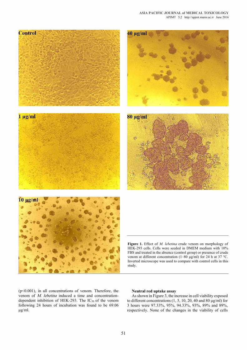
homogeneous distribution in the culture field exhibiting a polygonal shape with distinct boundaries. However, the occurrence of various morphological abnormalities was observed in cells exposed to various concentrations of venom and showed loss of their common polygonal shape, appearing as numerous roughly rounded cells of variable size. Moreover, the M. lebetina crude venom induced detachment of the cells from the plate. Areas devoid of cells were also recorded (Figure1).
MTT Assay The cytotoxic effects of various concentrations of crude
venom on cultured HEK-293 cells exposed for 3 and 24 hours were measured by the MTT assay. The results showed concentration-dependent reductions in the number of viable HEK-293 cells in response to treatment with M. lebetina venom. As shown in Figure 2, the decreased cell viability at various concentrations (1, 5, 10, 20, 40 and 80 μg/ml) for 3 hours exposure to the venom were 97%, 93.66%, 98.33%, 93.66%, 91.33% and 87.33%, respectively; these were not statistically significant. However, changes in viability after 24 hours exposure of the cells to the above mentioned concentrations of venom were 94%, 80.33%, 72.33%, 69.33%, 60.33% and 50.33%, respectively. The changes to the cells, viability were significant (p<0.05), (p<0.01), _______________
MATERIALS AND METHODS
RESULTS
ASIA PACIFIC JOURNAL of MEDICAL TOXICOLOGY APJMT 5;2 http://apjmt.mums.ac.ir June 2016
(p<0.001), in all concentrations of venom. Therefore, the venom of M. lebetina induced a time and concentration-dependent inhibition of HEK-293. The IC50 of the venom following 24 hours of incubation was found to be 69.06 μg/ml.
51
Neutral red uptake assay As shown in Figure 3, the increase in cell viability exposed
to different concentrations (1, 5, 10, 20, 40 and 80 μg/ml) for 3 hours were 97.33%, 95%, 94.33%, 93%, 89% and 89%, respectively. None of the changes in the viability of cells __________
Figure 1. Effect of M. lebetina crude venom on morphology of HEK-293 cells. Cells were seeded in DMEM medium with 10% FBS and treated in the absence (control group) or presence of crude venom at different concentration (1–80 μg/ml) for 24 h at 37 °C. Inverted microscope was used to compare with control cells in this study.

52
Macrovipera lebetina venom cytotoxicity H. E. Jahromi et al.
were found to be statistically significant. However, exposure of cells for 24 hours to various concentrations of venom caused significant reduced viability of cells. The IC50 of the venom following 24 hours incubation was 67.18 μg/ml.
LDH release assay Figure 4 shows the results of cytosolic LDH release of
HEK-293 cells when exposed to M. lebetina venom for 3 and 24 hours. Although there was an increase in LDH activity of culture medium, none of changes were statistically significant.
Macrovipera lebetina, a venomous snake in Iran, has
several enzymes, proteins and peptides that cause local and systemic damage. The venom can target the hemostatic system; prevent blood coagulation/platelet aggregation degrading fibrinogen; disrupt the extracellular matrix of the vascular subendothelium; increase the permeability of blood capillaries and promote hypotension; exert hemolytic and myotoxic effects; and lead to acute kidney injury (16, 17). In the present study, we evaluated the in vitro cytotoxicity of M. lebetina crude venom by exposing HEK-293 cells to various concentrations of crude venom for 3 and 24 hours. In our study, the crude venom induced detachment of cells from the plate. This could be due to metalloproteinases and disintegrin (18, 19). Our data demonstrated cytotoxic effects of the venom on HEK-293 cells only at 24 hours exposure, showing that the effect of the venom is time dependent. At 24 hours exposure the results also revealed that the effect is dose dependent. The cytotoxic effects of M. lebetina venom on cancer cells have been previously reported. Samel et al. studied the effect of the venom on the human prostate cancer ____________
(PC-3) cell-line and found that it can inhibit cell proliferation in a concentration and time-dependent manner (6). M. lebetina crude venom cytotoxicity has also been investigated in other cell lines such as mouse fibroblastic (L929) cells, human lung adenocarcinoma (A549) cells, human cervix adenocarcinoma (HeLa) cells, human colorectal adenocarcinoma (CaCo- 2) cells, human glioblastoma–astrocytoma (U-87MG) cells, human breast adenocarcinoma (MCF-7) cells, human umbilical vein endothelial cells
____________
Concentration (g/ml)
0 1 5 10 20 40 80
Cel
l via
bilit
y (%
of
cont
rol)
0
20
40
60
80
100
120
HEK-293 - 3 hours
HEK-293 - 24 hours
*
**
*
*****
**
Figure 2. Cytotoxic effects of M. lebetina crude venom on HEK-293 cells. Cell viability of HEK-293 cells treatment with different concentrations of M. lebetina (1, 5, 10, 20, 40&80 μg/ml) venom at 3 and 24 h, as measured by MTT assay. The control value (without venom) was set as 100%, Data are means ±SD from three independent determinations, in triplicate.*p<0.05, **p<0.01and ***p<0.001 were considered statistically significant, compared with controls.
Concentration (g/ml)
0 1 5 10 20 40 80
Cel
l vi
abili
ty (
% o
f co
ntro
l)
0
20
40
60
80
100
120
HEK-293 - 3 hours
HEK-293 - 24 hours
*
**
**
***
***
***
Figure 3. Cytotoxic effects of M. lebetina crude venom on HEK-293 cells. Cell viability of HEK-293 cells treatment with different concentrations of M. lebetina (1, 5, 10, 20, 40&80 μg/ml) venom at 3and 24 h, as measured by neutral red uptake assay. The control value (without venom) was set as 100%, Data are means ±SD from three independent determinations, in triplicate.*p<0.05, **p<0.01and ***p<0.001were considered statistically significant, compared with controls.
Concentration (g/ml)
0 1 5 10 20 40 80
LD
H R
elea
se (
U/L
)
0
100
200
300
400
500HEK-293 - 3 hours
HEK-293 - 24 hours
Figure 4. Cytotoxic effects of M. lebetina crude venom on HEK-293 cells by using LDH release. Activity of cytosolic enzyme Lactate Dehydrogenase (LDH) after treatment of HEK-293 cells in the presence and in the absence (control), treatment with different concentrations of M. lebetina (1, 5, 10, 20, 40 & 80 μg/ml) venom at 3 and 24 hrs. The cells were cultured in DMEM medium supplemented with 10% FBS for 3 and 24 h at 37˚C. Data are mean ± SD from three independent determinations in triplicate.
DISCUSSION

ASIA PACIFIC JOURNAL of MEDICAL TOXICOLOGY APJMT 5;2 http://apjmt.mums.ac.ir June 2016
(HUVECs) and kidney epithelial (Vero) cells (3, 20, 21). Some studies have recently reported the effect of Viperidae snake venom on the kidney and have shown that its cytotoxic effect plays a major role in ARF pathogenesis (13, 22, 23). In order to characterize the mechanism of crude M. lebetina venom cytotoxicity, we studied lactate dehydrogenase (LDH) activity in HEK-293 cells. LDH is a cytoplasmic enzyme retained by viable cells with intact plasma membranes, but it is released from necrotic cells with damaged membranes (8, 24). Our results showed an increase in LDH activity in cultured media when HEK-293 cells were exposed to venom at concentrations greater than 20 μg for 24 hours of exposure, which was not statistically significant when compared with the control group (Figure 4). This indicated that the cytotoxic effect of the venom is not necrotic but rather apoptotic in nature. However, it was determined that the IC50 of M. lebetina crude venom on HEK-293 cells, using MTT assay and neutral red uptake assay for 24 hours, was about 69.06 μg/ml and 67.18 μg/ml, respectively. It is generally accepted that apoptosis is a time-consuming process. Induction of apoptosis may be one of the mechanisms responsible for the cytotoxic effect of M. lebetina venom observed in the present study (3, 18, 25-29). Some reports indicate that M. lebetina venom causes local hemorrhage and necrosis, and may lead to permanent limb loss (7). It is also reported that principal causes of mortality after snakebites are ARF and hemorrhage (7). Various mechanisms have been incriminated in the pathogenesis of snakebite-related ARF, including hemorrhage, hypotension, hemolysis and hemoglobinuria, rhabdomyolysis and myoglobinuria, DIC, and the direct effect of the venom (13, 23, 30-32). Although hypotension, hemolysis, and DIC are likely to be important pathogenic factors, a direct cytotoxic effect on the kidney in producing ARF cannot be excluded (13, 23, 32, 33). The cytotoxic effect on the kidney is considered by some investigators to play a major role in the pathogenesis of ARF (13, 31). Raab and Kaiser observed a significant increase in the urinary alkaline phosphatase and aminopeptidase activities in rats following administration of Agkistrodon piscivorous venom and attributed these elevated urinary enzymes to the effect of venom on the tubular epithelial cells (34).
Based on the results obtained in this study, it can be concluded that the Iranian snake M. lebetina venom causes a cytotoxic effect on kidney tissue not by necrotic mechanism but rather by secondary effects, including hypotension, hemolysis, hemoglobinuria, rhabdomyolysis, myoglobinuria and disseminated intravascular coagulation (DIC), which may lead to ARF.
Conflict of interest: None to be declared. Funding and support: This project was supported by
Razi Vaccine and Serum Research Institute, Hesarak, Karaj, Iran.
53
1. Adukauskiene D, Varanauskiene E, Adukauskaite A.
Venomous Snakebites. Medicina (Kaunas). 2011;47(8):461-7.
2. Dehghani R, Fathi B, Panjeh Shahi M, Jazayeri M. Ten years of snakebites in Iran. Toxicon 2014;90:291-8.
3. Kakanj M, Ghazi-Khansari M, Mirakabadi AZ, Daraei B, Vatanpour H. Cytotoxic Effect of Iranian Vipera lebetina Snake Venom on HUVEC Cells. Iranian Journal of Pharmaceutical Research. 2015(14):109-14.
4. Moradi N, Rastegar-Pouyani N, Rastegar-Pouyani E. Geographic variation in the morphology of Macrovipera lebetina (Linnaeus, 1758) (Ophidia: Viperidae) in Iran. Acta Herpetologica 2014;9(2):187-202.
5. Fatehi-Hassanabad Z, Fatehi M. Characterisation of some pharmacological effects of the venom from Vipera lebetina. Toxicon. 2004;43:385–91.
6. Samel M, Trummal K, Siigur E, Siigur J. Effect of HUVEC apoptosis inducing proteinase from Vipera lebetina venom (VLAIP) on viability of cancer cells and on platelet aggregationq. Toxicon 2012;60:648–55.
7. Bennacef-Heffar N, Laraba-Djebari F. Evaluation of the effect of gamma rays on the venom of Vipera lebetina by biochemical study. Can J Physiol Pharmacol. 2003;81:1110–7.
8. Omran MAA, Fabb SA, Dickson G. Biochemical and morphological analysis of cell death induced by Egyptian cobra (Naja haje) venom on cultured cells. J Venom Anim Toxins incl Trop Dis. 2004;10(3):219-41.
9. Balali M. Protection, Diagnosis and Treatment of the Venomous Animals Bites. Mashhad University of Medical Sciences Press, Mashhad, Iran. 1999. (In Persion)
10. Siigur J, Vija H, Samela M, Tõnismägi K, Trummala K, Aaspõllu A, et al. Separation and analysis of peptides and proteins from Vipera lebetina snake venom. Procedia Chemistry. 2010 Jan;2(1):109–15.
11. Collares-Buzato CB, de Paula Le Sueur L, da Cruz-Höfling MA. Impairment of the Cell-to-Matrix Adhesion and Cytotoxicity Induced by Bothrops moojeni Snake Venom in Cultured Renal Tubular Epithelia. Toxicol Appl Pharmacol. 2002 Jun;181(2):124-32.
12. Mello CP, Morais ICO, Menezes RRPPB, Pereira GJS, Torres AFC, Lima DB, et al. Bothropoides insularis venom cytotoxicity in renal tubular epithelia cells. Toxicon 2014;88:107-14.
13. Sitprija V. Snakebite nephropathy. Nephrology (Carlton). 2006 Oct;11(5):442-8.
14. Mosmann T. Rapid colorimetric assay for cellular growth and survival: application to proliferation and cytotoxicity assays. J Immunol Methods. 1983;65:55-63.
15. Repetto G, del Peso A, Zurita JL. Neutral red uptake assay for the estimation of cell viability/cytotoxicity. NATURE PROTOCOLS. 2008;3(7):1125-31.
16. Siigur J, Aaspõllu A, Tõnismägi K, Trummal K, Samel M, Vija H, et al. Proteases from Vipera lebetina Venom Affecting Coagulation and Fibrinolysis. Haemostasis. 2001;31:123–32.
17. Bazaa A, Marrakchi N, Ayeb ME, Sanz L, Calvete JJ. Snake venomics: Comparative analysis of the venom proteomes of the Tunisian snakes Cerastes cerastes, Cerastes vipera and Macrovipera lebetina. Proteomics. 2005(5):4223–35.
CONCLUSION
REFERENCES
54
Macrovipera lebetina venom cytotoxicity H. E. Jahromi et al.
18. Trummal K1, Tõnismägi K, Siigur E, Aaspõllu A, Lopp A, Sillat T, et al. A novel metalloprotease from Vipera lebetina venom induces human endothelial cell apoptosis. Toxicon. 2005 Jul;46(1):46–61.
19. Siigur J, Tõnismägi K, Trummal K, Aaspõllu A, Samel M, Vija H, et al. Vipera lebetina Venom Contains All Types of Snake Venom Metalloproteases. Pathophysiol Haemost Thromb. 2005;34(4-5):209–14.
20. Nalbantsoy A, Karabay-Yavasoglu NU, Sayım F, Deliloglu-Gurhan I, Gocmen B, Arıkan H, et al. Determination of in vivo toxicity and in vitro cytotoxicity of venom from the Cypriot blunt-nosed viper Macrovipera lebetina lebetina and antivenom production. J Venom Anim Toxins Incl Trop Dis. 2012;18(2):208-16.
21. Ozena MO, ˙I ˘gci N, Yalçinc HT, Goçmend B, Nalbantsoya A. Screening of cytotoxic and antimicrobial activity potential of Anatolian Macrovipera lebetina obtusa (Ophidia: Viperidae) crude venom. Frontiers in Life Science. 2015:1-8.
22. Safe IP, Galvão AM, Ohnish YDO, Oliveira VC, Santos VDS, Silva VMFDQ, et al. Acute renal failure secondary to snakebite by Bothrops: a case report and critical analysis of treatment. Rev Panam Infectol 2014;16(3):187-90.
23. Kohli HS, Sakhuja V. Snake Bites and Acute Renal Failure. Saudi J Kidney Dis Transplant 2003;14(2):165-76.
24. Ka-Ming Chan F, Moriwaki K, José De Rosa M. Detection of Necrosis by Release of Lactate Dehydrogenase (LDH) Activity. Methods Mol Biol. 2013;979:65-70.
25. Elmore S. Apoptosis: a review of programmed cell death. Toxicol Pathol. 2007;35:495-516.
26. Park M, Jo M, Won D, Song HS, Han SB, Song MJ, et al. Snake
venom toxin from Vipera lebetina turanica induces apoptosis of colon cancer cells via upregulation of ROS- and JNK-mediated death receptor expression. BMC Cancer. 2012.
27. Park MH, Son DJ, Kwak DH, Song HS, Oh K-W, Yoo H-S, et al. Snake venom toxin inhibits cell growth through induction of apoptosis in neuroblastoma cells. Arch Pharm Res. 2009;32:1545–54.
28. Shebl RI, Mohamed AF, Ali AE, Amin MA. Cerastes cerastes and Vipera lebetina snake venoms apoptotic – stimulating activity to human breast cancer cells and related gene modulation. J Cancer Sci Ther. 2012;4:317–23.
29. Samel M, Vija H, Kurvet I, Kunnis-Beres K, Trummal K, Subbi J, et al. Interactions of PLA2-s from Vipera lebetina, Vipera berus berus and Naja naja oxiana venom with platelets, bacterial and cancer cells. Toxins. 2013.
30. Linardi A, Rocha e Silva AAT, Miyabara HE, Franco-Penteado CF, Cardoso KC, Boer PA, et al. Histological and functional renal alterations caused by Bothrops alternatus snake venom: Expression and activity of Na+/K+-ATPase. Biochimica et Biophysica Acta 2011:895–906.
31. Kanjanabuch T, Sitprija V. Snakebite Nephrotoxicity in Asia. Seminars in Nephrology. 2008;25(4): 363-72.
32. Harshavardhan L, Lokesh AJ, Tejeshw ari HL, Halesha BR, Siddharama SM. A Study on the Acute Kidney Injury in Snake Bite Victims in A Tertiary Care Centre. Journal of Clinical and Diagnostic Research. 2013;7(5):853-6.
33. Kumar V. Snake Bite Induced Acute Kidney Injury. Health Sciences 2012;1(8):1-13.
34. Raab W, Kaiser E. Nephrotoxic action of snake venoms. Mem Inst Butantan 1966;33:1017-20