Embed Size (px)
Citation preview
EVALUATION OF THE ANATOMICAL CHANGES OF THE WRIST IN NORMAL PREGNANCY BY
HIGH RESOLUTION ULTRASONOGRAPHY
by
DR. MOHD EZANE AZIZ
Dissertation Submitted In Partial Fulfillment of the Requirement For
The Degree of Master of Medicine (Radiology)
UNIVERSITI SAINS MALAYSIA 2001
DECLARATION
"I hereby declare that this dissertation entitled Evaluation of the Anatomical Changes
of the Wrist in Normal Pregnancy by High Resolution Ultrasonography is a result of
my own work~ except for the works that have been cited clearly in the references.'"
..
Signature: -.-.=:=4---+---f---f-----
Name: Dr. Mohd
Date: 20th November, 2001
SUPERVISOR'S RECOGNITION
The Dissertation Report Has Been Read and Certified By
Signature: ___ ---f-__ .....L...-____ _
Name: Assoc. Prof (Dr) Ibrahim Lutfi Shuaib
Date: 20th November. 2001.
Specially Dedicated For
My Wife
My Children
NilvNur SyaheeraJv NilvNurSy~
NilvAhmadSyameeb
My Parents
A~ Mohcunedt Ni1vS~WaYVMCY£d,
& T~tt~Ab-'R~ WaYVZ~AbU/Bakctr
Acknowledgement iii

ACKNOWLEDGEMENTS
'B~~~
A~~w.b-:"t
Acknowledgement iv
Praise to Allah s.w.t the most compassionate and most merciful, whose blessings
have helped me through the entire completion of this paper. The author would like to
express deepest gratitude to the following individuals during the preparation of this
dissertation and during the course for pursue the Masters in Medicine (Radiology) in
School of Medical Sciences, Universiti Sains Malaysia, Kelantan. The author also would
like to thank to these individuals who make a dream become reality.
• Dr. Ibrahim Lutfi Shuaib, Supervisor and the Head Department of Radiology,
School of Medical Sciences., USM for his enthusiastic support, encouragement,
valued advice and comments throughout the completion of this paper.
• Dr. Nurul Azman Ahmad Alias, Dr. Mahayidin Muhamad, Dr. Abdul
Rahman M. Ariff, Dr. Hj. Abdul Kareem, Dr. Noreen Noorfaraheen
Abdullah and Dr. Latifah Basheer, lecturers in USM who have given guidance.,
knowledge and support during the course of Master in Medicine (Radiology).
• Dr. Tengku Norbanee Tengku Hamzah, lovely wife and trainee of Master in
Community Medicine, who has given a great support and took care of my
children during the course, and has given an excellent taught in statistic during
data analysis and interpretation.

Acknowledgement v
• All staffs in the Antenatal Clinic, Department of Obstetric & Gynecology USM
who have given full cooperation for selection of the samples.
• Colleagues and all staffs in Radiology Department USM, who has involved either
direct or indirectly.
CONTENTS
ACKNOWLEDGEMENTS
CONTENTS
LIST OF TABLES AND FIGURES
LIST OF SYMBOLS AND ABREVIATIONS
ABSTRACT
Bahasa Melayu
English
CHAPTER ONE: INTRODUCTION
CHAPTER TWO: LITERATURE REVIEW
2.1 Anatomy of the Wrist
2.2 Radiological Evaluation of the Wrist
2.2.1 Conventional Radiograph
2.2.2 Computed tomography (CT)
2.2.3 Magnetic Resonance Imaging (MRI)
2.2.4 Ultrasonography
2.2 Assessment of median nerve and carpal tunnel by ultrasound
2.3. I. Characterization of the Normal Median
Nerve and Carpal Tunnel
Contents vi
Page
iii
IV
IX
XIll
XV
xviii
1
9
11
11
11
11
12
16
18
Contents vii
Page
2.3.2 Dimensions of Normal Median Nerve and Carpal Tunnel 19
2.3.3 Ultrasound Characteristic of the Abnormal Median Nerve 20
and Carpal Tunnel associated with Carpal Tunnel Syndrome.
2.3.4 Efficacy of Ultrasound in Carpal Tunnel
Syndrome
CHAPTER THREE: OBJECTIVES AND HYPOTHESES
3.1 General Objective
3.2 Specific Objectives
3.3 Hypotheses
CHAPTER FOUR: METHODOLOGY
4.1 Research Design
4.2 Inclusion Criteria
4.3 Exclusion Criteria
4.4 Sample Size
4.5 Sampling Method
4.6 Method
4.7 Statistic
21
23
23
24
25
25
25
26
26
27
33
CHAPTER FIVE: RESULTS
5.1 Determining of Demography
5.2 Wrist Changes in Pregnancy
5.3 Wrist Changes with Duration and Different Trimesters
of Pregnancy
5.4 Wrist Changes with Age, Parity and Gravida
CHAPTER SIX: DISCUSSION
6.1 Determining of Demography
6.1.1 Ultrasound Wrist
6.1.2. Ultrasound Characteristic and Dimensions of
Normal Median Nerve
Contents viii
Page
35
44
49
53
59
60
61
6.2 Wrist Changes in Pregnancy 62
6.3 Wrist Changes with Duration and Different Trimesters of Pregnancy 66
6.5 Wrist Changes with Age, Parity and Gravida 68
CHAPTER SEVEN: SUMMARY AND CONCLUSION 70
CHAPTER EIGHT: RECOMMENDATIONS 72
REFERENCES 73
APPENDICES 78
Lists o/figures and tables ix
LIST OF FIGURES AND TABLES
Figures
No. o.f Figure Page
Figure 2.1. Diagram of normal carpal tunnel. 10
Figure 4.1: Longitudinal sonogram of the wrist. 28
Figure 4.2: Axial sonogram of the wrist at the level of 29
distal radio-ulna joint.
Figure 4.3: Axial sonogram of the wrist at the level of pisiform. 30
Figure 4.4: Axial sonogram of the wrist at the level of hamate. 31
Figure 4.5: Sonogram of flexor retinaculum. 32
Figure 5.1: Histogram of the ultrasound wrist examination in 35
pregnant women according to age.
Figure 5.2: Pie Chart of the percentage of ultrasound wrist 36
examination in pregnant women according to race group.
Figure 5.3: Bar Chart of the ultrasound wrist examination in 37
pregnant women according to gravida.
Figure 5.4: Bar Chart of the ultrasound examination 38
according to side of wrist.
Figure 5.5: Pie Chart of the percentage of ultrasound wrist examination 39
in pregnant women according to trimester.
Figure 5.6: Histogram of the ultrasound wrist examination in pregnant 40
women with duration of pregnancy in week.
Lists of figures and tables x
No. of Figure Page
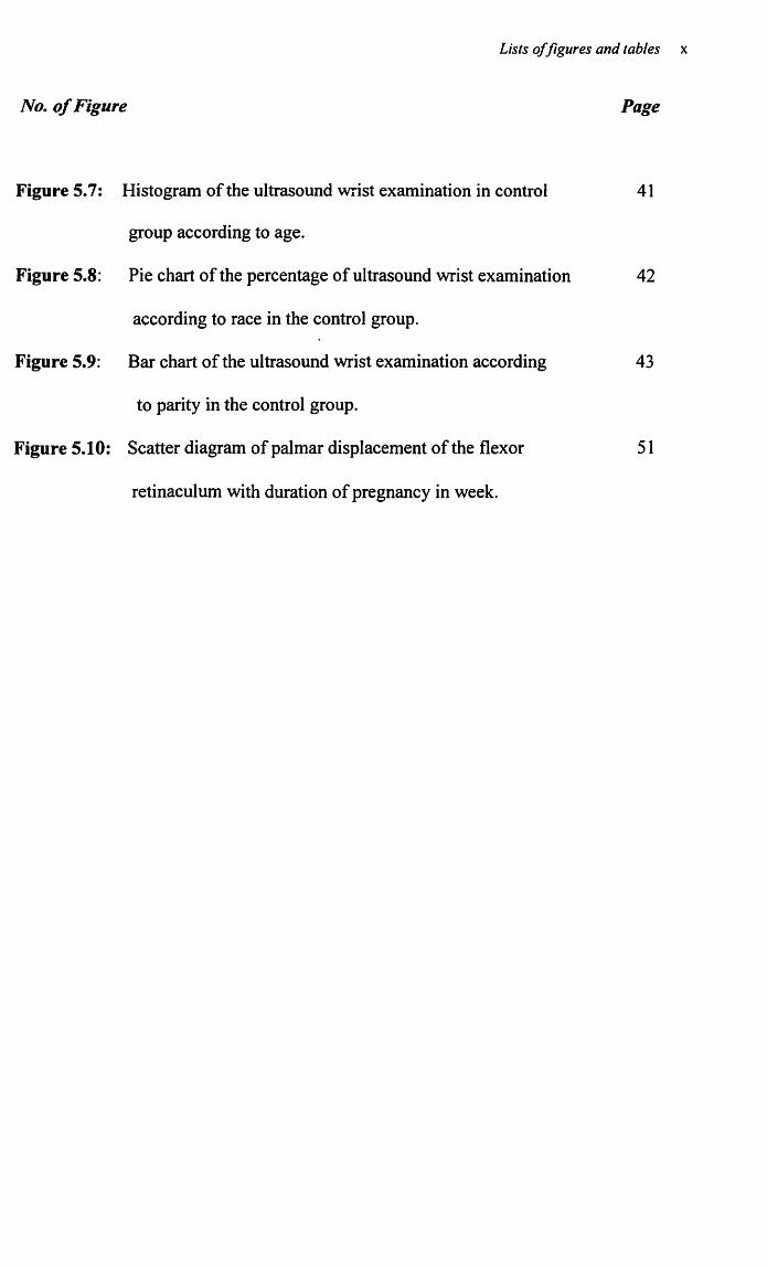
Figure 5.7: Histogram of the ultrasound wrist examination in control 41
group according to age.
Figure 5.8: Pie chart of the percentage of ultrasound wrist examination 42
according to race in the control group.
Figure 5.9: Bar chart of the ultrasound wrist examination according 43
to parity in the control group.
Figure 5.10: Scatter diagram of palmar displacement of the flexor 51
retinaculum with duration of pregnancy in week.
No. of Table
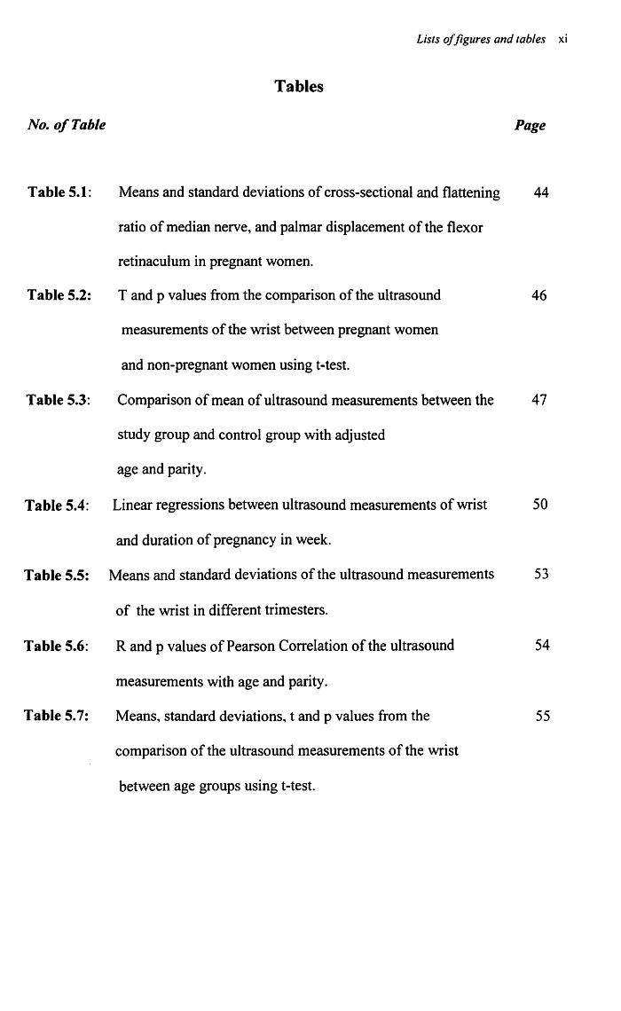
Table 5.1:
Table 5.2:
Table 5.3:
Table 5.4:
Table 5.5:
Table 5.6:
Table 5.7:
Lists offigures and tables xi
Tables
Means and standard deviations of cross-sectional and flattening
ratio of median nerve, and palmar displacement of the flexor
retinaculum in pregnant women.
T and p values from the comparison of the ultrasound
measurements of the wrist between pregnant women
and non-pregnant women using t-test.
Comparison of mean of ultrasound measurements between the
study group and control group with adjusted
age and parity.
Linear regressions between ultrasound measurements of wrist
and duration of pregnancy in week.
Means and standard deviations of the ultrasound measurements
of the wrist in different trimesters.
R and p values of Pearson Correlation of the ultrasound
measurements with age and parity.
Means, standard deviations, t and p values from the
comparison of the ultrasound measurements of the wrist
between age groups using t-test.
Page
44
46
47
50
53
54
55

Lists offigures and tables xii
No. of Table
Table 5.8:
Table 5.9:
Means., standard deviations, t and p values from the
comparison of the ultrasound measurements of the wrist
between parity groups using t-test.
R and p values of Pearson Correlation of the ultrasound
measurements with gravida.
Table 5.10: Means and standard deviations of the ultrasound
measurements of the wrist according to gravida statuses.
Page
56
58
58
Lists of abbreviations and symbols xiii
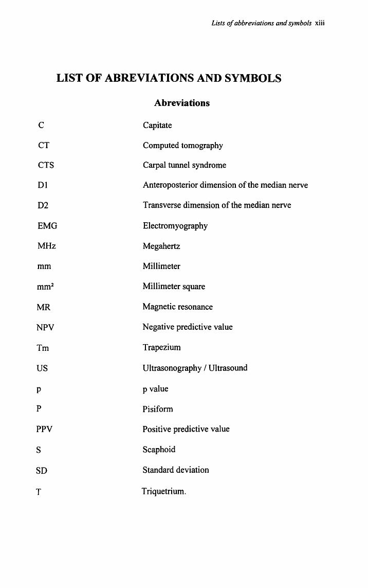
LIST OF ABREVIATIONS AND SYMBOLS
C
CT
CTS
DI
D2
EMG
MHz
mm
MR
NPV
Tm
US
p
P
PPV
S
SD
T
Abreviations
Capitate
Computed tomography
Carpal tunnel syndrome
Anteroposterior dimension of the median nerve
Transverse dimension of the median nerve
Electromyography
Megahertz
Millimeter
Millimeter square
Magnetic resonance
Negative predictive value
Trapezium
Ultrasonography / Ultrasound
p value
Pisiform
Positive predictive value
Scaphoid
Standard deviation
Triquetrium.
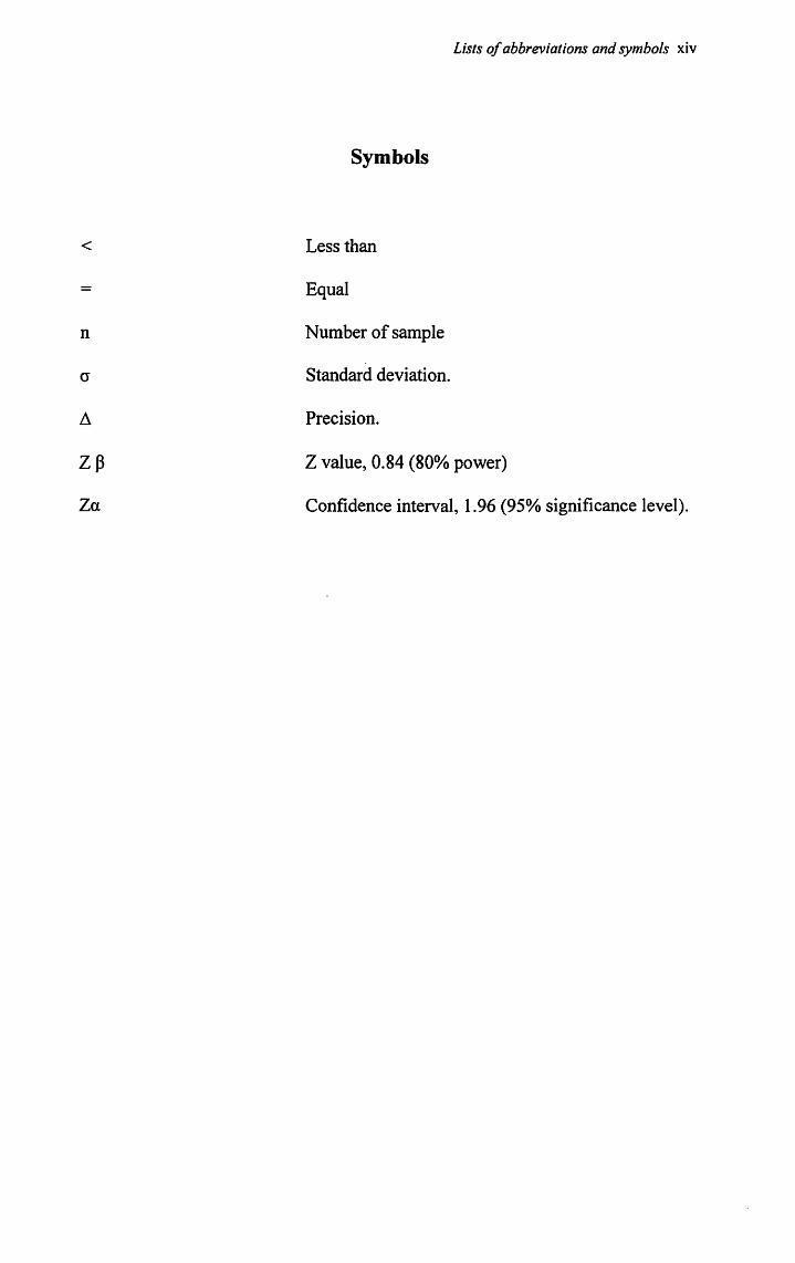
<
=
n
a
11
Z~
Za
Symbols
Less than
Equal
Number of sample
Standard deviation.
Precision.
Lists of abbreviations and symbols xiv
Z value, 0.84 (80% power)
Confidence interval, 1.96 (95% significance level).

Bahasa Melayu
TAJUK:
ABSTRAK
Abstract xv
Mengkaj i perubahan anatomi pada pergelangaan tangan semasa kehamilan normal
menggunakan ultrasound beresolusi tinggi.
PENGENALAN:
Perubahan fisiologi semasa kehamilan boleh menyebabkan perubahan pada pergelangan
tangan~ samada di dalam atau di luar terowong karpal. Perubahan di dalam terowong
sering dikaitkan dengan penghimpitan saraf median. Perubahan hormon dikatakan
sebagai puncanya. Ultrasound yang menggunakan alat berfrekuensi tinggi menawarkan
kaedah yang tepat dan kurang invasif untuk mengkaj i pergelangan tangan, baik yang
normal mahupun yang bermasalah.
OBJEKTIF:
U ntuk menentukan perubahan pada pergelangan tangan yang berlaku semasa kehamilan
normal iaitu dengan mengkaji terowong carpal dan saraf median menggunakan
ultrasound beresolusi tinggi dan menentukan peranan yang dimainkan oleh hormon.
KAEDAH:
Di dalam kajian prospektif dari bulan Januari 1999 hingga Mei 2000. pemeriksaan
ultrasound pad a pergelangan telah dilakukan pada tangan bukan utama ke atas dua
kumpulan - wanita hamil dan wanita tidak hamil (sebagai kontrol). Ukuran lebar dan
ketebalan saraf median telah diambil pada aras sendi hujung radio-ulna., tulang pisifom
dan penyangkut tulang hamat, seterusnya keluasan dan kadar penipisan saraf dikira.
Anjakan palmar flexor retinaculum juga dicatatkan. Ukuran-ukuran ini telah
Abstract xvi
dibandingkan antara dua kumpulan, umur, bilangan anak, trimester dan tempoh
kehamilan.
KEPUTUSAN:
Pemeriksaan dilakukan ke atas 56 pergelangan tangan wanita hamil dan 31 pergelangan
tangan wanita tidak hamil. Ciri-ciri normal saraf median dan flexor retinaculum
dijelaskan. Tiada atau sedikit peningkatan purata penebalan atau penipisan saraf median
dan juga perientukan flexor retinaculum terjadi pada kumpulan wanita hamil (p> 0.025).
Julat normal ukuran saraf median dikira dari kumpulan wanita tidak hamil. Dua wanita
hamil mempunyai gejala pada tangan. Seorang wanita hamil yang mengalami gejala
tangan mempunyai peningkatan pada luas keratan rentas saraf median pada aras sendi
hujung radio-ulna, pada aras tulang pisiform and tulang hamat dan juga keganjilan pada
anjakan palmar flexor retinaculum. Tujuh yang lain mempunyai keganjilan samada pada
saraf median atau flexor retinaculum tidak menghadapi gejala tangan. Seorang wanita
hamil yang mengalami gejala tangan didapati tidak mempunyai kelainan pada
pergelangan tangan. Semasa hamil, terdapat korelasi di antara peningkatan perientukan
palmar dengan peningkatan tempoh hamil (R = 0.43) dan ketara dalam trimester ketiga.
Luas permukaan saraf median meningkat di dalam wan ita berumur lebih 30 tahun, tetapi
ianya tiada hubung-kait dengan jumlah anak yang ada.
KESIMPULAN:
Keluasan dan kadar penipisan saraf median, dan juga anjakan palmar tidak bertambah
semasa kehamilan. Anjakan palmar adalah satu-satunya perubahan yand didapati beriaku
semasa kehamilan. Perubahan hormon yang dikaitkan dengan kehamilan tidak
menyebabkan perubahan pada pergelangan tangan. Ini mencadangkan bahawa hormon

A bstract xvii
bukanlah penyebabkan kepada sindrom terowong canal tetapi kemungkinan pemudarat
keadaan yang telah sedia ada.

English
TOPIC:
ABSTRACT
Abstract xviii
Evaluation of the anatomical changes of the wrist in normal pregnancy by high-resolution
ultrasonography.
INTRODUCTION:
Physiological changes in pregnancy can alter the anatomy of the wrist either within or
outside carpal tunnel. Changes in the carpal tunnel are always related to the compression
of the median nerve. The hormonal changes were thought to be a causative factor.
Ultrasound using high frequency transducers offers an accurate and non invasive method
for assessment of the wrist both in the normal and pathology conditions.
OBJECTIVE:
To determine the wrist changes in normal pregnant women by evaluation of the carpal
tunnel and median nerve with high-resolution ultrasonography and to establish the role of
hormone to these changes.
METHODOLOGY:
In this prospective study carried out from January 1999 to May 2000., ultrasound
examinations of the wrist were performed on the non-dominant hand in two groups of
women - the pregnant women and the non-pregnant women (as a control). The medio
lateral and antero-posterior diameters of median nerve were measured at the distal radio
ulna joint the pisiform bone and the hook of hamate bone following which the cross
sectional area and flattening ratio were calculated. The palmar displacement of the flexor
Abstract xix
retinaculum was also measured. Measurements were then compared between two groups.
by their age, parity, trimester and duration of pregnancy.
RESULTS:
56 wrists of pregnant and 31 wrists of non-pregnant women were examined. The normal
sonographic characteristics of the median nerve and flexor retinaculum were described.
No or minimal increase in mean thickeness or flattening of the median nerve as well as
bowing or the flexor retinaculum occurs in normal pregnancy, but are statistically not
significant (p>0.025). The normal range of the median nerve and flexor retinaculum were
derived from non-pregnant women. Two pregnant women had hand symptoms. One
pregnant woman with hand symptoms had an increased in cross-sectional area of median
nerve at the distal radio-ulna joint, pisiform and hamate bone, and also had abnormal
palmar displacement of flexor retinaculum. Seven had abnormal median nerve or flexor
retinaculum, which were sub-clinical. Another woman with hand symptoms had no wrist
changes. Palmar displacement of the flexor retinaculum correlated well with duration of
pregnancy (R= 0.48) and increase in the third trimester (p< 0.05). The surface area of
median nerve showed a significant increase in women above 30 years of age., but not with
parity and gravida.
CONCLUSION.
The cross-sectional area and flattening ratio. and palmar bowing were not increased in
pregnancy. Palmar displacement is the only change that occurs during pregnancy.
Pregnancy related hormonal changes do not cause any alteration of the wrist. These
suggest that the hormonal changes do not a cause CTS but probably aggravate the pre
existing condition.

CfiAPTER ONE:
INTRODUCTION

I ntroduct ion
1. INTRODUCTION
Pregnancy can alter the normal anatomy of the wrist. Changes can occur either
within the carpal tunnel including the median nerve or outside the carpal tunnel. Changes
in the carpal tunnel is always related to the compression of the median nerve that gives
rise to the symptom of tingling sensation over the innervation of median nerve known as
'Carpal Tunnel Syndrome'. The symptoms related to ulna nerve distribution also had
been reported (Fuente & Ellitsgaard, 1998; McLennan et al.~ 1987; Voitk et al., 1983).
Carpal tunnel syndrome (CTS) or compression neuropathy of the median
nerve at the wrist is a common, chronic and disabling condition afflicting many peoples
(Buchberger et al.; Rankin; Phalen, cited in Chen et al. 1997). This condition was first
describes by Sir James Paget in 1854 (Pfeiffer et al., cited in Lee et al., 1999) and later by
Moersch in 1938 (Rankin, cited in Chen et al., 1997). It is defined as a spectrum of
disease involving the hand and the wrist originating from problems related to the median
nerve (Amadio, cited in Lee et al ... 1999). In the general popUlation, the prevalence of
CTS is approximately 9.6% (Wand, 1990). However, De Krom et al. (1990) found that
the prevalence of CTS was only 3.90/0.
CTS affect primarily individuals who are 40-60 years old (Vessey et al., 1990).
Women are afflicted two to five times more often than men (Buchberger et al.; Phalen.
cited in Chen et al., 1997; De Krom et al., 1990; Armstrong & Chaffin, cited in Stolp
Smith et al ... 1998). This might be due to a smaller tunnel in women than men. CTS more
commonly involved the right hand but bilateral involvement was not uncommon
(Buchberger et al., cited in Chen et al... 1997). In most cases the dominant hand was

Introduction 2
involved first and more severe (Resnick & Boutin, 1999). The association between CTS
with cigarette smoking, oral contraceptive use, Quetelet's obesity index (weight
(g)/height (cm)2) and menstrual disorder had been described by Valley et al. (1990). The
risk of carpal tunnel syndrome was found to increase with the duration of activities of
flexed or extended wrist (De Krom et al., 1990).
The pathogenesis ofCTS is not completely understood (Buchberger et al.~ cited in
Chen et al., 1997) and several mechanisms have been suggested including hereditary
predisposition (Tanzer, cited in Chen et al., 1997). The possibility of the role of hormonal
changes as a causative factor was raised by the higher incidence among women who are
in their 50's and 60's and association of CTS with acromegaly, pregnancy, menopause
and women on oral contraceptive. The proposed mechanism was an increase in extra
cellular fluid volume surrounding the median nerve (Phalen; Tanzer; Schiller & Kolb,
cited in Chen et 01., 1997). Other proposed predisposing factors for CTS during
pregnancy include previously unrecognized and asymptomatic median neuropathy at the
wrist, body habitus (Leblhuber et 01.; Wand, cited in Stolp-Smith et 01., 1998) and carpal
tunnel size (Ekman-Ordeberg et 01.. 1987; McLennan et 01., 1987). The CTS in
association with collagen vascular disease and systemic illness such as multiple
myeloma, amylodosis. myxoedema, diabetis mellitus and sarcoidosis may be caused by
an increased in fluid, synovial proliferation or excess protein deposition (Buchberger;
Phalen. cited in Chen et 01., 1997; Fenves et 01., 1986). Anoxia from vascular spasm of an
inter-connected blood supply to the median nerve that results in neural ischaemia has
been suggested in the diabetics and in patients on chronic heamodialysis (Blunt. 1959).
Space-occupying lesions such as synovial cyst. ganglionic cyst. lipoma and haematoma
Introduction 3
can result in compression of the median nerve. Decreased carpal tunnel size e.g., carpal
bone malaligment, displaced fractures, and hyperthrophic changes (Gelberman et al.;
Phalen et al., cited in Chen et al., 1997) and congenital variants such as abberant
lumbrical, anomalous tendinous insertion, and persistent median artery (Ametewee et al.;
Luyendijk, cited in Chen et al., 1997) are rare causes of CTS. A1though~ CTS is not
primarily an occupational disorder, the symptoms are often produced or aggravated by
sudden increase in manual activity especially those involved in flexion or repeated
motion and stresses of the wrist and hand (Phalen, cited in Chen et al., 1997; Fenves et
aI., 1986). Compression of the median nerve within the fixed or decreased space of the
carpal tunnel is the final pathway for development of CTS, regardless of the cause. This
role is equally valid with regard to the production of ulna nerve symptoms~ since this
nerve may be compressed at the thoracic outlet, the elbow (in the groove of the medial
condyle of the humerus) or at the wrist (round the hook of the hamate bone).
During the course of pregnancy, women of average weight (55-60 kg) normally
increase their extra-cellular fluid by over 2500 ml. The degree of fluid retention may
cause slight thickening of the skin and, if the carpal tunnel is restricted. oedema of the
sheath of the median nerve may cause paraesthesia of the fingers, which is not
uncommon during pregnancy. Later, it will be followed by weakness of the thenar
muscles (wasting of the abductor pollicis brevis) with sensory loss of the palm and radial
three-and-a-half fingers. Tinel' s sign may be positive and electrical studies may show
slowing of nerve conduction across the wrist. These changes account for development of
the carpal tunnel syndrome in pregnancy.

I Introduction 4
I'
1:
I i The incidence of carpal tunnel syndrome or hand symptoms is increased during
pregnancy. Approximately 2.3% to 4.6% of patients with CTS were pregnant (Armstrong
& Chaffin; Dekel et al., cited in Stolp-Smith et al., 1998). In 1957. Wallace and Cook
described two pregnant patients with carpal tunnel syndrome. Since then an incidence in
pregnancy of 1 % to 50% has been reported (Voitk et al .. 1983). Twenty-one percent of
pregnant women reported paresthesia or hyperesthesia in the median nerve sensory
distribution of the hand during the pregnancy (Gould & Wissinger. 1978). Voitk et al.
(1983) reported 34% of pregnant women had hand symptoms (25% had symptoms of
carpal tunnel syndrome, 2% symptoms of ulna nerve compression and 7% ill defined
hand symptoms). Other study by McLennan et al. (1987), 35% of pregnancies reported
hand symptoms, but less than 20% of the affected patients described a classic median-
nerve symptom distribution (carpal tunnel syndrome). while 12% of patients described an
ulna nerve distribution. In 69% of patients, hand symptoms were generalised. The
prevalence of the median nerve symptoms in pregnancy in their study was only 7%. From
their study, they also found that the prevalence of the hand symptoms of the same
distribution and quality was 300/0 in non-pregnant women. although invariably mild. these
symptoms suggested that pregnancy might aggravate a pre-existing condition. In the
prospective study by Ekman-Ordeberg et al. (1987), only 2.3% (56 women of2.358~ 47%
nUlliparous and 53% multiparous) delivered during a 12-month period at the Department
of Obstetrics and Gynecology. Malmo General HospitaL had symptoms of carpal tunnel
syndrome during pregnancy. A large retrospective study was conducted by Stolp-Smith et
al. (1998) revealed the incidence of carpal tunnel syndrome in pregnancy is 0.340/0. In the
study by Fuente & Ellitsgaard (1998). the hand symptoms had been noted in 16% of

Introduction 5
pregnancy. Among these, 30% described a classic median nerve symptom distribution
and 24% of patients described an ulna nerve distribution .. and most of the symptoms are
bilateral.
The CTS or hand symptoms tend to occur during the third trimester (Fuente &
Ellitsgaard, 1998; McLennan et al., 1987; Seror, 1997; Wand, 1990; Voitk et al .. 1983).
However, Stolp-Smith et ale (1998) found that the symptom of onset occurred with even
distribution during each trimester but the diagnosis of CTS was diagnosed most
frequently during the third trimester. Voitk et ale (1983) and Stolp-Smith et al (1998)
found no correlation with gestational age or gestational interval. CTS in pregnancy occur
generally between 30 and 40 years of age (Wand, 1990) and in multi-parous women
(Seror. 1997~ Stolp-Smith, 1998; Wand, 1990). There was a significant correlation of
development of carpal tunnel syndrome to parity (Fuente & Ellitsgaard, 1998). In
contrast .. Ekman-Ordeberg et ale (1987) in their study found that carpal tunnel syndrome
during pregnancy was most common in primipara with generalised oedema. However.
McLennan et al. (1987) and Voitk et ale (1983) found no significant correlation of hand
symptoms in pregnancy with age and parity. The hand symptoms in pregnancy usually
affected both hands (Fuente & Ellitsgaard .. 1998; McLennan et al .. 1987; Stolp-Smith et
al .. 1998; Voitk et al., 1983). Other conditions that have been found to have association
with hand symptoms were tight rings (McLennan et al .. 1987~ Voitk et al .• 1983). pre
menstrual bloating (McLennan et al... 1987), pre-eclampsia (McLennan et al.. 1987~
Voitk et al... 1983). hypertension (Voitk et al... 1983). oedema (Ekman-Ordeberg et
a/.J 987~ Fuente & Ellitsgaard .. 1998; Voitk et al .. 1983). weight at confinement and birth
weight (McLennan et al... 1987). No correlation of CTS was found with weight gain
Introduction 6
(McLennan et al.~ 1987; Stolp-Smith et al., 1998; Voitk et ai" 1983), diabetes mellitus
(McLennan et al., 1987), hypertension (McLennan et al., 1987), renal disease (McLennan
et ai., 1987) and history of arthritis (McLennan et ai., 1987; Voitk et al.. 1983).
About half of the pregnant women who developed CTS during pregnancy
continued to have it after delivery. Most of the cases resolved soon after delivery (Fuente
& Ellitsgaard, 1998; Massey, 1978; McLennan et ai., 1987; Wand, 1990; Voitk et ai.,
1983). In a few of them, the symptoms will persist up to several months after delivery
(Gould & Wissenger, 1978).
Symptoms of CTS are usually burning pain, numbness and paresthesia in the
distribution of the median nerve (Buchberger; Phalen; Tanzer, cited in Chen et al .• 1997).
Classically, the distribution of symptoms involves the thumb. index finger. and middle
fingers. Anatomic variations occur, and the median nerve may join with ulnar and radial
nerver. causing variability in the motor and sensory changes of CTS. Symptoms may be
referred proximally as high as the shoulder (Phalen, cited in Chen et al.. 1997). Other
classically described symptoms include nocturnal burning and pain. presumably from
venous engorgement, and hypoesthesia of the tip of middle finger, area which has
isolated sensory supply of the median nerve (Phalen, cited in Chen et al .. 1997).
Clinical findings include reproduction of the patient's symptoms with gentle
percussion of the median nerve. which is called a positive Tinel's sign (Fenves et al ..
1986). In wrist flexion or Phalen's test (which involved unforced complete flexion of the
wrist for 30-60 sec). the reproduction or exaggeration of the patient's symptoms indicates
a positive test (Phalen, cited in Chen ef al .• 1997). Chronic median nerve compression
can cause thenar atrophy with weakness or paralysis of the abductor pollicis brevis.
I Introduction 7
opponens pollicis or flexor pollicis brevis. The thenar atrophy was usually pronounced
when the hands are viewed in profile (Phalen, cited in Chen et al., 1997. Clinically~ CTS
in pregnancy is very different from idiopathic CTS. Paraesthesia frequently occurs
during the daytime or is permanent and usually more troublesome than when it occurs at
night (Seror, 1997). Study by Seror (1998), revealed a higher incidence of persistence,
painful diurnal symptoms in pregnancy related CTS than idiopathic CTS.
The diagnosis of CTS is usually made by clinical examination and
electromyography (EMG). Electrodiagnostic studies are usually not necessary but may be
helpful for confirmation and for exclusion of the other conditions that may mimic CTS
such as cervical disc, demyelination or polyneuritis (Kimura~ cited in Chen e/ al., 1997)
Traditionally, EMG is the deciding factor in determining the shift of treatment from
conservative to surgical intervention, largely based on the shift from mildly abnormal to
markedly abnormal EMG result. Of the few tests available .. only the EMG of the median
nerve is considered the gold standard. The test is conducted by inserting the electrodes
into the muscle along the pathway of the median nerve and passing a small voltage
through them, thus allowing the measurement of velocity latencies as an indicator of the
severity of the disease. The nerve conduction studies reflect the status of the nerve fibers.
Although they are integral to the evaluation and diagnosis of the CTS~ they have inherent
disadvantages that limit their accuracy. In the diseased nerve, if there are nerve fibers
unaffected by disease or injury~ the test results may appear normal. Therefore~ a normal
conduction velocity does not exclude the presence of compression .. which may be related
in part to the chronicity and severity of median nerve compression. The limitation of
EMG is not only in its accuracy.. it is determined largely by the experience and
Introduction 8
interpretation of the reader. In addition, there is also the significant discomfort of the
examination itself.
Traditionally, imaging has little role in the diagnosis of carpal tunnel syndrome.
However, with the latest technology such as magnetic resonance imaging and
introduction of high frequency ultrasound transducer, it trigger the interest of many
authors to study the role of imaging for evaluation of the carpal tunnel and in the
diagnosis of CTS (Buchberger et ai., 1992; Fornage et ai., 1985; Fomage and Rifkin,
1988; Fomage, 1988; Fomage 1989; Lee et al., 1999). Fomage (1989) used ultrasound to
evaluate the soft tissue changes in the hand in rheumatoid arthritis. The ultrasound
changes in the wrist and hand in haemodialysis patients have been studied by Lanteri et
al. (1997). The anatomical changes of the wrist in pregnancy had never been studied so
far. The purpose of this study is to evaluate the anatomical changes in the wrist that might
occur in pregnancy and also to compare these changes with different trimester of
pregnancy. The information that will be obtained from this study probably can explain
why the pregnant women have a higher risk to develop CTS and to establish to role of
hormone for these changes.

CHAPTER TWO:
LITERA TURE REVIEW
Literature review 9
2. LITERATURE REVIEW
2.1 Anatomy of the Wrist
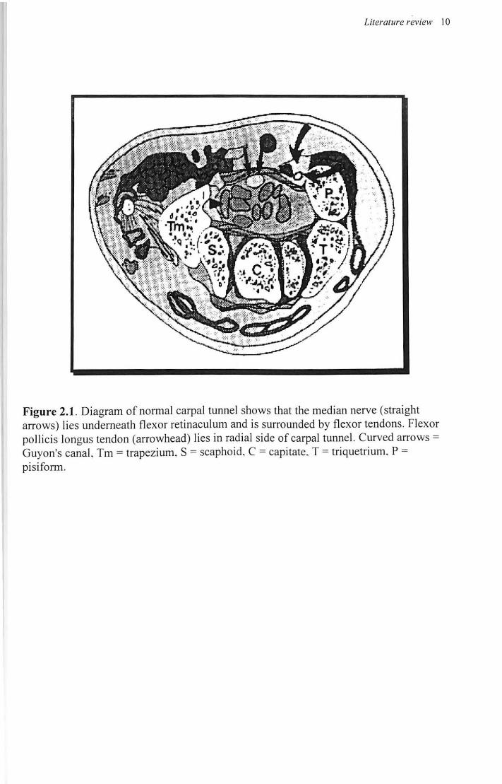
The carpus is the part of the upper extremity between hand and forearm. The wrist
comprises several joints or compartments, including the radiocarpal, midcarpal, common
carpometacarpal, first carpometacarpal, inferior radio-ulna and pisiform-triquetral
compartments (Resnick & Boutin, 1999). The carpal tunnel is a restricted space lying
between the flexor retinaculum (volar or transverse carpal ligament) ventrally and the
carpal bones dorsally (Figure 2.1). The flexor retinaculum extends from the pisiform and
hamate to the scaphoid and trapezium and is normally fairly straight or slightly convex.
The radial aspect of the flexor retinaculum splits into a superficial and deep layer to
accommodate the flexor carpi radialis. The ulna aspect joins the pisiform-hamate
ligament. The transverse carpal ligament is thickest in its mid portion near the base of the
capitate. The flexor carpi ulnaris tendon lies separately within the canal of Guyon, along
side the ulna nerve. The palmaris longus tendon lies just superficial to the median nerve
outside the carpal tunnel. The carpal tunnel contains four flexor digitorum superficial
tendons, four flexors digitorum profundus, the flexor pollicis longus tendon, and median
nerve. The median nerve is covered with a cellulo-adipose layer that is difficult to
separate from the ulna bursa that surrounds the flexor tendons. The median nerve is
intimately related to the flexor retinaculum, lying just deep to this ligament, and courses
ventral and parallel to the flexor tendons (Buchberger, cited in Chen et al., 1997~
Mesgarzadeh et ai., 1989; Robbins, 1963; Weiss et ai., 1986~ Middleton et al., 1987).
Literature review 10
Figure 2.1. Diagram of normal carpal tunnel shows that the median nerve (straight arrows) lies underneath fl exor retinaculum and is surrounded by flexor tendons. Flexor pollicis longus tendon (arrowhead) lies in radial side of carpal tunnel. Curved arrows = Guyon's canal. Tm = trapezium. S = scaphoid. C = capitate. T = triquetrium. P = pisiform.
Literature review 1 1
2.2 Radiological Evaluation of the Wrist
The diagnosis of CTS is often based on clinical findings alone. Historically.,
imaging studies have not played an important role in the evaluation of CTS.
2.2.1 Plain Radiograph
The role of conventional radiology in imaging of the nerve was limited, being
confined for the most part to imaging the sequelae of nerve damage or disease (e.g.,
muscle atrophy or degeneration associated with nerve disease). Imaging of the wrist still
relies first on plain radiograph, although it provides very little information on soft tissue.
Conventional radiographs are of little help if the bony structures are intact.
2.2.2 Computed Tomography (CT)
CT is of limited value because of the similar attenuation values of the contents of
the carpal tunnel (John et al.~ Zucker-Pinchoff et al., cited in Chen et al., 1997).
2.2.3 Magnetic Resonance Imaging (MRI)
MR imaging has been shown to be the most helpful. Anatomic relations of the
median nerve and underlying flexor tendons have been extensively studied with MR
imaging (Mesgarzadeh et al., 1989a; Middleton et al., 1987; Zeiss et al., 1981; Foo et al . .,
1992). MRI provides excellent tissue contrast and allows excellent appreciation of the
structures of the bones, ligaments, tendons and even nerves. However, evaluation of the
course of peripheral nerves away from its spinal roots remains severely limited. But. MR
imaging has been used successfully for the evaluating the carpal tunnel syndrome

Literature review 12
(Mesgarzadeh, 1989a; Mesgarzadeh, 1989b; Middleton, 1987). MR imaging of the wrist
for CTS is best perform~d on a high-field system, with a surface or dedicated wrist coil.
The patient can be in supine or prone position with wrist positioned transversely above
the patient's head and elbows flexed. A field of view of 8-14 cm and a matrix size of 128
x 256 or 256 x 256 provide excellent resolution. The median nerve is best visualised in
the axial plane, but sagittal and coronal sections are often helpful. Anatomic depiction is
best with Tl-weighted spin-echo sequences. Spin-echo or fast spin-echo T2 weighted
sequences are useful to detect signal abnormalities within or surrounding the median
nerve. MR may show subtle signal intensity changes and mild compression of median
nerve that may be missed on sonograms. MR imaging may be superior to sonography in
detecting the cause of CTS (Buchberger et al., 1992) and is a study of choice for
assessment of surrounding bony structures. MR imaging, although felt to be relatively
accurate in determining the changes in the carpal tunnel and morphology of median nerve
in patients with CTS, it has several disadvantages including claustrophobia and patient
motion artifact, which significantly degrades the signal. In addition, its high cost and time
requirement limit its application for routine clinical used.
2.2.4 Ultrasonography
Sonography may be a low cost alternative. Ultrasound is an important yet non
invasive tool in assessment of the wrist (Buchberger et al., 1992). Ultrasound of the wrist
requires knowledge of the normal anatomy and dedicated ultrasound equipment.
Sonography examination of the hand also requires high frequency linear transducers.
Although ultrasound imaging of the median nerve has been attempted in the past.

Literature review 13
technical limitations have largely precluded detailed study of its anatomy., structure, and
diseases intrinsic to and around the nerve. The advent of small high-frequency
transducers recently has resulted in the resurgence in the interest in using the sonography
for the evaluation of the musculoskeletal system (Buchberger et al., 1992; Fornage et al . .,
1985; Fornage, 1988; Fornage & Rifkin, 1988). These small high-frequency probes are
uniquely suited for small joints such as the wrist and are comfortable for sonographer to
use. Modem linear-array transducers in the 7 to 13-MHz range provided wide near field
view, allowing visualization of internal nerve anatomy as well as disease in the
surrounding structures that affect the nerve, such as oedema, inflammation, and tumour
(Silvestri, cited in Lee et ai, 1999).
The sonographic examination IS performed with the patient seated in a
comfortable position facing the sonographer. The wrist is placed in a slightly
hyperextended position. A IO-MHz compact linear transducer or IO-MHz linear
transducer is used to examine the wrist. Having the wrist in neutral position is important
in taking consistent measurements, because the carpal dimensions can significantly alter
with various wrist positions (Lee et al., 1999). The wrist is examined in the transverse
and longitudinal planes. The wrist crease is used as external landmark to simply scanning
by allowing consistent placement of the transducer at the carpal tunnel (Lee et al., 1999).
As the relevant structures are located very close to the surface, water standoff pad is used.
But standoff pad is not necessary when such high-frequency probes are used.
In the transverse plane, ulna artery is easily located and can ensure that the
orientation of the transverse images remains consistent. The sonographic beam needs to
be perpendicular to the surface of flexor tendons because of the anisotrophic effect
Literature review 14
(Buchberger et al., 1992). Otherwise, the tendons may appear relatively hypoechoiec and
mimic synovial fluids (Fornage & Rifkin, 1988; Fornage, 1989). The median nerve is
located superficial to the echo genic flexor tendons, and its size, shape, echogenicity, and
relationship to the underlying tendons and overlying retinaculum are noted. Finger and
wrist movements, and first clenching, can be performed to assess the mobility of the
median nerve. The amount of synovial fluid and presence or absence of masses should be
noted. The continuity of the median nerve and any area of constriction or swelling may
be better appreciated in a sagittal plane than in the transverse plane. The alignment of the
median nerve in the carpal tunnel, its relationship to the underlying flexor tendons, and
the shape of median nerve may be variable, depending on the position of the wrist (Zeiss
et al.~ 1989) Sonography, unlike MR imaging, is a dynamic study; therefore, the median
nerve can be evaluated with the wrist in different positions~ which may provide
information as to why certain wrist motions predispose the patient to symptom of CTS.
Real-time high frequency ultrasonography has been used to diagnose soft-tissue
lesions of the hand (Fomage et al., 1985; Fornage & Rifkin, 1988). The sonographic
appearance of various peripheral nerves of the extremities including the median nerve has
been described by Fornage et al. (1988). Since the distinction of CTS from the other
causes on pain (e.g., cervical root compression, thoracic outlet syndrome or nerve
entrapment in the forearm) is important to make, which is not always possible on the
basis of clinical findings and the results of the nerve conduction studies (Phalen; Rietz~
cited in Buchberger et al.~ 1992), sonography has been advocated as a noninvasive means
for evaluating the carpal tunnel. The ultrasound is a sensitive and reliable method in
detection of fluid. exudative synovitis. tenosynovitis. peritendinitis. tendon rupture versus
Literature review 15
tendon adhesion and ganglia (Fomage, 1989; MiIbradt el ai.~ 1990; Read el ai., 1996).
Ultrasound examination is a useful method in assessing of muscular atrophy and
alterations of the shape and echogenicity of the median nerve in patients with carpal
tunnel syndrome (Milbradt el ai., 1990). Further indications of sonographic examination
include suspected tumours, foreign bodies and synovial proliferation (Fomage, 1988;
Fomage, 1989; Milbradt et ai., 1990 and Middleton el ai., 1987). More work is needed to
determine the role (if any) of ultrasound in the evaluation of the peripheral nerve,
triangular fibro-cartilage, dorsal carpal tunnel ligament and bone pathology.
In light of the latest developments in ultrasound technology, application of this
modality was thought to be ideal for the evaluation of the median nerve and the carpal
tunnel. The advantages of the ultrasound technique include ease of scanning, patient
comfort~ short examination time and dynamic imaging (Foo el ai., 1992; Lee el at 1999).
In addition, sonography is less time-consuming than MR imaging, even though faster
imaging sequences have substantially reduced imaging time.
The limitation of the ultrasound is a small false negative, which are related to
variety of factors, including operator dependence, resolution threshold in the sub
millimeter range, image degradation and narrow field of view. Other shortcomings is that
the resolution in the edges of the image can be diminished by sub-optimal interface
between straight edge and curve wrist surface (Buchberger et ai., 1992)~ but this
difficulty is easily overcome by practicing transducer manipulation over the curved
surface (Lee et ai., 1999).

I i'
Literature review 16
2.3 Assessment of Median Nerve and Carpal Tunnel by Ultrasound
There are various methods used to assess the median nerve and carpal tunnel. In
the study by Lee et al (1999), measurements of the median nerve and the dimensions of
the carpal tunnel were obtained from the axial images of the carpal tunnel. The cross-
sectional area of the median nerve was calculated as an ellipse. The anterior-posterior
(AP) and transverse dimensions of the median nerve were measured and placed into the
equation for the ellipse (area = ® (Dl x D2) / 4). The AP diameter of the carpal tunnel
was measured as a distance from the posterior aspect of the flexor retinaculum to the
anterior surface of the capitate bone. The thickness of the flexor retinaculum was also
measured.
In the earlier study, Buchberger el al. (1992), have described their experience
with high resolution sonography for evaluation of the carpal tunnel, both in normal
volunteers and in symptomatic patients. where they measured the median nerve at three
different levels (distal radius, at the level of pisiform, and at the level of hamate bone).
Besides the anterior-posterior diameter, transverse diameter. and cross-sectional area,
they also measured the flattening ratio of the median nerve (ratio of the major axis of the
median nerve to its minor axis). The cross-sectional area and the flattening ratio can be
obtained by measuring the transverse and anterior-posterior diameters of the median
nerve at the proximal and distal carpal tunnel. respectively. For the assessment of the
carpal tunnel, the palmar displacement is obtained by measuring the distance from the
line drawn between the trapezium and the hamate to the top of flexor retinaculum.
Literature review 17
Lanteri et al. (1997) used four basic measurements taken in transverse and
longitudinal section at the level of pisiform bone; 1) carpal tunnel depth - the distance in
millimeters from the flexor retinaculum to the radial bone surface, 2) radius to tendon
thickness - the distance in millimeters separating the radial bone surface from the
overlying flexor tendons, 3) flexor retinaculum thickness in millimeters, and 4) median
nerve surface area index - the product of the width and depth in millimeter of the cross
section of the median nerve.
The normal carpal tunnel on MR imaging has been studied by Mesgarzadeh et al.
(1989). For the cross-sectional area of the median nerve in the study by Mesgarzadeh et
al. (1989), they used mean swelling ratio of the median nerve. The mean swelling ratio
was calculated by dividing the cross-sectional area of the median nerve at the pisiform
level and at the hamate level by that at the distal radius. They also expressed the palmar
displacement as a percentage of its unbowed length~ where the palmar displacement was
divided by the length of a straight line between the attachments of the flexor retinaculum
to the tubercle of the trapezium and the hook of hamate to determine 'the bowing ratio'.
The signal characterization of the median nerve has been studied and the
comparison was made with surrounding flexor tendons (Fomage, 1989; Lee et af., 1999).
The mobility of the median nerve is more readily evaluated with sonography than with
MR imaging and assessment can be done with both passive and active movements of the
patient's fingers and wrist. However, these assessments are subjective and harder to
quantify.
Literature review 18
2.3.1 Characterization of the Normal Median Nerve and Carpal Tunnel
Fornage (1988) described the appearance of normal nerves as markedly echogenic
tubular structures with parallel internal linear echoes (fibrillar texture) on longitudinally
orientated scans and as an oval-to-round echogenic section on transverse scans,
occasionally with internal punctuate echoes. Fornage (1988) also confirmed the
immobility of the nerve in relation to the surrounding musculotendinous structures at I I : i
dynamic examination during active or passive flexion/extension.
Lee et af (1999) studied in vitro ultrasound characteristics of the median nerve
through cadaver dissection and correlates with in vivo characteristics of the normal wrist
in 56 wrists of 28 normal volunteers. They found that in the cadaver, the flexor
retinaculum appeared as a band of alternating high and low echogenicity transversing the
carpal tunnel. The median nerve appeared as a structure of low echogenicity without
through transmission surrounded by a thin, hyperechoeic nerve sheath. The center of the
median nerve is of low echogenicity in relation to the surrounding tendons. Longitudinal
scans demonstrated the median nerve tapers as it coursed distally. Furthermore, the cross
sectional configuration of median nerve also changed along its path; nearly circular
proximal to the carpal tunnel, a flat ellipse within the carpal tunneL and wedge-like distal
to the tunnel. The tendons, however, did not taper distally and maintained a circular
cross-section. Adjacent to the median nerve were the radial and ulna arteries. both of low
echogenicity. The bones appeared highly echogenic and reflective. They also described
that. the anatomic details derived from scanning the cadaver correlated well in the normal
Literature review 19
wrists, where the carpal tunnel was bounded anteriorly by echogenic flexor retinaculum,
posteriorly by capitate, and laterally by scaphoid and pisiform. They noted that, the
hypoechoeic, elliptical median nerve was readily demonstrated among the hyperechoeic
tendons and the nerve showed no through transmission.
2.3.2 Dimensions of Normal Median Nerve and Carpal Tunnel
Buchberger et al. (1991) in a study of 28 normal wrist found the mean cross
i: sectional area of the median nerve was 7.9 mm2 (SD, 1.1 mm2) at the level of the distal
I radio-ulna joint, 8. I mm2 (SD, 1.3 mm2) at the level of pisiform bone, and 7.7 mm2 (SD,
1.1 mm2) at the level of hamate bone. The flattening ratio of the median nerve was 2.7
(SD, 0.3), 3.0 (SD, 0.5) and 3.2 (SD, 0.5) respectively. The mean palmar displacement of
the flexor reninaculum was 2.1 mm (SD, 0.8mm). They also found that the mean cross
sectional area of the median nerve is best obtained at the level of distal radius or pisiform,
as this is the level of proximal carpal tunnel and is the expected location for maximum
nerve swelling. The mean cross-sectional area of the median nerve at proximal carpal
tunnel should be no more than 10 mm2• The flattening ratio is best obtained at the level of
hamate, which is the level of the distal carpal tunnel and reflects the maximum flattening
and constriction of the nerve between the flexor tendons and transverse carpal ligament.
A normal flattening ratio at the level of the distal carpal tunnel should be less than 3.0
and the normal palmar displacement should not exceed 4.0 mm.
Lee et al. (1999) found that, the mean cross-sectional area of the median nerve
was 8.3mm2 (SD = 1.9) in men and 9.3mm2 (SD = 2.2) in women. The AP dimension of
the carpal tunnel was 10. 9mm (SD = 2. I) in men and 10.3mm (SD = 1.6) in women. The
Literature review 20
mean thickness of the flexor retinaculum was 1.1 mm (SD = 0.1) in men and 1.0mm (SD
= 0.1) in women. They also found that in 84% of the cases, the median nerve of the
dominant hand had a greater cross-sectional area but without a corresponding greater AP
carpal tunnel dimension.
Middleton et al. (1987) studied the carpal tunnel with MR imaging. They found
the cross-sectional area of the median nerve was 7.0 mm2 (SD, 1.4 mm2) at the pisiform
bone~ and 8.0 mm2 (SD, 1.9 mm2) at the hamate bone. Mesgarzadeh et al. (1989) found
the flattening ratio was 2.5 (SD, 1.0) at the distal radius, 3.3 at the pisiform bone and 2.9
(SD~ 0.9) at the hamate bone.
2.3.3 Ultrasound Characteristic of the Abnormal Median Nerve and Carpal Tunnel
associated with Carpal Tunnel Syndrome
Buchberger et aI, 1991 in their early study found that neither a significant increase
in size nor flattening of the median nerve in the carpal tunnel. They did another study in
the following year (Buchberger et al., 1992), and described three main objective findings
in CTS: swelling of the median nerve at proximal carpal tunnel with or without formation
of a pseudoneuroma, flattening of the median nerve at the distal carpal tunnel, and
increased bowing of the flexor retinaculum. These changes were statistically significant
(p<O.OI to p< 0.001). The findings were similar to those described with MR imaging
(Middleton et al .. 1986 & Mesgarzadeh et al.~ 1989). Similar findings were noted in the
study done by Lee et al. 1999, where the median nerve has consistent and statistically
significant increase in cross-sectional area. and the variations in the magnitude of
increases were empirically. corresponding to the severity of CTS. They also noted an
Literature review 21
abrupt contour changes along its course to varying degree, relative to the amount of
increase in cross-sectional area (i.e., the greater the increase, the greater the contour
deformity as the nerve flattens against unyielding flexor retinaculum. Other associated
findings such as synovitis and perineural oedema have been described (Lee et al., 1999).
2.3.4 Efficacy of Ultrasound in Carpal Tunnel Syndrome
The reliability of the sonography in the diagnosis of carpal tunnel syndrome on
the basis of sonography has been proven by several studies. The potential roles of
ultrasound as a primary diagnostic tool for determining the presence and the severity of
disease in the CTS was studied by Lee et ai., 1999. They found that the area
measurement of 15mm2 was an appropriate level for delineating the presence of
significant median neuropathy with respect to surgical treatment. There was excellent
correlation between the median nerve cross-sectional area and EMG findings. The
sensitivity, specificity, PPV and NPV of ultrasound were reported as 88%, 96%, 97% and
86%. respectively. From this study. they also reported that one could be confident of
determining the level or severity of median nerve neuropathy based on ultrasound
measurements of its cross-sectional area.
Buchberger et al. (1992) in their study used the quantitative analysis of the cross
sectional area and flattening ratio of the median nerve and of the palmar bowing of the
flexor retinaculum to calculate true-positive and false-positive percentages at different
critical values on a continuous scale. In this study, the diagnosis of CTS was made when
at least one of the following findings was shown: (1) increased cross-sectional area of the
median nerve at the pisiform and/or at the hamate bone. (2) increased flattening ratio of
Literature review 22
the median nerve at the hamate bone, or (3) increased palmar displacement of the flexor
retinaculum. Although they could not perform complete ROC analysis, from the
individual ROC curves, they found that the discrimination ability of these measurements
was sufficiently high to establish the diagnosis. They also found, the measurements of the
cross-sectional area and flattening ratio of the median nerve were correlated well with
MR imaging, but measurement of palmar displacement of flexor retinaculum correlated
less well. They concluded that, MR imaging may be superior to sonography in detecting
; the cause of CTS and may show subtle signal intensity changes and mild compression of I
I ;
, the median nerve that may be missed on sonography. In addition, MR is the study of
I
'\ choice for assessment of surrounding bony structures. However, no study was done to I
',determine the efficacy of sonography compared with MR imaging and the
complementary roles of these two techniques. In contrary, Chen et al. (1996) concluded
,that sonography evaluation of a large series of patients is still necessary to determine the
definitive role of sonography in CTS.
Ultrasound offers high diagnostic accuracy as indicated by high correlation with
EMG findings. It was recommended as the first step in diagnostic testing after the initial
physician evaluation of CTS and should be considered as a new, alternative diagnostic
modality. It also provides a reliable method for following response to therapy without
sacrificing patient comfort (Lee et al., 1999).

CHAPTER TflREE:
OBJECTIVES &
HYPO TflESES
Objectives and hypotheses 23
3. OBJECTIVES AND HYPOTHESES
3.1 General Objective
To evaluate the anatomical changes of the wrist in nonnal pregnancy using high
i resolution ultrasonography.
3.2 Specific Objectives
3.2.1 Detennining of the demography according to age, ethnic group, parity,
gravida, duration of pregnancy in weeks and trimester.
3.2.2 To determine the anatomical changes of the wrist in normal pregnancy, i.e.
the cross-sectional area and flattening of the median nerve and palmar
displacement of flexor retinaculum.
3.2.3 To determine the anatomical changes of the wrist during pregnancy by
comparing with duration of pregnancy (in weeks) and in different
trimesters of the pregnancy.
3.2.4 To determine the anatomical changes of the wrist with age, parity and
gravida.