Embed Size (px)
Citation preview

Zurich Open Repository andArchiveUniversity of ZurichMain LibraryStrickhofstrasse 39CH-8057 Zurichwww.zora.uzh.ch
Year: 2015
Evaluation of two miniplate systems and figure-of-eight bandages forstabilization of experimentally induced ulnar and radial fractures in pigeons
(Columba livia)
Bennert, Beatrice
Posted at the Zurich Open Repository and Archive, University of ZurichZORA URL: https://doi.org/10.5167/uzh-115671DissertationPublished Version
Originally published at:Bennert, Beatrice. Evaluation of two miniplate systems and figure-of-eight bandages for stabilization ofexperimentally induced ulnar and radial fractures in pigeons (Columba livia). 2015, University of Zurich,Vetsuisse Faculty.

1
Klinik für Zoo- Heim- und Wildtiere, Department für Kleintiere
der Vetsuisse-Fakultät Universität Zürich
Direktor: Prof. Dr. Jean-Michel Hatt
Evaluation of two miniplate systems and figure-of-eight bandages for stabilization of
experimentally induced ulnar and radial fractures in pigeons (Columba livia)
Inaugural-Dissertation
zur Erlangung der Doktorwürde der
Vetsuisse-Fakultät Universität Zürich
vorgelegt von
Beatrice Miriam Bennert
Tierärztin
von Rosenheim, Deutschland
genehmigt auf Antrag von
Prof. Dr. med. vet. Jean-Michel Hatt, Hauptreferent
Prof. Dr. med. vet. Maria-Elisabeth Krautwald-Junghanns, Korreferentin
Zürich 2015

2
Klinik für Zoo- Heim- und Wildtiere, Department für Kleintiere
der Vetsuisse-Fakultät Universität Zürich
Direktor: Prof. Dr. Jean-Michel Hatt
Evaluation of two miniplate systems and figure-of-eight bandages for stabilization of
experimentally induced ulnar and radial fractures in pigeons (Columba livia)
Inaugural-Dissertation
zur Erlangung der Doktorwürde der
Vetsuisse-Fakultät Universität Zürich
vorgelegt von
Beatrice Miriam Bennert
Tierärztin
von Rosenheim, Deutschland
genehmigt auf Antrag von
Prof. Dr. med. vet. Jean-Michel Hatt, Hauptreferent
Prof. Dr. med. vet. Maria-Elisabeth Krautwald-Junghanns, Korreferentin
Zürich 2015
3
Inhaltsverzeichnis
Zusammenfassung/ Summary 3
Artikel 6 - 31
Evaluation of two miniplate systems and figure-of-eight bandages for
stabilization of experimentally induced ulnar and radial fractures in pigeons
(Columba livia)
Bennert B, Kircher P, Gutbrod A , Riechert J, Hatt JM, Journal of Avian
Medicine and Surgery (submitted February 2015)
Danksagung 32
Curriculum Vitae 33

4
Vetsuisse-Fakultät Universität Zürich
Beatrice Bennert
Klinik für Zoo- Heim- und Wildtiere
Evaluation of two miniplate systems and figure-of-eight bandages for stabilization of
experimentally induced ulnar and radial fractures in pigeons (Columba livia)
Although plate fixation has advantages over other fixation methods for certain indications, it
is rarely used in avian surgery, especially in birds with a bodyweight below 1000 g.
Based on the results of the previous studies by Christen et al. (2005) and Gull et al. (2012),
two miniplate systems were evaluated in 27 pigeons (Columba livia) divided in 4 groups (A,
B, C and D) of 6 to 7 birds each. The left ulna and radius of the pigeons were transected and
the ulna was repaired with a bone plate. The plate systems were used in combination with or
without a figure-of-eight bandage for 10 days. In group A and B, an adaption plate 1.3 was
applied without and with bandage; in group C and D a compression plate 1.0 was applied
without and with bandage, respectively. Healing was evaluated with radiographs after 3, 14
and 28 days, a flight tests after 14, 21 and 28 days, and a macroscopic examination of the
wing after euthanizing the animals at day 28. Fractures healed without bending or distortion
of the plate in all 27 birds.
Without statistically significant differences 23 pigeons showed good or very good flight
ability at the end of the study.
In conclusion, the adaption plate 1.3 and the compression plate 1.0 met the requirements for
avian osteosynthesis and can be recommended for fracture repair of the ulna or other long
bones in birds weighing fewer than 500 g.
Key words: miniplate, compression plate 1.0, adaption plate 1.3, fracture, osteosynthesis,
ulna, radius, bird, avian, pigeon, Columba livia
Obwohl Frakturversorgung mit Verplattung anderen Methoden bei bestimmten Indikationen
überlegen ist, wird sie in der Vogelchirurgie kaum eingesetzt, besonders bei Vögeln mit
einem Körpergewicht von weniger als 1000g.
Ausgehend von den Resultaten früherer Studien von Christen et al. (2005) und Gull et al.
(2012), wurden zwei Miniaturplattensysteme an 27 Tauben (Columba livia), die in 4 Gruppen
(A, B, C und D) von jeweils 6 bis 7 Vögeln eingeteilt wurden, untersucht. Die linke Ulna und
Radius der Tauben wurden durchtrennt und die Ulna wurde mit einer Knochenplatte fixiert.
Die Plattensysteme wurden ohne und mit Kombination eines Achterschlingenverbandes für
10 Tage eingesetzt. In Gruppe A und B wurde jeweils eine adaption plate 1.3 ohne und mit
Verband angewendet; in Gruppe C und D wurde jeweils eine compression plate 1.0 ohne und
mit Verband angewendet. Die Heilung wurde mittels Röntgenbildern nach 3, 14 und 28
Tagen, Flugbeobachtungen nach 14, 21 und 28 Tagen sowie makroskopischer
Untersuchungen des Flügels nach Euthanasie der Tiere nach 28 Tagen beurteilt. Die
Frakturen heilten bei allen 27 Vögeln ohne Verbiegung oder Deformation. Ohne statistisch
signifikante Unterschiede waren 23 Tauben zum Ende der Studie gut oder sehr gut flugfähig.
Fazit: Die adaption plate 1.3 und die compression plate 1.0 genügen den Erfordernissen für
Osteosynthese beim Vogel und können zur Frakturversorgung der Ulna oder anderer
Röhrenknochen von Vögeln, die leichter als 500g sind, empfohlen werden.

5
Schlüsselwörter: Miniaturplatte, compression plate 1.0, adaption plate 1.3, Fraktur,
Osteosynthese, Ulna, Radius, Vogel, aviär, Taube, Columba livia

6
Evaluation of two miniplate systems and figure-of-eight bandages
for stabilization of experimentally induced ulnar and radial fractures
in pigeons (Columba livia)
Beatrice M. Bennert1, med. vet.
Patrick R. Kircher2, Prof. Dr. med. vet., PhD, Dipl. ECVDI
Andreas Gutbrod3, Dr. med. vet., Dipl. ECVS
Juliane Riechert4, Dr.
Jean-Michel Hatt1, Prof. Dr. med. vet., MSc, Dipl ACZM, Dipl ECZM (Avian)
1. Clinic for Zoo Animals, Exotic Pets and Wildlife, Department of Small Animals,
Vetsuisse Faculty, University of Zurich, Winterthurerstrasse 260, CH-8057, Zurich,
Switzerland; [email protected], phone numer: +41 44 635 83 42, fax number:
+41 44 635 89 01
2. Division of Diagnostic Imaging, Department of Small Animals, Vetsuisse Faculty,
University of Zurich, Winterthurerstrasse 260, CH-8057, Zurich, Switzerland
3. Clinic for Small Animal Surgery, Department of Small Animals, Vetsuisse Faculty,
University of Zurich, Winterthurerstrasse 260, CH-8057, Zurich, Switzerland
4. Hauptstr. 18, CH-8415 Berg am Irchel, Switzerland

7
Abstract: Although plate fixation has advantages over other fixation methods for certain
indications, it is rarely used in avian surgery, especially in birds with a bodyweight below
1000 g. Exceptionally small plating systems for these birds are required which are relatively
expensive and difficult to insert. In addition it was shown that bending of these small plates
frequently occurs. Based on the results of the previous studies by Christen et al.1 and Gull et
al.2, in the present study 2 miniplate systems were evaluated in 27 pigeons (Columba livia)
divided in 4 groups (A, B, C and D) of 6 to 7 birds each. The left ulna and radius of the
pigeons were transected and the ulna was repaired with a bone plate. In group A and B, an
adaption plate 1.3 was applied without and with a figure-of-eight bandage; in group C and D
a compression plate 1.0 was applied without and with bandage, respectively. Healing was
evaluated with radiographs after 3, 14 and 28 days, flight tests after 14, 21 and 28 days, and
macroscopic examination of the wing after euthanizing the animals on day 28. Fractures
healed without bending or distortion of the plate in all 27 birds. There were no major
statistically significant differences between the treatment groups. At the end of the study, 23
pigeons showed good or very good flight ability. In conclusion, the adaption plate 1.3 and the
compression plate 1.0 meet the requirements for avian osteosynthesis and can be
recommended for fracture repair of the ulna or other long bones in birds weighing fewer than
500 g. The application of a figure-of-eight bandage might be beneficial.
Key words: fracture, osteosynthesis, ulna, radius, compression plate 1.0, adaption plate 1.3,
bird, Columba livia

8
9
Introduction
Although plate fixation has advantages over other fixation methods for certain indications, it
is rarely used in avian surgery, especially in birds with a body mass below 1000 g.3 In larger
birds, plating appears to be used regularly, because metal and acrylic plates are easily
available in sizes that are useful for such animals, as reviewed in Gull et al.2 Montgomery et
al.4 used a locking compression plate system to treat a comminuted tarsometatarsal fracture
with delayed union after 1 month of external coaptation in a bald eagle (Haliaeetus
leucocephalus); Dal-Bó et al.5 treated a blue-yellow-macaw (Ara ararauna) with an old mid-
diaphyseal fracture of the left tibiotarsus successfully using a titanium miniplate 2.0 mm; and
Sá et al.6 stabilized a simple, complete, spiral-third fractured right tibia in a goose (Anser
anser) with a miniplate.
In birds with a body weight below 1000 g, exceptionally small plating systems (i.e. miniplate
systems < 2 mm) are required, which are relatively expensive and more challenging to insert.
So far, none of the tested implants have proven entirely satisfactory for the use in small avian
species.1, 2, 7
Christen et al.1 used a maxillofacial titanium adaptation miniplate 1.0 mm to treat induced
ulna and radius fractures in pigeons (Columba livia); all implates bent, and there was
exuberant callus formation. The authors recommended the use of stronger and longer plates
in a future study, because an increase in plate size and length may increase the strength of
an implant and reduce the risk of fatigue failure. The hypothesis that longer plates may be
more favorable was supported by findings in the study by Gouvêa et al.7 evaluated the use
of titanium miniplates in the treatment of experimentally induced mid-diaphyseal fractures of
the right tibiotarsus in 30 pigeons. Although the forces acting on the pelvic limb differ from
those acting on the thoracic limb, the most common complication in this study was also
bending of the implants; no implant loosening was observed. Best results were seen in that
study using the 1.0 titanium miniplate with 8 holes and a central spacer, resulting in bone
healing within 27 days. Bending occurred in 20% of patients treated with the titanium

10
miniplate with 8 holes and a central spacer compared to 60% with the titanium miniplate with
6 holes and a central spacer, and 40% with the titanium miniplate with 8 holes without a
central spacer.
Based on the results of Christen et al.1, Gull et al.2 performed a study on ulnar fracture repair
in pigeons using a longer 1.0 titanium miniplate (8 holes with a spacer) and compared it to a
steel 1.3 adaptation plate. Bending occurred in all titanium miniplates. The steel plate was
superior to the titanium plate with respect to bending; however, loosening of screws
occurred. The authors concluded that not the length of the plate, but the stability of the plate
material determines occurrence of bending. Regarding the risk of screw loosening, they
recommended further trials with smaller drill bits and with screws having a smaller thread
pitch to improve the system. In addition, the authors suggested the use of a figure-of-eight
bandage postoperatively to improve healing.
Recently a steel compression plate system 1.0 has been marketed that includes screws with
a thread pitch as proposed by Gull et al.2 The aim of the present study was to evaluate this
new plate in comparison with the 1.3 adaptation plate used by Gull et al.2 for the stabilization
of experimental ulna and radius fractures in pigeons. Additionally, the effect of a figure-of-
eight bandage was tested. We predicted that fracture healing with less callus proliferation
and better ability of flight could be achieved using a 1.0 compression plate compared to a 1.3
adaptation plate. Due to additional stability provided by a figure-of-eight bandage, an
improved healing with either plate system was expected in birds receiving a bandage
compared to those treated without a bandage.
Materials and Methods
The present study was carried out with 30 birds. All animal procedures were approved by the
cantonal Animal Care and Use Committee (license number 143/2013). The animals were
divided randomly into 4 groups of 7 pigeons each (A, B, C and D). Two additional birds

11
served as control group with physiologic ability of flight. In group B 1 animal died.
Pathological diagnosis revealed severe gout of the viscera and kidneys.
Animals were obtained from a private breeder. Prior to surgery, all birds were examined
clinically and marked individually with coloured plastic rings at the feet and plumage colour
spray (RAIDEX Animal Marking Spray®, RAIDEX GmbH, Dettingen/E., Germany). The body
mass of the pigeons ranged from 0.36 - 0.45 kg. The birds were kept in 2 aviaries (aviary
size 2.5 x 1.5 x 2.4 m) on a commercial diet for homing pigeons (vita-balance® Taubenfutter
Universal, Meliofeed AG, Herzogbuchsee, Switzerland). Food and water was available ad
libitum. Groups were arranged according to social interaction, with one control bird in each
aviary. All birds tested negative for Chlamydia psittaci and Salmonella spp. They had been
vaccinated by the breeder against Paramyxovirus 1 (Nobilis® PARAMYXO P201, MSD
Animal Health GmbH, Luzern, Switzerland). All pigeons were treated against trichomoniasis
with dimetridazole (500 mg/l drinking water, Chevicol®, Chevita GmbH, Pfaffenhofen,
Germany) for 6 days, once against ectoparasites with pyrethrum (Vinx nature Farmers Anti-
Insect-Spray®, A. Ziegler AG, Stallikon, Switzerland) and coccidia with toltrazuril (75 mg/l
drinking water Baycox®, Provet SA, Lyssach, Switzerland) for 5 days. Radiographic
examinations of the wings were performed and revealed no abnormalities.
The manual skills necessary for the intended surgery were acquired by practicing on dead
pigeons and in a preliminary trial with 4 animals intended for euthanasia.
Approximately 30 minutes prior to surgery, the pigeons received tramadol (5 mg/kg IM;
Tramadol-Mepha®, Mepha Pharma AG, Aesch, Switzerland) and meloxicam (2 mg/kg IM;
Metacam®, Boehringer Ingelheim GmbH, Basel, Switzerland). The pigeons were pre-
medicated with ketamine (20 mg/kg IM; Ketanarkon®, Streuli Pharma AG, Uznach,
Switzerland) and medetomidine (0.2 mg/kg IM; Dorbene®, Graeub AG, Bern, Switzerland).
Anaesthesia was induced with 5% isoflurane (IsoFlo®, Aboot, Baar, Switzerland) in oxygen
via facemask. Once anesthetized, the birds were intubated with a 20 or 25 AT sized uncuffed
endotracheal tube (SurgiVet, Waukesha, USA) dependent on the size of the individual

12
pigeon. The cornea were kept moist by covering with retinol palmitate eye ointment
(Vitamin® A, Bausch & Lomb Swiss SA, Zug, Switzerland). An intravenous catheter was
placed in the Vena metatarsalis plantaris medialis or the right Vena ulnaris and lactated
Ringer’s solution (Ringer-Acetat Fresenius®, Fresenius Kabi AG, Oberndorf, Switzerland)
mixed equally with 5% glucose solution (Glucose 5% Fresenius®, Fresenius Kabi AG,
Oberndorf, Switzerland) was administered at a rate of 10 ml/kg per hour during surgery.
Patient monitoring included observation of reflexes, electrocardiography, auscultation of
heart and respiration, body temperature and pulse-oxymetry.
Birds were placed sternally and the wings and the head were kept slightly elevated by
positioning the patient in a U-shaped board that provided a stable background for drilling and
sawing. The cover feathers of the left distal wing were plucked from the dorsal side of the
antebrachium. The surgical site was aseptically prepared (Octenisan® and Kodan® forte
farblos; Schülke & Mayr GmbH, Norderstedt, Germany) and covered with a sterile
transparent plastic sheet (Plastic-tac®; Plasti-Pac Zürich AG, Obfelden, Switzerland). At the
end of surgery, the pigeons received atipamezole (1 mg/kg IM, Alzane®, Graeub AG, Bern,
Switzerland). In addition, doxycyclin (75 mg/kg IV, Vibravenös®, Pfister AG, Zürich,
Switzerland) in 4 ml 0.9% sodium chloride solution (NaCl 0,9% B.Braun®, B.Braun Medical
AG, Sempach, Switzerland) mixed equally with 5% glucose solution was administered during
surgery.
Surgeries were performed in a randomized order of birds from the different groups. The
surgeries of the 4 groups differed in the implant and the use of a figure-of-eight bandage.
Birds in group A were treated with an eight-hole stainless steel adaption plate 1.3 (Synthes
GmbH, Oberdorf, Switzerland) and 6 self-taping screws with a 1.3 mm thread diameter, 8
mm length, and 0.5 mm thread pitch. For drilling, a 1.0 mm drill bit was used with a mini air
drill (Synthes GmbH, Oberdorf, Switzerland). In group B the same plate and screws were
used, but additionally a figure-of-eight bandage was applied postoperatively for 10 days. The
figure-of-eight bandage consisted of a non-adherent absorbent dressing (TelfaTM, Covidien

13
Ilc, Mansfiled, USA) applied directly on the suture, followed by a layer of synthetic orthopedic
padding (Soffban® Synthetic, BSN medical GmbH, Hamburg, Germany) and covered by a
stretched cohesive bandage (VetrapTM Henry Schein INC., Melville, USA). Birds in group C
were treated with a stainless steel compression plate 1.0 (Veterinary Instrumentation,
Sheffield, UK) and 6 self-taping screws with 1.0 mm thread diameter, 8 mm length and 0.25
mm thread pitch. For drilling a 0.7 mm drill bit was used. This drill bit was bought at a
hardware store, because surgical drill bits in this size were not available for the mini air drill
used. In group D the same plate and screws were used, but additionally a figure-of-eight
bandage was applied postoperatively for 10 days.
The general aspects of surgical procedure were according to the study of Christen et al.1 and
Gull et al.2 A dorsal approach to the radius and ulna was used. The skin incision was made
just cranial to the insertion point of the secondary flight feathers of the left ulna. The fractures
were produced by transecting the diaphysis of the radius and the ulna with an oscillating
bone saw (blade width 6 mm, thickness 0.25 mm) (Synthes GmbH, Oberdorf, Switzerland).
The ulnar osteotomy was stabilized using one of the plate systems while the radius was not
stabilized. The skin was closed with a single interrupted suture using 5-0 PDS*II (Ethicon
GmbH, Norderstedt, Germany).
Following surgery, the birds had individual cage rest (cage size 0.6 x 0.6 x 0.4 m) for up to 14
days. The animals were handled as little as possible and only while being wrapped in a towel
to minimize uncontrolled wing movements. Postoperative analgesia was provided with
tramadol (5 mg/kg IM q12h; Tramadol-Mepha®, Mepha Pharma AG, Aesch, Switzerland) for
1 day and meloxicam (2 mg/kg PO q12h; Metacam®, Boehringer Ingelheim GmbH, Basel,
Switzerland) for 5 days. If the bird did not receive intravenous doxycycline intra operationem
as described above, oral doxycycline (250 mg/l drinking water; Streuli Pharma AG, Uznach,
Switzerland) was administered for 7 days. If signs of pain were observed (such as fluffed up
feathers in hunched posture, shivering slightly with the left wing), the application of
analgesics was prolonged. Recovery food (12 ml PO BID, Avifood®, Harrison’s Bird Foods,

14
West Palm Beach FL, USA) was given in addition to the regular diet if a bird lost more than
10% of its body mass.
Figure-of-eight bandages in group B and D were changed after 3 days under general
isoflurane anesthesia. The affected limb was carefully stretched to reduce the risk of
shortening of the propatagium and the wound was disinfected (Octenisept® farblos/incolore,
Schülke & Mayr GmbH, Norderstedt, Germany). Changes of bandage were repeated
according to necessity, on average 2 (1 - 7) times per bird.
The position of the affected wing was noted every other day during cage rest and at day 10,
14 and 28 post-surgery. Mediolateral and caudocranial radiographic studies were taken from
the wing 3, 14 and 28 days after surgery. For evaluation and measurements, the OsiriX
Imaging Software (OsiriX Foundation, Geneva, Switzerland) was used. The length of radius
and ulna, the step between the fracture margins of the ulna, and the maximal fracture gap
were measured. The percentage of pigeons of a group in which all screws were bicortical
was evaluated. Signs of osteomyelitis (bone lucency, irregular fracture margins, periosteal
reactions), the occurrence of synostosis, loosening of screws or additional fractures were
noted. The angle of the fracture ends of the ulna was measured at the intersection of a line
from the distal metaphyseal corticalis of the ulna to the distal corticalis of the osteotomy site,
and a line from the corticalis of the osteotomy site to the metaphyseal corticalis of the
proximal ulna. The alignment of the fracture ends of the radius and the ulna was evaluated.
The width of the mineralized callus, classified as maximal callus width, was measured at the
caudal surface of the ulna and radius at the fracture site. The bone width was measured at
the distal end of the ulna and radius, classified as distal bone width. The ratio of the maximal
callus width to the distal bone width was calculated and recorded as callus ratio.
Flight ability was assessed by two methods. At least twice daily, the birds in the aviaries were
checked and it was noted if a pigeon perched in the upper half of the aviary (at 110 - 240 cm)
that had not been seen there before. In addition, two cameras (DAY&NIGHT COLOR CCD
CAMERA 3.6mm lens, Visor Tech®, PEARL GmbH, Buggingen, Germany) linked to a video

15
recorder (D7704HT, Visor Tech®, PEARL GmbH, Buggingen, Germany) were installed in
each aviary, one pointing at the feeding/drinking station at 110 cm height and the ground, the
other pointing at the upper perches. Flight ability at day 14, 21 and 28 days post-surgery was
classified into 4 categories: very good flight ability equal to physiologic flight ability of the
control animals (no problems reaching perches located at a height of 220 cm), good flight
ability (no problems reaching perches located at 200 cm), moderate flight ability (no
problems reaching perches located at 30 – 110 cm), and poor flight ability (not reaching a
height of 30 cm).
Twenty-eight days after surgery the pigeons were anesthesized with ketamine (25 - 30 mg/kg
IM; Ketanarkon®, Streuli Pharma AG, Uznach, Switzerland) and medetomidine (0.25 - 0.30
mg/kg IM; Dorbene®, Graeub AG, Bern, Switzerland) and euthanized with pentobarbital (750
mg/kg IV; Eskonarkon®, Streuli Pharma AG, Uznach, Switzerland). The treated wings were
dissected and signs of distortion and bending of the plate, osteomyelitis, and the callus
formation were noted.
Groups were compared by one-way ANOVA with Sidak post hoc tests if measurements had
normal distribution, and by Kruskal-Wallis-test and subsequent pair-wise Mann-Whitney U-
tests (with Sidak adjustment for multiple testing) if not. All analyses were performed in SPSS
21.0 (SPSS Inc. Chicago, IL). Significance was set at 0.05.
Results
In the subjective opinion of the surgeon (B.B.) the surgical technique differed only minimally
between the plate systems, but the ease in plate application was not similar. As the tip of the
1.0 mm cruciate screwdriver has a taper fit that holds the screw without a sleeve, the screws
used to install the compression plate 1.0 were prone to fall off the screwdriver. Despite this,
mean anesthesia duration (87.2 ± 14.9 min) did not differ significantly between groups (F3, 23
= 0.341, P = .796). The mean surgery duration (51.7 ± 12.0 min) did not differ significantly
between groups (F3, 23 = 1.022, P = .401), either (Tab. 1).

16
Several difficulties occurred during surgery (Tab.1). In 1 pigeon from group B an axis
deviation of 1 screw resulted in fracturing of the ulna, and 4 instead of 6 screws were used in
this animal. In group A and C, screw holes needed to be redrilled in 5 animals following
application of the plate (in group A:1, C:4). In 3 pigeons (1 each in group B, C, D) the position
order of 1 screw each had to be changed.
Post-surgery, in 3 of the birds accidental fractures of the ulna occurred. One pigeon each of
group A and group C needed to be euthanatized because they suffered from comminuted
fractures due to wing flapping during handling. One bird in group C fell on the left wing during
recovery from anesthesia. As it had a monocortical stable fracture, it remained participant of
the study. One pigeon in group A developed instability of the ulna and required prolonged
cage rest until day 14 after surgery. By 28 days after surgery, the fractures of all remaining
birds were stable at palpation of the wing. Three pigeons (in group B:1, C:1, D:1) showed
depression and anorexia post-surgery and were housed separately for a prolonged period
and their flight ability was not evaluated on the corresponding date.
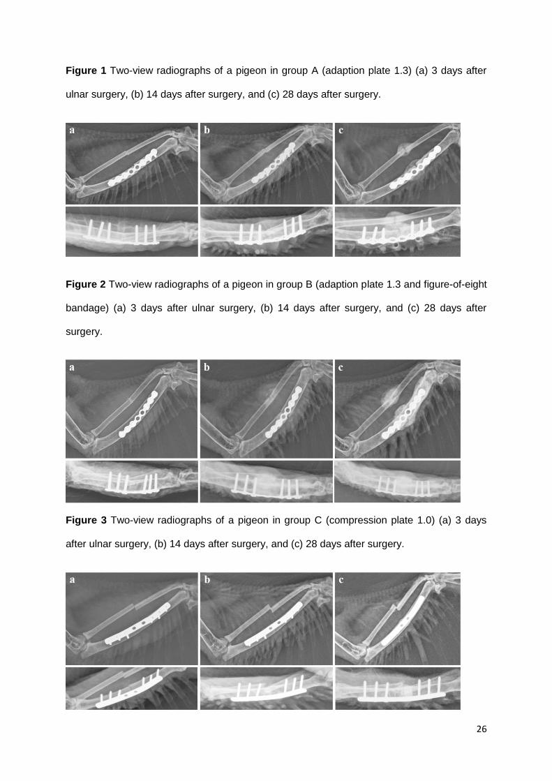
Radiological evaluation (Fig. 1 – 4, Tab. 2) revealed no signs of plate bending or screw
loosening. All screws remained bicortical until the end of the study except for 1 screw each in
1 pigeon of group B and D. The pigeon in group B was the bird that suffered from a
committed fracture during surgery. In this bird, 1 screw was unicortical at radiographs taken
14 days after surgery and remained in that position until the end of the study. The screws of
the pigeon in group D were bicortical including all radiographs taken before 28 days post-
surgery, but 1 screw was unicortical at the radiographs 28 days post-surgery. The wings of
these birds were stable on palpation.
Results of radiographic measurements are summarized in Table 2. The only significant
radiographic difference between groups was regarding maximal callus width of the radius.
Pigeons of group A (5.9 ± 0.9 mm) and C (5.8 ± 0.8 mm) showed significantly less maximal
callus width of the radius (Kruskal-Wallis-test, P = .034) than those of group B (7.1 ± 1.0) and
D (6.8 ± 0.6) on radiographs taken 28 days post-surgery. The callus ratio of the ulna did not

17
differ between groups but the mean ratio in all animals (2.04 ± 0.43) was significantly smaller
than that of the radius (2.42 ± 0.49; paired t-test t = 3.782, P = .001, n = 25).
The length of the ulna (pre OP 59.5 ± 2.2mm) remained unchanged (3 days post OP 59.1 ±
2.2 mm), whereas the length of the unfixed radius was reduced by 2.0 mm to 51.3 ± 1.8 mm
at 28 days after surgery, as the radial fracture ends were dislocated in the majority of animals
(in group A:4, B:4, C:5, D:5).
In 8 pigeons (29,6%) postoperative signs of fractures or fissures occurred (in group A:2, B:2,
C:3, D:1). This led to the euthanasia of 2 animals (1 pigeon each in group A and C) for
humane reasons as mentioned earlier. The cage rest of the other animals was prolonged
until 14 days post-surgery, but no further treatment was needed. The wings of these birds
were stable on palpation. Radiographically, 1 bird of group A showed signs of osteomyelitis
and 1 bird of group D showed the development of a synostosis between radius and ulna.
Thirteen pigeons (group B:6, D:7) were treated with a figure-of-eight bandage. It was noted
that the position of the left wing did differ significantly at day 10 post-surgery (Kruskal-Wallis-
test, P = .041) in group C compared to group D. The wing tip of pigeons of group D touched
the ground or the pigeons let their left wing droop mildly without the wing tip touching the
ground whereas pigeons of group C held their wings in physiologic position or let their left
wing droop mildly without the wing tip touching the ground. However, this difference was no
longer evident at 14 days or 28 days after surgery, when most of the pigeons showed only a
slight drop of the wing without the wing tip touching the ground, or held the wing in
physiologic position. There was no correlation between slight wing drop or physiologic wing
position and flight ability.
Evaluation of flight ability on day 28 post-surgery in 23 birds (group A: 6, B: 5, C: 5, D: 7) is
represented in Table 1 and 3. There were no significant differences in flight ability between

18
the treatment groups at any time point. Most pigeons began to flap their wings and fly short
distances in the lower half of the aviaries at 14 days post-surgery and regained good to very
good flight ability between 21 and 28 days post-surgery. The combination of both methods
assessing flight ability (personal observation and video observation) allowed defining the
mean number of days when the pigeons were seen in the upper half of the aviary for the first
time (21.7 (± 5.9) days post-surgery), respectively (Tab. 3). It is also interesting to note that
results gained by camera observation differed from those gained during
approaching/handling of the birds at 28 days post-surgery (Tab. 1): In presence of a human,
12 and 11 birds showed very good and good flight ability, respectively.
At necropsy evaluation, no significant differences among the treatment groups were found.
The bones of 3 pigeons showed signs of osteomyelitis (in group A:1, C:2). In 7 birds, the
implant was still visible (in group B:2, C:2, D:3), in 15 birds, the plate and screws were only
partially visible (in group A:6, B:4, C:2 D:3), and in 3 birds, no implant was visible due to the
callus formation (in group C:2, D:1). In one pigeon of group D, the synostosis diagnosed by
radiography was confirmed.
Discussion
In this study, we evaluated 2 different miniplates, with and without use of a figure-of-eight
bandage post operatively for birds weighing less than 500 g. Taking into account the results
described above, there was, in contrast to our predictions, no significant difference between
the treatment groups. There was no evidence that fracture healing with less callus
proliferation and better ability of flight could be achieved using a 1.0 compression plate
compared to a 1.3 adaptation plate. The use of a figure-of-eight bandage post operatively for
10 days did not improve fracture healing.
In contrast to similar studies,1, 8 there was neither distortion nor bending of the plates in the
present study (Fig. 5). This finding is explained by the material of the different plate systems.

19
The maxillofacial miniplate compact 1.0 with 11 holes evaluated by Gull et al.2 consisted of
titanium, whereas the adaption plate 1.3 as well as the compression plate 1.0 evaluated in
the present study consisted of stainless steel. Taking into account the results of all studies1, 2
evaluating these plates for their applicability for wing fracture repair, we conclude that
stainless steel plates are required to withhold the in vivo stresses for fracture repair of the
ulna in pigeons. Stainless steel plates as implanted in the present study are suited for single
plating of fractures of the avian antebrachium. With respect to plate choice it should be noted
that the compression plate system used in this study provides fracture fixation with equal
clinical results at an economically preferable prize in comparison with the adaption plate
system.
An additional finding of this paper is regarding the importance of screw length.
In the study performed by Gull et al.2 1 experimental group of 6 pigeons (group A) was
treated with an eight hole adaption plate 1.3 using 4 screws of 6 mm length. In our study in
group A the same plate with 6 screws with 8 mm length were used. In the study of Gull et al.2
1 out of 6 pigeons had to be euthanatized due to screw loosening and 3 out of 6 pigeons had
screws which were not bicortical. In the present study in contrast, screw loosening was only
observed in 1 out of 7 pigeons in the corresponding treatment group. The choice of longer
screws is recommended to ensure bicortical placement. This is especially critical when
performing surgery in small animals as no deep gauge is available for such small implants
which would allow measuring the length of the screw holes intra surgery.
In contrast to the predictions of Gull et al.2 thread pitch of screws was not as critical as screw
length. In this study screws with a thread pitch of 0.25 and 0.5 mm were used. Gull et al.2
suggested that the use of screws with a thread pitch of less than 0.5 mm may be preferable
due to better holding power. However, in the present study no signs of a difference in holding
power were observed. Nevertheless, the screws with a thread pitch of 0.25 mm may prove to
be advantageous in birds with a thinner cortex than the ones used in this study.
20
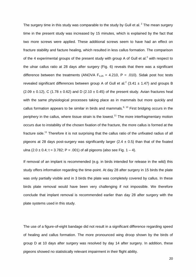
The surgery time in this study was comparable to the study by Gull et al. 2 The mean surgery
time in the present study was increased by 15 minutes, which is explained by the fact that
two more screws were applied. These additional screws seem to have had an effect on
fracture stability and facture healing, which resulted in less callus formation. The comparison
of the 4 experimental groups of the present study with group A of Gull et al.2 with respect to
the ulnar callus ratio at 28 days after surgery (Fig. 6) reveals that there was a significant
difference between the treatments (ANOVA F4,25 = 4.210, P = .010). Sidak post hoc tests
revealed significant differences between group A of Gull et al.2 (3.41 ± 1.47) and groups B
(2.09 ± 0.12), C (1.78 ± 0.62) and D (2.10 ± 0.45) of the present study. Avian fractures heal
with the same physiological processes taking place as in mammals but more quickly and
callus formation appears to be similar in birds and mammals.9, 10 First bridging occurs in the
periphery in the callus, where tissue strain is the lowest.11 The more interfragmentary motion
occurs due to instability of the chosen fixation of the fracture, the more callus is formed at the
fracture side.11 Therefore it is not surprising that the callus ratio of the unfixated radius of all
pigeons at 28 days post-surgery was significantly larger (2.4 ± 0.5) than that of the fixated
ulna (2.0 ± 0.4; t = 3.782; P = .001) of all pigeons (also see Fig. 1 – 4).
If removal of an implant is recommended (e.g. in birds intended for release in the wild) this
study offers information regarding the time-point. At day 28 after surgery in 15 birds the plate
was only partially visible and in 3 birds the plate was completely covered by callus. In these
birds plate removal would have been very challenging if not impossible. We therefore
conclude that implant removal is recommended earlier than day 28 after surgery with the
plate systems used in this study.
The use of a figure-of-eight bandage did not result in a significant difference regarding speed
of healing and callus formation. The more pronounced wing droop shown by the birds of
group D at 10 days after surgery was resolved by day 14 after surgery. In addition, these
pigeons showed no statistically relevant impairment in their flight ability.

21
Therefore one may deduct that the use of a figure-of-eight bandage does not bring any
advantages. Nevertheless we consider the application of a figure-of-eight bandage to be an
advantage and we found two reasons for it.
At necropsy signs of osteomyelitis were only found in groups treated without bandage. This
finding might indicate that the application of dressings is advantageous to reduce the risk of
infection and the development of bacterial osteomyelitis.
The figure-of-eight bandage may also have reduced the risk of fracture occurrence after
surgery. No bird of group B and D suffered from additional fractures due to wing flapping
while handling or recovery from anesthesia, whereas two birds treated without bandage
needed to be euthanized because of additional fractures.
Therefore the authors do recommend the use of a figure-of-eight bandage post-surgery for
up to 10 days. The bandage should be changed every 2 to 3 days for inspection of the
wound. Bandage changes should also be used to perform physical therapy as described by
Wimsatt et al.12 to prevent complications from immobilization such as muscle atrophy, joint
ankylosis, tendon contraction, and patagial constriction. In the present study, the wings were
very gently stretched and mobilized during bandage changes.
Regarding the radiographic diagnosis of osteomyelitis this study revealed that radiography
only detected osteomyelitis in 1 out of 3 birds. This finding emphasizes the fact that it may
take days or weeks until bacterial osteomyelitis becomes evident with plain radiography.13
In the post-surgical radiographic examinations 29.6% of the pigeons of our study showed
fissures or fracture lines. Fissures may occur intraoperatively during manipulation with
surgical equipment and implant. As the avian bone with its specific structure is more prone to
brittle than a mammalian bone, one may easily damage the thin avian corticalis while placing
the screws or pins.9, 14-16 Post-surgery, suboptimal placement of the implant may result in too
high strain on the bone due to wrong positioning or wrong size of the implant. A recently

22
stabilized bone may fracture due to a trauma in the recovery and reconvalescence period as
described for the 4 birds mentioned earlier. Unnoticed trauma due to too much pressure on
the stabilized bone post-surgery, maybe during handling, cannot be ruled out completely for
the 4 remaining pigeons but seemed less likely. As a limitation of the study the first X-rays
postoperatively were not taken immediately after, but at 3 days post-surgery, thus these
possible reasons could not be definitely narrowed down further.
The occurrence of iatrogenic fracture formation as a result of pin insertion was reported by
Ferraz et al.17 In that study, 18 experimental induced distal fractures of the humerus of 12
pigeons were stabilized with an articulated external fixator consisting of 3 titanium-rods with 6
mm diameter and 5 (3 humeral placed / 2 ulnar placed) Shunz Pins with 1 mm or 1.5 mm
diameter. Iatrogenic fractures of humerus and/or ulna occurred in 33.3% of osteotomies
during insertion of the pins. The authors concluded the large pin size to be the most likely
cause for the iatrogenic fractures. This could be the case in present study as well, as the
ulnar diameter did vary according to the size of the individual pigeon.
Comparing the results in regard to flight ability of that study17 with the present study is
interesting, too, as the study design in that aspect is similar. Ferraz et al.17 state adequate
flight capacity in all 6 birds by at least 13 weeks post-surgery, 2 weeks after being put in
aviaries (2.5 m x 2.5 m x 3 m) allowing unrestricted movement. In the present study, the
mean of days for all pigeons evaluated for their flight ability to regain good flight ability is 21.8
(± 4.4) days post-surgery, 11.8 (± 4.4) days after being put in aviaries allowing unrestricted
movement.
The 2 methods to evaluate flight ability in the present study yielded different results, direct
observation scored birds in better flight ability than camera observation. This may reflect the
tendency of birds to hide a weakened condition in sight of a possible threat as the sight of
humans approaching, even though the pigeons used were raised as companion animals.

23
These differences would possibly be even more distinct in a wild animal with no possibility to
get familiar with the housing and might confuse the clinical assessment of the patient, thus
provoking a premature release. This discrepancy could be surely minimized by examination
without human input, i.e. the installation of cameras in the aviaries in question, thereby
reducing the influence of stress on the evaluated behaviour.
Conclusion
The present data suggest that stainless steel miniplate systems are suitable for ulna fracture
repair in birds weighing less than 500 g using screws with a thread pitch of either 0.25 mm or
0.5 mm. If plates are to be removed, this must be done before day 28 after surgery. The use
of a post-surgical figure-of-eight bandage for up to 10 days may be advantageous, and
appears to reduce the risk of postoperative wound infection. When evaluating flight ability it
should be noted that birds may feign better ability. This is of special importance in birds
which are not accustomed to humans.
Acknowledgments: We thank Marcus Clauss for his assistance during manuscript
preparation, the keepers and the students of the Clinic for Zoo Animals, Exotic Pets and
Wildlife for their help in the care for the animals, the technicians of the Division of Diagnostic
Imaging for their practical advice and Urs Freiburghaus for providing the animals, as well as
Lukas Sprenger for his technical advice regarding the video observation. The authors would
also like to acknowledge the financial support by the Schwyzer-Stiftung and the Baugarten-
Stiftung.

24
LITERATURE CITED
1. Christen C, Fischer I, von Rechenberg B, et al. Evaluation of a maxillofacial miniplate
compact 1.0 for stabilization of the ulna in experimentally induced ulnar and radial fractures
in pigeons (Columba livia). J Avian Med Surg. 2005;19:185-190.
2. Gull J, Saveraid T, Szabo D, et al. Evaluation of three miniplate systems for fracture
stabilization in pigeons (Columba livia). J Avian Med Surg. 2012;26:203-211.
3. Hatt J-M. Hard tissue surgery. In: Chitty J, Lierz M, eds. Raptors, Pigeons and Passerine
Birds. Gloucester: British Small Animal Veterinary Association; 2008:157-175.
4. Montgomery R, Crandall E, Bellah J. Use of a locking compression plate as an external
fixator for repair of a tarsometatarsal fracture in a bald eagle (Haliaeetus leucocephalus). J
Avian Med Surg. 2011;25:119-125.
5. Dal-Bó IS, Alievi MM, Silva LM, et al. Tibiotarsus osteosynthesis' in blue-yellow-macaw
(Ara ararauna) using titanium miniplate. Arq Bras Med Vet Zoo. 2011;63:1003-1006.
6. Sá SS, Filho JC, Souza FS, et al. Osteosynthesis in tibiae fractures with mini plate, screws
and cerclage wire in goose (Anser anser): case report. Acta Vet Bras. 2012;6:61.
7. Gouvêa A, Meller M, Noriega V, et al. Titanium microplates for treatment of tibiotarsus
fractures in pigeons. Cienc Rural. 2011;41:476-482.
8. Gull J. Comparision of three miniplate systems in experimentally induced ulnar and radial
fractures in pigeons (Columba livia). Doctoral Thesis, University of Zurich., Zürich: University
of Zurich; 2010.
9. Coles B. Essentials of avian medicine and surgery. Oxford: Blackwell Publishing; 2007.
10. Howard M, Ritchie B. Orthopedic surgical techniques. In: Ritchie B, Harrison G, Harrison
L, eds. Avian medicine: Principles and Application. Lake Worth: Wingers Publishing;
1994:1137-1156.
11. Brown S, Kramers P. Indirect (secondary) bone healing. In: Bojrab M, ed. Disease
Mechanisms in Small Animal Surgery. Philadelphia: Lea & Febiger; 1993:671-677.
12. Wimsatt J, Dressen P, Clinton D, et al. Ultrasound therapy for the prevention and
correction of contractures and bone mineral loss associated with wing bandaging in the
domestic pigeon (Columba livia). J Zoo Wildl Med. 2000;31:190-195.
13. Gold RH, Hawkins RA, Katz RD. Bacterial osteomyelitis: findings on plain radiography,
CT, MR, and scintigraphy. Am J Roentgenol. 1991;157:365-370.
14. Degernes L, Roe S, Abrams CF. Holding power of different pin designs and pin insertion
methods in avian cortical bone. Vet Surg. 1998;27:301-306.
15. Koranyi E, Bowman CE, Knecht CD, et al. Holding power of orthopedic screws in bone.
Clin Orthop Relat R. 1970;72:283-286.
16. Numaker DM. Experimental models of fracture repair. Clin Orthop Relat R. 1998:S56-
S65.

25
17. Ferraz VCM, Ferrigno SRG, Cortopassi R, et al. Radiologic and flight function evaluation
after fixation of distal humeral osteotomies in pigeons, with model of articulated external
fixator. Pesquisa Vet Brasil. 2008;28:351-357.
26
Figure 1 Two-view radiographs of a pigeon in group A (adaption plate 1.3) (a) 3 days after
ulnar surgery, (b) 14 days after surgery, and (c) 28 days after surgery.
Figure 2 Two-view radiographs of a pigeon in group B (adaption plate 1.3 and figure-of-eight
bandage) (a) 3 days after ulnar surgery, (b) 14 days after surgery, and (c) 28 days after
surgery.
Figure 3 Two-view radiographs of a pigeon in group C (compression plate 1.0) (a) 3 days
after ulnar surgery, (b) 14 days after surgery, and (c) 28 days after surgery.

27
Figure 4 Two-view radiographs of a pigeon in group C (compression plate 1.0 and figure-of-
eight bandage) (a) 3 days after ulnar surgery, (b) 14 days after surgery, and (c) 28 days after
surgery.
Figure 5 Mean angle of osteotomy ends of the ulna measured in pigeons immediately after
surgery (osteotomy of ulna and radius, fixation of ulna) and/or at 3, 14 and 28 days later.
Note that in contrast to group C of Gull et al. (2012)2, treated with a titanium miniplate, the
steel plate systems used in both studies showed no evidence of bending irrespective of
number of screws or additional bandaging after surgery.
Figure 6 Mean ulnar callus ratio measured in pigeons (osteotomy of ulna and radius, fixation
of ulna) 28 days post-surgery. There were significant differences between group A of Gull et
al. (2012)2 and groups B, C and D of the present study due to the increased number of
screws applied.

28

29
Table 1 In 4 groups of pigeons (A: n=7, B: n=6, C: n=7, D: n=7) fractures of the radius and
the ulna were induced, and the ulna was repaired with a bone plate (Ap = adaption plate 1.3
or Cp = compression plate 1.0) and with a figure-of-eight bandage (+) or without (-). The
most important findings and differences between the groups A, B, C, and D are listed. No
statistically significant differences were measured between the groups.
Parameter Days
after
surgery
Group A
Ap -
Group B
Ap+
Group C
Cp-
Group D
Cp+
Surgical
time (mean ±
SD), min
- 50.6
± 6.7
43.9
± 4.8
56.7
± 16.5
53.1
± 15.2
Surgical
procedure:
Problems
-
Redrill of
holes
necessary,
soft tissue
trauma
Accidental
fracture
while
inserting 1
screw,
variation in
order of
screws
Redrill of
holes
necessary,
variation in
order of
screws
Soft tissue
trauma,
variation in
order of
screws
Euthanized
because of
screw
loosening, %
1 until 28 0 0 0 0
Euthanized
because of
fracture, %
1 until 28 14 0 14 0
Plate bent
and twisted,
%
14 0 0 0 0
28 0 0 0 0

30
Callus width
(mean ± SD),
mm
14 Signs of bone remodelling, but not measurable
28 9.5 ± 1.9 9.6 ± 0.5 7.9 ± 2.6 9.4 ± 1.6
Flight ability,
%1
very good 28 43 / 71 17 / 17 14 / 43 14 / 43
good 28 29 / 14 50 / 67 14 / 29 14 / 57
moderate 28 0 / 0 17 / 0 29 / 0 43 / 0
poor 28 14 / 0 0 / 0 14/ 0 29 / 0
not evaluted
28 14 / 14 17 / 17 29 / 29 0 / 0
Signs of
osteomyeliti
s at
necropsy, %
28 14 0 29 0
1Observations gained by video observation / direct observation in 23 pigeons, not evaluated
refers to pigeons euthanized for humane reasons or housed outside aviaries.
Table 2 Mean radiographic values from 4 groups of pigeons (A: n=7, B: n=6, C: n=7, D: n=7)
following experimental fracture of the radius and the ulna.
Days after
surgery mean (±SD)
Length of radius, mm 0 53.3 (± 2.2)a
3 50.5 (± 2.2)b
28 51.3 (± 1.8)c
Length of ulna, mm 0 59.5 (± 2.2)
3 59.1 (± 2.2)

31
28 59.5 (± 1.8)
Maximal fracture gap of the ulna, mm 3 0.2 (± 0.17)
Step between the fracture margins of
the ulna, mm 28 0.6 (± 0.7)
Angle of the fracture ends of the ulna,
degree 3 165.9 (± 4.7)
14 163.6 (± 4.9 )
28 162.6 (± 4.9 )
Callus ratio ulna 28 2.0 (± 0.4)
Callus ratio radius 28 2.4 (± 0.5)
a,b,c different superscripts indicate significant differences (paired t-test with Sidak
adjustment) in the length of the radius; there were no significant differences in the length of
the ulna
Table 3 Mean (±SD) number of days following experimental fracture of the radius and the
ulna at which pigeons were first seen in the upper half of the aviary.
Group Good flight ability Very good flight ability
mean (d)
% of
pigeons mean (d)
% of
pigeons
A
19.9
(± 4.4) 100
23.6
(± 4.4) 83.3
B
21.2
(± 1.1) 83.3
24.5
(± 4.9) 40
C
22.4
(± 4.0) 83.3
25.7
(± 2.5) 60
D
23.4
(± 6.1) 100
25.0
(± 5.2) 42.9

32
Danksagung
An dieser Stelle möchte ich mich bei allen bedanken, die das Erstellen dieser Dissertation erst
möglich gemacht haben. Mein besonderer Dank gilt:
Prof. Dr. Jean-Michel Hatt für das Überlassen des Themas, die geduldige Betreuung und
Unterstützung bei den Versuchen und der Erstellung des Manuskriptes.
Prof. Dr. Marcus Clauss für die Unterstützung bei der Erstellung des Mauskriptes und die
statistische Analyse der Daten des Versuches.
Allen Mitarbeitern der Klinik für Zoo- Heim- und Wildtiere bei der tatkräftigen Unterstützung
bei der Durchführung meiner Versuche.
Allen Studenten der Vetsuisse-Fakultät, die mit mir im Rahmen meiner Versuche
zusammengearbeitet haben, für Ihr besonderes Engagement und Ihre tatkräftige Unterstützung
(auch am Wochenende).
Urs Freiburghaus für das Überlassen der Versuchstiere.
Lukas Sprenger für seine technische Expertise.
Jeanne Peter für die Hilfe mit dem Bildmaterial.
Den Co-Autoren Prof. Dr. med. vet., PhD, Dipl. ECVDI Patrick R. Kircher, Dr. med. vet.,
Dipl. ECVS Andreas Gutbrod und Dr. Juliane Riechert.
Meinen Bürokollegen und Mitdoktoranden für zahlreiche Gespräche und das grosszügige
Teilen Ihrer Erfahrung.
Meinen Eltern, Ute und Stephan Schmidt, meiner Schwester Annabell Bennert und Anna
Bonsmann für die moralische Unterstützung.

33
Curriculium Vitae
Name, Vorname: Bennert, Beatrice Miriam Geburtsdatum: 06.06.1988
Geburtsort:
Rosenheim
Nationalität:
Deutsch
Schulausbildung
09/1994- 07/1998
Grundschule Bad Aibling, Bad Aibling,
Deutschland
09/1998- 06/2007
Gymnasium Bad Aibling, Bad Aibling,
Deutschland
höchster Schulabschluss
29.06.2007
Allgemeine Hochschulreife
Studium
10/2007- 05/2013 Tiermedizin, Ludwig-Maximilians-Universität, München,
Deutschland
22.04.2013
Abschlussprüfung vet. med.
Ludwig-Maximilians-Universität, München, Deutschland
10/2013- 10/2014 Anfertigung der Dissertation
unter Leitung von Prof. Dr. med. vet.,
MSc, Dipl. ACZM, Dipl. ECZM (Avian) Jean-Michel Hatt
am Department of Small Animals Clinic for Zoo Animals,
Exotic Pets and Wildlife,
der Vetsuisse-Fakultät Universität Zürich
Direktor Prof. Dr. med. vet., MSc, Dipl. ACZM, Dipl.
ECZM (Avian)
Jean-Michel Hatt