Embed Size (px)
DESCRIPTION
MDT
Citation preview

C H A P T E R 8
Evaluating the Patient Using the McKenzie Approach
GARY JACOB, DC, LAc, DIP.MDT, STEVEN HEFFNER, DC, DIP.MDT, ROBERT MEDCALF, PT, DIP.MDT, DOUGLAS BARENBURG, DC, CRED.MDT,
& SUZAN STARLER, DC, CRED.MDT
I N T R O D U C T I O N
The main purpose of this chapter is to explore the clinical reasoning resulting from McKenzie's paradigm regarding the nature and solution of common activity-related spinal complaints. Clinical competency in the McKenzie approach is greatly aided by study of McKenzie's original texts13,14 and supervised clinical training. Supervised clinical training is particularly important in order to develop the skills to best manage the fears faced by patients and clinicians when therapy involves promoting movement of patients' painful spines. Our primary purpose is to communicate the conceptual structure of the McKenzie paradigm.
Many approaches exist for patients with common spinal complaints. Each approach is based upon certain criteria. By choosing a particular evaluation or treatment approach, the practitioner (consciously or not) chooses certain criteria or paradigms upon which evaluation or treatment is predicated. The spinal evaluation and treatment paradigms employed often inculcate patients with belief systems regarding the nature of and solution to their spinal problems.
Rehabilitation philosophies argue passive therapies turn patients into passive receptacles of care. Intrinsic to the clinical reasoning, problem solving and goal setting of a rehabilitation pro-gram is the emphasis on motivating patients to participate actively in their own care. This is gen-erally done through a conditioning program targeting posture, balance, cardiovascular, flexibility, and/or strength status.
The McKenzie paradigm distinguishes itself among re-habilitation approaches by means of the criteria upon which it predicates evaluation and treatment methods. The McKenzie paradigm accounts for common spinal complaints by classifying the mechanical and symptomatic responses to spinal loading (forces applied by means of movement and positioning). A McKenzie evaluation requires the clinician to look and listen for clues regarding patterns of mechanical and symptomatic responses to loading. Patients report movements and positionings of their spines affect them in a variable manner. Provocative spinal loading strategically performed in the clinical setting reveals different reactions to different loading strategies. The McKenzie approach ex-
plores activity-related solutions to activity-related complaints, employing the criteria of mechanical and symptomatic responses to loading to evaluate, treat, and provide prophylaxis.
McKenzie protocols permit the employment of a reha-bilitation approach in the acute phase. Rehabilitation em-ploys activity therapies to achieve restoration, self-sufficiency, and independent functioning skills. The clinician's responsibility is to introduce activity therapy as soon as possible and to identify cases where activity therapy is in-effective or inappropriate as soon as possible. The patient's responsibility is to participate actively in exploring the potential benefits of self-generated movements.

Evaluating the Patient Using the McKenzie Approach 148
A PRIORI NOTIONS THAT LIMIT THERAPEUTIC POSSIBILITIES Certain paradigms of spinal evaluation and treatment are based on preconceived notions regarding the cause of common spinal complaints. Two of the most common preconceived notions are those of inflammation and pain of muscular origin. Many passive therapies are employed for the purpose of reducing inflammation and/or pain of muscular origin. Promotion of rest and use of medication are often the preferred strategies to treat inflammation or pain of muscular origin. Exercise is considered contraindicated. Treatment predicated on such a priori notions prevents the exploration of the potential benefits of activity therapies.
INFLAMMATION
Relief by means of NSAIDs does not confirm the presence of inflammation. NSAIDs have analgesic as well as antiinflammatory properties and may suppress pain in the absence of inflammation. With joint complex inflammation (e.g., inflammatory arthritis), relief cannot be found by means of movement or positioning. Inflammation is thought of as a chemical event that would not be relieved greatly by mechanical factors. Contrary to this, acute and chronic spinal complaints commonly include a history of certain activities, movements, or positionings that relieve complaints, suggesting that the role of inflammation may not be significant. Prolonged rest used to treat purported in-flammation promotes disability.'
MUSCLE SPASM
Most commonly, pain purported to be of muscular origin is ascribed to muscle spasm. This notion leads to inactivity as the strategy to relax the spasm. Thermal agents, electrical stimulation, massage, or other passive procedures are employed to this end. A diagnosis of "chronic spasm" condemns patients to prolonged inactivity, i.e., disability.
Medically, spasm is understood to be a violent, invol-untary contraction of a muscle, something not seen com-monly. What is commonly referred to as spasm in the clinical setting is curious:
1. When patients present with antalgia contralateral
to the symptomatic side, it is often ascribed to muscle spasm even though the side of symptoms (spasm) would be expected to cause ipsilateral antalgia.
2. The ascription of kyphotic antalgia to spasm of posterior paraspinal muscles is not clear thinking, as paraspinal muscles are extensors, not flexors.
3. Most patients with spinal complaints are worse when sitting, during which time spinal muscles are recruited less, contrary to what would be expected from muscle spasm.
4. Expert physicians cannot reliably identify muscle spasm by means of palpation.'
5. A survey of the literature concluded that muscles are not the primary cause of spinal complaints.)
SPINAL MANIPULATION AS EVIDENCE OF THE EFFICACY OF MOVEMENT The efficacy of spinal manipulation casts doubt on the wis-dom of universally applying the models of inflammation, spasm, immobility, and rest to common, activity-related spinal complaints.
Chiropractic has successfully employed manipulation for the past 100 years, in spite of periods of aggressive med-ical opposition to manipulative therapies. Medicine accused those who "adjusted" the spine of adding insult to in-jury and prematurely introducing aggressive movement when rest, immobility, and inactivity were needed. Medical objections were predicated on the criteria that inflammation and spasm were the culpable physiologic processes, es-pecially for the acute spine. Inflammation and spasm would be expected to respond poorly to movement, especially the aggressive end range loading movements of manipulation.
Current wisdom views manipulation as an efficacious approach worthy of consideration for acute, activity-related spinal complaints.4,23,24 The same cannot be said for passive nonmovement modalities that target inflammation and/or spasm.
The efficacy of spinal manipulation for acute conditions suggests:
Movement is of potential benefit for the acute spine. Aggressive movement is of potential benefit for the acute spine. Aggressive movement to end range is of potential benefit for the acute spine.
Manipulation, as in exercise, is a movement therapy. Both have been classified as forms of "activation."24
DETERMINING WHETHER LOADING STRATEGIES ARE APPROPRIATE McKenzie protocols determine, early in the course of care, who are the responders and nonresponders to mechanical therapies. Mechanical therapies include postural training, all prescriptive exercises, mobilizations, manipulations, and advice concerning activities of daily living. If self-generated loading and/or clinical mobilizations in all movement plane directions fail to provide relief, manipulation or other mechanical therapies are less likely to be of benefit. If self-generated loading and/or clinical mobilizations in all movement plane directions appear to be to the patient's detriment, "red flags" go up raising suspicions of pernicious processes not amenable to mechanical therapies. If patient-generated end range loading or positioning is of benefit, these conditions are less likely. For those identified as being amenable to mechanical therapies, the clinician's task is to
Evaluating the Patient Using the McKenzie Approach 149
determine the preferred loading strategy, to institute activity therapies immediately, and to avoid passive care and rest. This entails determining which loading tactics to avoid, which to pursue, and when it is safe to introduce previously avoided loading tactics.
SPINAL MANIPULATION, REHABILITATION AND McKENZIE PROTOCOLS Spinal manipulation and spinal rehabilitation are activity therapies that complement each other. Spinal manipulation involves passive movement. Rehabilitation involves active movement. The success of spinal manipulation for "acutes" suggests self-generated movements may benefit acute pa-tients, something not entertained commonly. McKenzie em-ploys a system of prescriptive exercises based on principles similar to that of spinal manipulation, i.e., end range loading. McKenzie protocols explore the mechanical and symptomatic responses to end range loading prior to introducing ma-nipulative forces.
The McKenzie protocols employ manipulation when patient self-generated movement to end range is demon-strated to be of some, but not complete, benefit. If complete recovery is realized from self-generated end range loading, manual therapies are not needed. If a partial response is realized, the practitioner has a good understanding of which movement plane directions to manipulate. To "test the waters," the clinician would provide mobilization to end range to ensure the correct movement plane direction was being explored. If mobilization resolves complaints, there is no need to proceed to manipulation. If a full response is not realized, the progression of forces then considers manipulation. Premanipulative end range mobilization testing, whether performed by the patient or the clinician, heralds the mechanical and symptomatic responses one can expect from end range manipulation. These herald responses are evoked by means of progressive forces, from patient-generated to clinician mobilization to clinician manipulation.
McKENZIE EVALUATION PROTOCOLS BASELINE MOTION MEASUREMENTS
Movement is first assessed without regard to symptomatic response or to how one movement may affect another, after which these responses are evaluated closely. Movements employed that differ from those conducted during usual range of motion examinations will he illustrated.
Baseline measurements are taken in the following order for the respective spinal areas:
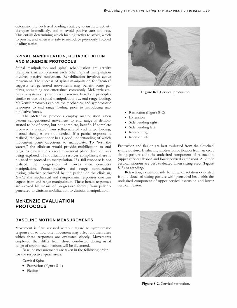
Cervical Spine • Protrusion (Figure 8–1) • Flexion
Figure 8-1. Cervical protrusion.
• Retraction (Figure 8–2) • Extension • Side bending right • Side bending left • Rotation right • Rotation left
Protrusion and flexion are best evaluated from the slouched sitting posture. Evaluating protrusion or flexion from an erect sitting posture adds the undesired component of re-traction (upper cervical flexion and lower cervical extension). All other cervical motions are best evaluated when sitting erect (Figure 8–3) or standing.
Retraction, extension, side bending, or rotation evaluated from a slouched sitting posture with protruded head adds the undesired component of upper cervical extension and lower cervical flexion.
Figure 8-2. Cervical retraction.

Evaluating the Patient Using the McKenzie Approach 150
Lumbar Spine • Flexion • Extension • Side gliding right (Figure 8—4) • Side gliding left
For the lumbar spine, lateral flexions and rotations are not employed. Instead, side gliding (which combines these two movements) is utilized. Side gliding involves a translational movement in the coronal plane, whereby the pelvis and trunk are moved in opposite directions while attempting to keep the shoulders and iliac crests level.
Patients do not present with lateral flexion or rotation antalgias, but do present with antalgias resulting from side gliding. These antalgias or lists are known within McKenzie protocols, for the low back, as lateral shifts and indicate the relevance of considering side gliding as a potentially thera-peutic movement to explore.
D Y N A M I C A N D STATIC EVALUATIONS Subsequent to obtaining baseline motion measurements, the mechanical and symptomatic responses to loading are explored. McKenzie protocols have a preference for explor-ing dynamic (repetitive movement) loading within the sagittal plane first. If dynamic loading in the sagittal plane is of benefit, loading strategies are not explored in the coronal or transverse planes. If dynamic loading within the sagittal plane does not appear to be of any benefit, coronal and then transverse planes are explored. If dynamic loading fails to demonstrate benefit, static loading is then explored.
All cervical spine loading procedures except for flex-ion and protrusion are performed from the retracted position in order to promote maximum end range loading. Evaluations are performed in the following order for the respective spinal areas:
Figure 8—5. Cervical extension (from retraction).
Figure 8—3. Sitting erect.
Figure 8—4. Side gliding right.
t

Evaluating the Patient Using the McKenzie Approach 151
Figure 8–8. Cervical side bending (from retraction).
Cervical Spine Dynamic Tests • Flexion sitting • Retraction sitting (Figure 8–2) • Retraction-then-extension sitting (Figures 8–2 and 8–5)
• Retraction lying (head off the edge of treatment table) (Figure 8–6)
• Retraction-then-extension lying (head off edge of treatment table) (Figures 8–6 and 8–7)
Other Cervical Spine Dynamic Tests (if required) • Protrusion sitting (Figure 8–1) • Retraction-then-side bending right or left sitting
(Figure 8–8)
• Retraction-then-rotation right or left sitting (Figure 8—9)
Cervical Spine Static Tests • Protrusion (Figure 8–1) • Flexion • Retraction (sitting or supine) (Figures 8–2 and 8–6) • Retraction-then-extension (sitting, prone) (Figures 8—2 and 8—7)
• Retraction-then-side bending right or left (Figure 8–8)
• Retraction-then-rotation right or left (Figure 8–9)
Figure 8–6. Cervical retraction lying.
Figure 8–9. Cervical rotation (from retraction). Figure 8–7. Cervical extension lying (from retraction).

Evaluating the Patient Using the McKenzie Approach 152
Figure 8—12. Lumbar extension in lying (McKenzie "press-up").
Lumbar Spine Dynamic Tests • Flexion standing • Extension standing (Figure 8—10) • Flexion in lying (supine knee to chest) (Figure 8—11) • Extension in lying (prone McKenzie press-up(Fig-
ure 8—12) Other Lumbar Spine Dynamic Tests (if required)
• Side gliding right or left standing (Figure 8—4) • Extension from prone right or left lateral shift posi-
tion (Figure 8—13) Lumbar Spine Static Tests • Sitting slouched (Figure 8—1) • Sitting erect (Figure 8—3) • Standing slouched • Standing erect • Lying on elbows (Figure 8—14)
Figure 8—10. Lumbar extension standing.
Figure 8—11. Lumbar flexion in lying.

Evaluating the Patient Using the McKenzie Approach 153
Long sitting Lateral shift right or left (Figure 8–4) Rotation in flexion (Figure 8–15)
THE USE OF OVERPRESSURE FOR EVALUATION A N D TREATMENT
Overpressure represents strategies that may be employed by the patient or the clinician in order to realize further end range positioning. Chiropractors may think of overpressure as strategies that "take the slack out."
Overpressure may be used to obtain diagnostic informa-tion or as part of a therapeutic progression. Typically, over-pressure exaggerates the mechanical and symptomatic re-sponses of the joint complex it is applied to. The concept of overpressure is intimately connected to the McKenzie phi-losophy of introducing forces in a progressive manner. Pa-tients may exert overpressure on joint complexes by strategic movements of their own. Patients must be encouraged to achieve as much end range as possible. If the therapist pas-sively moves a joint to end range, this may be construed as mobilization. The next and final progression of overpressure forces toward end range would be manipulation.
Some examples of overpressure follow: Prone extensions offer more overpressure to the low back than do standing
Figure 8–14. Lying on elbows ("lying prone in extension").
Figure 8–13. Lumbar prone extension (from right lateral shift). Figure 8–15. Lumbar rotation in flexion.

Evaluating the Patient Using the McKenzie Approach 154
Figure 8-16. Therapist overpressure: prone extension.
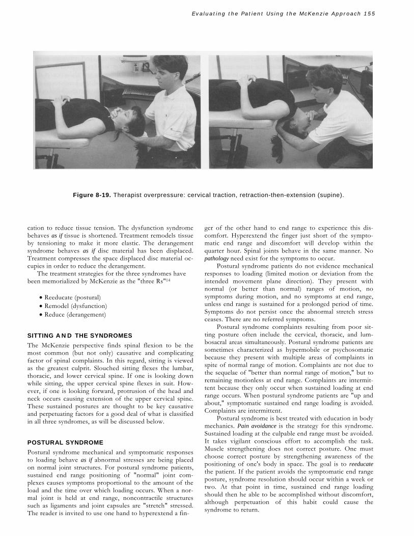
extensions. A prone extension performed while the clinician's hands hold the patient's hips down, creates greater overpres-sure (Figure 8–16). For example, therapist overpressure is commonly employed to assist lumbar side gliding (in the di-rection opposite the presenting lateral shift) (Figure 8–17) and cervical retraction (Figure 8–18) and for cervical traction, retraction-then-extension when supine (Figure 8–19).
Figure 8–17. Therapist overpressure: left side gliding (to reverse right lateral shift).
Figure 8-18. Therapist overpressure: cervical retraction.
McKENZIE'S SYNDROMES
McKenzie organizes mechanical and symptomatic responses to loading into three "syndrome patterns" that classify the manner in which mechanical and symptomatic responses occur in tandem. McKenzie protocols recognize the loading in one movement plane direction may affect the mechanical responses in that direction, the opposite movement plane direction, or an entirely different movement plane. The mechanical responses are range of motion and the ability to accomplish the movement in the intended movement plane without deviation. McKenzie protocols recognize the ability of loading in one movement plane direction to affect symptomatic responses in the same or other movement planes. Symptoms include the topography and severity of symptoms, the various "-esthesia," and subjectively perceived disabilities. The three syndromes are:
• Postural syndrome • Dysfunction syndrome • Derangement syndrome
The names given to the syndromes serve as functional, op-erational, and metaphorical definitions. Syndrome names are similes or "as if" identifications of what mechanical and symptomatic response patterns to loading behave "like."
The postural syndrome behaves as if postural stresses on normal tissue are culpable. Treatment is postural reedu-
Evaluating the Patient Using the McKenzie Approach 155
Figure 8-19. Therapist overpressure: cervical traction, retraction-then-extension (supine).
cation to reduce tissue tension. The dysfunction syndrome behaves as if tissue is shortened. Treatment remodels tissue by tensioning to make it more elastic. The derangement syndrome behaves as if disc material has been displaced. Treatment compresses the space displaced disc material oc-cupies in order to reduce the derangement.
The treatment strategies for the three syndromes have been memorialized by McKenzie as the "three Rs"14
• Reeducate (postural) • Remodel (dysfunction) • Reduce (derangement)
SITTING A N D THE SYNDROMES The McKenzie perspective finds spinal flexion to be the most common (but not only) causative and complicating factor of spinal complaints. In this regard, sitting is viewed as the greatest culprit. Slouched sitting flexes the lumbar, thoracic, and lower cervical spine. If one is looking down while sitting, the upper cervical spine flexes in suit. How-ever, if one is looking forward, protrusion of the head and neck occurs causing extension of the upper cervical spine. These sustained postures are thought to be key causative and perpetuating factors for a good deal of what is classified in all three syndromes, as will be discussed below.
POSTURAL SYNDROME Postural syndrome mechanical and symptomatic responses to loading behave as if abnormal stresses are being placed on normal joint structures. For postural syndrome patients, sustained end range positioning of "normal" joint com-plexes causes symptoms proportional to the amount of the load and the time over which loading occurs. When a nor-mal joint is held at end range, noncontractile structures such as ligaments and joint capsules are "stretch" stressed. The reader is invited to use one hand to hyperextend a fin-
ger of the other hand to end range to experience this dis-comfort. Hyperextend the finger just short of the sympto-matic end range and discomfort will develop within the quarter hour. Spinal joints behave in the same manner. No pathology need exist for the symptoms to occur.
Postural syndrome patients do not evidence mechanical responses to loading (limited motion or deviation from the intended movement plane direction). They present with normal (or better than normal) ranges of motion, no symptoms during motion, and no symptoms at end range, unless end range is sustained for a prolonged period of time. Symptoms do not persist once the abnormal stretch stress ceases. There are no referred symptoms.
Postural syndrome complaints resulting from poor sit-ting posture often include the cervical, thoracic, and lum-bosacral areas simultaneously. Postural syndrome patients are sometimes characterized as hypermobile or psychosomatic because they present with multiple areas of complaints in spite of normal range of motion. Complaints are not due to the sequelae of "better than normal range of motion," but to remaining motionless at end range. Complaints are intermit-tent because they only occur when sustained loading at end range occurs. When postural syndrome patients are "up and about," symptomatic sustained end range loading is avoided. Complaints are intermittent.
Postural syndrome is best treated with education in body mechanics. Pain avoidance is the strategy for this syndrome. Sustained loading at the culpable end range must be avoided. It takes vigilant conscious effort to accomplish the task. Muscle strengthening does not correct posture. One must choose correct posture by strengthening awareness of the positioning of one's body in space. The goal is to reeducate the patient. If the patient avoids the symptomatic end range posture, syndrome resolution should occur within a week or two. At that point in time, sustained end range loading should then he able to be accomplished without discomfort, although perpetuation of this habit could cause the syndrome to return.

Evaluating the Patient Using the McKenzie Approach 156
Postural Syndrome and Sitting Due to common poor sitting posture, the upper cervical spine extends and the rest of the spine slouches into flex-ion. A sustained extension postural syndrome develops at the upper cervical spine. A sustained flexion postural syndrome develops anywhere from the lower cervical spine to the lumbosacral junction.
Postural Syndrome and Spinal Manipulation The postural syndrome represents abnormal stress placed on normal tissue. Range of motion is equal to or greater than normal in the postural syndrome. Complaints are only experienced when end range loading is performed for a pro-tracted period of time. Spinal manipulation is not an ap-propriate consideration for the postural syndrome because there is no motion to restore. Only postural correction re-solves the syndrome.
DYSFUNCTION SYNDROME Dysfunction syndrome mechanical and symptomatic re-sponses to loading behave as if adaptive shortening, loss of elasticity, scar tissue formation, etc., are restricting spinal movements. For dysfunction syndrome patients, symptom-free movement is accomplished until the end range of a short-ened structure is realized, at which point there is prohibition of further movement, often accompanied by symptoms. Within the course of an initial examination, no matter how many repetitions of the limited motion are pursued, a me-chanical impedance occurs at the same point in the movement arc. Responses are as if a shortened structure is present that cannot be lengthened during the course of the initial visit. It may take weeks or months of vigilant loading for this to be accomplished.
Like the postural syndrome, the dysfunction syndrome displays intermittent complaints. In the postural syndrome, complaints develop over time with sustained loading at end range. In the dysfunction syndrome, complaints are experi-enced immediately when the mechanically impeded, limited end range is realized and shortened tissue is challenged to lengthen. For both the postural and dysfunction syndromes, discomfort ceases once loading at the culpable end range ceases.
Postural syndrome patients do not present with symptom referral phenomena. Dysfunction syndrome patients ex-perience localized symptoms except for the case of the adherent nerve root. The adherent nerve root is the only instance in which a dysfunction syndrome refers symptoms. This has been referred to as adverse neural tension, 3 and may be due to postsurgical scarring or scar formation resulting from natural healing processes without surgical intervention.
Like all dysfunction syndromes, adherent nerve root phenomena conform to a typical pattern. Adherent nerve root signs and symptoms are reproduced or intensified by increasing the "tension" on the adherent root. Nerve root tension is increased by means of spinal flexion combined with strategic movements of the involved extremity. Cervi-
cothoracic adherent nerve root signs and symptoms are re-produced or intensified by cervical flexion, combined with cervical contralateral lateral flexion and abduction of the ipsilateral shoulder with extended elbow. Lumbosacral ad-herent nerve root signs and symptoms are reproduced or in-tensified by lumbosacral flexion, with ipsilateral hip flexion and knee extended. As with all dysfunction syndromes, the mechanical and symptomatic responses of an adherent nerve root occur at the limited end range of the "adherence." This is the point at which the adherence prevents any further movement as it anchors the root to resist the action of further spinal flexion. Mechanical and symptomatic responses occur immediately as the end range of the adherence is challenged. Responses fade once end range tensioning is terminated.
Unilateral dysfunctions may cause deviation from the intended movement plane direction as the body "accom-modates" by deviating toward the side of dysfunction to continue a semblance of the intended movement. If a lum-bosacral joint or nerve root is suffering dysfunction on the right of the spine, attempted flexion may be accompanied by movement toward the right without which further flex-ion would not be possible. Deviation to the right represents an attempt to "slacken" the adherence, thus permitting further flexion movement.
In the postural syndrome discomfort results from normal tissue being subjected to abnormal (sustained end range) forces whereas in the dysfunction syndrome abnormal (shortened) tissue causes discomfort when subjected to normal forces (movement within normal range of motion). For the postural syndrome, avoiding end range discomfort is the remedy; for the dysfunction syndrome, it perpetuates the problem. Pain pursual is the preferred strategy for the dysfunction syndrome. The loading strategy for the dys-function syndrome is to challenge shortened tissue fre-quently. Dysfunction patients are required to elicit discomfort at the mechanically impeded limited end range over the course of the day. Dysfunction syndrome resolution em-ploys the "use it or lose it" principle. Treatment behavior is conducted as if one were trying to remodel shortened tissue.
Dysfunction syndrome nomenclature employs the name of the movement plane direction limited by shortened tissue. If extension is limited due to adaptively shortened tissue, this would be referred to as an extension dysfunction.
Dysfunction Syndrome and Sitting Dysfunction (adaptive shortening of tissue) is thought to develop as a result of habitual positionings absent movement or positioning in opposite directions. Poor sitting posture results in habitual upper cervical extension and lower cervical, thoracic, and lumbosacral flexion. This pro-motes the development of flexion dysfunctions of the upper cervical spine and extension dysfunctions of the lower cervical, thoracic, and lumbosacral spinal regions.
Spinal Manipulation and Dysfunction Syndrome The dysfunction syndrome requires frequent end range load-ing during the course of the day in order to remodel short-
Evaluating the Patient Using the McKenzie Approach 157
ened tissue. Tissue that is "too short" may not respond fa-vorably to manipulation. If shortened tissue is over-stretched, it will become inflamed and change the clinical picture from intermittent mechanical pain to constant chemical pain. After patient-generated and/or clinician-as-sisted mobilizations partially remodel shortened tissue, dy-namic manipulative loading is less of a risk and may, in fact, be of further benefit to remodel the joint complex. Manipu-lation is appropriate in the' later stages of dysfunction re-modeling in order to stretch tissue beyond the range of the patient's own capabilities. Manipulation, without the per-formance of frequent patient-generated end range loading, cannot resolve this syndrome.
DERANGEMENT SYNDROME
Derangement syndrome mechanical and symptomatic re-sponses behave as if there has been displacement, accumu-lation, migration, or directional pressure changes of in-tradiscal materials. In postural and dysfunction syndromes, loading in one movement plane direction does not affect mechanical and symptomatic responses in other movement plane directions. In the derangement syndrome, loading in one movement plane direction may affect mechanical and symptomatic responses in the opposite movement plane di-rection or in a totally different movement plane. If repeti-tive flexion forces disc material to derange posteriorly, this results in a symptomatic obstruction to extension due to the amount of disc material "in the way." If flexion and ex-tension both cause lateral derangement of disc material (sagittal compressive forces causing coronal spread), a symptomatic mechanical obstruction to coronal movements will develop.
Derangement symptoms may be intermittent or con-stant. Intermittent symptoms occur as movements and po-sitionings cause disc materially to cyclically derange and re-duce. Constant symptoms occur if the derangement is too formidable to be reduced by movements and positionings.
Derangement syndrome patients must avoid spinal load-ings that result in mechanical and symptomatic responses as if disc material is deranging further. When symptoms move distal to the spine or into the extremities, this is referred to as peripheralization and is regarded as evidence of further de-rangement of disc material. The peripheralization of derange-ment may be constant, and contrary to the peripheralization of dysfunction, does not occur at end range spinal movements only. Derangement peripheralization can occur during any point of the movement plane and may remain after the cul-pable loading action ceases, contrary to adherent nerve root dysfunction peripheralization, which "is gone" once flexion end range loading ceases.
Consider the following regarding the low hack. If flexion in standing (bilateral hip flexion with extended knee) causes lower extremity symptoms that do not re-solve once end range flexion loading ceases, the behavior pattern is one of a derangement. Supine knee-to-chest exercises (bilateral hip and knee flexion) should not tension
an adherent nerve root dysfunction. Adherent nerve root dysfunction causes lower extremity complaints at end range when performing standing in flexion, not when per-forming supine knee-to-chest exercise. If supine knee-tochest exercises peripheralize low hack symptoms to the lower extremity, this is derangement behavior, not dysfunction behavior.
When peripheral symptoms resolve and/or "move to-ward the center" of the spine, this is referred to as central-ization. This is regarded as evidence of the reduction of disc derangement. The "movement of symptoms" toward the center of the spine may be accompanied by increased in-tensity of central discomfort. The phenomenon of in-creased central symptoms accompanying the resolution of peripheral complaints is considered the "good pain" (hurt, not harm) of intradiscal derangement reduction to a "more central location." This centralization phenomenon indicates the therapeutic movement is being pursued. Any in-crease of central symptom intensity is usually short-lived.
For the postural syndrome, symptoms are realized at a sustained normal end range. In the dysfunction syndrome, symptoms are realized at the "limited" end range because of shortened tissue. For the postural and dysfunction syn-dromes, there are no mechanical or symptomatic responses during midrange. Derangement syndromes exhibit me-chanical (deviation from the intended movement plane di-rection) and symptomatic responses during motion that are never evidenced by postural and dysfunction syndromes. Responses to loading may not cease once the precipitating loading tactic ceases, contrary to the postural and dysfunc-tion syndromes in which symptoms resolve once end range loading is terminated.
For the postural syndrome patient, avoidance of symp-toms resolves the syndrome. For the dysfunction patient, pursual of symptom resolves the syndrome. For the de-rangement syndrome, it is important to both avoid symp-toms (as in the postural syndrome) and to pursue symptoms (as in the dysfunction syndrome). A preferred loading strategy is devised to pursue symptoms representing the re-duction of deranged disc material. The remedy rule for the postural syndrome is to avoid stretching in a certain move-ment plane direction. The remedy rule for the dysfunction syndrome is stretching. The remedy rule for the derangement syndrome is compressing disc material to be driven in the direction opposite the compressive forces.
The strategy for the derangement syndrome, as for the dysfunction syndrome, is loading at a symptomatic, me-chanically impeded end range. For the derangement syn-drome, movement is not restricted by shortened tissue "holding the joint back" but by displaced disc material which is "in the way," or obstructing movement. Dysfunc-tion syndrome patients respond as if whatever is holding movement back can be stretched. Derangement syndrome patients respond as if "something in the way" can be "com-pressed" or "pushed out of the way."
Overpressure is a useful tool to differentiate between dysfunction and derangement. Overpressure in the thera-

Evaluating the Patient Using the McKenzie Approach 158
peutically correct direction reveals dysfunction and reduces derangement. That is, overpressure would increase the dis-comfort of dysfunction or reduce the derangement (which may be accompanied by the discomfort of a centralization). Overpressure, by promoting further end range loading, am-plifies or accelerates the mechanical and/or symptomatic responses. Overpressure in the therapeutically incorrect di-rection would have no effect on dysfunction but has the potential of making derangement worse.
Derangement syndrome patients may have multiple ob-structed movement plane directions. The goal is to find the key obstruction to reduce by compressive loading forces, so as to resolve the syndrome in the most time-efficient manner. Mechanical and symptomatic (especially centralization) re-sponses are monitored to determine this.
Derangement syndrome nomenclature indicates the direction of disc displacement. Derangement syndromes are named according to the direction in which the disc has moved, whereas dysfunction syndromes are named by the direction in which the patient cannot move and postural syndromes are named according to the direction the patient doesn't move from.
A posterior derangement behaves as if disc material has deranged in a posterior direction. The patient may have loss of lordosis or, if severe, fixed kyphosis. Extension is ob-structed. Flexion may be limited by symptoms but is not limited by mechanical obstruction. Sustained or repetitive flexion peripheralizes complaints and may cause greater ob-struction to extension. Extension loading strategies return lordosis and cause centralization of complaints. Considering the amount of flexion performed in industrial societies, posterior derangement is common.
An anterior derangement behaves as if disc material has deranged in an anterior direction. The patient typically has a fixed lordosis in the lumbar spine and may or may not have fixed lordosis in the cervical spine. Anterior derangements are rare in the thoracic spine. Flexion is obstructed. Extension may be limited by symptoms but not by me-chanical obstruction. Sustained or repetitive extension pe-ripheralizes complaints and may cause greater obstruction to flexion. Flexion loading strategies resolve the fixed lordosis and cause centralization. Because of the frequency of flexion in industrial societies, anterior derangements are less common.
A lateral derangement may cause central or unilateral symptoms. Lateral derangements may, nonetheless, be re-duced by loading strategies in the sagittal plane (flexion or extension). If sagittal loading tactics resolve unilateral com-plaints, those complaints are not considered to be caused by a relevant lateral component (i.e., one requiring coronal or transverse loading strategies). The extreme case of a lateral derangement is the fixed deformity of acute lumbar scoliosis or cervical torticollis. In general, when patients present with a fixed lateral deformity, McKenzie protocols do not require a full range of motion analysis, as the "writing is on the wall." Loading strategies are employed to reduce the fixed deformity in the coronal plane while monitoring for cen-tralization versus peripheralization.
McKenzie also classifies derangements numerically, or-ganized according to symptom topography and the pres-ence or absence of fixed deformities. Derangements one through six are posterior derangements, whereas all ante-rior derangements are classified as "derangement seven." The numerical classification of derangements is as follows.
Derangement One • Central or symmetrical symptoms about the spine • Rarely, shoulder/arm or buttocks/thigh symptoms • No deformity
Derangement Two • Central or symmetrical symptoms about the spine • With or without shoulder/arm or buttocks/thigh
symptoms • With deformity of kyphosis
Derangement Three • Unilateral or asymmetrical symptoms about the spine • With or without shoulder/arm or buttocks/thigh
symptoms • No deformity
Derangement Four • Unilateral or asymmetrical symptoms about the spine • With or without shoulder/arm or buttocks/thigh
symptoms • With deformity of torticollis or lumbar scoliosis
Derangement Five • Unilateral or asymmetrical symptoms about the
spine • With or without shoulder/arm or buttocks/thigh
symptoms • With symptoms extending below the elbow or knee • No deformity
Derangement Six • Unilateral or asymmetrical symptoms about the spine • With or without shoulder/arm or buttocks/thigh
symptoms • With symptoms extending below the elbow or knee • With deformity of acute kyphosis, torticollis, or
lumbar scoliosis
Derangement Seven • Symmetrical or asymmetrical symptoms about the
spine • With or without shoulder/arm or buttocks/thigh
symptoms • Deformity of accentuated lordosis may or may not
be present
For derangements one through six (posterior derange-ments), the even-numbered derangements have the same symptoms as the preceding odd-numbered derangements with the addition of a fixed deformity. A mnemonic de-vice to remember the numerical classification of derangements employed by the senior author is the "deranged" poem:
Evaluating the Patient Using the McKenzie Approach 159
One is central Two is kyphotic
Three is unilateral Four is scoliotic
Five is the arm or leg Six is all the worst
Seven to two is the reverse Derangement Syndrome and Sitting The McKenzie paradigm perceives posterior derangements as more common because of the frequent, sustained, and repetitive spinal flexions of daily life. For the same reason, anterior derangements are considered less common.
A scenario for the genesis of posterior derangement is as follows: Sustained and repetitive flexion and the lack of ex-
tension movements during daily activities eventually leads to loss of extension. An extension dysfunction may develop. Sustained and repetitive spinal flexions, over time, compress the anterior disc space, promoting migration of disc material to the posterior disc space. The absence of compressive forces on the posterior disc space and the increased tension on posterior annular structures, over time, further increase susceptibility to posterior derangement.
Most posterior derangement patients are worse with sit-ting. Sitting represents spinal flexion. Spinal flexion would be expected to make posterior derangements worse. In gen-eral, anterior derangements feel better when sitting because the flexion reduces the anterior migration of disc material. Exceptions to these generalities are considered in the sec-tion below entitled "Clinical Reasoning Scenarios."
A C
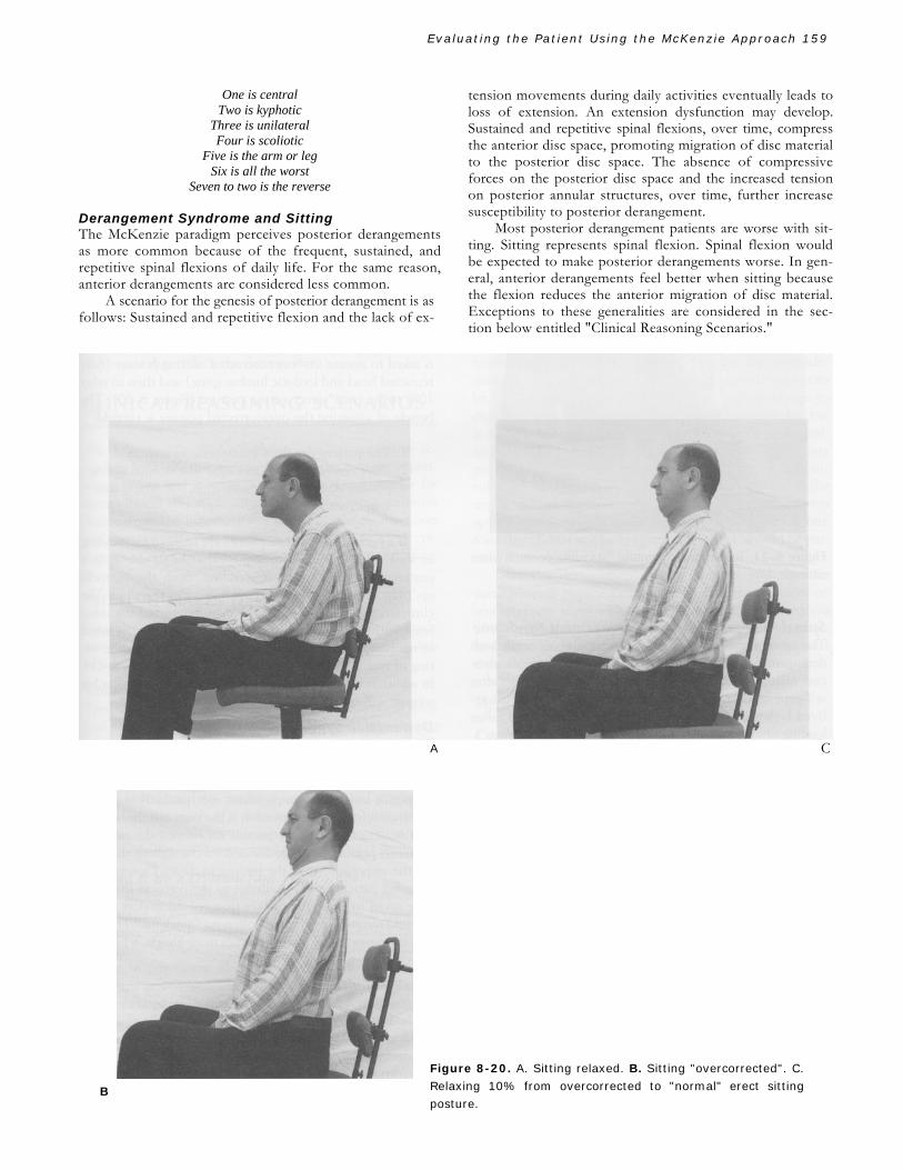
Figure 8-20. A. Sitting relaxed. B. Sitting "overcorrected". C. Relaxing 10% from overcorrected to "normal" erect sitting posture.
B

Evaluating the Patient Using the McKenzie Approach 160
Figure 8-21. Teaching to maintain "standing posture when sitting". Spinal Manipulation and Derangement Syndrome The most dramatic responses to manipulation occur with derangement syndrome patients. McKenzie protocols advo-cate manipulation for derangement syndrome patients after self-generated movements demonstrate partial or short-lived benefits. Full recovery from self-generated end range loading obviates the need for manipulation.
Responses to patient-generated end range loading herald the responses to be expected if the patient were to be po-sitioned, moved, or manipulated at that same end range. If self-generated end range loading in a particular movement plane direction is beneficial, manipulation to the same end range will most likely be as, if not more, beneficial. If self-generated end range loading, in a particular movement plane direction, is detrimental, manipulation to the same end range will most likely be as, if not more, detrimental. THERAPEUTIC PROGRESSIONS AND RESPONSIBILITIES The same movements employed to evaluate mechanical and symptomatic responses are employed for therapeutic purposes. Responses dictate which movements and positions are to be pursued or avoided.
Postural Syndrome The goal is to reeducate the patient to avoid the culpable end range. When a patient frequently stresses the culpable end
range, symptoms are provoked sooner and more often. When the culpable end range is avoided, symptoms are avoided.
Postural syndrome patients are not aware they are suf-fering the effects of sustained end range loading. Postural syndromes typically result from the poor, slouched sitting posture. Instruction in the body mechanics of sitting is es-sential. Vigilance is required on the part of the patient to avoid sustained loading at the culpable end range. The pos-tural patient may have difficulty believing the solution is "so simple." It is the responsibility of the practitioner to educate the patient concerning the need to avoid end range discomfort. It is the patient's responsibility to avoid sus-tained end range loading long enough for the condition to resolve. This usually takes 1 or 2 weeks.
Proper sitting posture is taught as follows: The patient is asked to assume an "over corrected" sitting posture (fully retracted head and lordotic lumbar spine) and then to relax 10% to "normal" erect sitting posture (Figure 8-20). The benefit of assuming the overcorrected posture is twofold:
1. The patient develops kinesthetic awareness. 2. Over correction is so awkward that normal sitting
posture does not seem so strange, something that keeps patients from "sitting up."
If a patient's spinal complaints are reduced by standing
or walking, it is helpful to request that he or she sit in a posture identical to that of standing "from the buttocks up." The patient is asked to stand in front of the chair. The clinician places one hand on the manubrium and the other hand within the hollow of the back. The patient is asked to maintain the "relationship of the two hands" while sitting or making transitions to or from sitting (Figure 8-21). In addition to education in proper sitting posture, a lumbar support will enhance compliance.
Dysfunction Syndrome The goal is to remodel shortened tissue by frequently pro-voking the discomfort of loading at the restricted end range. Dysfunction syndrome patients tend to avoid their discom-forts at end range, perpetuating the condition. Complaints will persist unless the symptomatic, mechanically impeded end range is frequently pursued. It is the responsibility of the practitioner to educate the patient concerning the need to frequently pursue end range discomforts throughout the day. It is the responsibility of the patient to do so. Dysfunction syndrome patients are a challenge to motivate, as they are being asked to cause themselves frequent discomfort with-out rapid results. The clinician offers no "quick fix." The pa-tient improves as the tissue becomes more elastic, a process that can take months.
Derangement Syndromes The goal is to reduce displacement of disc material by load-ing the key obstructed end range. Symptoms of peripheral-ization are avoided. Symptoms of centralization are pursued. Derangement syndrome patients often avoid movements and positionings accompanied by the symptoms of central-
Evaluating the Patient Using the McKenzie Approach 161
ization. The centralization response occurs as the obstruc-tion to end range is reduced (relocated). It is the responsi-bility of the practitioner to educate the patient that in-creased central complaints are beneficial, especially when accompanied by diminished peripheral complaints. It is the practitioner's responsibility to make clear which movements and positionings are to be pursued and which are to be avoided. It is the patient's responsibility to comply. The successful resolution of a derangement syndrome requires:
• Reduction of the deranged disc material • Maintenance of the reduction • Reintroduction of movements previously considered
to promote derangement (recovery of function) • Prophylaxis by periodically pursuing loading strate-
gies that reduce the derangement
CLINICAL REASONING SCENARIOS
The following "scenarios" explore clinical reasoning ac-cording to McKenzie protocols. How the patient experi-ences sitting and standing serves as posture examples for these clinical reasoning scenarios. The following scenarios demonstrate the importance of basing McKenzie diagnoses and treatments upon mechanical and symptomatic re-sponses to repetitive movements and sustained positioning.
"SITTING MAKES IT WORSE"
Consider a patient presenting with spinal complaints made worse with sitting. This symptom alone does not tell you which syndrome is involved.
Could It Be a Postural Syndrome? Typical poor sitting posture extends the upper cervical spine and flexes lower spinal levels. Sustained end range loading causes symptoms in one or more of these areas. Examination of the patient provokes no symptoms with end range dy-namic loading, no symptoms during motion, no symptom re-ferrals and no mechanical responses. Considering the way most individuals sit, resulting upper cervical complaints would be the result of a sustained extension postural syn-drome, whereas all lower levels of spinal complaints would be manifestations of a sustained flexion postural syndrome.
Could It Be a Dysfunction Syndrome? Poor sitting posture typically extends the upper cervical spine and flexes lower levels, promoting the development of upper cervical flexion dysfunction and extension dys-functions of lower spinal levels. Dysfunctions typically de-velop in movement plane directions opposite that of the habitual poor posturing. There would be symptoms at an early limited end range. There are no symptoms during mo-tion. The patient is essentially no better or worse as a result of provocative testing or therapeutic movement. Repetition of movement does not result in further gain of range of motion until weeks have transpired.
Could It Be a Derangement Syndrome? Considering typical poor sitting posture, loading the upper cervical spine at sustained end range extension and lower spinal levels at sustained end range flexion, causes one to consider the possibility of anterior derangement of the upper cervical spine or posterior derangement at lower spinal levels. Dynamic or static loading in the direction that promotes the derangement may result in peripheralization of complaints that remain worse after loading ceases. There may be symptoms during motion. Dynamic or static loading at the key obstructed end range would be accompanied by centralization, improved mechanics, and maintained benefit.
However, the derangement syndrome patient's com-plaint of being "worse when sitting" may not be due to pro-motion of derangement. It may, in fact, be due to inadequate reduction of derangement. Recall centralization can be quite uncomfortable. When spinal loading begins to challenge the obstruction resulting from derangement but is not applied with enough intensity to reduce it, centralization-like discomforts may be experienced without any net benefit. Therefore, the patient who is worse with sitting may, in fact, have a posterior derangement of the upper cervical spine, or anterior derangement of lower spinal levels, for which sitting symptoms represent discomforts that should not be avoided and, in fact, should be pursued more vigorously.
Anterior derangements require flexion, and posterior derangements require extension. Treatment predicates on one symptom along with failure to perform careful analysis of the mechanical and symptomatic responses to dynamic and static loading may lead to a loading strategy opposite that which is required.
"STANDING MAKES IT WORSE"
Could It be a Postural Syndrome? Postural syndromes are due to sustained end range loading of normal spinal joints. A variety of standing postures can be responsible (e.g., protruded head, hyperkyphotic thorax, hy-perlordotic lumbars, standing with one's weight on one leg). The diagnosis is confirmed when dynamic loading is without mechanical or symptomatic responses and complaints are reproduced with sustained end range loading only.
Could It be a Dysfunction Syndrome? Relative to sitting, standing involves retraction (flexion) of the upper cervical spine and relative extension of all lower spinal levels. Typically, a patient who is asymptomatic when sitting but experiences dysfunction symptoms when standing is suffering from upper cervical flexion (retraction) dysfunction. These conclusions, of course, would be reached after exploring the effects of mechanical and symptomatic responses to loading.
Could It be a Derangement Syndrome? Relative to sitting, standing involves retraction (flexion of the upper cervical spine) and relative extension of all lower

Evaluating the Patient Using the McKenzie Approach 162
spinal levels, causing one to consider the possibilities of posterior derangement of the upper cervical spine or anterior derangement at lower spinal levels.
Lower cervical and low back derangement symptoms that worsen with standing most often are due to an anterior, not a posterior, derangement. Dynamic and static loading responses are the key to whether or not this is true for a particular patient. However, the derangement syndrome patient's complaint "worse when standing" may not be due to promotion of derangement. It may be due to the discomfort of centralization accompanying inadequate reduction of derangement. The patient avoids the centralization discomfort, not realizing its potential benefit. There-fore, the patient who is worse with standing may, in fact, have an anterior derangement of the upper cervical spine or posterior derangement of lower spinal levels, for which standing symptoms represent discomfort that should not be avoided and, in fact, should be pursued more vigorously.
MIXED SYNDROME SCENARIOS
It is possible for syndromes to coexist. We have considered how habitually flexed lumbosacral posture can at first result in postural syndrome symptoms and then lead to extension dysfunction and posterior migration (derangement) of disc material. Accordingly, the individual experiences all three of these syndromes simultaneously. Consider the following regarding mixed syndrome scenarios.
Mixed Syndrome Scenario 1 The patient presents complaining of low back symptoms after sitting slouched for 15 minutes. Examination reveals no symptoms during motion. All ranges of motion are full and unrestricted except for extension, which is limited by one-third. When the patient reaches two-thirds normal ex-tension, discomfort is experienced immediately but resolves upon returning to neutral posture. No matter how many extension movements are performed during the initial exam, the range of motion or symptoms do not change appreciably. There are no symptoms during motion.
What McKenzie syndrome best accounts for presenting complaints? The patient obviously suffers from an extension dysfunction. However, an extension dysfunction does not account for the presenting complaint of pain with slouched sitting. The pain with slouched sitting is best explained by the postural syndrome. The dysfunction syndrome should be ad-dressed; however, it is not responsible for the complaint that brought the patient in. In order to treat the presenting complaint, postural syndrome protocols are required.
Mixed Syndrome Scenario 2 A patient presents complaining of low back symptoms that begin after sitting for 15 minutes. After sitting 30 minutes symptoms peripheralize to the buttock and right lower ex-tremity. Standing, walking, and lying down are not prob-lematic. Flexion in standing or supine knee-to-chest move-ments peripheralize complaints to the lower extremity. All ranges of motion are full and unrestricted except for exten-sion, which is two-thirds normal. When the patient reaches two-thirds normal extension, discomfort is experienced im-mediately that resolves upon returning to neutral posture. Centralization does not occur with extension. No matter how many extension movements are performed, symptoms and range of motion do not change.
What McKenzie syndromes best account for presenting complaints? The patient exhibits mechanical and symptomatic responses evidencing derangement peripheralization behavior. However, centralization responses are not achieved. The patient has an extension dysfunction syndrome in which the limited end range occurs before and prevents loading from "reaching" the obstructed end range of the derangement. It is not until the dysfunction is "worked through" that loading can be expected to reduce the derangement.
McKENZIE PROTOCOLS' IMPACT ON PATIENT BEHAVIOR Consistent with the rehabilitation philosophies, McKenzie protocols promote self-sufficiency and independent func-tioning. The potentials of self-treatment are explored on the initial visit, even if the patient is acute. Patient re-sponsibilities include the pursuit and/or avoidance of specific positions and exercises. The emphasis is placed on control of symptoms rather than actively avoiding symptoms. Fear of activity, symptom magnification, and practitioner dependence are discouraged.
A significant goal of component of treatment is to affect the mind set of the patient. If patients are taught to participate in their own care, the chance of recovery is greater and the chance of recurrence is less. In McKenzie's own words:
"If there is the slightest chance that a patient can be educated in a method of treatment that enables him to reduce his own pain and disability using his own understanding and resources, he should receive that education. Every patient is entitled to this information, and every therapist should be obliged to provide it."14

Evaluating the Patient Using the McKenzie Approach 163
C l i n i c a l A p p l i c a t i o n 8 — 1
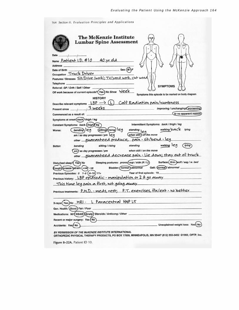
SUBJECTIVE HISTORY A 40-year-old truck driver (Patient ID 10) presents with low back and left lower extremity pain to the midcalf (see Figure 8-22A). His postures and stresses are pro-longed sitting while driving at work, and at home his activities include watching television, yardwork, and cutting wood.
The pain has been present for 3 weeks and is worsening. He has been off work for 1 week because of the symptoms. The pain commenced for no apparent reason and began in the lumbar region. He has intermit-tent low back pain, but constant thigh and leg pain.
Sitting and forward bending are guaranteed to in-crease his peripheral symptoms. Lying supine is guar-anteed to decrease the leg symptoms, but it does not last. Rising from sitting is positive, and standing and walking make the back worse at times. He is better when awaking and gets worse as the day progresses. Walking makes the leg better at times, and he feels bet-ter on the move and worse when still.
His sleep is disturbed. He sleeps on his sides and back and on a firm mattress. Coughing and sneezing is positive for leg pain, but bowel, bladder, and gait are normal.
He has had back pain many times in the past, but a chiropractic manipulation or two provided sympto-matic relief. The leg pain is now present for the first time, and it won't resolve.
Previous treatments for this episode included pre-scription pharmaceuticals, rest, and passive physical therapy ( heat and exercises, including flexion and ex-tension). None of the treatment interventions have had any lasting effect.
Diagnostic testing involved an (MRI) that revealed a left paracentral herniated nucleus pulposis of the L5 lumbar disc. His general health is good. He is currently taking antiinflammatory and analgesic medicine. He denies any recent or major surgery, accidents, or unexplained weight loss.
CLINICAL EVALUATION
His sitting posture is poor, standing posture is fair, lor-dosis is reduced, and he has a right lateral shift present (Figure 8-22B).
loss is as follows:
Movement loss is as follows:
Flexion: moderate to major loss with right deviation in flexion Extension: moderate loss Side gliding (R): nil Side gliding (L): minimal to moderate loss
Pretest pain in standing was low back and left thigh
pain. Flexion in standing (FIS) produced pain during movement (PDM) and end range pain (ERP). Repeated FIS increased thigh pain and produced leg pain and remained worse. Extension in standing (EIS) produced ERP, and repeated EIS increased leg and back pain but it did not remain worse.
Pretest pain in lying supine increased thigh and leg pain. Flexion in lying (FIL) produced PDM and ERP with repeated FIL increasing thigh and leg pain, and it remained worse. Extension in lying (EIL) produced ERP, and repeated EIL increased his low back pain but de-creased his thigh and leg symptoms. They remained better. Both side gliding movements had no effect on the symptoms.
Static tests of sitting slouched increased and worsened the leg pain but had no effect on his low back pain. Sitting erect decreased the leg pain and increased his back pain. Lying prone abolished his low back pain and decreased his leg symptoms. Lying prone in extension only decreased his leg symptoms.
Neurological testing was all within normal limits (WNL), as was testing of the hip joints and SI joints. Review Questions for Cl in i ca l App l i ca t i on 8–1 *
1. Based on the subjective history, what is your working hypothesis as to his condition and why?
2. What would be the first clinical test you would per-form? Why?
3. What is your working diagnosis? Why? 4. What would you recommend this patient do at home?
How often? When would you see him for follow-up and what would you look for in the second-day assessment? What are the reasons for your answers?
*Answers appear in Appendix at end of chapter.
Evaluating the Patient Using the McKenzie Approach 164
C l i n i c a l A p p l i c a t i o n 8 — 2

Evaluating the Patient Using the McKenzie Approach 165

Evaluating the Patient Using the McKenzie Approach 166
C l i n i c a l A p p l i c a t i o n 8 — 2
SUBJECTIVE HISTORY This patient is a 20-year-old secretary (Patient ID 20) who sits most of the day in front of a computer and performs clerical duties. At home she is very active, which includes walking and riding a bicycle daily (see Figure 8-23A).
Her pain pattern is central low back pain present for no apparent reason and of 3 or 4 months' duration. It is gradually worsening and is described as an un-comfortable ache, and she has lost no time from work or stopped activities because of it. The onset was central low back pain, and it is of intermittent frequency.
The worse/better section revealed that what would guarantee to produce the pain was to sit for long peri-ods. She has no pain or stiffness when rising from a sit-ting position. It worsens as the day progresses and when she is still. Guaranteed to decrease the pain is to get up and move around. She is better bending, walking, when waking up, when getting home from work, and when she is on the move.
She has had no disturbed sleep. Dejerine's triad is negative and her bladder and bowel function, as well as her gait patterns, are all normal.
She had no previous episodes of low back pain, and her previous history was unremarkable. She sought no treatment for this until now and had no diagnostic testing done.
Her general health is good. She uses no medicine, and denies previous surgeries or accidents. She denies the presence of unexplained weight loss.
CLINICAL EVALUATION Sitting posture is poor, standing posture good, lordosis normal, and she has no lateral shift present (Figure 8-23B).
Movement loss is as follows:
Flexion: no loss Extension: no loss Side gliding (R): no loss Side gliding (L): no loss
There was no pretest pain while standing. The following
movements were tested: FIS, EIS, FIL, EIL, side gliding in standing right [SGIS(R)], and side gliding in standing left [SGIS(L)]. None of the movements had any PDM or ERP. All of the movements done repetitively had no effect on her condition.
Static tests performed included sitting slouched, sitting erect, standing slouched, standing erect, lying prone, lying prone in extension, and long sitting. The only positions that had any effect on her condition were sitting slouched, which reproduced her low back pain, and sitting erect, which decreased her low back pain.
Neurological testing was not performed because there were no radicular symptoms. Hip and SI joint testing was unremarkable. Review Questions for C l i n i ca l App l i ca t i on 8—2
1. What is your working hypothesis? What may this be? What are the reasons to support your hypotheses? If any of the three syndromes are ruled out, why?
2. What will be the first clinical test you perform and why? 3. After reviewing the results of the movement loss sec-
tion, what syndromes are ruled out? If you picked dysfunction, why?
4. What is your provisional diagnosis? Why? 5. Why would you rarely see patients with this type of
syndrome in your office?

Evaluating the Patient Using the McKenzie Approach 167

Evaluating the Patient Using the McKenzie Approach 168
Evaluating the Patient Using the McKenzie Approach 169
C l i n i c a l A p p l i c a t i o n 8 — 3
SUBJECTIVE HISTORY
This 35-year-old laborer (Patient ID 30) sits and assem-bles parts all day. At home he watches television and likes to ride his four-wheeler. He has central low back pain that began 8 weeks ago when he fell off a chair at work. He originally missed 5 weeks of work and was virtually in bed during this time with short periods of sitting as tolerated. The condition has been unchanging for the past 2 weeks and is of intermittent frequency (see Figure 8-24A).
The better/worse section is unremarkable except that bending forward will reproduce the pain. Guaranteed to produce the pain is to bend forward and guaranteed to reduce the pain is to avoid bending forward.
He has no disturbed sleep. His bladder, bowel, and gait are normal. Dejerine's triad is negative. Previous history reveals a few bouts of low back pain over the past several years but each episode in the past self-resolved.
Previous treatments during this episode included no bending, twisting, lifting, or walking for 5 weeks. This resulted in the low back pain gradually decreasing, and it remained better. He then returned to restricted duty at work that was mentioned above. He was treated by the company doctor with prescription medication and rest. He had passive physical therapy (moist heat and electrical stimulation) performed with no relief.
Diagnostic tests included x-rays that were unre-markable. His general health is good. He continues taking antiinflammatory medicine. He has had no recent or major surgery, no accidents, and no unexplained weight loss.
CLINICAL EVALUATION
Sitting posture is poor. Standing is good. He has a normal lordosis and no lateral shift (see Figure 8-24B).
Movement loss is as follows:
Flexion: Moderate loss, no deviation Extension: no loss Side gliding (R): no loss Side gliding (L): no loss
Pretest pain on standing was absent. FIS produced
ERP but no worse. Repeated FIS produced ERP, but was no worse after. EIS had no effect. Repeated EIS had no effect.
Pretest pain in lying was absent. FIL produced ERP. Repeated FIL produced ERP but did not remain worse. EIL had no effect. Repeated EIL had no effect. Side glid-ing right and left had no effect, as did repeated move-ments of both.
Static testing had no effect for sitting slouched/erect, standing slouch/erect, lying prone in extension, and long sitting.
No neurological testing was performed because of the absence of radicular symptoms. Hip and SI joint testing was normal. Review Questions for C l i n i ca l App l i ca t i on 8—3
1. With the better/worse section being so unremarkable, what additional questions could you have asked to further clarify the section, including but not limited to the bending movement?
2. What are your hypotheses of this condition? Why? 3. With the movement loss data available, what syn-
drome is ruled out? Why? 4. Can this be a posterior derangement? Defend your
answer. 5. What is your working diagnosis? What are two re-
peated test movement clues that would initially indi-cate only one of the three syndromes based on the McKenzie system?

Evaluating the Patient Using the McKenzie Approach 170
C l i n i c a l A p p l i c a t i o n 8 — 4

Evaluating the Patient Using the McKenzie Approach 171
Evaluating the Patient Using the McKenzie Approach 172
C l i n i c a l A p p l i c a t i o n 8 — 4
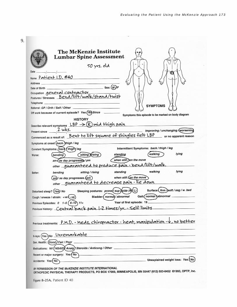
SUBJECTIVE HISTORY This 50-year-old male general contractor (Patient ID 40) assumes work-related postures/stresses that involve bending, lifting, walking, standing, and twisting through-out the day. At home he plays softball and basketball and watches the same on television (see Figure 8-25A).
His pain diagram shows central low back pain ra-diating to the posterior right midthigh. The pain has been present for 2 weeks and is worsening. Because he is self-employed, he has missed no work but believes the pain is bad enough at times that if he was not self-employed he would take some sick leave. The pain began 2 weeks ago while he was lifting a square of shingles and felt a sharp low back pain that gradually radiated down the buttock into the thigh. He states the back and thigh pain are constant but certain things do increase and decrease the pain.
The better/worse section illustrates that bending, sitting, rising from sitting, and walking make the condition worse and it lasts. He is worse as the day progresses and when still. Standing at times is painful. The things guaranteed to increase the pain are bending, lifting, and walking. He gets better lying on his right side and while on the move. When he wakes up he feels better and also before he goes to bed. Lying down on his right side is guaranteed to decrease his pain and it lasts until he does one of the worse movements.
His sleep is disturbed. He sleeps on his right side to start but ends up on his back or left side.
Cough, sneeze, and strain are negative. His bowel, bladder, and gait are all normal. He has a previous his-tory of many episodes of low back pain. In fact, he states he has had it one or two times per year and it usually self-limits.
He has been treated by his family doctor with anti-inflammatory medicine and by his doctor of chiropractic with manipulation and heat. His symptoms have de-creased but have not remained better.
He has had x-rays but they were nonrevealing. His general health is good. He denies any recent or major surgery. He denies any accidents or unexplained weight loss.
CLINICAL EVALUATION His sitting posture is poor. His standing posture is
fair. He has a reduced lordosis and a left lateral shift (see Figure 8-25B).
Movement loss is as follows:
Flexion: major loss with no deviation Extension: minimal loss with no deviation
Side gliding (R): major loss Side gliding (L): no loss
Pretest pain while standing is low back pain with pain
to right midthigh pain. Pretest pain in lying de-creases the symptoms but does not last. All sagittal standing and lying movements produce PDM and ERP. All sagittal repeated movements increase and worsen the symptoms. The frontal plane movement of SGIS(L) also produces PDM and ERP, and repeated movements increase the thigh pain and it remains worse. SGIS(R) has ERP, and repeated movements abolish the thigh pain and it remains better. But the low back pain increases and worsens.
Static tests reveal that sitting slouched increases and worsens the thigh. Erect sitting increases the thigh pain, but it is no worse after. Standing slouched in-creases and worsens both symptoms. Standing erect in-creases the thigh pain, but it is no worse after.
Neurological testing was done because he had thigh pain that was worsening. But all the tests were negative including dural tension tests. The SI and hip joints were normal. Review Questions for C l i n i ca l App l i ca t i on 8—4
1. Which syndrome can be ruled out based on the pain drawing or assessment sheet for this clinical applica-tion? Why?
2. What other syndrome is ruled out now because of what was described in the paragraph beginning with the sentence "The pain has been present for 2 weeks and is worsening."? Give three reasons why that syndrome is now not possible?
3. At this time what syndrome are you strongly sup-porting? Provide three reasons.
4. What are your hypotheses regarding his condition? 5. With regard to the lateral shift, what question can
you ask the patient to determine if this obvious shift is relevant?
6. If SGIS(R) is the preferred loading strategy for this to be a derangement, then after repeated SGIS(R) move-ments the reevaluation of the movement loss base-lines should show what? Why?
7. What is your conclusion? If you suspected derange-ment, could you disprove your hypotheses with the clinical testing?
8. If you suspected derangement, was a lateral compo-nent present? Was it a relevant lateral component or irrelevant? Explain.
Evaluating the Patient Using the McKenzie Approach 173
9.

Evaluating the Patient Using the McKenzie Approach 174

Evaluating the Patient Using the McKenzie Approach 175
SUMMARY
The following is a summary of the literature support for the theoretical and practical aspects of the McKenzie approach. The McKenzie approach, as noted in the 1981 text T h e L u m -b ar Sp in e 1 3 pays particular attention to symptom topography. In 1987, The Quebec Task Force on Activity Related Spinal Disorders concluded that the majority of patients with non-specific spinal symptoms should be classified based on symptom topography.23 The McKenzie approach is somewhat more sophisticated in subdividing these patients based upon the presence or absence of relevant spinal deformity and most importantly on the mechanical and symptomatic responses to dynamic and static end-range loading. Spratt et al recommended the use of the patient's response to repetitive test movements based upon documented reliability.'
Kilby et al9 found good intertester reliability in docu-menting symptom response to McKenzie's repeated test movements but fair to poor intertester reliability in docu-menting the presence or absence of spinal deformity. Rid-dle and Rothstein18 found less than acceptable intertester reliability in reaching a mechanical diagnosis based upon the McKenzie assessment process. It should be noted that none of the therapists in the study had undergone more than basic instruction in the McKenzie approach.
A common occurrence noted by practitioners of the McKenzie approach is that of centralization of symptoms. The frequency of centralization in common practice has ranged from 47% to 87%.6,12 A two-part randomized, prospective study demonstrated that the referred pain of 58% of low back pain subjects could be centralized with a single direction of repeated test movements. It also demon-strated superior treatment outcomes in patients whose symptoms can be centralized compared to noncentralizers.
The mechanism responsible for centralization of symp-toms is not fully understood currently. McKenzie has attrib-uted this response to nuclear movement. Specifically he pro-poses that displacement of nuclear and/or annular material within the intervertebral disc causes a progressive increase in the intensity and distribution of symptoms. Conversely, in response to other movements and positions the nu-clear/annular displacement is reversed, thereby causing the symptoms to reduce in distribution and intensity. This could occur only if the hydrostatic mechanism of the painful disc is intact and functional. If the symptoms could only be pe-ripheralized but not centralized, the theoretical model would propose that the disc was responsible for the symp-toms but that the painful disc is not intact and, therefore, the hydrostatic mechanism is no longer functional. Symp-
toms that cannot be centralized or peripheralized would be considered nondiscogenic in origin.
If the above proposal is accurate, then the following must be proven: (1) The disc would have to be implicated as the symptom generator in patients whose symptoms can be centralized or peripheralized only. (2) Nuclear movement would have to be demonstrated in vitro and in vivo. (3) A correlation between centralization and simultaneous nuclear movement would have to be demonstrated. The following will address the scientific evidence to support the theoretical model.
A recent study by Medcalf et al15 found that examiners utilizing the McKenzie assessment process displayed a high level of accuracy in predicting the outcome of discography, including the symptomatic level, annular containment, and internal disc fissure pattern. A strong relationship between the occurrence of centralization and a positive discogram with a contained functional annulus was shown. During the McKenzie assessment, the referred symptoms of 50% cen-tralized with 74% having positive discograms, of which 91% had an intact annulus. The conclusion from this study is that the McKenzie assessment process reliably differentiates discogenic from nondiscogenic symptoms (p<.001) and a competent from an incompetent annulus (p<.042).
Nuclear movement has been documented in vitro in several studies.11,20-22 Schnebel et al19 documented nuclear movement in vivo in normal discs. No clear pattern of nu-clear movement was documented in discographically abnor-mal discs. Centralization was not monitored during repeated movements in the study by Schnebel. Currently, research is underway to assess the presence or absence of nuclear movement in discographically abnormal discs during the centralization process.
From a practical standpoint several studies have demonstrated the superiority of the McKenzie approach as compared to other treatments. Nwuga and Nwuga16 and Ponte et al17 found the McKenzie approach to be superior to the Williams approach to back treatment. However both studies do suffer from methodological flaws.
Recently, Stankovic and Johnell26'27 found the McKenzie approach to be superior in terms of rapid resolution of symptoms and long-term recurrence rate to a "mini back school" at 1- and 5-year follow-ups. Kopp et al10 and Alexan-der et all found the McKenzie assessment process identified patients with surgical disc pathology. Patients with nonsurgi-cal disc pathology were treated successfully with the McKen-zie approach, and at 5-year follow-up 91% were maintaining a good to excellent treatment outcome. Several studies are currently underway around the world to assess the clinical and theoretical aspects of the McKenzie approach.

Evaluating the Patient Using the McKenzie Approach 176
REFERENCES
1. Alexander AH, Jones AM, Rosenbaum DH Jr. Nonoperative management of herniated nucleus pulposus: Patient selection by the extension sign long-term follow-up. Presented at North American Spine Society Annual Meeting, Monterey, California, August 8-11, 1990.
2. Allan, DB, Waddell G: An historical perspective on low back pain and disability. Acta Orthop Scand. 1989;60(Suppl 234).
3. Andersson GBJ. Evaluation of muscle function. In: Frymoyer JW, et al, eds. The Adult Spine, Principles and Practice. New York: Raven Press, 1991;269.
4. Bigos S, Bowyer 0, Braen G, et al. Acute low back problems in adults. Clinical Practice Guideline No. 14. AHCPR Publication No. 95-0642. Rockville, MD: Agency for Healthcare Policy and Research, Public Health Service, U.S. Department of Health and Human Services, December 1994.
5. Butler D. Mobilization of the Nervous System. Melbourne: Churchill Livingstone, 1991. 6. Donelson R, Murphy K, Silva G. The centralization phenomenon: Its usefulness in evaluating and treating
referred pain. Spine. 1990;15:211-215. 7. Donelson R, Grant W, Kamps C, Medcalf R. Pain response to sagittal end-range spinal motion: A
prospective randomized multicentered trial. Spine. 1991;16:S206-S212. 8. Donelson R, Grant W, Kamps C, Medcalf R. Pain response to end-range spinal motion in the frontal plane: A
multi-centered, prospective trial. Presented at International Society for the Study of the Lumbar Spine, Heidelberg, Germany, May 1991.
9. Kilby J, Stignani M, Roberts A. The reliability of back pain assessment by physiotherapists, using a McKenzie algorithm. Physiotherapy. 1990;76(9):379-383.
10. Kopp JR, Alexander AH, Torocy RH, Levrini MG, Lichtman DM. The use of lumbar extension in the evaluation and treatment of patients with acute herniated nucleus pulposus. A preliminary report. Clin Orthop. 1986;202:211-218.
11. Krag MH, Seroussi RE, Wilder DG, et al. Internal displacements from vitro loading of human spinal motion segments. Spine. 1987;12(10):1001-1007.
12. Long AL. The centralization phenomenon: Its usefulness as a predictor of outcome in conservative treatment of chronic low back pain (a pilot study). Spine. 1995;20:2513-2521.
13. McKenzie RA. The Lumbar Spine. Mechanical Diagnosis and Therapy, 1st ed. Lower Hutt, New Zealand: Spinal Publications, 1981.
14. McKenzie RA. The Cervical and Thoracic Spine. Mechanical Diagnosis and Therapy, 1st ed. Walkanae, New Zealand: Spinal Publications, 1990.
15. Medcalf R, Aprill CA, Donelson R, Grant WA, Incorvaia K. Discographic outcomes predicted by pain centralization and "directional preference": A prospective, blinded study. Presented at Annual Meeting of the North American Spine Society, Washington, DC, October 1995.
16. Nwuga G, Nwuga V. Relative therapeutic efficacy of the Williams and McKenzie protocols in back pain management. Physiother Pract. 1985;1:99-105.
17. Ponte DJ, Jensen GJ, Kent BE. A preliminary report on the use of the McKenzie protocol versus Williams protocol in the treatment of low back pain.] Orth Sports Phys Ther. 1984;6(2):130-139.
18. Riddle DL, Rothstein JM. Intertester reliability of McKenzie's classifications of the syndrome types present in patients with low-back pain. Spine. 1994;21:1333-1344.
19. SchnebeL BE, Simmons JW, Chowning J, et al. A digitizing technique for the study of movement of in- tradiscal dye in response to flexion and extension of the lumbar spine. Spine. 1988;13(3):309-312.
20. Seroussi RE, Krag MH, Muller DL, et al. Internal deformations of intact and denucleated human lumbar discs subjected to compression, flexion and extension loads. J Orthop Res. 1989;7(1):122-123.
21. Shah JS, Hampson WGJ, Jayson MIV. Distribution of surface strain in the cadaveric lumbar spine. J Bone Joint Surg. 1978;60B:246-251.
22. Sheppard J. Patterns of internal disc dynamics, cadaver motion studies. Abstract. The International Society for the Study of the Lumbar Spine, Boston, June 1990.
23. Spitzer WO, LeBlanc FE, Dupuis M, et al. Scientific approach to the assessment and management of ac-tivity-related spinal disorders (The Quebec Task Force). Spine. 1987;12(7S):22-30.
24. Spitzer WO, et al. Scientific monograph of the Quebec Task Force on whiplash-associated disorders (WAD); Redefining "whiplash" and its management. Spine. 1995;Suppl 20:8S.
25. Spratt KF, Lehmann TR, Weinstein JN, Sayze HA. A new approach to the low hack physical examination: Behavioral assessment of mechanical signs. Spine. 1990;15:96-102.

Evaluating the Patient Using the McKenzie Approach 177
26. Stankovic R, Johnell O. Conservative treatment of acute low-back pain. A perspective randomized trial: McKenzie method of treatment versus patient education in "Mini Back School." Spine. 1990;15:2.
27. Stankovic R, Johnell O. Conservative treatment of acute low back pain: A 5-year follow-up study of two methods of treatment. Spine. 1985;20(4):469–477.
28. Waddell G, et al. Normality and reliability in the clinical assessment of backache. Br Med J 1984;284:1519-1523.
Additional References Of Interest
Belanger AY, Depres MC, Goulet H, Troftier F. The McKenzie approach: How many clinical trials support its effectiveness? Proceedings of the World Confederation for Physical Therapy 11th International Congress, London, July 28–August 2, 1991.
DiMaggio A, Mooney V. Conservative care for low hack pain: What works? J Musculoskel Med. 1987;4(9):27–34. DiMaggio A, Mooney V. The McKenzie program: Exercise effective against hack pain. J Musculoskel Med. 1987;4(12):63-74. Donelson R. The McKenzie approach to evaluating and treating low back. Orthop Rev. 1990;XIX(5). Donelson RG. Identifying appropriate exercises for your low back pain patient. J Musculoskel Med. 1991;8:14–29. Donelson RG, McKenzie R. Mechanical assessment and treatment of spinal pain. In: Frymoyer JW, ed. The
Adult Spine: Principles and Practice. New York: Raven Press, 1991;1627–1639. Donelson R, McKenzie R. Letter to the Editor. Spine. 1992;17(10):1267. Elnaggar IM, Nordin M, Shikhzadeh A, Parnianpour M, Kahanovitz N. Effects of spinal flexion and extension
exercises on low-hack pain and spinal mobility in chronic mechanical low-back pain patients. Spine. 1991;16(8):967-972.
Fredrickson BE, Murphy K, Donelson R, Yuan H. McKenzie Treatment of Low Back Pain: A correlation of significant factors in determining prognosis. Annual Meeting of International Society for the Study of the Lumbar Spine, Dallas, 1986.
Jacob G. Specific application of movement and positioning technique to the lumbar spine, considering theo- retical formulation and therapeutic application. Today's Chiro. 1989–1990; Part I 118(6)/Part II 19(1).
Jacob G. The McKenzie protocol and the demand of rehabilitation. Calif Chiro Assn J. 1991;16:10. Kay MA, Helowa A. The effects of Maitland and McKenzie techniques in the musculoskeletal management of
low hack pain: A pilot study. Abstract. Phys Ther. 1994;74:5 (Suppl) FS59. Kuslich, SD, Ulstrom CL, Michael CJ. The tissue origin of low back pain and sciatica: A report of pain response to
tissue stimulation during operations on the lumbar spine using local anesthesia. Orthop Clin North Am. 1991;22(2):181-187.
McKenzie RA. Manual correction of sciatic scoliosis. NZ Med J. 1972;76(484):194–199. McKenzie RA. Prophylaxis in recurrent low back pain. NZ Med J. 1979;89(627):22–23. McKenzie RA. Treat Your Own Back. Lower Hutt, New Zealand: Spinal Publications, 1981. McKenzie RA. The Lumbar Spine. Mechanical Diagnosis and Therapy, 1st ed. Lower Hutt, New Zealand: Spinal
Publications, 1981. McKenzie RA. Treat Your Own Neck. Lower Hutt, New Zealand: Spinal Publications, 1983. McKenzie RA. Mechanical diagnosis and therapy for low back pain: Toward a better understanding. In:
Twomey LT, Taylor JR eds. Clinics in Physical Therapy. Physical Therapy of the Low Back. New York: Churchill Livingstone, 1987:157.
McKenzie RA. Early mobilization and outcome in acute sprains of the neck. Br Med J. 1989;299:1005. McKenzie RA. Perspective on manipulative therapy. Physiotherapy. 1989;75(8):440–444. McKenzie RA. The Cervical and Thoracic Spine. Mechanical Diagnosis and Therapy, 1st ed. Walkanae, New
Zealand: Spinal Publications, 1990. McKenzie RA. Mechanical diagnosis and therapy for low hack pain: Toward a better understanding. In: Weinstein
JN, Weisel SW, eds. The Lumbar Spine. The International Society for the Study of the Lumbar Spine. Philadelphia: WB Saunders, 1990:792-805.
McKenzie RA. A physical therapy perspective on acute spinal disorders. In: Mayer TO, Mooney V, Gatchel RJ, eds. Contemporary Conservative Case for Painful Spinal Disorders: Concepts, Diagnosis and Treatment. Malvera, PA: Lea & Febiger, 1991:211-220.
McKenzie RA. Mechanical diagnosis and therapy for disorders of the lower hack. In: Twomey LT, Taylor JR, eds. Clinics in Physical Therapy. Physical Therapy of the Low Back. 2nd ed. New York: Churchill Livingstone (In Press).
Evaluating the Patient Using the McKenzie Approach 178
McKenzie RA. Spinal assessment and therapy based on the behavior of pain and mechanical response to dynamic and static loading. In: Ernst E, ed. Proceedings of Advances in Idiopathic Low Back Pain Symposium. Vienna, November 27-28, 1992.
McKenzie RA. REPEX in acute and subacute low back pain. In: Ernst E, ed. Proceedings of Advances in Idiopathic Low Back Pain Symposium. Vienna, November 27-28, 1992.
McKinney LA, Doman JO, Ryan M. The role of physiotherapy in the management of acute neck sprains following road-traffic accidents. Arch Emerg Med. 1989;6:27-33.
McKinney MB. Treatment of dislocations of the cervical vertebrae in so-called whiplash injuries. Altnagelvin Hospital, Londonberry, Northern Ireland. Orthopade. 1994;23(4):287-290.
Poulter DC, McKenzie RA. The management of work related back pain. In: Patient Management. Auckland: Adio International Medical Publishers (In Press).
Rath WW, Rath JND, Duffy CG. A comparison of pain location and duration with treatment outcome and frequency. Presented at First International McKenzie Conference, Newport Beach, CA, July 1989.
Riddle DL, Rothstein JM. Intertester reliability of McKenzie's classifications of the syndrome types present in patients with low hack pain. Spine. 1993;18(10):1333.
Schnebel BE, Watkins RG, Dillin W. The role of spinal flexion and extension in changing nerve root compression in disc herniations. Spine. 1989;14(8):835-837.
Spratt RF, Weinstein JN, Lehmann TK, Woody J, Bayne H. Efficacy of flexion and extension treatments incorporating braces for low-back pain patients with retrodisplacement, spondylolisthesis, or normal sagittal translation. Spine. 1993;18(13):1839-1849.
Stankovic R, Johnell O. Conservative treatment of acute low-hack pain. A 5 year follow-up study of two methods of treatment. Spine. 1995;20:4.
Stevens BJ, McKenzie RA. Mechanical diagnosis and self treatment of the cervical spine, vol 17. In: Grant R, ed. Clinics in Physical Therapy, Physical Therapy of the Cervical and Thoracic Spine. New York: Churchill Livingstone, 1988.
Van Wijmen PM. Lumbar pain syndromes. In: Grieve GP, ed. Modern Manual Therapy of the Vertebral Column. New York: Churchill Livingstone, 1986:442-462.
Van Wijmen PM. The management of recurrent low hack pain. In: Grieve GP, ed. Modern Manual Therapy of the Vertebral Column. New York: Churchill Livingstone, 1986:756-776.
Vanharanta H, Videman T, Mooney V. Comparison of McKenzie exercises hack trac and back school in lumbar syndrome preliminary results. Annual Meeting of International Society for the Study of the Lumbar Spine, Dallas, 1986.
Williams M, Grant R. Effects of a McKenzie spinal therapy and rehabilitation program: Preliminary findings. Abstract. Presented at Society for Back Pain Research Annual Scientific Meeting, London, October 30, 1992.
Williams MM, Grant RN. A comparison of low-back and referred pain responses to end-range lumbar movement and position. Conference Proceedings at International Society for the Study of the Lumbar Spine, Chicago, May 20-24, 1992.
Williams MM, Hawley JA, McKenzie RA, Van Wijmen PM. A comparison of the effects of two sitting postures on back and referred pain. Spine. 1991;16(10):1185-1191.
Williams MM, Wright DGR, Mugglestone AA, Lynch GB, Spekreijse SA. Psychological distress in chronically disabled workers attending a Mckenzie spinal therapy and rehabilitation program. Abstract. Conference Proceedings at New Zealand Pain Society Annual Scientific Meeting, 1993.

Evaluating the Patient Using the McKenzie Approach 179
A P P E N D I X
Answers to Review Questions
for Clinical Applications
CLINICAL APPLICATION 8–1 1. Posterior derangement and I don't know. Posterior de-
rangement because it is the only syndrome remaining. It cannot be a postural syndrome because the pain is ra-diating and he has pain affected by movement. It can-not be a dysfunction syndrome because there are con-stant symptoms and most importantly the condition has only been present for 3 weeks. You need a minimum of 6 weeks for the healing to conclude. I don't know is a hypothesis because it could always be something you never saw before.
2. See what effect sitting unsupported on the exam table has on his symptoms. Then have him sit erect and see if any change takes place. You do this now because: (1) It is a natural progression because he has already been sitting slouched through the subjective history. (2) It is a great time for postural correction education because all syndromes involve posture correction.
3. Posterior derangement versus I don't know. Derange-ment because he worsens with flexion movements and positions and centralizes with EIL. Derangement is the only syndrome that can centralize. The I don't know hy-pothesis could still be it until you cannot disprove pos-terior derangement over the next few days.
4. [IL, as often as he agrees to perform the exercise. Dis-cuss when good times may be to perform these exer-cises, regularly 10 to 15 every 2 to 3 hours or when he feels the pain increase. If you can get him to decide when and how many, then he is in charge. Remember it is self-treatment, and he should be in charge.
See him back in 24 hours if possible to recheck all baselines and see if centralization is taking place. If it is, you should see an abolishment of the most distal symp-toms and an increase in the mobility of any or all the movement loss, especially extension. If extension is the preferred loading strategy, then attempt to get him doing EIS because it is much easier to do and more functional.
CLINICAL APPLICATION 8–2 1. Postural syndrome, derangement syndrome, and I don't
know because derangement can be almost anything and is hard to disprove. At present she does fit all the categories of postural syndrome. It cannot be a dys-function syndrome because it is worsening and she gets symptoms from the gradual onset of sitting. A dysfunc-
tion has no pain except at end range stretch when the pain is immediate and no worse after.
2. Check to see if any movement loss is present. If not, then it is not a derangement. Flex with intent to help rule out derangement because a posterior derangement will increase and worsen with flexion.
3. Dysfunction, because there is no movement loss in that section. There must be movement loss for a dysfunction syndrome to be present.
4. Postural syndrome versus I don't know. Postural syn-drome because derangement is ruled out since there is no movement loss and no effect with any repetitive test movements. Also, it indicates postural syndrome be-cause only static positions produce the symptoms and she is better whenever she is not in those positions.
5. You rarely see this type of person because the pain is not severe, it comes on gradually, it is so easily de-creased or prevented, and they have not had to stop any activities of daily living because of the condition. Also, when at home they can avoid the prognosticator and generally have no symptoms.
CLINICAL APPLICATION 8–3
1. If the guarantee to produce the pain is bending over, is it every time? Does it last? Is the pain at the same point every time?
2. Dysfunction syndrome, derangement syndrome, postural syndrome, or I don't know.
Dysfunction because the pain is intermittent, over 6 weeks present, unchanging, only produced through one movement, and no other movements or positions affect the symptoms.
Derangement because it can be almost anything. It has many characteristics and resembles many conditions.
It probably is not a postural syndrome because he states there is movement loss but keep it in mind until verifying in the objective evaluation.
I don't know because it could always be something you never saw before.
3. Postural syndrome it is ruled out because there is move-ment loss present; postural syndrome has no movement loss.
4. It can be but probably not. Most postural derangements will increase and worsen with flexion. This did not, but it could be a healing derangement or a small enough one that the test flexion movements were not of the

Evaluating the Patient Using the McKenzie Approach 180
quantity to affect it yet. To confirm, you can try one of two methods over a 24 to 48-hour treatment process: (1) Flex with intent to see if the condition (symptom or movements) changes; if it does change, then it is not a dysfunction. (2) Treat with postural correction and ex-tensions exercises to see if the condition changes (symptoms or movements). If it does change, then it is not a dysfunction. (This is a safer method because the extension movements usually will not worsen the con-dition if it is a derangement, but will improve it.)
5. Flexion dysfunction. The FIS and FIL movements pro-duced ERP, no worse.
CLINICAL APPLICATION 8—4 1. Postural syndrome because the symptoms radiate. 2. Dysfunction syndrome because the condition is of less
than 6 weeks' duration, it is worsening, and the symp-toms are constant.
3. Posterior derangement. You can use the same answers to question 2 to support this syndrome as to disprove dysfunction. Also, certain repetitive movements make the condition worse and it lasts; certain positions de-crease the symptoms and it lasts.
4. Posterior derangement or I don't know.
5. How long has this shift been present? If it came during this episode, then it is relevant.
6. After repeated SGIS(R), if there is a derangement, then the mechanics should improve through increased mobility of the FF, SGIS(R), or both. For a condition to be a derangement, there must be both mechanics and pain.
7. Posterior derangement. No, because certain movements increased and worsened the condition and others de-creased the symptoms and they remained better. He had a preferred loading strategy, and centralization did take place. These must be present for a derangement to be the syndrome. Remember derangement is the only syndrome in which centralization is possible during the mechanical evaluation.
8. Yes. Relevant because the condition only responded (preferred loading strategy) to frontal plane movements, in this instance SGIS(R). If sagittal movements would have decreased the symptoms and they remained better, then it would still be a lateral component but irrelevant because then it could have been treated the same as a straight posterior derangement.
NOTE: The references for the criteria for the above answers are The Lumbar Spine and The Cervical and Thoracic Spine, both by Robin McKenzie.13,,14