Embed Size (px)
Citation preview

Molecular Microbiology (2003) doi:10.1046/j.1365-2958.2003.03814.x
© 2003 Blackwell Publishing Ltd
Blackwell Science, LtdOxford, UKMMIMolecular Microbiology1365-2958Blackwell Publishing Ltd, 2003Original Article
S. M. Kraemer and J. D. SmithGenetic structuring of PfEMP1 adhesion groups
Accepted 8 September, 2003. *For correspondence. [email protected]; Tel. (
+
1) 206 284 8846, ext. 384; Fax (
+
1) 206284 0313.
Evidence for the importance of genetic structuring to the structural and functional specialization of the
Plasmodium falciparum var
gene family
Susan M. Kraemer
1,2
and Joseph D. Smith
1,2
*
1
Seattle Biomedical Research Institute, 4 Nickerson Street, Seattle, WA 98109, USA.
2
Department of Pathobiology, University of Washington, Seattle, WA 98195, USA.
Summary
The
var
gene family encodes
Plasmodium falciparum
erythrocyte membrane 1 (PfEMP1) proteins that actas virulence factors responsible for both antigenicvariation and cytoadherence of infected erythrocytes.These proteins orchestrate infected erythrocytesequestration from blood circulation and contributeto adhesion-based complications of
P. falciparum
malaria infections. For this study, we analysed thegenetic organization and strain structure of
var
genesand present evidence for three separately evolvinggroups that have, in part, functionally diverged anddiffer between subtelomeric and central chromo-somal locations. Our analyses suggest that a recom-bination hierarchy limits reassortment betweengroups and may explain why some
var
genes areunusually conserved between parasite strains. Thisrecombination hierarchy, coupled with binding andimmune selection, shapes the variant antigen reper-toire and has structural, functional and evolutionaryconsequences for the PfEMP1 protein family that aredirectly relevant to malaria pathogenesis.
Introduction
Because of their clonally variant and cytoadherent prop-erties, members of the
Plasmodium falciparum
erythro-cyte membrane protein 1 (PfEMP1) family play a centralrole in malaria pathogenesis (Miller
et al
., 2002). ThePfEMP1 proteins, encoded by
var
genes (Baruch
et al
.,1995; Smith
et al
., 1995; Su
et al
., 1995), are expressedat the infected erythrocyte surface and allow parasitizederythrocytes to sequester from blood circulation to avoidspleen-dependent killing mechanisms. Sequestration is a
P. falciparum
virulence determinant. Although most infec-tions do not cause severe disease, disease severity isincreased when infected erythrocytes accumulate in vitalorgans, such as the brain or the placenta (Miller
et al
.,2002). The surface location of PfEMP1 proteins exposesthem to intense antibody responses (Bull and Marsh,2002). By transcriptional switching between
var
genes,parasites evade immunity and extend opportunities to betransmitted to mosquitoes.
Var
genes have a two-exon structure with an
ª
170 bpto 1.2 kb intron. The first exon is highly polymorphic,encodes the extracellular binding region and transmem-brane domain and varies in size between 3.5 and 9.0 kb(Gardner
et al
., 2002). The second exon is more con-served, encodes an acidic cytoplasmic tail and is between1.0 and 1.5 kb. There is an enormous diversity of
var
genes in the parasite population. Whereas each parasitegenotype encodes approximately 60 different
var
loci(Thompson
et al
., 1997; Gardner
et al
., 2002), gene rep-ertoires differ extensively between parasite strains (Peter-son
et al
., 1995; Kyes
et al
., 1997; Ward
et al
., 1999Fowler
et al
., 2002). However, a small number of excep-tional
var
genes have been described that are unusuallyconserved between parasite genotypes (Fried and Duffy,2002; Rowe
et al
., 2002; Salanti
et al
., 2002; 2003; Winter
et al
., 2003) Strain-transcendent
var
may have importantroles in malaria pathogenesis, as both serological andepidemiological investigations suggest that a limited sub-set of particularly virulent proteins is responsible for dis-ease in pregnant mothers and severe childhood malaria(Gupta
et al
., 1994; 1999; Fried and Duffy, 1996; Fried
et al
., 1998; Bull
et al
., 1999; Nielsen
et al
., 2002).It has been suggested that
var
loci could become rela-tively fixed in the parasite population if a gene has anunusual flanking sequence or gene orientation that limitsreassortment with other
var
family members. This concepthas important implications for variant antigen diversifica-tion and parasite evasion of host immunity, and may bedirectly relevant to malaria pathogenesis if caused by‘conserved’ variants, but has not been investigateddirectly. Besides the 3D7 parasite strain that wassequenced completely for the Malaria Genome Project(Gardner
et al
., 2002), there is only sketchy informationabout variant antigen repertoires from different parasitestrains. In general, a greater understanding of the
var
2
S. M. Kraemer and J. D. Smith
© 2003 Blackwell Publishing Ltd,
Molecular Microbiology
gene family conservation and diversity, the factors regu-lating variant antigen gene diversification and the expres-sion of particular
var
during disease could provide criticalinsights into malaria pathogenesis and aid vaccine devel-opment against PfEMP1 proteins.
PfEMP1-binding regions contain multiple receptor-likedomains called Duffy binding-like (DBL) domains and cys-teine-rich interdomain regions (CIDR). These adhesiondomains can be grouped by sequence similarity (Smith
et al
., 2000) into seven types of DBL domains (
a
,
a
1
,
b
,
g
,
d
,
e
and x) and four types of CIDR domains (
a
,
a
1
,
b
and
g
). Adhesion domain sequence criteria have been used asa basis for dissecting PfEMP1 protein forms and mayrelate to binding function. For example, the 59 PfEMP1proteins encoded by the 3D7 parasite strain have beenarbitrarily assigned one of 16 different type designationsbased upon their domain organization (Gardner
et al
.,2002). In a genome-wide binding analysis, the majority ofthe 3D7 PfEMP1 proteins encoded CD36 binding capacityin the semi-conserved protein head structure, whereas asubset did not (Robinson
et al
., 2003). CD36 binding headstructures were designated type 1 head structures andcontained a DBL-
a
domain and a CD36-binding CIDR-
a
type of domain. In contrast, type 2 head structures had aDBL-
a
or DBL-
a
1
subtype domain plus a non-CD36 bind-ing CIDR-
a
1
or CIDR-
g
domain, whereas atypical headstructures lacked a CIDR domain altogether and werepredicted not to bind CD36 (Fig. 1). Thus, PfEMP1 pro-teins are categorized by type of domain organization anddistinguished further by type of protein head structure (thefirst two adhesion domains), the latter of which has beenrelated to CD36 binding function. The functional diver-gence in CIDR domain binding to CD36 is clinically sig-nificant in that non-CD36 binding variants are responsiblefor pregnancy malaria (Fried and Duffy, 1996; Beeson
et al
., 2000; Flick
et al
., 2001).For this study, we analysed how CD36 binding and non-
binding
var
evolve in the highly recombinogenic
var
genefamily. We describe a unique subtelomeric
var
gene archi-tecture that is entirely composed of PfEMP1 proteins thatare not predicted to bind CD36. The implications of thesefindings for
var
gene evolution and investigation of adhe-sion-based pathogenesis are discussed.
Results
A distinctive subtelomeric
var
gene architecture is associated with PfEMP1 proteins predicted not to bind CD36
To study the relationship between PfEMP1 protein func-tion and gene context, results from a genome-wide CIDR–CD36 binding analysis (Robinson
et al
., 2003) wererelated to the 3D7 parasite genomic map. Interestingly,
although
var
genes encoding CD36 binding capacity werepresent in central and subtelomeric chromosomal regions,nearly all PfEMP1 proteins predicted not to bind CD36localized to the telomere ends and were transcribedtowards the telomere (Fig. 1A).
From the genome analysis and other recent studies, the
var
upstream non-coding sequences have been shown tocluster into three distinct sequence families, UpsA, UpsBand UpsC. These three sequence groups specifically dif-fer between central and subtelomeric
var
genes and havedistinct chromosomal orientations (Voss
et al
., 2000;Gardner
et al
., 2002; Vazquez-Macias
et al
., 2002). Thenon-CD36 binding group described above associated withUpsA.
To understand better how the genomic compartmental-ization of CD36 binding and non-CD36 binding PfEMP1adhesion groups occurred, the genetic sequence contextsof 3D7
var
genes were studied here. As
var
promoterelements exist at least 2000 bp upstream of the codingsequence (Voss
et al
., 2003), 2000 bp of 5
¢
and 3
¢
flankingsequence were collected from 3D7
var
genes for phylo-genetic and bootstrap analysis. This analysis confirmedthat the majority of 3D7
var
upstream elements groupedinto one of three major clades named UpsA, UpsB andUpsC (Fig. 1B), with a few exceptions described below.As reported earlier (Gardner
et al
., 2002), all UpsA andmost UpsB sequences were associated with subtelomeric
var
genes. UpsC sequences were associated with central
var
genes (Fig. 1).Within the UpsA clade defined by Gardner
et al
. (2002),most sequences were closely related to each otherwith the exceptions of PFL0030c and the PFE1640wpseudovar (also referred to as 3D7 chr5var). The lattertwo sequences, although they associated with the UpsAclade, were statistically distinct from other UpsAsequences by bootstrap analysis (Fig. 1B; data notshown). Of the two sequences, the PFL0030c was moredivergent whereas PFE1640w had a limited amount ofsequence identity to the UpsA consensus that was prima-rily confined to the 100–200 bp upstream of the predictedprotein start (data not shown;
Supplementary material
,Fig. S1). In addition, none of the other UpsA sequencescontained the two different repetitive motifs of 150 bp and60 bp characteristic of PFE1640w (Vazquez-Macias
et al
.,2002). Because of their distinctiveness, the PFL0030cand PFE1640w upstream sequences were designatedUpsD and UpsE (Fig. 1B).
The UpsB clade also contained some sequences thatdid not group tightly with the main cluster (Fig. 1B). Thesedivergent sequences had relatively good nucleotide iden-tity to the UpsB consensus residues in the first 600 bpupstream (data not shown). For this reason, there was nofirm support to assign them to a distinct upstreamsequence type. Thus, at least five different types of
var
Genetic structuring of PfEMP1 adhesion groups
3
© 2003 Blackwell Publishing Ltd,
Molecular Microbiology
upstream sequences (UpsA to UpsE) could be distin-guished (Fig. 1B).
By comparison, the downstream regions of the twosubtelomeric
var
genes grouped into two distinct families,DownsA and the extremely conserved DownsB (Fig. 1C).The downstream region of central
var
genes, however,was not conserved (Fig. 1C; Gardner
et al
., 2002). In
some notable exceptions, the PFE1640w downstreamwas not analysed because the gene is truncated in thefirst exon and ends in a telomere repeat. The PFL0030csequence that has a distinct upstream sequence (UpsE)also groups separately from the DownsA cluster.
Based on these analyses, three broad categories of
var
gene architectures were defined as subtelomeric
var
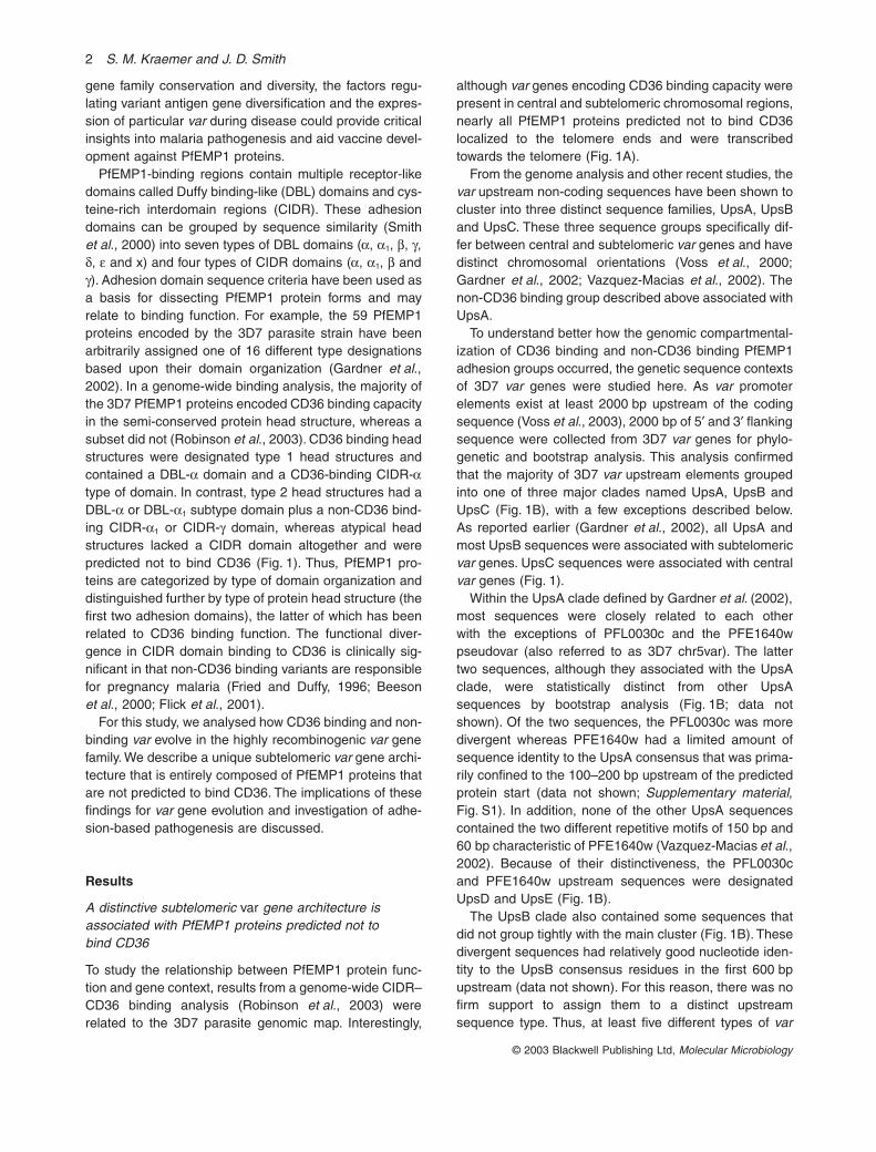
Fig. 1.
Structural and functional specialization of
var
architectural groups.A. Chromosome schematic of the two subtelomeric and central
var
groups. For simplicity, the tandemly arranged subtelomeric
var
genes are depicted as though there were no intervening genes. However, it is not uncommon for one or two
rif
genes to be between these
var
genes (Gardner
et al
., 2002). Nearly all group 1 and central
var
genes would be predicted to bind CD36, whereas group 2
var
genes are predicted not to bind CD36. Listed are the number of genes in each group and type of protein head structure. Type 1 head structures contain a DBL1-
a
domain and a CD36 binding CIDR1-
a
domain. Type 2 head structures contain either a DBL1-
a
or a DBL1-
a
1
subtype domain and a non-CD36 binding CIDR1-
a
1
subtype or CIDR-g domain. Atypical head structures lack a CIDR domain. Phylogenetic analysis of 2000 bp of 5¢ (B) and 3¢ (C) flanking sequences from 3D7 var genes (Gardner et al., 2002). Circles indicate distinct clades with the corresponding bootstrap value. Genes are coloured according to chromosomal location and types of 5¢ flanking sequences. Green and blue sequences are located in the subtelomeric region and oriented towards the centromere (subtelomeric var group 1), black sequences are in central chromosomal locations, and orange, burgundy, purple and pink sequences are located in the subtelomeric regions and oriented towards the telomere (subtelomeric var group 2). Sequences are shaded according to grouping. Five types of var upstream sequences were designated UpsA to UpsE and two types of downstream sequences DownsA and Downs B. For the boxed sequences, there were regions of overlap between the 2000 bp analysed and the 5¢, 3¢ or coding sequence of adjacent var genes. The distribution of sequences on the tree did not change when the overlapping regions were excluded from the phylogenetic comparison (data not shown). N, CIDR1 domain does not bind CD36; B, CIDR1 domain binds CD36; N*, protein head structures do not contain CIDR domains and are not predicted to bind CD36 (Robinson et al., 2003).

4 S. M. Kraemer and J. D. Smith
© 2003 Blackwell Publishing Ltd, Molecular Microbiology
groups 1 (flanked by UpsB, DownsB), subtelomeric vargroup 2 (flanked by UpsA, UpsD or UpsE and DownsA)and central var genes (flanked by UpsC) (Fig. 1).
Of the 59 var genes present in the 3D7 genome, 36mapped to subtelomeric regions and 23 to centralregions (Gardner et al., 2002). Twenty-five of the subte-lomeric var genes were transcribed towards the cen-tromere (var group 1), and 11 were transcribed towardsthe telomere (var group 2) (Fig. 1A). Altogether, 22 ofthe 28 telomere ends had a var group 1 gene as thetelomere-proximal var gene (Gardner et al., 2002). Asubset of these, eight chromosome ends, had the var 1/var 2 group arrangement illustrated in Fig. 1A. For sim-plicity, this tandem arrangement is depicted as thoughthere were no intervening genes. However, it is notuncommon for one or two rif genes to be between thetwo var genes (Gardner et al., 2002). A ninth chromo-some end, chromosome 12, had this arrangementexcept that there were two var group 1 genes next to thevar group 2 gene PFL0030c (Gardner et al., 2002). Theremaining var group 2 genes, MAL7P1.1 and PF11-0521, were not flanked by a var group 1 gene. Becausetelomere-proximal genes are frequently deleted when P.falciparum parasites are adapted to in vitro cultivation, itis not clear whether single var group 2 genes are naturalarrangements or if a var group 1 gene was deleted fromthese chromosome ends.
The subtelomeric var group 2 was distinguished fromother var groups by encoding proteins that were pre-dicted not to bind CD36. All the var group 2 proteins hadeither a type 2 protein head structure (seven genes plusthe PFE1640w pseudovar) or atypical head structures(four genes) that lacked a CIDR domain (Fig. 1A). Of thefour PfEMP1 proteins that lack a CIDR domain, threehad a binding region domain organization referred to as‘type 3’, and the fourth (PFL0030c) had a domain organi-zation referred to as ‘type 13’ (Fig. 1A) (Gardner et al.,2002). For convenience, the three 3D7 var genes(PFA0015c, MAL6p1.314 and PFI1820w) and othersrelated to them are referred to collectively as ‘type 3-like’var in this paper.
Remarkably, in the whole-genome analysis (Robinsonet al., 2003), only two PfEMP1 proteins that were notpredicted to bind CD36 did not localize with the group 2vars. The two exceptions, PF08-0140 and MAL6P1.316(blue sequences in Fig. 1B), had a slightly different type2 head structure from var group 2 proteins. Whereas theyhad CIDR1-a1 subtype domains that did not bind CD36,their DBL1 domain was the ‘a type’ rather than the ‘DBL1-a1’ subtype (Fig. 1A) (Robinson et al., 2003). Thus, pro-teins in the subtelomeric var group 2 were structurally andfunctionally distinct from central and subtelomeric vargroup 1 and may be selected for different adherenceproperties.
The subtelomeric var group 2 contains several unusually conserved, ‘strain-transcendent’ var genes
To assess the extent of var gene overlap between differentparasite strains, most previous investigations have com-pared small DBL1 domain fragments amplified by degen-erate primers with semi-conserved motifs. These studieshave documented an enormous diversity of var genesequences (Taylor et al., 2000a; Fowler et al., 2002; Tamiet al., 2003) and a limited number of unusually conservedvar fragments. To date, two 3D7 var group 2 loci, thePFE1640w and the PFL0030c (or var2csa), have beenshown to be unusually conserved between parasitestrains (Salanti et al., 2003; Winter et al., 2003). TheFCR3-CSA var gene (or var1csa), which is chimeric to thePFE1640w pseudovar and may share a common ances-tral origin, is a third highly conserved var group 2 locus(Gardner et al., 2002; Rowe et al., 2002; Salanti et al.,2002; Vazquez-Macias et al., 2002). To test the idea thatthe unique gene orientation of the subtelomeric var group2 would minimize recombination and cause other genesin this group to be more conserved between parasitestrains, primers based upon the 3D7 UpsA, UpsE andDownsA sequences were used for amplification.
For this comparison, three parasite strains (IT4/25/5,Dd2 and MC) collected at different times and from distantgeographical locations were investigated, and severalcomplete var genes totalling nearly 100 kb of new varsequence were cloned and sequenced. To facilitate thisanalysis, long polyymerase chain reactions (PCRs) wereoptimized. The longest var product that was cloned suc-cessfully was ª10 kb, but products of 6 kb and greaterwere routinely amplified and cloned. Larger P. falciparumDNA fragments are notorious for undergoing recombina-tion in bacteria. Recombination did not occur for larger vargene fragments as confirmed by multiple, independentshorter PCRs repeated on genomic DNA for each newlycloned var (data not shown). Overall, three different typesof subtelomeric var group 2 gene products were studied;(i) the ‘type 3’ var; (ii) the PFL0030c-like var genes; and(iii) var genes encoding type 2 head structures (Fig. 1A).
To clone and sequence ‘type 3’ var genes from theMalayan Camp and It4/25/5 strains, type 3-tailoredupstream and downstream primers were used to amplifyª6 kb PCR products that were sequenced rapidly usingoligonucleotides based upon the 3D7 ‘type 3’ var genes.A comparison of the genes amplified with those from the3D7 strain showed that at least 800 bp of 5¢ flankingsequence and 400 bp of downstream sequence werehighly conserved (Fig. 2A, Supplementary material,Fig. S1). In addition, each of the genes had a conservedand unusually short intron, ª150–170 bp (Fig. 2A; datanot shown). As the intron region has been implicated invar gene silencing (Deitsch et al., 2001), possible implica-

Genetic structuring of PfEMP1 adhesion groups 5
© 2003 Blackwell Publishing Ltd, Molecular Microbiology
tions on gene regulation or switching exist. The degree ofcoding region similarity has not been reported for ‘type 3’var. A comparison of the five sequences (Fig. 2A) showeda remarkable degree of sequence conservation bothwithin and between parasite strains. These proteins werenearly identical over much of their predicted bindingregion and only diverged at the amino-terminus and theintracytoplasmic region (Fig. 2A).
As shown recently (Salanti et al., 2003), PFL0030c-likevar genes are also highly conserved between parasitestrains. In addition, the 3D7 strain also contains a second,PFL0030c-like pseudogene at the end of chromosome13 (MAL13P1-354) that is transcriptionally orientatedtowards the telomere and has a nearly identical upstreamsequence to PFL0030c. To clone and sequencePFL0030c-like var genes from other parasite strains,these ª10 kb genes were amplified in two separate PCRsusing gene-specific primers positioned at approximately-800 and +500 bp relative to the coding sequence andinternal primers in the third DBL domain (Fig. 2). Theinitial PCR products overlapped at the primers. To confirmtheir connection, a third PCR product from DBL domains2–4, which straddled these PCR products, was clonedand sequenced. For Dd2, only the 5¢ half of the gene wascloned, but complete genes were cloned and sequencedfrom Malayan Camp and It4/25/5. As was done with the‘type 3’ var, oligos based upon the 3D7 var sequencewere used to sequence the new genes.
Although it is known that the coding sequences ofPFL0030c-like var are conserved, this analysis demon-strated significant conservation of the gene flankingsequence (Fig. 2B). The upstream regions were nearlyidentical except that the It4/25/5 and Dd2 strains had aninsertion of ª250 bp, accounting for the lower sequenceidentity values for It4 var4 and Dd2 var6 (Fig. 2B; data notshown). PFL0030c-like var have a truncated N-terminalsegment at the beginning of the protein and lack a con-
ventional head structure because they do not have a CIDRdomain (Fig. 1A; data not shown). The first three DBLdomains do not cluster with other DBL types but formunique clades of their own (Fig. 4B). In addition, the inter-domain regions after DBL domains 1 and 2 were notpresent in other var genes, but both had several con-served cysteine residues (data not shown). These distinc-tive features suggest that the PFL0030c-like var do notrecombine frequently with other var genes.
Lastly, to test whether UpsA sequences were associ-ated with type 2 head structures in non-3D7 parasitegenotypes, an UpsA primer was paired with a degenerateDBL-b primer. In large PfEMP1 proteins, DBL-bc2 tandemdomains are the most common domains after either type1 or type 2 head structures (Smith et al., 2000; Gardneret al., 2002). Using the UpsA primer, each of five ran-domly cloned var genes from Malayan Camp, Dd2 and It4/25/5 had DBL1 and CIDR1 domains characteristic of type2 head structures (Fig. 4B and C). By comparison, whenan UpsC primer was paired with the same degenerateDBL-b primer, only PfEMP1 proteins with type 1 headstructures were amplified from IT4/25/5 genomic DNA(Kraemer et al., 2003; data not shown).
The downstream gene regions of the new genes withtype 2 head structures were cloned using gene-specificDBL-b primers paired with a DownsA primer (Table 1).These var were more diverse than the ‘type 3 var’ and thePFL0030c-like homologues (Figs 2 and 3). To aid theirsequencing, ‘universal’ DBL type-specific primers devel-oped for PCR amplification (Kraemer et al., 2003) wereadapted to sequencing reactions. When adhesiondomains were not repeated in the same cloned product,several contigs were produced that could be rapidlyclosed by ‘walking’. The new proteins included two exam-ples of a DBL-gc2 domain pairing, where previously thec2 domain has only been associated with the DBL-b type(Smith et al., 2000; Gardner et al., 2002).
Fig. 2. Comparison of ‘type 3’ and PFL0030c-like var genes from different parasite genotypes. Percentage nucleotide identity (bold type) or amino acid identity (normal type) is shown for (A) type 3 var and (B) PFL0030c-like var. Accession numbers of genes amplified for this paper are listed in parentheses.
6 S. M. Kraemer and J. D. Smith
© 2003 Blackwell Publishing Ltd, Molecular Microbiology
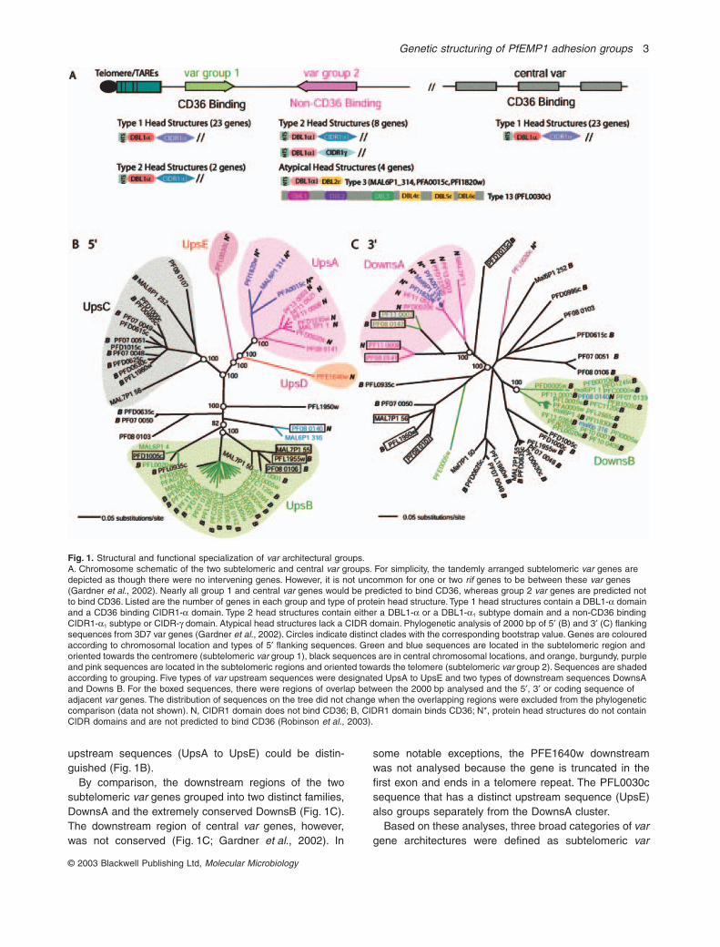
This analysis demonstrated that subtelomeric group 2-like vars were broadly distributed between parasite geno-types and had related features. Some group 2 vars, suchas the PFL0030c-like var and ‘type 3’ var, were extremelyconserved (Fig. 2). Others were less strictly conservedbetween parasite genotypes (Fig. 3) but had type 2 headstructures rather than type 1 head structures.
Evidence for three separately evolving var groups across parasite genotypes
The unusual interstrain conservation of the var group 2-like genes is consistent with the hypothesis that genelocation, orientation and non-coding flanking sequence
limit var recombinational possibilities. To test further theidea that a recombination hierarchy exists between vargroups, additional sequence comparisons were per-formed. Initially, the 10 new var genes amplified for thisstudy were compared with var group 2 genes from the3D7 strain using phylogenetic criteria. As mentionedabove, the ‘type 3’ var and PFL0030c-like var were highlyrelated between strains (Figs 2 and 4). However, even themore divergent var genes encoding type 2 head structureswere similar between parasite strains, as evidenced bytwo approaches. First, for the domains comprising theprotein’s head structure and cytoplasmic regions, each ofthe subtelomeric var group 2-like sequences tended togroup together in the NTS, DBL, CIDR and exon 2
Table 1. Oligos used in cloning var genes from different genotypes.
Oligo name Sequence Fragment cloned
PFL0030c-like varPFL0030c 5p 5.1 GTCTGTGAATGCAATGACAG Gene flankingPFL0030c DBL3 3.1 CGTGATATAATTGCTGTACC 5¢ to DBL3PFL0030c DBL3 5.1 GGTACAGCAATTATATCACG DBL3 to genePFL0030c 3p 3.1 GTAAGATGTAACAAGATATTAC flanking 3¢PFL0030c DBL2 5.2 GTAAGTCGTGTAAGGAAAGTG DBL2 to DBL4PFL0030c DBL4 3.2 GCAACTATTTGTAATGTTTCC
Type 3 varType3 5p 5.1 GGATAAGTGATRACATAATRT Gene flanking 5¢Var gr2 3p 3.1 (DownsA) CAAATAATCAMATGTGTCAAAYAR to gene flanking 3¢
Var containing type 2 head structuresVar gr2 5p 5.1 (UpsA) ATKTATTAYATTTGTTGTAGGTGA Gene flanking 5¢ to DBLbdegenDBLb 3.2 AATCKTTGDGG RATRTARTC
Fig. 3. Comparison of var group 2-like proteins that contain type 2 head structures. PfEMP1 sequences amplified from different parasite genotypes are compared with PfEMP1 proteins from the 3D7 parasite strain. Percentage amino acid identity to the 3D7 sequence with greatest BLAST identity is listed. Accession numbers are listed in parentheses.

Genetic structuring of PfEMP1 adhesion groups 7
© 2003 Blackwell Publishing Ltd, Molecular Microbiology
Fig. 4. Sequence comparison of the subtelomeric var group 2 genes with other var genes. Phylogenetic trees were generated based upon domains in PfEMP1 proteins. The sequences compared included the 59 3D7 PfEMP1 proteins (Gardner et al., 2002), eight PfEMP1 proteins from different parasite genotypes that had previously been mapped to chromosome locations (FCR3T11-1, A4var, Dd2var1, FCR3var2, FCR3var3, Dd2var7a, Dd2var7b, ItR29) and the 10 new var genes amplified for this study (indicated by asterisks). Amino acid sequences are compared. Genes and gene groups are coloured as in Fig. 1. For simplicity, only the DBL1 and CIDR1 head structure domains were coloured in the DBL and CIDR trees. In addition, in the DBL and CIDR trees, most of the gene names were removed, except for the subtelomeric var group 2, to make the figure legible.

8 S. M. Kraemer and J. D. Smith
© 2003 Blackwell Publishing Ltd, Molecular Microbiology
(cytoplasmic region) trees (Fig. 4A–D). In addition, nearlyall the predicted DBL and CIDR adhesive domains in thenew genes were most similar to 3D7 subtelomeric vargroup 2 genes by BLAST analysis (Fig. 3). The only excep-tion was the Dd2 var4 DBL6gc2.
The similarity between var group 2 genes also includedthe non-coding flanking sequence. When the upstream,downstream and intron sequences were compared acrossparasite strains, groups of sequences that were definedbased upon the 3D7 genome remained intact (Supple-mentary material, Fig. S1; data not shown). As var 5¢regions have promoter activity and co-operate with theintron to silence var genes (Deitsch et al., 1999; 2001;Calderwood et al., 2003; Voss et al., 2003), these uniquefeatures of var group 2 sequences could have regulatoryimplications.
To extend this analysis to non-var group 2 genes, sev-eral previously mapped var genes from non-3D7 parasitegenotypes were added to the phylogenetic analyses.Based on chromosomal location, FCR3T11-1 and A4varare members of subtelomeric var group 1 (Hernandez-Rivas et al., 1997; Horrocks et al., 2002); Dd2var1,FCR3var2, FCR3var3, Dd2var7a and Dd2var7b are cen-tral var (Su et al., 1995; Deitsch et al., 1999); and ItR29is a member of subtelomeric var group 2 (Horrocks et al.,2002). For each of these var, the non-coding flankingsequences grouped according to prediction (Supplemen-tary material, Fig. S1). Moreover, the coding sequence ofall central and subtelomeric var group 1 genes had type1 head structures and grouped together in the NTS andexon 2 analyses, whereas the R29 var had a type 2 headstructure and grouped with other var group 2 genes in theNTS tree (Fig. 4; data not shown). Therefore, this analysisshowed that central and subtelomeric var groups are sur-rounded by specific and distinct flanking sequences andhave diverged in characteristic patterns in their proteincoding sequence. These data suggest that the differentgroups may be recombining and evolving separately fromeach other.
Discussion
The availability of a complete P. falciparum genomicsequence creates dramatic new opportunities to investi-gate the parasite’s biology. For this study, we analysed thegene structure and chromosomal organization of vargenes and used this information to gain insight into howthe gene family evolves and causes disease. In commonwith other organisms, P. falciparum variant genes are con-centrated at the telomere ends (Fischer et al., 1997;Thompson et al., 1997; Gardner et al., 2002). However,unlike many other variant gene families that function onlyas clonally variant targets of immunity, var genes alsoencode cytoadherent activity. Thus, the var gene reper-
toire is shaped by selective forces acting to both maintainbinding capability and escape immunity (Roberts et al.,1993). At the genomic level, these varied pressures havebeen manifested in an apparent genetic compartmental-ization of PfEMP1 binding phenotypes to particular vargene sequence contexts.
In the 3D7 genome, at least three broadly distinct vargene groups were defined based upon chromosomal loca-tion, gene orientation and 5¢ and 3¢ flanking sequences(Fig. 1). Subtelomeric var group 1 consists of telomericvar genes that are transcribed towards the centromereand are flanked by UpsB and DownsB; subtelomeric vargroup 2 contains telomeric var genes that are orientedtowards the telomere and are flanked by either UpsA,UpsD or UpsE and DownsA; and central var are flankedby UpsC and a non-conserved 3¢ region (Fig. 1). Thesefeatures appear to establish a recombination hierarchythat limits reassortment between var groups and maycreate opportunities for the parasite to evolve specializedfunctions and activities relative to the different groups. Astriking example is the subtelomeric var group 2 that con-tains several unusually conserved var genes and consistsof non-CD36 binding variants. Although CD36 is a keymicrovasculature receptor used by most parasite isolates(Newbold et al., 1997), non-CD36-binding PfEMP1 pro-teins have important roles in placental sequestration(Fried and Duffy, 1996; Beeson et al., 2000; Flick et al.,2001) and possibly other vascular sites, such as brainendothelium, where CD36 is poorly or not expressed.
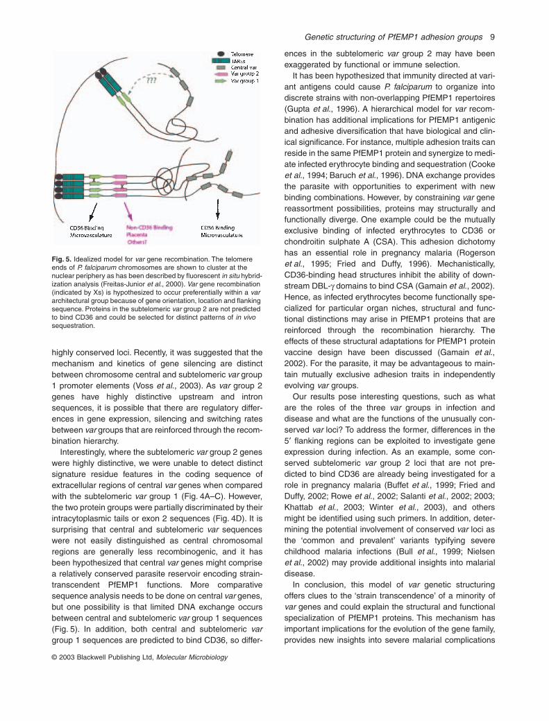
It is surprising that a subtelomeric var group wouldcontain so many broadly conserved loci, as this chromo-somal region is highly recombinogenic (Lanzer et al.,1995; Freitas-Junior et al., 2000; Taylor et al., 2000b;Figueiredo et al., 2002). Recently, the telomere ends of P.falciparum chromosomes were shown to cluster togetherin the nuclear periphery, suggesting a potential mecha-nism for facilitating var gene recombination (Freitas-Junioret al., 2000; Figueiredo et al., 2002). Our comparativeanalysis of the subtelomeric var group 2 builds on thisanalysis and suggests a model in which gene orientation,location and flanking sequences are crucial to the straintranscendence of some var genes and favour the prefer-ential reassortment of genes within architectural groups(Fig. 5). Conserved subtelomeric var group 2 loci, such asPFE1640w (Winter et al., 2003) and PFL0030c, havehighly distinctive gene flanking sequences (Fig. 1B andC), which may reinforce their withdrawal from generalfamily recombination. This mechanism would promote‘self–self’ recombination and interstrain gene conserva-tion. Other group 2 var genes encoding type 2 head struc-tures were more diverse between strains. This diversitymay occur because there are more of these to recombinewith (Fig. 1) and/or the individual genes are under differ-ent functional and immune selective pressures from the
Genetic structuring of PfEMP1 adhesion groups 9
© 2003 Blackwell Publishing Ltd, Molecular Microbiology
highly conserved loci. Recently, it was suggested that themechanism and kinetics of gene silencing are distinctbetween chromosome central and subtelomeric var group1 promoter elements (Voss et al., 2003). As var group 2genes have highly distinctive upstream and intronsequences, it is possible that there are regulatory differ-ences in gene expression, silencing and switching ratesbetween var groups that are reinforced through the recom-bination hierarchy.
Interestingly, where the subtelomeric var group 2 geneswere highly distinctive, we were unable to detect distinctsignature residue features in the coding sequence ofextracellular regions of central var genes when comparedwith the subtelomeric var group 1 (Fig. 4A–C). However,the two protein groups were partially discriminated by theirintracytoplasmic tails or exon 2 sequences (Fig. 4D). It issurprising that central and subtelomeric var sequenceswere not easily distinguished as central chromosomalregions are generally less recombinogenic, and it hasbeen hypothesized that central var genes might comprisea relatively conserved parasite reservoir encoding strain-transcendent PfEMP1 functions. More comparativesequence analysis needs to be done on central var genes,but one possibility is that limited DNA exchange occursbetween central and subtelomeric var group 1 sequences(Fig. 5). In addition, both central and subtelomeric vargroup 1 sequences are predicted to bind CD36, so differ-
ences in the subtelomeric var group 2 may have beenexaggerated by functional or immune selection.
It has been hypothesized that immunity directed at vari-ant antigens could cause P. falciparum to organize intodiscrete strains with non-overlapping PfEMP1 repertoires(Gupta et al., 1996). A hierarchical model for var recom-bination has additional implications for PfEMP1 antigenicand adhesive diversification that have biological and clin-ical significance. For instance, multiple adhesion traits canreside in the same PfEMP1 protein and synergize to medi-ate infected erythrocyte binding and sequestration (Cookeet al., 1994; Baruch et al., 1996). DNA exchange providesthe parasite with opportunities to experiment with newbinding combinations. However, by constraining var genereassortment possibilities, proteins may structurally andfunctionally diverge. One example could be the mutuallyexclusive binding of infected erythrocytes to CD36 orchondroitin sulphate A (CSA). This adhesion dichotomyhas an essential role in pregnancy malaria (Rogersonet al., 1995; Fried and Duffy, 1996). Mechanistically,CD36-binding head structures inhibit the ability of down-stream DBL-g domains to bind CSA (Gamain et al., 2002).Hence, as infected erythrocytes become functionally spe-cialized for particular organ niches, structural and func-tional distinctions may arise in PfEMP1 proteins that arereinforced through the recombination hierarchy. Theeffects of these structural adaptations for PfEMP1 proteinvaccine design have been discussed (Gamain et al.,2002). For the parasite, it may be advantageous to main-tain mutually exclusive adhesion traits in independentlyevolving var groups.
Our results pose interesting questions, such as whatare the roles of the three var groups in infection anddisease and what are the functions of the unusually con-served var loci? To address the former, differences in the5¢ flanking regions can be exploited to investigate geneexpression during infection. As an example, some con-served subtelomeric var group 2 loci that are not pre-dicted to bind CD36 are already being investigated for arole in pregnancy malaria (Buffet et al., 1999; Fried andDuffy, 2002; Rowe et al., 2002; Salanti et al., 2002; 2003;Khattab et al., 2003; Winter et al., 2003), and othersmight be identified using such primers. In addition, deter-mining the potential involvement of conserved var loci asthe ‘common and prevalent’ variants typifying severechildhood malaria infections (Bull et al., 1999; Nielsenet al., 2002) may provide additional insights into malarialdisease.
In conclusion, this model of var genetic structuringoffers clues to the ‘strain transcendence’ of a minority ofvar genes and could explain the structural and functionalspecialization of PfEMP1 proteins. This mechanism hasimportant implications for the evolution of the gene family,provides new insights into severe malarial complications
Fig. 5. Idealized model for var gene recombination. The telomere ends of P. falciparum chromosomes are shown to cluster at the nuclear periphery as has been described by fluorescent in situ hybrid-ization analysis (Freitas-Junior et al., 2000). Var gene recombination (indicated by Xs) is hypothesized to occur preferentially within a var architectural group because of gene orientation, location and flanking sequence. Proteins in the subtelomeric var group 2 are not predicted to bind CD36 and could be selected for distinct patterns of in vivo sequestration.

10 S. M. Kraemer and J. D. Smith
© 2003 Blackwell Publishing Ltd, Molecular Microbiology
and needs to be considered in PfEMP1 vaccine develop-ment and implementation.
Experimental procedures
Parasites
The A4, Dd2 and Malayan Camp (MC) clonal lines werecultured using standard practices, and genomic DNA wasprepared. The A4 clone was originally derived by microman-ipulation from P. falciparum line IT4/25/5, and the Dd2 strainis a clone of W2-MEF.
PCR amplification and sequencing of var genes
PCRs were done using TaKaRa LA TaqTM polymerase (Fisher)with 50 ng of genomic DNA, 1¥ buffer, 0.4 mM dNTPs (each),2.5 mM MgCl2, 0.5 mM primers and 2.5 U of enzyme run ina DNA Engine DyadTM Peltier thermal cycler from MJResearch. Oligonucleotide primers are listed in Table 1. PCRconditions were one cycle of 94∞C for 1 min followed by 35cycles of 98∞C for 1 min, annealing for 1 min, extension at62∞C for 6–18 min, depending upon fragment size, and a finalextension for 10 min at 68∞C. Products were cloned into thepCR®4-TOPO vector or pCR®-XL-TOPO from Invitrogen andsequenced to provide at least double coverage. In caseswhere degenerate DBL-type specific oligos (Kraemer et al.,2003) were adapted to sequencing reactions, oligo concen-trations were increased 10-fold to account for primer degen-eracy. Sequences were assembled using the PHRED/PHRAP/CONSED software suite (Gordon et al., 1998).
Sequence analysis
Phylogenetic analyses were done using CLUSTALX for multiplealignments and PAUP*4.0b10 (* phylogenetic analysis usingparsimony and other methods) to generate neighbour-joiningtrees with 1000 bootstrap replicates. For tree generation, thenew sequences amplified in the study were compared with3D7 PfEMP1 sequences and others from non-3D7 parasitegenotypes that had previously been mapped to chromosomallocations. 3D7 PfEMP1 sequences are identified by genenames in the figures, and their sequences can be accessedusing this identifier at the PlasmoDB (http://plamodb.org/).Gene names and accession numbers for previouslypublished non-3D7 genotypic sequences were: Dd2var(L40608), FCR3T11-1 (U67959), FCR3var2 and FCR3var3(L40609), MCvar1 (U27338), MCvar2 (U27339), Dd2var7(L42636), Dd2var7a (AF041422), Dd2var7b (AF041423),ItvarR29 (Y13402) and IT-A4var (3540145). Gene sequenceswere divided into respective domains using publishedapproaches (Smith et al., 2000). BLAST analysis of eachregion was done against the PlasmoDB. Percentagesequence identities were calculated using the algorithm inDNAStar MEGALIGN, version 5.0.
Acknowledgements
The authors thank Siri Nelson, Martin Pentony and PeterMyler for guidance in using CONSED and advice concerning
sequencing reactions using degenerate oligos. In addition,we thank Zoe Christodoulou, Alex Rowe and Sue Kyes forsharing their unpublished R29 upstream sequence with us.The authors also thank Leia Smith for a construct containingpart of IT4 var2. J.D.S. is supported by an Ellison MedicalFoundation New Scholars Award in Global Infectious Dis-ease, the Bill and Melinda Gates Foundation and a NationalInstitutes of Health grant (RO1 AI47953-01A1).
Supplementary material
The following material is available from http://www.blackwellpublishing.com/products/journals/suppmat/mmi/mmi3814/mmi3814sm.htmFig. S1. Sequence comparison of the var genes 5¢ and 3¢flanking regions. The flanking sequences of var genes amplified from MalayanCamp, It4/25/5, and Dd2 strains were compared to 3D7 vargenes after trimming to the same length. Circles indicatedistinct tree branches with the corresponding bootstrapvalue. Genes are coloured according to Fig. 1. and sequencegroups are shaded.
References
Baruch, D.I., Pasloske, B.L., Singh, H.B., Bi, X., Ma, X.C.,Feldman, M., et al. (1995) Cloning the P. falciparum geneencoding PfEMP1, a malarial variant antigen and adher-ence receptor on the surface of parasitized human eryth-rocytes. Cell 82: 77–87.
Baruch, D.I., Gormely, J.A., Ma, C., Howard, R.J., andPasloske, B.L. (1996) Plasmodium falciparum erythrocytemembrane protein 1 is a parasitized erythrocyte receptorfor adherence to CD36, thrombospondin, and intercellularadhesion molecule 1. Proc Natl Acad Sci USA 93: 3497–3502.
Beeson, J.G., Rogerson, S.J., Cooke, B.M., Reeder, J.C.,Chai, W., Lawson, A.M., et al. (2000) Adhesion of Plasmo-dium falciparum-infected erythrocytes to hyaluronic acid inplacental malaria. Nature Med 6: 86–90.
Buffet, P.A., Gamain, B., Scheidig, C., Baruch, D., Smith,J.D., Hernandez-Rivas, R., et al. (1999) Plasmodium falci-parum domain mediating adhesion to chondroitin sulfateA: a receptor for human placental infection. Proc Natl AcadSci USA 96: 12743–12748.
Bull, P.C., and Marsh, K. (2002) The role of antibodies toPlasmodium falciparum-infected-erythrocyte surface anti-gens in naturally acquired immunity to malaria. TrendsMicrobiol 10: 55–58.
Bull, P.C., Lowe, B.S., Kortok, M., and Marsh, K. (1999)Antibody recognition of Plasmodium falciparum erythro-cyte surface antigens in Kenya: evidence for rare and prev-alent variants. Infect Immun 67: 733–739.
Calderwood, M.S., Gannoun-Azki, L., Wellems, T.E., andDeitsch, K.W. (2003) Plasmodium falciparum var genesare regulated by two regions with separate promoters, oneupstream of the coding region and a second within theintron. J Biol Chem 278: 34125–34132.
Cooke, B.M., Berendt, A.R., Craig, A.G., MacGregor, J.,Newbold, C.I., and Nash, G.B. (1994) Rolling and station-ary cytoadhesion of red blood cells parasitized by Plasmo-
Genetic structuring of PfEMP1 adhesion groups 11
© 2003 Blackwell Publishing Ltd, Molecular Microbiology
dium falciparum: separate roles for ICAM-1, CD36 andthrombospondin. Br J Haematol 87: 162–170.
Deitsch, K.W., del Pinal, A., and Wellems, T.E. (1999) Intra-cluster recombination and var transcription switches in theantigenic variation of Plasmodium falciparum. Mol Bio-chem Parasitol 101: 107–116.
Deitsch, K.W., Calderwood, M.S., and Wellems, T.E. (2001)Malaria. Cooperative silencing elements in var genes.Nature 412: 875–876.
Figueiredo, L.M., Freitas-Junior, L.H., Bottius, E., Olivo-Marin,J.C., and Scherf, A. (2002) A central role for Plasmodiumfalciparum subtelomeric regions in spatial positioning andtelomere length regulation. EMBO J 21: 815–824.
Fischer, K., Horrocks, P., Preuss, M., Wiesner, J., Wunsch,S., Camargo, A.A., and Lanzer, M. (1997) Expression ofvar genes located within polymorphic subtelomericdomains of Plasmodium falciparum chromosomes. MolCell Biol 17: 3679–3686.
Flick, K., Scholander, C., Chen, Q., Fernandez, V., Pouvelle,B., Gysin, J., and Wahlgren, M. (2001) Role of nonimmuneIgG bound to PfEMP1 in placental malaria. Science 293:2098–2100.
Fowler, E.V., Peters, J.M., Gatton, M.L., Chen, N., andCheng, Q. (2002) Genetic diversity of the DBLalpha regionin Plasmodium falciparum var genes among Asia-Pacificisolates. Mol Biochem Parasitol 120: 117–126.
Freitas-Junior, L.H., Bottius, E., Pirrit, L.A., Deitsch, K.W.,Scheidig, C., Guinet, F., et al. (2000) Frequent ectopicrecombination of virulence factor genes in telomericchromosome clusters of P. falciparum. Nature 407: 1018–1022.
Fried, M., and Duffy, P.E. (1996) Adherence of Plasmodiumfalciparum to chondroitin sulfate A in the human placenta.Science 272: 1502–1504.
Fried, M., and Duffy, P.E. (2002) Two DBLgamma subtypesare commonly expressed by placental isolates of Plasmo-dium falciparum. Mol Biochem Parasitol 122: 201–210.
Fried, M., Nosten, F., Brockman, A., Brabin, B.J., and Duffy,P.E. (1998) Maternal antibodies block malaria. Nature 395:851–852.
Gamain, B., Gratepanche, S., Miller, L.H., and Baruch, D.I.(2002) Molecular basis for the dichotomy in Plasmodiumfalciparum adhesion to CD36 and chondroitin sulfate A.Proc Natl Acad Sci USA 99: 10020–10024.
Gardner, M.J., Hall, N., Fung, E., White, O., Berriman, M.,Hyman, R.W., et al. (2002) Genome sequence of thehuman malaria parasite Plasmodium falciparum. Nature419: 498–511.
Gordon, D., Abajian, C., and Green, P. (1998) CONSED: agraphical tool for sequence finishing. Genome Res 8: 195–202.
Gupta, S., Hill, A.V., Kwiatkowski, D., Greenwood, A.M.,Greenwood, B.M., and Day, K.P. (1994) Parasite virulenceand disease patterns in Plasmodium falciparum malaria.Proc Natl Acad Sci USA 91: 3715–3719.
Gupta, S., Maiden, M.C., Feavers, I.M., Nee, S., May, R.M.,and Anderson, R.M. (1996) The maintenance of strainstructure in populations of recombining infectious agents.Nature Med 2: 437–442.
Gupta, S., Snow, R.W., Donnelly, C.A., Marsh, K., and New-bold, C. (1999) Immunity to non-cerebral severe malaria is
acquired after one or two infections. Nature Med 5: 340–343.
Hernandez-Rivas, R., Mattei, D., Sterkers, Y., Peterson,D.S., Wellems, T.E., and Scherf, A. (1997) Expressed vargenes are found in Plasmodium falciparum subtelomericregions. Mol Cell Biol 17: 604–611.
Horrocks, P., Pinches, R., Kyes, S., Kriek, N., Lee, S.,Christodoulou, Z., and Newbold, C.I. (2002) Effect of vargene disruption on switching in Plasmodium falciparum.Mol Microbiol 45: 1131–1141.
Khattab, A., Kremsner, P.G., and Klinkert, M.Q. (2003) Com-mon surface-antigen var genes of limited diversityexpressed by Plasmodium falciparum placental isolatesseparated by time and space. J Infect Dis 187: 477–483.
Kraemer, S.M., Gupta, L., and Smith, J.D. (2003) New toolsto identify var sequence tags and clone full-length genesusing type-specific primers to Duffy binding-like domains.Mol Biochem Parasitol 129: 91–102.
Kyes, S., Taylor, H., Craig, A., Marsh, K., and Newbold, C.(1997) Genomic representation of var gene sequences inPlasmodium falciparum field isolates from different geo-graphic regions. Mol Biochem Parasitol 87: 235–238.
Lanzer, M., Fischer, K., and Le Blancq, S.M. (1995) Parasit-ism and chromosome dynamics in protozoan parasites: isthere a connection? Mol Biochem Parasitol 70: 1–8.
Miller, L.H., Baruch, D.I., Marsh, K., and Doumbo, O.K.(2002) The pathogenic basis of malaria. Nature 415: 673–679.
Newbold, C., Warn, P., Black, G., Berendt, A., Craig, A.,Snow, B., et al. (1997) Receptor-specific adhesion andclinical disease in Plasmodium falciparum. Am J Trop MedHyg 57: 389–398.
Nielsen, M.A., Staalsoe, T., Kurtzhals, J.A., Goka, B.Q.,Dodoo, D., Alifrangis, M., et al. (2002) Plasmodium falci-parum variant surface antigen expression varies betweenisolates causing severe and nonsevere malaria and ismodified by acquired immunity. J Immunol 168: 3444–3450.
Peterson, D.S., Miller, L.H., and Wellems, T.E. (1995) Isola-tion of multiple sequences from the Plasmodium falciparumgenome that encode conserved domains homologous tothose in erythrocyte-binding proteins. Proc Natl Acad SciUSA 92: 7100–7104.
Roberts, D.J., Biggs, B.A., Brown, G., and Newbold, C.I.(1993) Protection, pathogenesis, and phenotypic plasticityin Plasmodium falciparum malaria. Parasitol Today 9: 281–286.
Robinson, B.A., Welch, T.L., and Smith, J.D. (2003) Wide-spread functional specialization of Plasmodium falciparumerythrocyte membrane protein 1 family members to bindCD36 analysed across a parasite genome. Mol Microbiol47: 1265–1278.
Rogerson, S.J., Chaiyaroj, S.C., Ng, K., Reeder, J.C., andBrown, G.V. (1995) Chondroitin sulfate A is a cell surfacereceptor for Plasmodium falciparum-infected erythrocytes.J Exp Med 182: 15–20.
Rowe, J.A., Kyes, S.A., Rogerson, S.J., Babiker, H.A., andRaza, A. (2002) Identification of a conserved Plasmodiumfalciparum var gene implicated in malaria in pregnancy. JInfect Dis 185: 1207–1211.
Salanti, A., Jensen, A.T., Zornig, H.D., Staalsoe, T., Joer-

12 S. M. Kraemer and J. D. Smith
© 2003 Blackwell Publishing Ltd, Molecular Microbiology
gensen, L., Nielsen, M.A., et al. (2002) A sub-family ofcommon and highly conserved Plasmodium falciparum vargenes. Mol Biochem Parasitol 122: 111–115.
Salanti, A., Staalsoe, T., Lavstsen, T., Jensen, A.T., KordaiSowa, M.P., Arnot, D.E., et al. (2003) Selective upregula-tion of a single distinctly structured var gene in chon-droitin sulphate A-adhering Plasmodium falciparuminvolved in pregnancy-associated malaria. Mol Microbiol49: 179–191.
Smith, J.D., Chitnis, C.E., Craig, A.G., Roberts, D.J.,Hudson-Taylor, D.E., Peterson, D.S., et al. (1995) Switchesin expression of Plasmodium falciparum var genes corre-late with changes in antigenic and cytoadherent pheno-types of infected erythrocytes. Cell 82: 101–110.
Smith, J.D., Subramanian, G., Gamain, B., Baruch, D.I., andMiller, L.H. (2000) Classification of adhesive domains inthe Plasmodium falciparum erythrocyte membrane protein1 family. Mol Biochem Parasitol 110: 293–310.
Su, X.Z., Heatwole, V.M., Wertheimer, S.P., Guinet, F., Her-rfeldt, J.A., Peterson, D.S., et al. (1995) The large diversegene family var encodes proteins involved in cytoadher-ence and antigenic variation of Plasmodium falciparum-infected erythrocytes. Cell 82: 89–100.
Tami, A., Ord, R., Targett, G.A., and Sutherland, C.J. (2003)Sympatric Plasmodium falciparum isolates from Venezuelahave structured var gene repertoires. Malar J 2: 7.
Taylor, H.M., Kyes, S.A., Harris, D., Kriek, N., and Newbold,C.I. (2000a) A study of var gene transcription in vitro usinguniversal var gene primers. Mol Biochem Parasitol 105:13–23.
Taylor, H.M., Kyes, S.A., and Newbold, C.I. (2000b) Var genediversity in Plasmodium falciparum is generated by fre-
quent recombination events. Mol Biochem Parasitol 110:391–397.
Thompson, J.K., Rubio, J.P., Caruana, S., Brockman, A.,Wickham, M.E., and Cowman, A.F. (1997) The chro-mosomal organization of the Plasmodium falciparum vargene family is conserved. Mol Biochem Parasitol 87: 49–60.
Vazquez-Macias, A., Martinez-Cruz, P., Castaneda-Patlan,M.C., Scheidig, C., Gysin, J., Scherf, A., and Hernandez-Rivas, R. (2002) A distinct 5¢ flanking var gene regionregulates Plasmodium falciparum variant erythrocyte sur-face antigen expression in placental malaria. Mol Microbiol45: 155–167.
Voss, T.S., Thompson, J.K., Waterkeyn, J., Felger, I.,Weiss, N., Cowman, A.F., and Beck, H.P. (2000)Genomic distribution and functional characterization oftwo distinct and conserved Plasmodium falciparum vargene 5¢ flanking sequences. Mol Biochem Parasitol 107:103–115.
Voss, T.S., Kaestli, M., Vogel, D., Bopp, S., and Beck, H.P.(2003) Identification of nuclear proteins that interact differ-entially with Plasmodium falciparum var gene promoters.Mol Microbiol 48: 1593–1607.
Ward, C.P., Clottey, G.T., Dorris, M., Ji, D.D., and Arnot, D.E.(1999) Analysis of Plasmodium falciparum PfEMP-1/vargenes suggests that recombination rearranges constrainedsequences. Mol Biochem Parasitol 102: 167–177.
Winter, G., Chen, Q., Flick, K., Kremsner, P., Fernandez, V.,and Wahlgren, M. (2003) The 3D7var5.2 (var (COMMON)type var gene family is commonly expressed in non-placental Plasmodium falciparum malaria. Mol BiochemParasitol 127: 179–191.