Embed Size (px)
Citation preview
of April 10, 2019.This information is current as
Extracellular Trapsthe Antimicrobial Activity of Neutrophil
Promastigotes EvadeLeishmania donovani
Albert DescoteauxChristelle Gabriel, W. Robert McMaster, Denis Girard and
http://www.jimmunol.org/content/185/7/4319doi: 10.4049/jimmunol.1000893September 2010;
2010; 185:4319-4327; Prepublished online 8J Immunol
Referenceshttp://www.jimmunol.org/content/185/7/4319.full#ref-list-1
, 23 of which you can access for free at: cites 50 articlesThis article
average*
4 weeks from acceptance to publicationFast Publication! •
Every submission reviewed by practicing scientistsNo Triage! •
from submission to initial decisionRapid Reviews! 30 days* •
Submit online. ?The JIWhy
Subscriptionhttp://jimmunol.org/subscription
is online at: The Journal of ImmunologyInformation about subscribing to
Permissionshttp://www.aai.org/About/Publications/JI/copyright.htmlSubmit copyright permission requests at:
Email Alertshttp://jimmunol.org/alertsReceive free email-alerts when new articles cite this article. Sign up at:
Print ISSN: 0022-1767 Online ISSN: 1550-6606. Immunologists, Inc. All rights reserved.Copyright © 2010 by The American Association of1451 Rockville Pike, Suite 650, Rockville, MD 20852The American Association of Immunologists, Inc.,
is published twice each month byThe Journal of Immunology
by guest on April 10, 2019
http://ww
w.jim
munol.org/
Dow
nloaded from
by guest on April 10, 2019
http://ww
w.jim
munol.org/
Dow
nloaded from
The Journal of Immunology
Leishmania donovani Promastigotes Evade the AntimicrobialActivity of Neutrophil Extracellular Traps
Christelle Gabriel,*,† W. Robert McMaster,‡,x Denis Girard,* and Albert Descoteaux*,†
Upon their recruitment to a site of infection and their subsequent activation, neutrophils release DNA and a subset of their granule
content to form filamentous structures, known as neutrophil extracellular traps, which capture and kill microorganisms. In this
study, we show that Leishmania promastigotes induced the rapid release of neutrophil extracellular traps from human neutrophils
and were trapped by these structures. The use of Leishmania mutants defective in the biosynthesis of either lipophosphoglycan or
GP63 revealed that these two major surface promastigote virulence determinants were not responsible for inducing the release of
the surface protease neutrophil extracellular traps. We also demonstrate that this induction was independent of superoxide
production by neutrophils. Finally, in contrast to wild-type Leishmania donovani promastigotes, mutants defective in lipophos-
phoglycan biosynthesis were highly susceptible to the antimicrobial activity of neutrophil extracellular traps. Altogether, our data
suggest that neutrophil extracellular traps may contribute to the containment of L. donovani promastigotes at the site of in-
oculation, thereby facilitating their uptake by mononuclear phagocytes. The Journal of Immunology, 2010, 185: 4319–4327.
Protozoan parasites of the genus Leishmania infect millionsof people worldwide, causing leishmaniases, a spectrumof clinical manifestations that range from self-healing
ulcers to potentially fatal visceral leishmaniasis. Transmission ofthe parasite is mediated by the blood-sucking sand fly of eitherthe genus Phlebotomus or the genus Lutzomyia. Upon feeding,infected sand flies inoculate infective promastigote forms into themammalian host, where they evade and resist nonspecific defensemechanisms, such as complement-mediated lysis, to ultimatelyenter mononuclear phagocytes by a receptor-mediated process.After their uptake by macrophages, promastigotes of Leishmaniadonovani and Leishmania major inhibit phagolysosome biogen-esis to evade the microbicidal phagolysosomal environment (1–5).This inhibition is mediated by lipophosphoglycan (LPG), anabundant surface virulence glycolipid consisting of a polymer ofGalb1,4Mana1-PO4 units anchored into the promastigote mem-brane via an unusual glycosyl phosphatidylinositol (1, 6).Because neutrophils are rapidly recruited to infection sites, there
has been growing evidence for the potential role of these im-mune cells during the early stages of Leishmania promastigotesinfection. However, data obtained in various experimental modelsled to conflicting conclusions regarding the role of neutrophilsin experimental leishmaniasis (7). Hence, although several stud-ies indicated that neutrophils contribute to a protective response
against Leishmania (8, 9), others provided evidence that inter-nalization of Leishmania promastigotes by neutrophils may favorthe establishment of infection (10–12). The latter observations lentweight to the concept that apoptotic neutrophils harboring Leish-mania promastigotes may act as “Trojan horses,” because they areinternalized by macrophages (7, 13).Phagocytosis is the best characterized killing mechanism in
neutrophils, whereby microorganisms are efficiently internalized inphagosomes were large amounts of toxic reactive oxygen species(ROS) are generated (14, 15). Furthermore, fusion with variousintracellular granules results in the rapid delivery of proteases,antimicrobial peptides, and myeloperoxidase, culminating in theformation of a highly microbicidal phagolysosome (16, 17). Themechanisms by which Leishmania promastigotes evade killing byneutrophils may be related to their ability to block the oxidativeburst (18) and to enter a nonlytic compartment unable to fuse withlysosomes and displaying endoplasmic reticulum features (10).Recently, evidence was provided that neutrophils use an additional
microbicidal mechanism that consists of the release of structurescomposed of chromatin and specific granule proteins in responseto various stimuli, including IL-8, LPS, bacteria, and fungi (19, 20).These structures, called neutrophil extracellular traps (NETs), formextracellular fibers able to ensnare and kill extracellularly variousmicroorganisms (19, 21–25), possibly through the action of one orseveral microbicidal proteins associated with these structures inhigh concentrations (19, 23). Additionally, NETs may also preventthe spread of pathogens by confining microorganisms to the site ofinfection (26). In this study, we show that although L. donovanipromastigotes are trapped by NETs released by human neutrophils,they resist their microbicidal activity.
Materials and MethodsHuman neutrophils
Neutrophils were isolated from venous blood of healthy volunteers by dex-tran sedimentation followed by centrifugation over Ficoll-Hypaque (Phar-macia Biotech, Quebec, Canada). Blood donations were obtained frominformed and consenting individuals according to institutionally approvedprocedures. Cell viability was monitored by trypan blue exclusion, and thepurity (.98%) was verified by cytology from cytocentrifuged preparationsstainedbyDiff-Quick staining (FisherScientific,Ottawa,Ontario,Canada) (27).
*Institut National de la Recherche Scientifique, Institut Armand-Frappier; †Center forHost–Parasite Interactions, Laval, Quebec; ‡Department of Medical Genetics, Universityof British Columbia; and xImmunity and Infection Research Centre, Vancouver CoastalHealth Research Institute, Vancouver, British Columbia, Canada
Received for publication March 18, 2010. Accepted for publication August 2, 2010.
This work was supported by Canadian Institutes of Health Research Grant MOP-12933. A.D. is the holder of a Canada Research Chair, and A.D. and D.G. wereChercheur-Boursier from the Fonds de la Recherche en Sante du Quebec. C.G. waspartly supported by the Center for Host-Parasite Interactions (Fonds Quebecois de laRecherche sur la Nature et les Technologies).
Address correspondence and reprint requests to Dr. Albert Descoteaux, Institut Na-tional de la Recherche Scientifique, Institut Armand-Frappier, 531 Boulevard des Prai-ries, Laval, Quebec, Canada H7V 1B7. E-mail address: [email protected]
Abbreviations used in this paper: DPI, diphenyleneiodonium; LPG, lipophosphogly-can; MOI, multiplicity of infection; NET, neutrophil extracellular trap; RLU, relativelight unit; ROS, reactive oxygen species; WT, wild-type.
Copyright� 2010 by The American Association of Immunologists, Inc. 0022-1767/10/$16.00
www.jimmunol.org/cgi/doi/10.4049/jimmunol.1000893
by guest on April 10, 2019
http://ww
w.jim
munol.org/
Dow
nloaded from
Parasites
Promastigotes of the L. donovani strains 1S and LV9 and the L. majorstrains LV39 and NIH S clone A2 were cultured at 26˚C in M199 sup-plemented with 10% heat-inactivated FBS, 100 mM adenine, HEPES,5 mM hemin, 3 mM biopterin, 1 mM biotin, and antibiotics. The L.donovani 1S isogenic mutants lpg1-KO and lpg2-KO were described pre-viously (28). The lpg1-KO mutant secretes repeating Galb1,4Mana1-PO4–containing molecules but lacks the ability to assemble a functionalLPG glycan core (29), precluding synthesis of LPG. The rescued lpg1-KOadd-back was generated by transfection of the pLeishZeo-LPG1 expres-sion vector. The lpg2-KO mutant expresses the truncated LPG Gal(a1,6)Gal(a1,3)Galf(b1,3)[Glc(a1-P)]Man(a1,3)Man(a1,4)GN(a1,6)-PI and doesnot synthesize repeating Galb1,4Mana1-PO4 units (30). The L. majorNIH S clone A2 isogenic gp63-KO mutant and gp63-KO add-back werepreviously described (31). L. donovani 1S and L. major NIH S clone A2promastigotes expressing luciferase (32) and L. donovani 1S promasti-gotes expressing GFP (3) were grown in the presence of 50 mg/ml G418(Life Technologies, Rockville, MD). The L. donovani lpg1-KO add-backwas grown in the presence of 100 mg/ml Zeocin (Invitrogen). Promas-tigotes were used in the late stationary phase of growth. Unless otherwisestated, promastigotes were opsonized by a 30-min incubation at 37˚Cin the presence of 10% human C8-deficient serum (Sigma-Aldrich,St. Louis, MO).
Confocal immunofluorescence microscopy for the detectionand quantification of NETs
Human neutrophils (106 per well) were seeded on poly-L-lysine–coatedglass coverslips (BD Biosciences, San Jose, CA) in RPMI 1640 sup-
plemented with 10 mM HEPES, penicillin/streptomycin, and 2% heat-inactivated human serum and were allowed to adhere for 30 min at 37˚Cin a humidified incubator with 5% CO2. Adherent neutrophils were in-cubated in the absence or presence of either Leishmania promastigotes,zymosan, or LPG-coated zymosan during 10, 30, or 60 min at 37˚C. Theywere then fixed with 4% paraformaldehyde (Sigma-Aldrich), blockedovernight in blocking solution (PBS with 10% normal goat serum [JacksonImmunoResearch Laboratories, West Grove, PA], 5% cold fish gelatin[Sigma-Aldrich], 1% BSA [Sigma-Aldrich], 0.05% Tween 20 [Sigma-Aldrich]), and incubated with primary Abs either directed against humanneutrophil-elastase (Santa Cruz Biotechnology, Santa Cruz, CA) or againstrepeated subunits of LPG (CA7AE Ab). Secondary Abs coupled to AlexaFluor 568 or 488 (Invitrogen/Molecular Probes, Eugene, OR) were used todetect primary Abs, and DRAQ5 (Biostatus Limited, Shepshed, Leices-tershire, U.K.) was used for DNA labeling. Coverslips were mounted onslides with Fluoromount-G (Southern Biotechnology Associates, Bir-mingham, AL). Specimens were analyzed with an oil immersion NikonPlan Apo 100 (Toronto, Ontario, Canada) (numerical aperture 1.4) ob-jective mounted on a Nikon Eclipse E800 microscope equipped with a Bio-Rad Radiance 2000 confocal imaging system (Bio-Rad, Hercules, CA).Percentages of cells producing NETs were determined by counting at least100 cells per condition. Neutrophils that release DNA and elastase to formfilamentous structures were considered as producing NETs.
Quantification of DNA released from neutrophils
Human neutrophils adhered to poly-L-lysine–coated glass coverslips wereincubated with either Leishmania promastigotes or zymosan at a multi-plicity of infection (MOI) of 10:1 for 30 min in RPMI 1640 supplementedwith 10 mM HEPES, penicillin/streptomycin, and 2% heat-inactivated
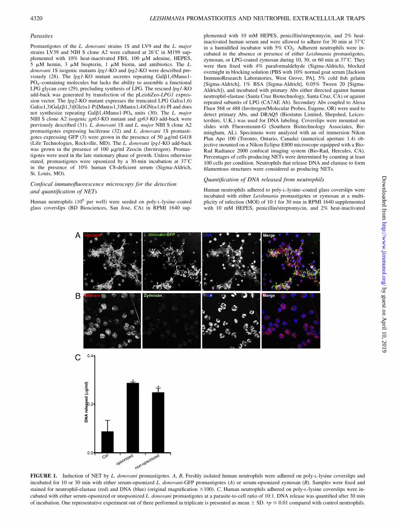
FIGURE 1. Induction of NET by L. donovani promastigotes. A, B, Freshly isolated human neutrophils were adhered on poly-L-lysine coverslips and
incubated for 10 or 30 min with either serum-opsonized L. donovani-GFP promastigotes (A) or serum-opsonized zymosan (B). Samples were fixed and
stained for neutrophil-elastase (red) and DNA (blue) (original magnification 3100). C, Human neutrophils adhered on poly-L-lysine coverslips were in-
cubated with either serum-opsonized or unopsonized L. donovani promastigotes at a parasite-to-cell ratio of 10:1. DNA release was quantified after 30 min
of incubation. One representative experiment out of three performed in triplicate is presented as mean 6 SD. pp # 0.01 compared with control neutrophils.
4320 LEISHMANIA PROMASTIGOTES AND NEUTROPHIL EXTRACELLULAR TRAPS
by guest on April 10, 2019
http://ww
w.jim
munol.org/
Dow
nloaded from
human serum at 37˚C in a humidified incubator with 5% CO2. For neu-trophils in suspension, cells were distributed in a 48-well plates in the sameculture medium (detailed above) and were incubated with either zymosanor L. donovani LV9 promastigotes at a MOI of 10:1 for 120 min at 37˚C ina humidified incubator with 5% CO2. Neutrophils in suspension weregently resuspended every 10 min. After incubation, 1 U/ml micrococcalnuclease (Worthington Biochemical, Lakewood, NJ) in the presence of 1mM Ca2+ was added for 1 h at 37˚C. The nuclease activity was stoppedwith 5 mM EDTA, and samples were collected. Released DNA wasquantified using the Quant-iT PicoGreen dsDNA Assay Kit (Invitrogen/Molecular Probes). Samples were distributed into 96-well plates and wereread in a spectrofluorometer reader (SpectraMax M5; Molecular Devices,Sunnyvale, CA) with a filter setting of 480 nm (excitation)/520 nm(emission).
Phagocytosis assay in suspension
Human neutrophils (4 3 106 per well) were incubated with serum-opsonized zymosan or with serum-opsonized L. donovani LV9 promasti-gotes at a MOI of 3:1 in a 48-well plate in RPMI 1640 supplemented with10 mM HEPES, penicillin/streptomycin, and 2% heat-inactivated humanserum at 37˚C in a humidified incubator with 5% CO2. Cells were gentlyresuspended every 10 min. After 30 min of incubation, cells were washedwith PBS and cytospinned. Cytocentrifuged slides were stained with Diff-Quick, and phagocytosed particles were counted by light microscopy. Eachcondition was analyzed in triplicate, and at least 100 cells were scored persample. Results were expressed as the number of ingested particles per 100neutrophils.
Effect of NETs on Leishmania promastigote survival
Human neutrophils were adhered to poly-L-lysine–coated coverslips (BDBiosciences) in RPMI 1640 supplemented with 10 mM HEPES, penicillin/streptomycin, and 2% heat-inactivated human serum. Cells pretreated or notwith DNase-1 (100 U/ml; Worthington Biochemical) were incubated with
luciferase-expressing L. donovani 1S or L. major NIH S clone A2 pro-mastigotes during 6 h in an incubator at 37˚C with 5% CO2. Controlsconsisted of promastigotes without neutrophils incubated in the absence orpresence of DNase-1. Luciferase activity was measured using a luciferaseassay system (Promega, Madison, WI). Briefly, after 6 h of incubation, cellswere lysed with 13 Cell Culture Lysis Reagent (Promega, Madison, WI)containing 23 protease inhibitor (Roche, Laval, Canada). Luciferase ac-tivity was quantified using a Lumat LB 9507 luminometer (EG&GBerthold,Nashua, NH). Leishmania promastigote survival in the presence of humanneutrophils was determined as the percentage of control values (luciferase-expressing Leishmania promastigotes incubated without neutrophils inpresence or not of DNase-1).
Influence of ROS on the induction of NETs by Leishmaniapromastigotes
Human neutrophils adhered to poly-L-lysine–coated were incubated withthe NADPH oxidase inhibitor diphenyleneiodonium (DPI) (5 or 10 mM) orcatalase (1000 or 2000 U) for 30 min. Leishmania promastigotes were thenadded, and after 30 min DNA release was quantified.
Incubation of neutrophils with Leishmania promastigotesupernatant
Human neutrophils adhered to poly-L-lysine–coated coverslips were incu-bated in the absence or presence of supernatants from L. donovani pro-mastigotes in the stationary phase of growth, and after 30 min DNA releasewas quantified.
Statistical analyses
Each experiment was performed at least with three different blood donors.Comparisons of the means within and between groups were tested with theStudent t test to determine statistical significance. A p value of #0.05 wasconsidered significant.
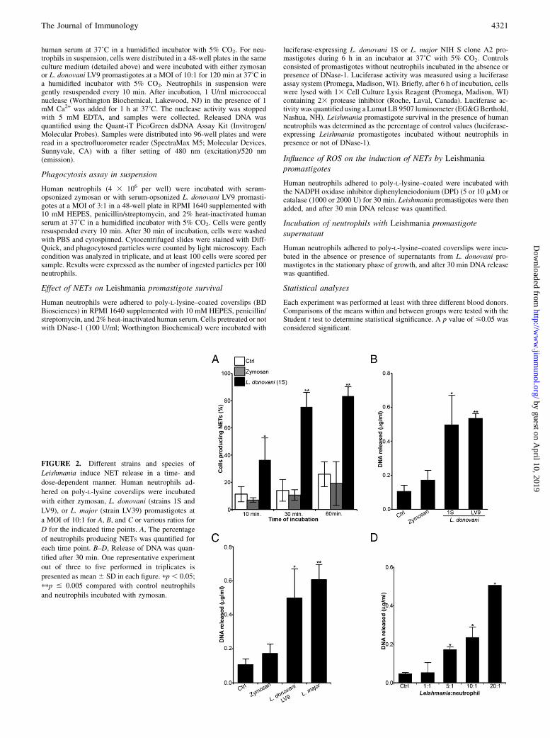
FIGURE 2. Different strains and species of
Leishmania induce NET release in a time- and
dose-dependent manner. Human neutrophils ad-
hered on poly-L-lysine coverslips were incubated
with either zymosan, L. donovani (strains 1S and
LV9), or L. major (strain LV39) promastigotes at
a MOI of 10:1 for A, B, and C or various ratios for
D for the indicated time points. A, The percentage
of neutrophils producing NETs was quantified for
each time point. B–D, Release of DNA was quan-
tified after 30 min. One representative experiment
out of three to five performed in triplicates is
presented as mean 6 SD in each figure. pp , 0.05;
ppp # 0.005 compared with control neutrophils
and neutrophils incubated with zymosan.
The Journal of Immunology 4321
by guest on April 10, 2019
http://ww
w.jim
munol.org/
Dow
nloaded from
ResultsLeishmania promastigotes induce the formation of NETs
To study one aspect of the interaction between Leishmania pro-mastigotes and human neutrophils, we assessed the potential ofthese parasites to induce the formation of NETs. Human neu-trophils were incubated for various time points with serum-opsonized L. donovani-GFP promastigotes (strain 1S), and thepresence of NETwas determined by confocal immunofluorescencemicroscopy. As early as 10 min after the addition of L. donovani-GFP promastigotes, neutrophils lost their typical rounded mor-phology and released NETs, which appear as filamentous structuresidentified by the presence of DNA and elastase (Fig. 1A). NET re-lease appears to be contact-dependent, because we observed thesestructures only where neutrophils were associated with L. donovani-GFP promastigotes (Fig. 1A). In contrast, the levels of NET inducedby serum-opsonized zymosan (Fig. 1B) were similar to the sponta-neous baseline of NET release observed with resting neutrophils. Weobserved no differences between serum-opsonized and unopsonizedpromastigotes for their ability to induce the formation of NETs, asassessed by quantification of DNA released by neutrophils (Fig. 1C).NET formation was time-dependent, with .80% of neutrophils
having released DNA and elastase after 1 h of contact with L.donovani-GFP promastigotes (Fig. 2A). We also assessed the abilityof other Leishmania strains and species to induce the formation ofNETs by quantifying the amount of DNA released by neutrophils30 min after the initial contact with the parasites. As shown in Fig. 2Band 2C, promastigotes from two strains of L. donovani (Sudanese 1Sand Ethiopian LV9) and the L. major strain LV39 induced compa-rable levels of DNA release. Confocal immunofluorescence micros-copy confirmed that these Leishmania strains and species inducedthe formation of NETs (data not shown). Using various ratios ofL. donovani LV9 promastigotes per neutrophil, we observed that therelease of neutrophil DNA was dose-dependent, with a 4-fold in-crease above baseline levels for a MOI of 5:1 and a 10-fold increaseabove baseline levels at aMOI of 20:1 (Fig. 2D). Similar results wereobtained with promastigotes of L. donovani 1S and L. major LV39(data not shown).
Leishmania promastigotes are trapped by NETs
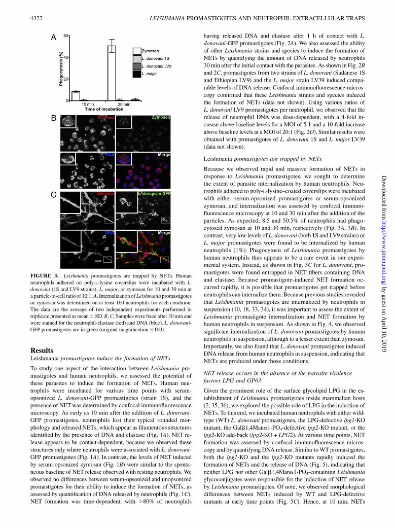
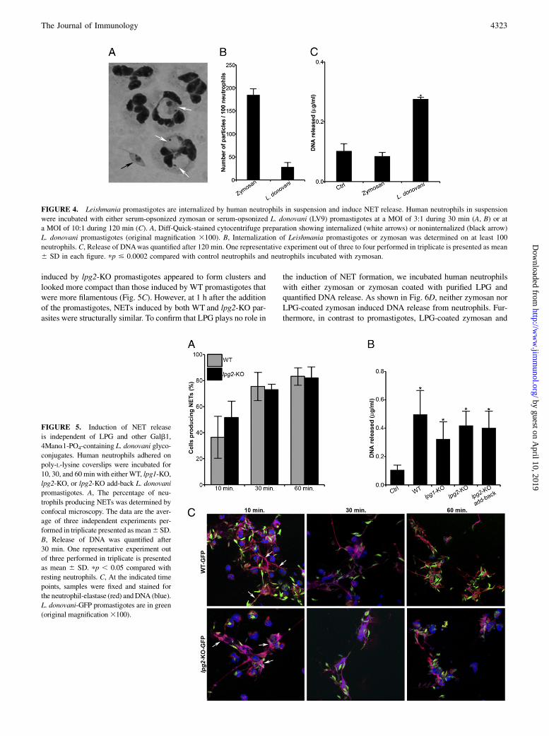
Because we observed rapid and massive formation of NETs inresponse to Leishmania promastigotes, we sought to determinethe extent of parasite internalization by human neutrophils. Neu-trophils adhered to poly-L-lysine–coated coverslips were incubatedwith either serum-opsonized promastigotes or serum-opsonizedzymosan, and internalization was assessed by confocal immuno-fluorescence microscopy at 10 and 30 min after the addition of theparticles. As expected, 8.5 and 50.5% of neutrophils had phago-cytosed zymosan at 10 and 30 min, respectively (Fig. 3A, 3B). Incontrast, very low levels of L. donovani (both 1S and LV9 strains) orL. major promastigotes were found to be internalized by humanneutrophils (1%). Phagocytosis of Leishmania promastigotes byhuman neutrophils thus appears to be a rare event in our experi-mental system. Instead, as shown in Fig. 3C for L. donovani, pro-mastigotes were found entrapped in NET fibers containing DNAand elastase. Because promastigote-induced NET formation oc-curred rapidly, it is possible that promastigotes get trapped beforeneutrophils can internalize them. Because previous studies revealedthat Leishmania promastigotes are internalized by neutrophils insuspension (10, 18, 33, 34), it was important to assess the extent ofLeishmania promastigote internalization and NET formation byhuman neutrophils in suspension. As shown in Fig. 4, we observedsignificant internalization of L. donovani promastigotes by humanneutrophils in suspension, although to a lesser extent than zymosan.Importantly, we also found that L. donovani promastigotes inducedDNA release from human neutrophils in suspension, indicating thatNETs are produced under those conditions.
NET release occurs in the absence of the parasite virulencefactors LPG and GP63
Given the prominent role of the surface glycolipid LPG in the es-tablishment of Leishmania promastigotes inside mammalian hosts(2, 35, 36), we explored the possible role of LPG in the induction ofNETs. To this end, we incubated human neutrophils with either wild-type (WT) L. donovani promastigotes, the LPG-defective lpg1-KOmutant, the Galb1,4Mana1-PO4-defective lpg2-KO mutant, or thelpg2-KO add-back (lpg2-KO + LPG2). At various time points, NETformation was assessed by confocal immunofluorescence micros-copy and by quantifying DNA release. Similar toWT promastigotes,both the lpg1-KO and the lpg2-KO mutants rapidly induced theformation of NETs and the release of DNA (Fig. 5), indicating thatneither LPG nor other Galb1,4Mana1-PO4-containing Leishmaniaglycoconjugates were responsible for the induction of NET releaseby Leishmania promastigotes. Of note, we observed morphologicaldifferences between NETs induced by WT and LPG-defectivemutants at early time points (Fig. 5C). Hence, at 10 min, NETs
FIGURE 3. Leishmania promastigotes are trapped by NETs. Human
neutrophils adhered on poly-L-lysine coverslips were incubated with L.
donovani (1S and LV9 strains), L. major, or zymosan for 10 and 30 min at
a particle-to-cell ratio of 10:1.A, Internalization ofLeishmania promastigotes
or zymosan was determined on at least 100 neutrophils for each condition.
The data are the average of two independent experiments performed in
triplicate presented asmean6 SD.B,C, Samples were fixed after 30min and
were stained for the neutrophil-elastase (red) and DNA (blue). L. donovani-
GFP promastigotes are in green (original magnification3100).
4322 LEISHMANIA PROMASTIGOTES AND NEUTROPHIL EXTRACELLULAR TRAPS
by guest on April 10, 2019
http://ww
w.jim
munol.org/
Dow
nloaded from
induced by lpg2-KO promastigotes appeared to form clusters andlooked more compact than those induced by WT promastigotes thatwere more filamentous (Fig. 5C). However, at 1 h after the additionof the promastigotes, NETs induced by both WT and lpg2-KO par-asites were structurally similar. To confirm that LPG plays no role in
the induction of NET formation, we incubated human neutrophilswith either zymosan or zymosan coated with purified LPG andquantified DNA release. As shown in Fig. 6D, neither zymosan norLPG-coated zymosan induced DNA release from neutrophils. Fur-thermore, in contrast to promastigotes, LPG-coated zymosan and
FIGURE 5. Induction of NET release
is independent of LPG and other Galb1,
4Mana1-PO4-containing L. donovani glyco-
conjugates. Human neutrophils adhered on
poly-L-lysine coverslips were incubated for
10, 30, and 60 min with eitherWT, lpg1-KO,
lpg2-KO, or lpg2-KO add-back L. donovani
promastigotes. A, The percentage of neu-
trophils producing NETs was determined by
confocal microscopy. The data are the aver-
age of three independent experiments per-
formed in triplicate presented asmean6 SD.
B, Release of DNA was quantified after
30 min. One representative experiment out
of three performed in triplicate is presented
as mean 6 SD. pp , 0.05 compared with
resting neutrophils. C, At the indicated time
points, samples were fixed and stained for
the neutrophil-elastase (red) andDNA (blue).
L. donovani-GFP promastigotes are in green
(original magnification3100).
FIGURE 4. Leishmania promastigotes are internalized by human neutrophils in suspension and induce NET release. Human neutrophils in suspension
were incubated with either serum-opsonized zymosan or serum-opsonized L. donovani (LV9) promastigotes at a MOI of 3:1 during 30 min (A, B) or at
a MOI of 10:1 during 120 min (C). A, Diff-Quick-stained cytocentrifuge preparation showing internalized (white arrows) or noninternalized (black arrow)
L. donovani promastigotes (original magnification 3100). B, Internalization of Leishmania promastigotes or zymosan was determined on at least 100
neutrophils. C, Release of DNAwas quantified after 120 min. One representative experiment out of three to four performed in triplicate is presented as mean
6 SD in each figure. pp # 0.0002 compared with control neutrophils and neutrophils incubated with zymosan.
The Journal of Immunology 4323
by guest on April 10, 2019
http://ww
w.jim
munol.org/
Dow
nloaded from
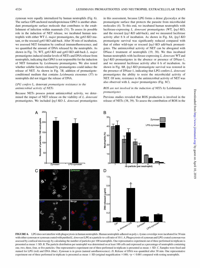
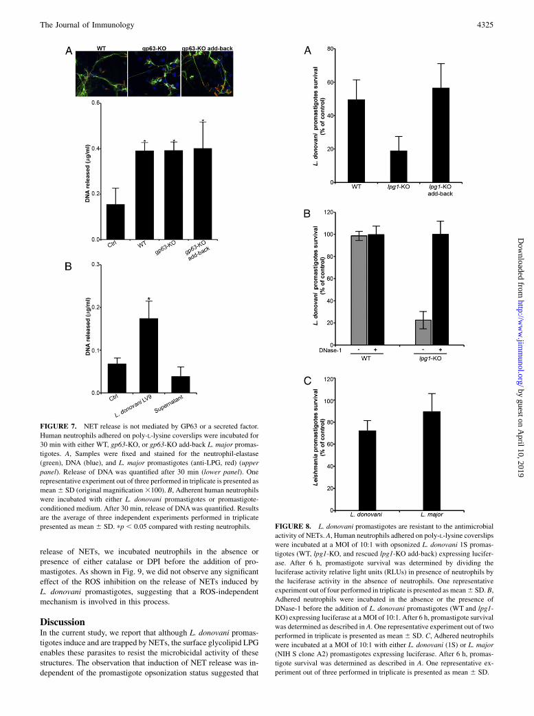
zymosan were equally internalized by human neutrophils (Fig. 6).The surface GPI-anchored metalloproteinase GP63 is another abun-dant promastigote surface molecule that contributes to the estab-lishment of infection within mammals (31). To assess its possiblerole in the induction of NET release, we incubated human neu-trophils with either WT L. major promastigotes, the gp63-KO mu-tant, or the rescued gp63-KO add-back. After 30 min of incubation,we assessed NET formation by confocal immunofluorescence, andwe quantified the amount of DNA released by the neutrophils. Asshown in Fig. 7A, WT, gp63-KO and gp63-KO add-back L. majorpromastigotes induced similar levels of NETs and DNA release fromneutrophils, indicating that GP63 is not responsible for the inductionof NET formation by Leishmania promastigotes. We also testedwhether soluble factors released by promastigotes could induce therelease of NET. As shown in Fig. 7B, addition of promastigote-conditioned medium that contains Leishmania exosomes (37) toneutrophils did not trigger the release of DNA.
LPG confers L. donovani promastigote resistance to theantimicrobial activity of NETs
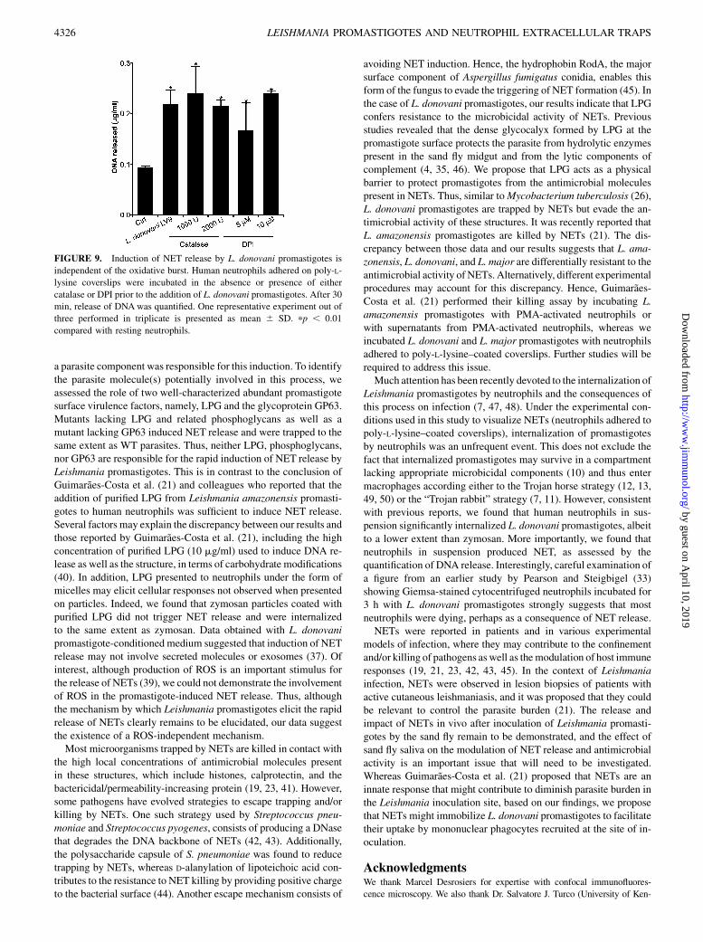
Because NETs possess potent antimicrobial activity, we deter-mined the impact of NET release on the viability of L. donovanipromastigotes. We included lpg1-KO L. donovani promastigotes
in this assessment, because LPG forms a dense glycocalyx at thepromastigote surface that protects the parasite from microbicidalmolecules (4). To this end, we incubated human neutrophils withluciferase-expressing L. donovani promastigotes (WT, lpg1-KO,and the rescued lpg1-KO add-back), and we measured luciferaseactivity after 6 h of incubation. As shown in Fig. 8A, lpg1-KOpromastigote survival was significantly reduced compared withthat of either wild-type or rescued lpg1-KO add-back promasti-gotes. The antimicrobial activity of NET can be abrogated withDNase-1 treatment of neutrophils (19, 38). We thus incubatedhuman neutrophils with luciferase-expressing L. donovaniWTandlpg1-KO promastigotes in the absence or presence of DNase-1,and we measured luciferase activity after 6 h of incubation. Asshown in Fig. 8B, lpg1-KO promastigote survival was restored inthe presence of DNase-1, indicating that LPG confers L. donovanipromastigotes the ability to resist the microbicidal activity ofNET. Of note, resistance to the antimicrobial activity of NET wasalso observed with L. major promastigotes (Fig. 8C).
ROS are not involved in the induction of NETs by Leishmaniapromastigotes
Previous studies revealed that ROS production is involved in therelease of NETs (38, 39). To assess the contribution of ROS in the
FIGURE 6. LPGdoes not interferewith phagocytosis in human neutrophils. Human neutrophils adhered on poly-L-lysine coverslips were incubated for 30min
with either zymosan or zymosan coatedwith purifiedL. donovaniLPGat a particle-to-cell ratio of 10:1.A, Phagocytosis of zymosan andLPG-coated zymosanwas
assessed by confocal microscopy by calculating the number of particles per 100 neutrophils. One representative experiment out of three performed in triplicate is
presented as mean6 SD. B, The particle distribution per neutrophil was determined on at least 100 cells and expressed as a percentage of neutrophils containing
one, two, three, four, or five particles. One representative experiment out of three performed in triplicate is presented as mean6 SD. C, Samples were fixed and
stained for LPG (red) and DNA (blue). Zymosan is in green (natural autofluorescence). D, Release of DNA was quantified after 30 min. One representative
experiment out of three performed in triplicate is presented as mean6 SD (original magnification3100). pp, 0.001 compared with resting neutrophils.
4324 LEISHMANIA PROMASTIGOTES AND NEUTROPHIL EXTRACELLULAR TRAPS
by guest on April 10, 2019
http://ww
w.jim
munol.org/
Dow
nloaded from
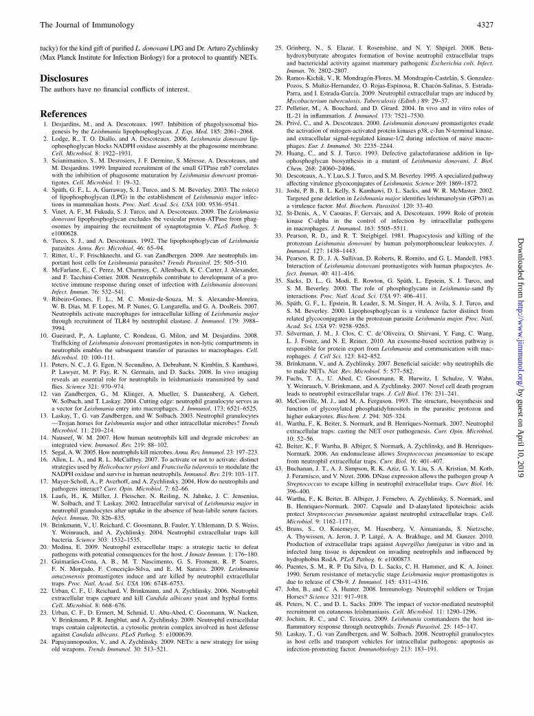
release of NETs, we incubated neutrophils in the absence orpresence of either catalase or DPI before the addition of pro-mastigotes. As shown in Fig. 9, we did not observe any significanteffect of the ROS inhibition on the release of NETs induced byL. donovani promastigotes, suggesting that a ROS-independentmechanism is involved in this process.
DiscussionIn the current study, we report that although L. donovani promas-tigotes induce and are trapped by NETs, the surface glycolipid LPGenables these parasites to resist the microbicidal activity of thesestructures. The observation that induction of NET release was in-dependent of the promastigote opsonization status suggested that
FIGURE 7. NET release is not mediated by GP63 or a secreted factor.
Human neutrophils adhered on poly-L-lysine coverslips were incubated for
30 min with either WT, gp63-KO, or gp63-KO add-back L. major promas-
tigotes. A, Samples were fixed and stained for the neutrophil-elastase
(green), DNA (blue), and L. major promastigotes (anti-LPG, red) (upper
panel). Release of DNA was quantified after 30 min (lower panel). One
representative experiment out of three performed in triplicate is presented as
mean6 SD (original magnification3100). B, Adherent human neutrophils
were incubated with either L. donovani promastigotes or promastigote-
conditioned medium. After 30 min, release of DNAwas quantified. Results
are the average of three independent experiments performed in triplicate
presented as mean 6 SD. pp , 0.05 compared with resting neutrophils. FIGURE 8. L. donovani promastigotes are resistant to the antimicrobial
activity of NETs. A, Human neutrophils adhered on poly-L-lysine coverslips
were incubated at a MOI of 10:1 with opsonized L. donovani 1S promas-
tigotes (WT, lpg1-KO, and rescued lpg1-KO add-back) expressing lucifer-
ase. After 6 h, promastigote survival was determined by dividing the
luciferase activity relative light units (RLUs) in presence of neutrophils by
the luciferase activity in the absence of neutrophils. One representative
experiment out of four performed in triplicate is presented as mean6 SD. B,
Adhered neutrophils were incubated in the absence or the presence of
DNase-1 before the addition of L. donovani promastigotes (WT and lpg1-
KO) expressing luciferase at a MOI of 10:1. After 6 h, promastigote survival
was determined as described in A. One representative experiment out of two
performed in triplicate is presented as mean 6 SD. C, Adhered neutrophils
were incubated at a MOI of 10:1 with either L. donovani (1S) or L. major
(NIH S clone A2) promastigotes expressing luciferase. After 6 h, promas-
tigote survival was determined as described in A. One representative ex-
periment out of three performed in triplicate is presented as mean 6 SD.
The Journal of Immunology 4325
by guest on April 10, 2019
http://ww
w.jim
munol.org/
Dow
nloaded from
a parasite component was responsible for this induction. To identifythe parasite molecule(s) potentially involved in this process, weassessed the role of two well-characterized abundant promastigotesurface virulence factors, namely, LPG and the glycoprotein GP63.Mutants lacking LPG and related phosphoglycans as well as amutant lacking GP63 induced NET release and were trapped to thesame extent as WT parasites. Thus, neither LPG, phosphoglycans,nor GP63 are responsible for the rapid induction of NET release byLeishmania promastigotes. This is in contrast to the conclusion ofGuimaraes-Costa et al. (21) and colleagues who reported that theaddition of purified LPG from Leishmania amazonensis promasti-gotes to human neutrophils was sufficient to induce NET release.Several factors may explain the discrepancy between our results andthose reported by Guimaraes-Costa et al. (21), including the highconcentration of purified LPG (10 mg/ml) used to induce DNA re-lease as well as the structure, in terms of carbohydrate modifications(40). In addition, LPG presented to neutrophils under the form ofmicelles may elicit cellular responses not observed when presentedon particles. Indeed, we found that zymosan particles coated withpurified LPG did not trigger NET release and were internalizedto the same extent as zymosan. Data obtained with L. donovanipromastigote-conditioned medium suggested that induction of NETrelease may not involve secreted molecules or exosomes (37). Ofinterest, although production of ROS is an important stimulus forthe release of NETs (39), we could not demonstrate the involvementof ROS in the promastigote-induced NET release. Thus, althoughthe mechanism by which Leishmania promastigotes elicit the rapidrelease of NETs clearly remains to be elucidated, our data suggestthe existence of a ROS-independent mechanism.Most microorganisms trapped by NETs are killed in contact with
the high local concentrations of antimicrobial molecules presentin these structures, which include histones, calprotectin, and thebactericidal/permeability-increasing protein (19, 23, 41). However,some pathogens have evolved strategies to escape trapping and/orkilling by NETs. One such strategy used by Streptococcus pneu-moniae and Streptococcus pyogenes, consists of producing a DNasethat degrades the DNA backbone of NETs (42, 43). Additionally,the polysaccharide capsule of S. pneumoniae was found to reducetrapping by NETs, whereas D-alanylation of lipoteichoic acid con-tributes to the resistance to NET killing by providing positive chargeto the bacterial surface (44). Another escape mechanism consists of
avoiding NET induction. Hence, the hydrophobin RodA, the majorsurface component of Aspergillus fumigatus conidia, enables thisform of the fungus to evade the triggering of NET formation (45). Inthe case of L. donovani promastigotes, our results indicate that LPGconfers resistance to the microbicidal activity of NETs. Previousstudies revealed that the dense glycocalyx formed by LPG at thepromastigote surface protects the parasite from hydrolytic enzymespresent in the sand fly midgut and from the lytic components ofcomplement (4, 35, 46). We propose that LPG acts as a physicalbarrier to protect promastigotes from the antimicrobial moleculespresent in NETs. Thus, similar toMycobacterium tuberculosis (26),L. donovani promastigotes are trapped by NETs but evade the an-timicrobial activity of these structures. It was recently reported thatL. amazonensis promastigotes are killed by NETs (21). The dis-crepancy between those data and our results suggests that L. ama-zonensis, L. donovani, and L. major are differentially resistant to theantimicrobial activity of NETs. Alternatively, different experimentalprocedures may account for this discrepancy. Hence, Guimaraes-Costa et al. (21) performed their killing assay by incubating L.amazonensis promastigotes with PMA-activated neutrophils orwith supernatants from PMA-activated neutrophils, whereas weincubated L. donovani and L. major promastigotes with neutrophilsadhered to poly-L-lysine–coated coverslips. Further studies will berequired to address this issue.Much attention has been recently devoted to the internalization of
Leishmania promastigotes by neutrophils and the consequences ofthis process on infection (7, 47, 48). Under the experimental con-ditions used in this study to visualize NETs (neutrophils adhered topoly-L-lysine–coated coverslips), internalization of promastigotesby neutrophils was an unfrequent event. This does not exclude thefact that internalized promastigotes may survive in a compartmentlacking appropriate microbicidal components (10) and thus entermacrophages according either to the Trojan horse strategy (12, 13,49, 50) or the “Trojan rabbit” strategy (7, 11). However, consistentwith previous reports, we found that human neutrophils in sus-pension significantly internalized L. donovani promastigotes, albeitto a lower extent than zymosan. More importantly, we found thatneutrophils in suspension produced NET, as assessed by thequantification of DNA release. Interestingly, careful examination ofa figure from an earlier study by Pearson and Steigbigel (33)showing Giemsa-stained cytocentrifuged neutrophils incubated for3 h with L. donovani promastigotes strongly suggests that mostneutrophils were dying, perhaps as a consequence of NET release.NETs were reported in patients and in various experimental
models of infection, where they may contribute to the confinementand/or killing of pathogens aswell as themodulation of host immuneresponses (19, 21, 23, 42, 43, 45). In the context of Leishmaniainfection, NETs were observed in lesion biopsies of patients withactive cutaneous leishmaniasis, and it was proposed that they couldbe relevant to control the parasite burden (21). The release andimpact of NETs in vivo after inoculation of Leishmania promasti-gotes by the sand fly remain to be demonstrated, and the effect ofsand fly saliva on the modulation of NET release and antimicrobialactivity is an important issue that will need to be investigated.Whereas Guimaraes-Costa et al. (21) proposed that NETs are aninnate response that might contribute to diminish parasite burden inthe Leishmania inoculation site, based on our findings, we proposethat NETs might immobilize L. donovani promastigotes to facilitatetheir uptake by mononuclear phagocytes recruited at the site of in-oculation.
AcknowledgmentsWe thank Marcel Desrosiers for expertise with confocal immunofluores-
cence microscopy. We also thank Dr. Salvatore J. Turco (University of Ken-
FIGURE 9. Induction of NET release by L. donovani promastigotes is
independent of the oxidative burst. Human neutrophils adhered on poly-L-
lysine coverslips were incubated in the absence or presence of either
catalase or DPI prior to the addition of L. donovani promastigotes. After 30
min, release of DNAwas quantified. One representative experiment out of
three performed in triplicate is presented as mean 6 SD. pp , 0.01
compared with resting neutrophils.
4326 LEISHMANIA PROMASTIGOTES AND NEUTROPHIL EXTRACELLULAR TRAPS
by guest on April 10, 2019
http://ww
w.jim
munol.org/
Dow
nloaded from
tucky) for the kind gift of purified L. donovani LPG and Dr. Arturo Zychlinsky
(Max Planck Institute for Infection Biology) for a protocol to quantify NETs.
DisclosuresThe authors have no financial conflicts of interest.
References1. Desjardins, M., and A. Descoteaux. 1997. Inhibition of phagolysosomal bio-
genesis by the Leishmania lipophosphoglycan. J. Exp. Med. 185: 2061–2068.2. Lodge, R., T. O. Diallo, and A. Descoteaux. 2006. Leishmania donovani lip-
ophosphoglycan blocks NADPH oxidase assembly at the phagosome membrane.Cell. Microbiol. 8: 1922–1931.
3. Scianimanico, S., M. Desrosiers, J. F. Dermine, S. Meresse, A. Descoteaux, andM. Desjardins. 1999. Impaired recruitment of the small GTPase rab7 correlateswith the inhibition of phagosome maturation by Leishmania donovani promas-tigotes. Cell. Microbiol. 1: 19–32.
4. Spath, G. F., L. A. Garraway, S. J. Turco, and S. M. Beverley. 2003. The role(s)of lipophosphoglycan (LPG) in the establishment of Leishmania major infec-tions in mammalian hosts. Proc. Natl. Acad. Sci. USA 100: 9536–9541.
5. Vinet, A. F., M. Fukuda, S. J. Turco, and A. Descoteaux. 2009. The Leishmaniadonovani lipophosphoglycan excludes the vesicular proton-ATPase from phag-osomes by impairing the recruitment of synaptotagmin V. PLoS Pathog. 5:e1000628.
6. Turco, S. J., and A. Descoteaux. 1992. The lipophosphoglycan of Leishmaniaparasites. Annu. Rev. Microbiol. 46: 65–94.
7. Ritter, U., F. Frischknecht, and G. van Zandbergen. 2009. Are neutrophils im-portant host cells for Leishmania parasites? Trends Parasitol. 25: 505–510.
8. McFarlane, E., C. Perez, M. Charmoy, C. Allenbach, K. C. Carter, J. Alexander,and F. Tacchini-Cottier. 2008. Neutrophils contribute to development of a pro-tective immune response during onset of infection with Leishmania donovani.Infect. Immun. 76: 532–541.
9. Ribeiro-Gomes, F. L., M. C. Moniz-de-Souza, M. S. Alexandre-Moreira,W. B. Dias, M. F. Lopes, M. P. Nunes, G. Lungarella, and G. A. DosReis. 2007.Neutrophils activate macrophages for intracellular killing of Leishmania majorthrough recruitment of TLR4 by neutrophil elastase. J. Immunol. 179: 3988–3994.
10. Gueirard, P., A. Laplante, C. Rondeau, G. Milon, and M. Desjardins. 2008.Trafficking of Leishmania donovani promastigotes in non-lytic compartments inneutrophils enables the subsequent transfer of parasites to macrophages. Cell.Microbiol. 10: 100–111.
11. Peters, N. C., J. G. Egen, N. Secundino, A. Debrabant, N. Kimblin, S. Kamhawi,P. Lawyer, M. P. Fay, R. N. Germain, and D. Sacks. 2008. In vivo imagingreveals an essential role for neutrophils in leishmaniasis transmitted by sandflies. Science 321: 970–974.
12. van Zandbergen, G., M. Klinger, A. Mueller, S. Dannenberg, A. Gebert,W. Solbach, and T. Laskay. 2004. Cutting edge: neutrophil granulocyte serves asa vector for Leishmania entry into macrophages. J. Immunol. 173: 6521–6525.
13. Laskay, T., G. van Zandbergen, and W. Solbach. 2003. Neutrophil granulocytes—Trojan horses for Leishmania major and other intracellular microbes? TrendsMicrobiol. 11: 210–214.
14. Nauseef, W. M. 2007. How human neutrophils kill and degrade microbes: anintegrated view. Immunol. Rev. 219: 88–102.
15. Segal, A.W. 2005. How neutrophils kill microbes.Annu. Rev. Immunol. 23: 197–223.16. Allen, L. A., and R. L. McCaffrey. 2007. To activate or not to activate: distinct
strategies used by Helicobacter pylori and Francisella tularensis to modulate theNADPH oxidase and survive in human neutrophils. Immunol. Rev. 219: 103–117.
17. Mayer-Scholl, A., P. Averhoff, and A. Zychlinsky. 2004. How do neutrophils andpathogens interact? Curr. Opin. Microbiol. 7: 62–66.
18. Laufs, H., K. Muller, J. Fleischer, N. Reiling, N. Jahnke, J. C. Jensenius,W. Solbach, and T. Laskay. 2002. Intracellular survival of Leishmania major inneutrophil granulocytes after uptake in the absence of heat-labile serum factors.Infect. Immun. 70: 826–835.
19. Brinkmann, V., U. Reichard, C. Goosmann, B. Fauler, Y. Uhlemann, D. S. Weiss,Y. Weinrauch, and A. Zychlinsky. 2004. Neutrophil extracellular traps killbacteria. Science 303: 1532–1535.
20. Medina, E. 2009. Neutrophil extracellular traps: a strategic tactic to defeatpathogens with potential consequences for the host. J Innate Immun. 1: 176–180.
21. Guimaraes-Costa, A. B., M. T. Nascimento, G. S. Froment, R. P. Soares,F. N. Morgado, F. Conceicao-Silva, and E. M. Saraiva. 2009. Leishmaniaamazonensis promastigotes induce and are killed by neutrophil extracellulartraps. Proc. Natl. Acad. Sci. USA 106: 6748–6753.
22. Urban, C. F., U. Reichard, V. Brinkmann, and A. Zychlinsky. 2006. Neutrophilextracellular traps capture and kill Candida albicans yeast and hyphal forms.Cell. Microbiol. 8: 668–676.
23. Urban, C. F., D. Ermert, M. Schmid, U. Abu-Abed, C. Goosmann, W. Nacken,V. Brinkmann, P. R. Jungblut, and A. Zychlinsky. 2009. Neutrophil extracellulartraps contain calprotectin, a cytosolic protein complex involved in host defenseagainst Candida albicans. PLoS Pathog. 5: e1000639.
24. Papayannopoulos, V., and A. Zychlinsky. 2009. NETs: a new strategy for usingold weapons. Trends Immunol. 30: 513–521.
25. Grinberg, N., S. Elazar, I. Rosenshine, and N. Y. Shpigel. 2008. Beta-hydroxybutyrate abrogates formation of bovine neutrophil extracellular trapsand bactericidal activity against mammary pathogenic Escherichia coli. Infect.Immun. 76: 2802–2807.
26. Ramos-Kichik, V., R. Mondragon-Flores, M. Mondragon-Castelan, S. Gonzalez-Pozos, S. Muniz-Hernandez, O. Rojas-Espinosa, R. Chacon-Salinas, S. Estrada-Parra, and I. Estrada-Garcıa. 2009. Neutrophil extracellular traps are induced byMycobacterium tuberculosis. Tuberculosis (Edinb.) 89: 29–37.
27. Pelletier, M., A. Bouchard, and D. Girard. 2004. In vivo and in vitro roles ofIL-21 in inflammation. J. Immunol. 173: 7521–7530.
28. Prive, C., and A. Descoteaux. 2000. Leishmania donovani promastigotes evadethe activation of mitogen-activated protein kinases p38, c-Jun N-terminal kinase,and extracellular signal-regulated kinase-1/2 during infection of naive macro-phages. Eur. J. Immunol. 30: 2235–2244.
29. Huang, C., and S. J. Turco. 1993. Defective galactofuranose addition in lip-ophosphoglycan biosynthesis in a mutant of Leishmania donovani. J. Biol.Chem. 268: 24060–24066.
30. Descoteaux, A., Y. Luo, S. J. Turco, and S.M.Beverley. 1995.A specialized pathwayaffecting virulence glycoconjugates of Leishmania. Science 269: 1869–1872.
31. Joshi, P. B., B. L. Kelly, S. Kamhawi, D. L. Sacks, and W. R. McMaster. 2002.Targeted gene deletion in Leishmania major identifies leishmanolysin (GP63) asa virulence factor. Mol. Biochem. Parasitol. 120: 33–40.
32. St-Denis, A., V. Caouras, F. Gervais, and A. Descoteaux. 1999. Role of proteinkinase C-alpha in the control of infection by intracellular pathogensin macrophages. J. Immunol. 163: 5505–5511.
33. Pearson, R. D., and R. T. Steigbigel. 1981. Phagocytosis and killing of theprotozoan Leishmania donovani by human polymorphonuclear leukocytes. J.Immunol. 127: 1438–1443.
34. Pearson, R. D., J. A. Sullivan, D. Roberts, R. Romito, and G. L. Mandell. 1983.Interaction of Leishmania donovani promastigotes with human phagocytes. In-fect. Immun. 40: 411–416.
35. Sacks, D. L., G. Modi, E. Rowton, G. Spath, L. Epstein, S. J. Turco, andS. M. Beverley. 2000. The role of phosphoglycans in Leishmania-sand flyinteractions. Proc. Natl. Acad. Sci. USA 97: 406–411.
36. Spath, G. F., L. Epstein, B. Leader, S. M. Singer, H. A. Avila, S. J. Turco, andS. M. Beverley. 2000. Lipophosphoglycan is a virulence factor distinct fromrelated glycoconjugates in the protozoan parasite Leishmania major. Proc. Natl.Acad. Sci. USA 97: 9258–9263.
37. Silverman, J. M., J. Clos, C. C. de’Oliveira, O. Shirvani, Y. Fang, C. Wang,L. J. Foster, and N. E. Reiner. 2010. An exosome-based secretion pathway isresponsible for protein export from Leishmania and communication with mac-rophages. J. Cell Sci. 123: 842–852.
38. Brinkmann, V., and A. Zychlinsky. 2007. Beneficial suicide: why neutrophils dieto make NETs. Nat. Rev. Microbiol. 5: 577–582.
39. Fuchs, T. A., U. Abed, C. Goosmann, R. Hurwitz, I. Schulze, V. Wahn,Y. Weinrauch, V. Brinkmann, and A. Zychlinsky. 2007. Novel cell death programleads to neutrophil extracellular traps. J. Cell Biol. 176: 231–241.
40. McConville, M. J., and M. A. Ferguson. 1993. The structure, biosynthesis andfunction of glycosylated phosphatidylinositols in the parasitic protozoa andhigher eukaryotes. Biochem. J. 294: 305–324.
41. Wartha, F., K. Beiter, S. Normark, and B. Henriques-Normark. 2007. Neutrophilextracellular traps: casting the NET over pathogenesis. Curr. Opin. Microbiol.10: 52–56.
42. Beiter, K., F. Wartha, B. Albiger, S. Normark, A. Zychlinsky, and B. Henriques-Normark. 2006. An endonuclease allows Streptococcus pneumoniae to escapefrom neutrophil extracellular traps. Curr. Biol. 16: 401–407.
43. Buchanan, J. T., A. J. Simpson, R. K. Aziz, G. Y. Liu, S. A. Kristian, M. Kotb,J. Feramisco, and V. Nizet. 2006. DNase expression allows the pathogen group AStreptococcus to escape killing in neutrophil extracellular traps. Curr. Biol. 16:396–400.
44. Wartha, F., K. Beiter, B. Albiger, J. Fernebro, A. Zychlinsky, S. Normark, andB. Henriques-Normark. 2007. Capsule and D-alanylated lipoteichoic acidsprotect Streptococcus pneumoniae against neutrophil extracellular traps. Cell.Microbiol. 9: 1162–1171.
45. Bruns, S., O. Kniemeyer, M. Hasenberg, V. Aimanianda, S. Nietzsche,A. Thywissen, A. Jeron, J. P. Latge, A. A. Brakhage, and M. Gunzer. 2010.Production of extracellular traps against Aspergillus fumigatus in vitro and ininfected lung tissue is dependent on invading neutrophils and influenced byhydrophobin RodA. PLoS Pathog. 6: e1000873.
46. Puentes, S. M., R. P. Da Silva, D. L. Sacks, C. H. Hammer, and K. A. Joiner.1990. Serum resistance of metacyclic stage Leishmania major promastigotes isdue to release of C5b-9. J. Immunol. 145: 4311–4316.
47. John, B., and C. A. Hunter. 2008. Immunology. Neutrophil soldiers or TrojanHorses? Science 321: 917–918.
48. Peters, N. C., and D. L. Sacks. 2009. The impact of vector-mediated neutrophilrecruitment on cutaneous leishmaniasis. Cell. Microbiol. 11: 1290–1296.
49. Jochim, R. C., and C. Teixeira. 2009. Leishmania commandeers the host in-flammatory response through neutrophils. Trends Parasitol. 25: 145–147.
50. Laskay, T., G. van Zandbergen, and W. Solbach. 2008. Neutrophil granulocytesas host cells and transport vehicles for intracellular pathogens: apoptosis asinfection-promoting factor. Immunobiology 213: 183–191.
The Journal of Immunology 4327
by guest on April 10, 2019
http://ww
w.jim
munol.org/
Dow
nloaded from