Embed Size (px)
Citation preview
Evidence that Electromagnetic Radiation is Genotoxic:
The implications for the epidemiology of cancer and cardiac, neurological and reproductive effects
Dr Neil Cherry
2nd March 2001
Extended from a paper to the conference on Possible health effects on health of radiofrequency
electromagnetic fields European Parliament, Brussels.
Environmental Management and Design Division P.O. Box 84
Lincoln University Canterbury, New Zealand
Evidence that Electromagnetic Radiation is Genotoxic: The implications for the epidemiology
of cancer and cardiac, neurological and reproductive effects
Dr Neil Cherry
Lincoln University, New Zealand March 2001
"Our frame of reference determines what we look at and how we look. And as a
consequence, this determines what we find."
Burke J, The Day the Universe Changed, 1985. Introduction: The way we perceive things determines how we make decisions. To move from picturing ourselves as physical beings to biological beings significantly changes our view of health and fitness. Too much exercise and oxygen free radicals really damage our cells. To move from biological to bioelectrical incorporates intelligence and emotion and leads to a radically fundamental change in our way of seeing things that it forms the basis of a new paradigm. Principles of Approach: This paper attempts to follow basic classical scientific principles to counter the dismissive and biased approach of industry and many government and international authorities, including the WHO and ICNIRP. The principles found to be important are that biology reveals that brains, hearts and cells use electromagnetic signals, charged ions, voltage-gated ion channels, ion regulated gap junctions, all of which can be interfered with by external electromagnetic fields in subtle but vital ways in relation to health. A primary physical principle of resonant absorption explains why external and internal signals that share the same part of the spectrum, resonantly exchange energy at levels well below the thermal threshold. This is also true for radio and TV receivers. It involves tuned circuits and resonant absorption. Laboratory experiments provide evidence of effects. Replicated and/or extended studies provided confirmation and establish an effect. Multiple studies confirm and strengthen the cause and effect relationship. In assessing genotoxicity, any evidence of genetic damage, cell death or neoplastic transformation is evidence of genotoxicity. The genetic material is fundamentally the double
2
helix of the DNA molecule. During the cell cycle the helix unwinds and clones itself. They then fold themselves into the set of chromosomes that are so large that they can be seen in powerful microscopes. In the second half of the cell cycle the chromosomes clone themselves so that at mitosis, cell division, each cell has a full set of chromosomes. They then unfold themselves to form the DNA strands. Any substance that damages DNA or chromosomes, or changes genetic activity, is genotoxic because it is acting on the same material, i.e. the DNA molecule. A genotoxic substance is mutagenic, carcinogenic and teratogenic. Strength of evidence for public health has a classical hierarchy that has dose-response relationship at the top and biological mechanism at the bottom, Hill (1965). This is seen by considering Sir Austin Bradford Hill's descriptions of his 'view points' from which the question of cause and effect is being considered. Of dose-response he says: "The simple dose-response curve admits of a simple explanation and obviously puts the case in a clearer light", i.e. cause and effect. Sir Austin considers many other forms of evidence from which cause and effect can be decided in the absence of a dose-response. These include strength of association and consistency, although he points out that the lack of strength and apparent inconsistency, is not necessarily arguments against cause and effect. Of biological mechanism, or plausibility, he states: "It will be helpful if the causation we suspect is biologically plausible. But this is a feature I am convinced we cannot demand. What is biologically plausible depends upon the biological knowledge of the day." Thus biological plausibility has a low status and dose-response has a very high status. When epidemiological evidence is available it should be used to set public health standards, where possible, using the dose-response relationships. In the absence of these, the level of lowest observed effect, with a safety margin to allow for uncertainly, the vulnerable, the size of the population at risk, are appropriate. Dose response relationships for epidemiological studies of cancer are likely to be linear because of the cumulative cell damage/repair/mistake mechanism. At very high levels approaching lethal levels the curve become asymptotic. At very low levels, around the optimum homeostatic levels, curves can become "U" shaped. Thus, with the great sensitivity of the brain the neurological effects at extremely low exposure levels might be curved. EMR Spectrum Principle: It is observed that both biological effects and epidemiological effects appear to be the same or very similar from ELF exposure and from RF/MW exposures, including calcium ion efflux, melatonin reduction, DNA strand breakage, chromosome aberrations, leukaemia, brain cancer, breast cancer, miscarriage and neurological effects.
3
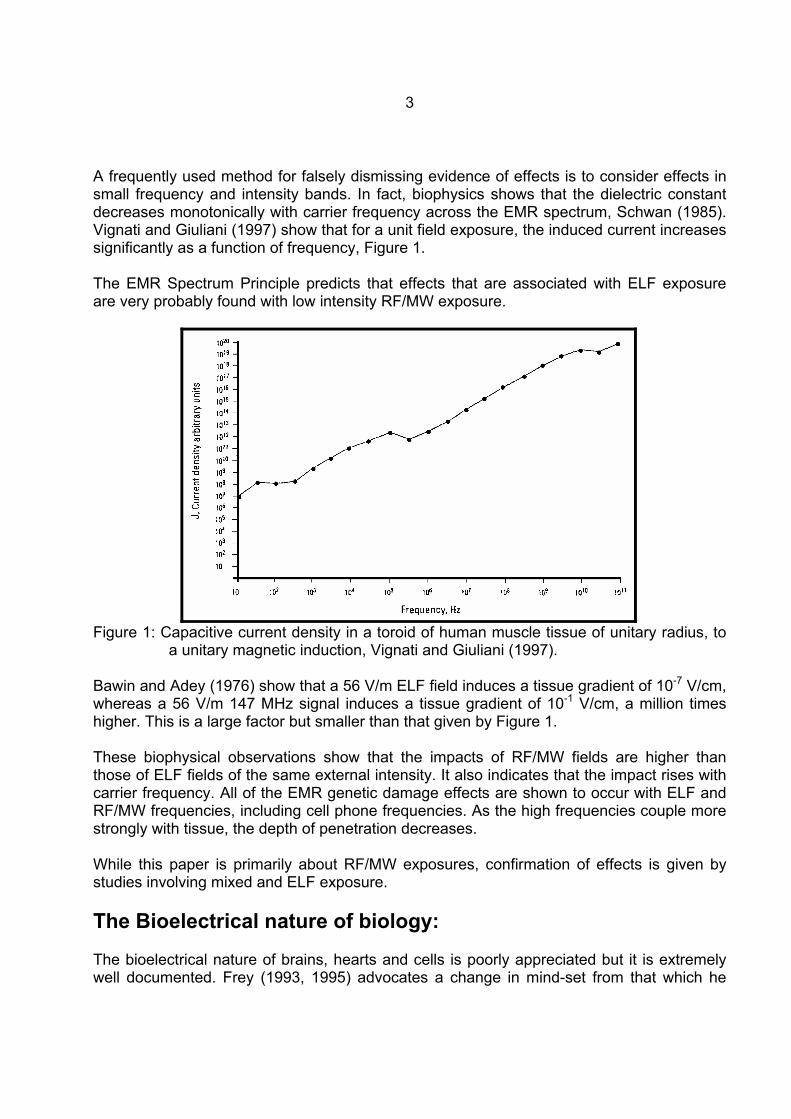
A frequently used method for falsely dismissing evidence of effects is to consider effects in small frequency and intensity bands. In fact, biophysics shows that the dielectric constant decreases monotonically with carrier frequency across the EMR spectrum, Schwan (1985). Vignati and Giuliani (1997) show that for a unit field exposure, the induced current increases significantly as a function of frequency, Figure 1. The EMR Spectrum Principle predicts that effects that are associated with ELF exposure are very probably found with low intensity RF/MW exposure.
Figure 1: Capacitive current density in a toroid of human muscle tissue of unitary radius, to
a unitary magnetic induction, Vignati and Giuliani (1997). Bawin and Adey (1976) show that a 56 V/m ELF field induces a tissue gradient of 10-7 V/cm, whereas a 56 V/m 147 MHz signal induces a tissue gradient of 10-1 V/cm, a million times higher. This is a large factor but smaller than that given by Figure 1. These biophysical observations show that the impacts of RF/MW fields are higher than those of ELF fields of the same external intensity. It also indicates that the impact rises with carrier frequency. All of the EMR genetic damage effects are shown to occur with ELF and RF/MW frequencies, including cell phone frequencies. As the high frequencies couple more strongly with tissue, the depth of penetration decreases. While this paper is primarily about RF/MW exposures, confirmation of effects is given by studies involving mixed and ELF exposure. The Bioelectrical nature of biology: The bioelectrical nature of brains, hearts and cells is poorly appreciated but it is extremely well documented. Frey (1993, 1995) advocates a change in mind-set from that which he
4
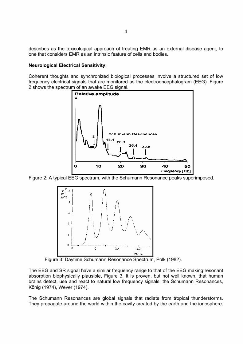
describes as the toxicological approach of treating EMR as an external disease agent, to one that considers EMR as an intrinsic feature of cells and bodies. Neurological Electrical Sensitivity: Coherent thoughts and synchronized biological processes involve a structured set of low frequency electrical signals that are monitored as the electroencephalogram (EEG). Figure 2 shows the spectrum of an awake EEG signal.
Figure 2: A typical EEG spectrum, with the Schumann Resonance peaks superimposed.
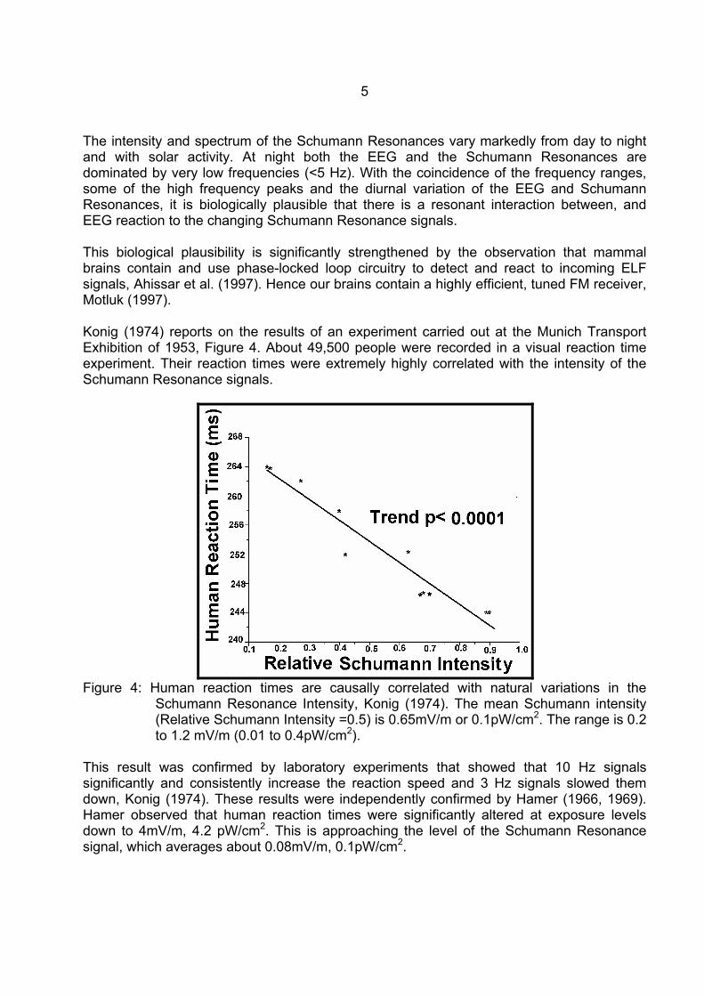
Figure 3: Daytime Schumann Resonance Spectrum, Polk (1982).
The EEG and SR signal have a similar frequency range to that of the EEG making resonant absorption biophysically plausible, Figure 3. It is proven, but not well known, that human brains detect, use and react to natural low frequency signals, the Schumann Resonances, König (1974), Wever (1974). The Schumann Resonances are global signals that radiate from tropical thunderstorms. They propagate around the world within the cavity created by the earth and the ionosphere.
5
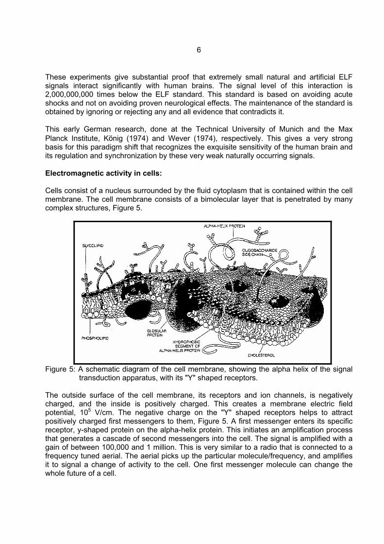
The intensity and spectrum of the Schumann Resonances vary markedly from day to night and with solar activity. At night both the EEG and the Schumann Resonances are dominated by very low frequencies (<5 Hz). With the coincidence of the frequency ranges, some of the high frequency peaks and the diurnal variation of the EEG and Schumann Resonances, it is biologically plausible that there is a resonant interaction between, and EEG reaction to the changing Schumann Resonance signals. This biological plausibility is significantly strengthened by the observation that mammal brains contain and use phase-locked loop circuitry to detect and react to incoming ELF signals, Ahissar et al. (1997). Hence our brains contain a highly efficient, tuned FM receiver, Motluk (1997). Konig (1974) reports on the results of an experiment carried out at the Munich Transport Exhibition of 1953, Figure 4. About 49,500 people were recorded in a visual reaction time experiment. Their reaction times were extremely highly correlated with the intensity of the Schumann Resonance signals.
Figure 4: Human reaction times are causally correlated with natural variations in the
Schumann Resonance Intensity, Konig (1974). The mean Schumann intensity (Relative Schumann Intensity =0.5) is 0.65mV/m or 0.1pW/cm2. The range is 0.2 to 1.2 mV/m (0.01 to 0.4pW/cm2).
This result was confirmed by laboratory experiments that showed that 10 Hz signals significantly and consistently increase the reaction speed and 3 Hz signals slowed them down, Konig (1974). These results were independently confirmed by Hamer (1966, 1969). Hamer observed that human reaction times were significantly altered at exposure levels down to 4mV/m, 4.2 pW/cm2. This is approaching the level of the Schumann Resonance signal, which averages about 0.08mV/m, 0.1pW/cm2.
6
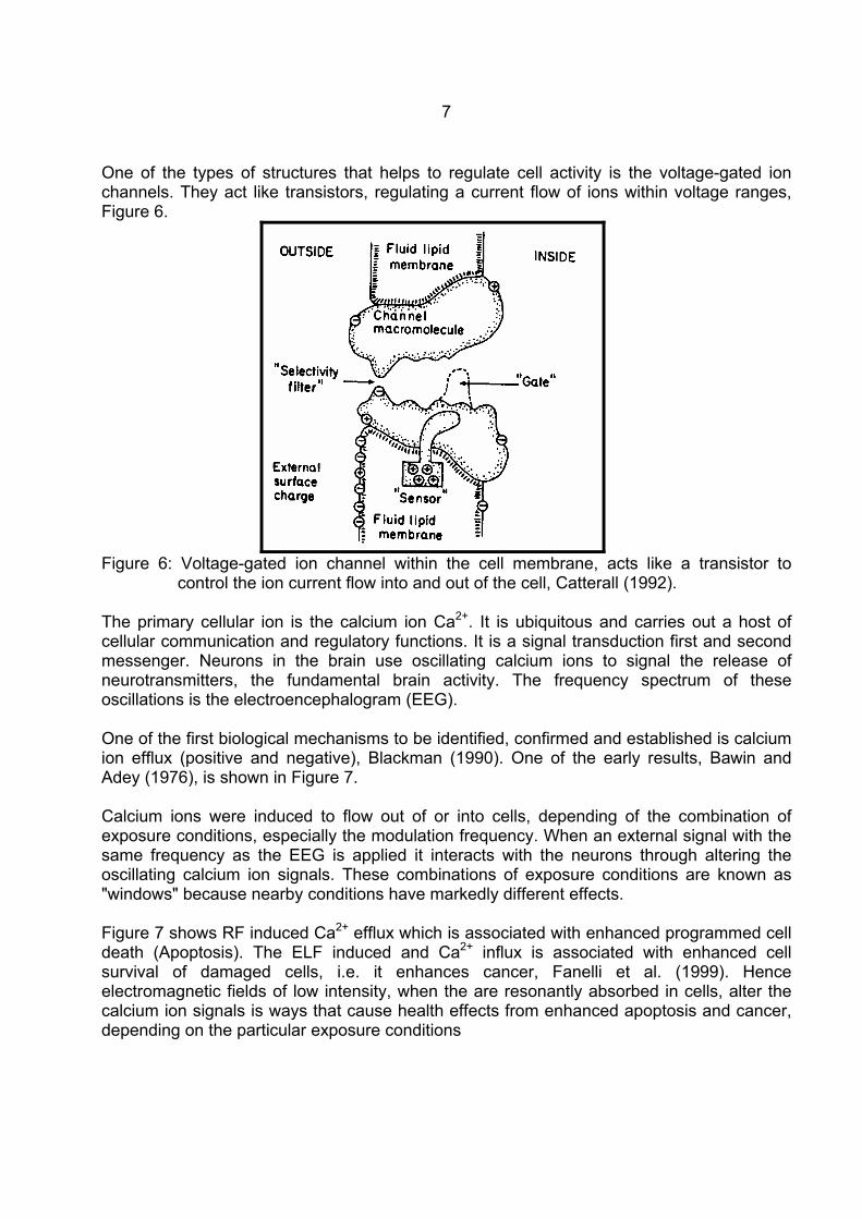
These experiments give substantial proof that extremely small natural and artificial ELF signals interact significantly with human brains. The signal level of this interaction is 2,000,000,000 times below the ELF standard. This standard is based on avoiding acute shocks and not on avoiding proven neurological effects. The maintenance of the standard is obtained by ignoring or rejecting any and all evidence that contradicts it. This early German research, done at the Technical University of Munich and the Max Planck Institute, König (1974) and Wever (1974), respectively. This gives a very strong basis for this paradigm shift that recognizes the exquisite sensitivity of the human brain and its regulation and synchronization by these very weak naturally occurring signals. Electromagnetic activity in cells: Cells consist of a nucleus surrounded by the fluid cytoplasm that is contained within the cell membrane. The cell membrane consists of a bimolecular layer that is penetrated by many complex structures, Figure 5.
Figure 5: A schematic diagram of the cell membrane, showing the alpha helix of the signal
transduction apparatus, with its "Y" shaped receptors. The outside surface of the cell membrane, its receptors and ion channels, is negatively charged, and the inside is positively charged. This creates a membrane electric field potential, 105 V/cm. The negative charge on the "Y" shaped receptors helps to attract positively charged first messengers to them, Figure 5. A first messenger enters its specific receptor, y-shaped protein on the alpha-helix protein. This initiates an amplification process that generates a cascade of second messengers into the cell. The signal is amplified with a gain of between 100,000 and 1 million. This is very similar to a radio that is connected to a frequency tuned aerial. The aerial picks up the particular molecule/frequency, and amplifies it to signal a change of activity to the cell. One first messenger molecule can change the whole future of a cell.
7
One of the types of structures that helps to regulate cell activity is the voltage-gated ion channels. They act like transistors, regulating a current flow of ions within voltage ranges, Figure 6.
Figure 6: Voltage-gated ion channel within the cell membrane, acts like a transistor to
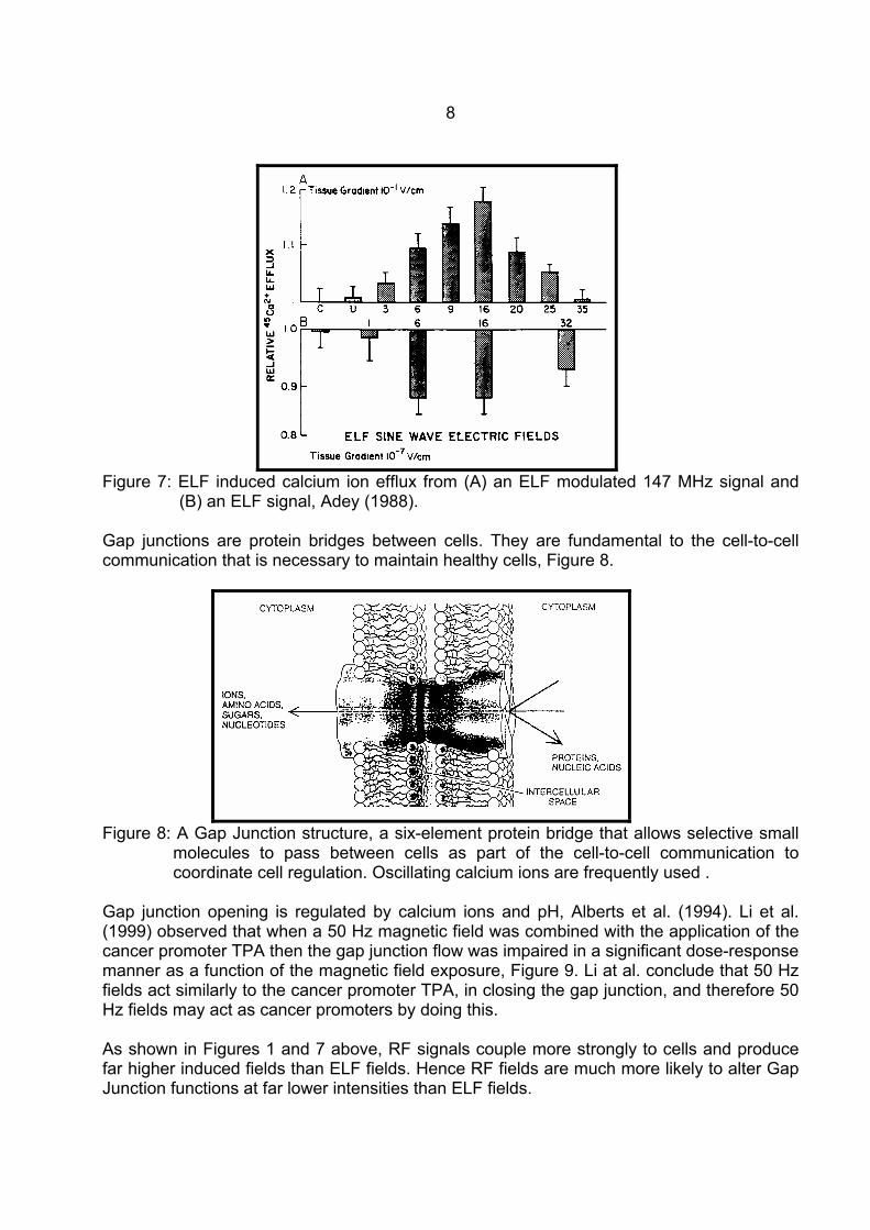
control the ion current flow into and out of the cell, Catterall (1992). The primary cellular ion is the calcium ion Ca2+. It is ubiquitous and carries out a host of cellular communication and regulatory functions. It is a signal transduction first and second messenger. Neurons in the brain use oscillating calcium ions to signal the release of neurotransmitters, the fundamental brain activity. The frequency spectrum of these oscillations is the electroencephalogram (EEG). One of the first biological mechanisms to be identified, confirmed and established is calcium ion efflux (positive and negative), Blackman (1990). One of the early results, Bawin and Adey (1976), is shown in Figure 7. Calcium ions were induced to flow out of or into cells, depending of the combination of exposure conditions, especially the modulation frequency. When an external signal with the same frequency as the EEG is applied it interacts with the neurons through altering the oscillating calcium ion signals. These combinations of exposure conditions are known as "windows" because nearby conditions have markedly different effects. Figure 7 shows RF induced Ca2+ efflux which is associated with enhanced programmed cell death (Apoptosis). The ELF induced and Ca2+ influx is associated with enhanced cell survival of damaged cells, i.e. it enhances cancer, Fanelli et al. (1999). Hence electromagnetic fields of low intensity, when the are resonantly absorbed in cells, alter the calcium ion signals is ways that cause health effects from enhanced apoptosis and cancer, depending on the particular exposure conditions
8
Figure 7: ELF induced calcium ion efflux from (A) an ELF modulated 147 MHz signal and
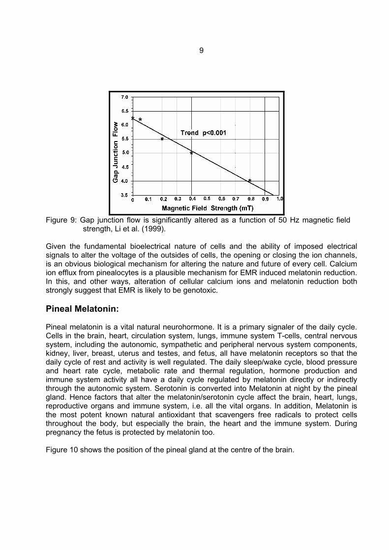
(B) an ELF signal, Adey (1988). Gap junctions are protein bridges between cells. They are fundamental to the cell-to-cell communication that is necessary to maintain healthy cells, Figure 8.
Figure 8: A Gap Junction structure, a six-element protein bridge that allows selective small
molecules to pass between cells as part of the cell-to-cell communication to coordinate cell regulation. Oscillating calcium ions are frequently used .
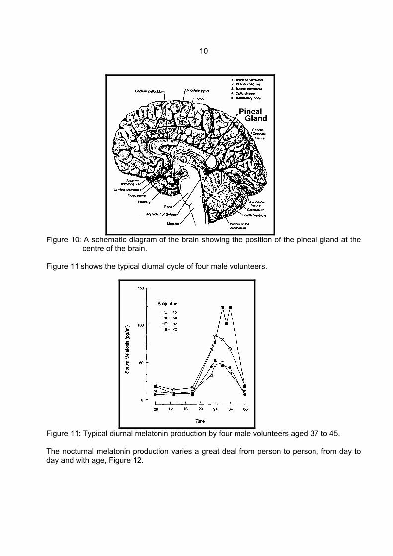
Gap junction opening is regulated by calcium ions and pH, Alberts et al. (1994). Li et al. (1999) observed that when a 50 Hz magnetic field was combined with the application of the cancer promoter TPA then the gap junction flow was impaired in a significant dose-response manner as a function of the magnetic field exposure, Figure 9. Li at al. conclude that 50 Hz fields act similarly to the cancer promoter TPA, in closing the gap junction, and therefore 50 Hz fields may act as cancer promoters by doing this. As shown in Figures 1 and 7 above, RF signals couple more strongly to cells and produce far higher induced fields than ELF fields. Hence RF fields are much more likely to alter Gap Junction functions at far lower intensities than ELF fields.
9
Figure 9: Gap junction flow is significantly altered as a function of 50 Hz magnetic field
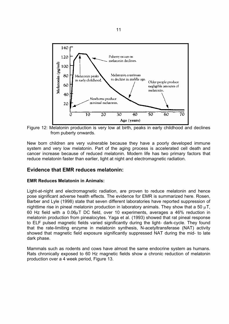
strength, Li et al. (1999). Given the fundamental bioelectrical nature of cells and the ability of imposed electrical signals to alter the voltage of the outsides of cells, the opening or closing the ion channels, is an obvious biological mechanism for altering the nature and future of every cell. Calcium ion efflux from pinealocytes is a plausible mechanism for EMR induced melatonin reduction. In this, and other ways, alteration of cellular calcium ions and melatonin reduction both strongly suggest that EMR is likely to be genotoxic. Pineal Melatonin: Pineal melatonin is a vital natural neurohormone. It is a primary signaler of the daily cycle. Cells in the brain, heart, circulation system, lungs, immune system T-cells, central nervous system, including the autonomic, sympathetic and peripheral nervous system components, kidney, liver, breast, uterus and testes, and fetus, all have melatonin receptors so that the daily cycle of rest and activity is well regulated. The daily sleep/wake cycle, blood pressure and heart rate cycle, metabolic rate and thermal regulation, hormone production and immune system activity all have a daily cycle regulated by melatonin directly or indirectly through the autonomic system. Serotonin is converted into Melatonin at night by the pineal gland. Hence factors that alter the melatonin/serotonin cycle affect the brain, heart, lungs, reproductive organs and immune system, i.e. all the vital organs. In addition, Melatonin is the most potent known natural antioxidant that scavengers free radicals to protect cells throughout the body, but especially the brain, the heart and the immune system. During pregnancy the fetus is protected by melatonin too. Figure 10 shows the position of the pineal gland at the centre of the brain.
10
Figure 10: A schematic diagram of the brain showing the position of the pineal gland at the
centre of the brain. Figure 11 shows the typical diurnal cycle of four male volunteers.
Figure 11: Typical diurnal melatonin production by four male volunteers aged 37 to 45. The nocturnal melatonin production varies a great deal from person to person, from day to day and with age, Figure 12.
11
Figure 12: Melatonin production is very low at birth, peaks in early childhood and declines
from puberty onwards. New born children are very vulnerable because they have a poorly developed immune system and very low melatonin. Part of the aging process is accelerated cell death and cancer increase because of reduced melatonin. Modern life has two primary factors that reduce melatonin faster than earlier, light at night and electromagnetic radiation. Evidence that EMR reduces melatonin: EMR Reduces Melatonin in Animals: Light-at-night and electromagnetic radiation, are proven to reduce melatonin and hence pose significant adverse health effects. The evidence for EMR is summarized here. Rosen, Barber and Lyle (1998) state that seven different laboratories have reported suppression of nighttime rise in pineal melatonin production in laboratory animals. They show that a 50 µT, 60 Hz field with a 0.06µT DC field, over 10 experiments, averages a 46% reduction in melatonin production from pinealocytes. Yaga et al. (1993) showed that rat pineal response to ELF pulsed magnetic fields varied significantly during the light- dark-cycle. They found that the rate-limiting enzyme in melatonin synthesis, N-acetyltransferase (NAT) activity showed that magnetic field exposure significantly suppressed NAT during the mid- to late dark phase. Mammals such as rodents and cows have almost the same endocrine system as humans. Rats chronically exposed to 60 Hz magnetic fields show a chronic reduction of melatonin production over a 4 week period, Figure 13.
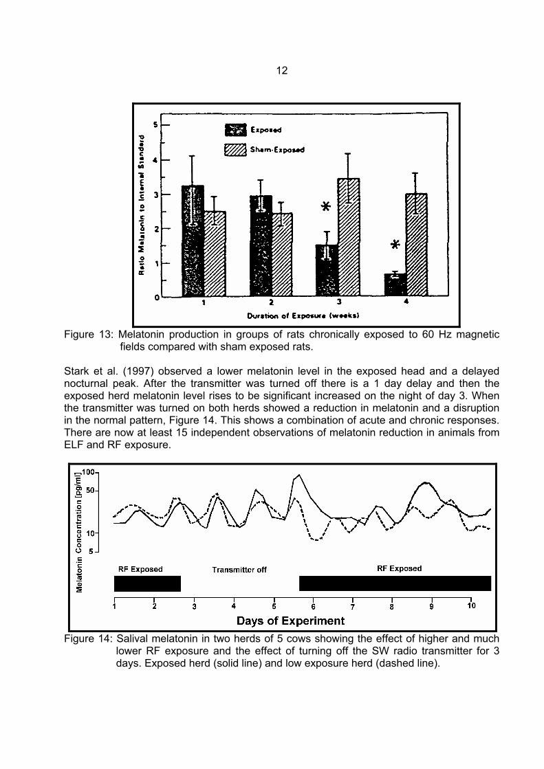
12
Figure 13: Melatonin production in groups of rats chronically exposed to 60 Hz magnetic
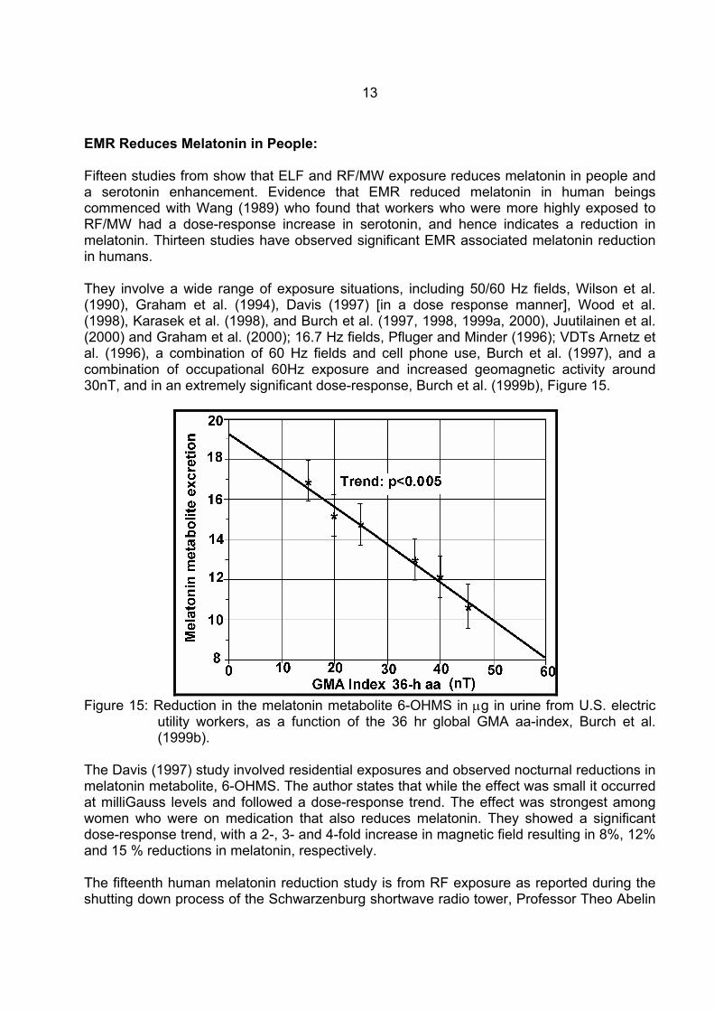
fields compared with sham exposed rats. Stark et al. (1997) observed a lower melatonin level in the exposed head and a delayed nocturnal peak. After the transmitter was turned off there is a 1 day delay and then the exposed herd melatonin level rises to be significant increased on the night of day 3. When the transmitter was turned on both herds showed a reduction in melatonin and a disruption in the normal pattern, Figure 14. This shows a combination of acute and chronic responses. There are now at least 15 independent observations of melatonin reduction in animals from ELF and RF exposure.
Figure 14: Salival melatonin in two herds of 5 cows showing the effect of higher and much
lower RF exposure and the effect of turning off the SW radio transmitter for 3 days. Exposed herd (solid line) and low exposure herd (dashed line).
13
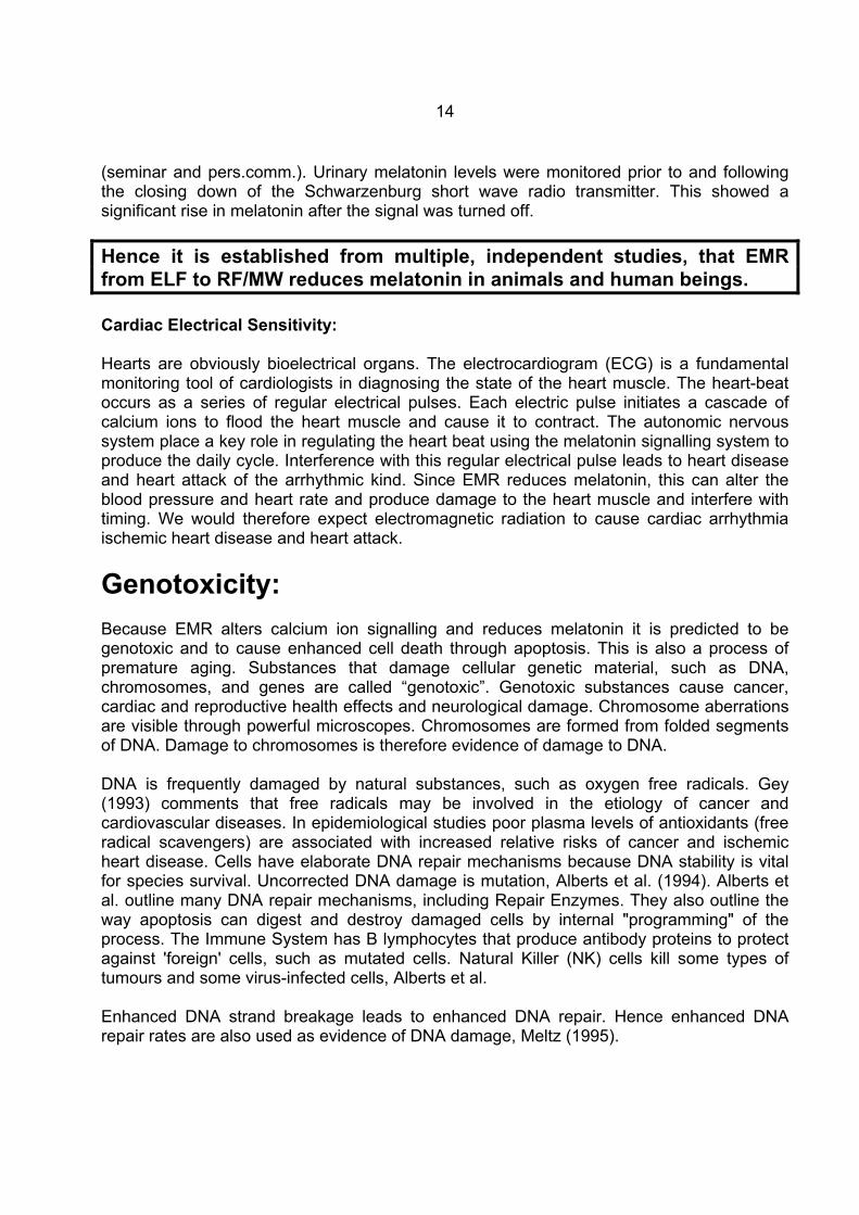
EMR Reduces Melatonin in People: Fifteen studies from show that ELF and RF/MW exposure reduces melatonin in people and a serotonin enhancement. Evidence that EMR reduced melatonin in human beings commenced with Wang (1989) who found that workers who were more highly exposed to RF/MW had a dose-response increase in serotonin, and hence indicates a reduction in melatonin. Thirteen studies have observed significant EMR associated melatonin reduction in humans. They involve a wide range of exposure situations, including 50/60 Hz fields, Wilson et al. (1990), Graham et al. (1994), Davis (1997) [in a dose response manner], Wood et al. (1998), Karasek et al. (1998), and Burch et al. (1997, 1998, 1999a, 2000), Juutilainen et al. (2000) and Graham et al. (2000); 16.7 Hz fields, Pfluger and Minder (1996); VDTs Arnetz et al. (1996), a combination of 60 Hz fields and cell phone use, Burch et al. (1997), and a combination of occupational 60Hz exposure and increased geomagnetic activity around 30nT, and in an extremely significant dose-response, Burch et al. (1999b), Figure 15.
Figure 15: Reduction in the melatonin metabolite 6-OHMS in µg in urine from U.S. electric
utility workers, as a function of the 36 hr global GMA aa-index, Burch et al. (1999b).
The Davis (1997) study involved residential exposures and observed nocturnal reductions in melatonin metabolite, 6-OHMS. The author states that while the effect was small it occurred at milliGauss levels and followed a dose-response trend. The effect was strongest among women who were on medication that also reduces melatonin. They showed a significant dose-response trend, with a 2-, 3- and 4-fold increase in magnetic field resulting in 8%, 12% and 15 % reductions in melatonin, respectively. The fifteenth human melatonin reduction study is from RF exposure as reported during the shutting down process of the Schwarzenburg shortwave radio tower, Professor Theo Abelin
14
(seminar and pers.comm.). Urinary melatonin levels were monitored prior to and following the closing down of the Schwarzenburg short wave radio transmitter. This showed a significant rise in melatonin after the signal was turned off. Hence it is established from multiple, independent studies, that EMR from ELF to RF/MW reduces melatonin in animals and human beings. Cardiac Electrical Sensitivity: Hearts are obviously bioelectrical organs. The electrocardiogram (ECG) is a fundamental monitoring tool of cardiologists in diagnosing the state of the heart muscle. The heart-beat occurs as a series of regular electrical pulses. Each electric pulse initiates a cascade of calcium ions to flood the heart muscle and cause it to contract. The autonomic nervous system place a key role in regulating the heart beat using the melatonin signalling system to produce the daily cycle. Interference with this regular electrical pulse leads to heart disease and heart attack of the arrhythmic kind. Since EMR reduces melatonin, this can alter the blood pressure and heart rate and produce damage to the heart muscle and interfere with timing. We would therefore expect electromagnetic radiation to cause cardiac arrhythmia ischemic heart disease and heart attack.
Genotoxicity: Because EMR alters calcium ion signalling and reduces melatonin it is predicted to be genotoxic and to cause enhanced cell death through apoptosis. This is also a process of premature aging. Substances that damage cellular genetic material, such as DNA, chromosomes, and genes are called “genotoxic”. Genotoxic substances cause cancer, cardiac and reproductive health effects and neurological damage. Chromosome aberrations are visible through powerful microscopes. Chromosomes are formed from folded segments of DNA. Damage to chromosomes is therefore evidence of damage to DNA. DNA is frequently damaged by natural substances, such as oxygen free radicals. Gey (1993) comments that free radicals may be involved in the etiology of cancer and cardiovascular diseases. In epidemiological studies poor plasma levels of antioxidants (free radical scavengers) are associated with increased relative risks of cancer and ischemic heart disease. Cells have elaborate DNA repair mechanisms because DNA stability is vital for species survival. Uncorrected DNA damage is mutation, Alberts et al. (1994). Alberts et al. outline many DNA repair mechanisms, including Repair Enzymes. They also outline the way apoptosis can digest and destroy damaged cells by internal "programming" of the process. The Immune System has B lymphocytes that produce antibody proteins to protect against 'foreign' cells, such as mutated cells. Natural Killer (NK) cells kill some types of tumours and some virus-infected cells, Alberts et al. Enhanced DNA strand breakage leads to enhanced DNA repair. Hence enhanced DNA repair rates are also used as evidence of DNA damage, Meltz (1995).
15
Many studies have shown that radiofrequency/microwave (RF/MW) radiation and extremely low frequency (ELF) fields cause increased DNA strand breakage and chromosome aberrations. This has been shown in cell lines, human blood, animals and living human beings. This means that epidemiological studies of people exposed to electromagnetic radiation (EMR) are likely to show increased cancer, miscarriage and reproductive adverse effects. In fact many epidemiological studies have shown these effects, Goldsmith (1995, 1996, 1997, 1997a), Szmigielski (1991, 1996). Two plausible biological mechanisms involving free radicals are involved in this effect. The first involves increased free radical activity and genetic damage as a response to exposure. The second involves increased free radical activity and genetic damage because of an induced reduction of a free radical scavenger, e.g. reduced melatonin, Reiter (1994). It is clear however, that both mechanisms have the same effect of damaging the DNA and chromosomes. Another established biological mechanism, EMR-induced alteration of cellular calcium ion homeostasis, Blackman (1990), is also involved in cell regulation, cell survival and apoptosis, DNA synthesis and melatonin regulation. Direct measurements of Chromosome aberrations: Direct evidence that EMR induces significant increases in chromosome damage, with significant dose response relationships, is evidence of a causal effect when replicated or extended by independent laboratories. Chromosome damage from RF/MW exposure: The first identified study that showed that pulsed RF radiation cause significant chromosome aberrations was Heller and Teixeira-Pinto (1959). Garlic roots were exposed to 27 MHz pulsed at 80 to 180 Hz. for 5 min and then they were examined 24 hrs later. The concluded that this RF signal mimicked the chromosomal aberration produced by ionizing radiation and c-mitotic substances. No increased temperature was observed. Blood samples were taken from the staff of the U.S. Embassy in Moscow. They had been chronically exposed to a low intensity radar signal. Significant increases in chromosome damage was reported, Tonascia and Tonascia (1966) cited in Goldsmith (1997a). Yao (1982) exposed rat kangaroo RH5 and RH16 cells to 2.45 GHz microwaves, maintaining the temperature at 37°C in the incubator. After 50 passages with microwave exposure there we 30 passages without. Significant chromosome aberrations were measured after 20 MW passages. Yao (1978) also found elevated chromosome damage in Chinese Hamsters. Elevated and significantly elevated (*) chromosome damage with RF/MW exposure has been observed by Manikowska et al. (1979), Berman Carter and House (1980), Nawrot, McRee and Staples (1981*), Kowalczuk, Saunders and Stapleton (1983), Banerjee, Goldfeder and Mitra (1983a,b*), Antipenko, Koveshnikova and Timchenko (1984), Manikowska-Czerska, Czerska and Leach (1985), Beechey et al. (1986), Garaj-Vrhovac et al. (1987) and Saunders et al. (1988). Thus before the end of the 1980's at least 15 studies
16
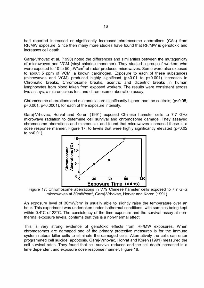
had reported increased or significantly increased chromosome aberrations (CAs) from RF/MW exposure. Since then many more studies have found that RF/MW is genotoxic and increases cell death. Garaj-Vrhovac et al. (1990) noted the differences and similarities between the mutagenicity of microwaves and VCM (vinyl chloride monomer). They studied a group of workers who were exposed to 10 to 50 µW/cm2 of radar produced microwaves. Some were also exposed to about 5 ppm of VCM, a known carcinogen. Exposure to each of these substances (microwaves and VCM) produced highly significant (p<0.01 to p<0.001) increases in Chromatid breaks, Chromosome breaks, acentric and dicentric breaks in human lymphocytes from blood taken from exposed workers. The results were consistent across two assays, a micronucleus test and chromosome aberration assay. Chromosome aberrations and micronuclei are significantly higher than the controls, (p<0.05, p<0.001, p<0.0001), for each of the exposure intensity. Garaj-Vrhovac, Horvat and Koren (1991) exposed Chinese hamster cells to 7.7 GHz microwave radiation to determine cell survival and chromosome damage. They assayed chromosome aberrations and micronuclei and found that microwaves increased these in a dose response manner, Figure 17, to levels that were highly significantly elevated (p<0.02 to p<0.01).
Figure 17: Chromosome aberrations in V79 Chinese hamster cells exposed to 7.7 GHz
microwaves at 30mW/cm2, Garaj-Vrhovac, Horvat and Koren (1991).
An exposure level of 30mW/cm2 is usually able to slightly raise the temperature over an hour. This experiment was undertaken under isothermal conditions, with samples being kept within 0.4°C of 22°C. The consistency of the time exposure and the survival assay at non-thermal exposure levels, confirms that this is a non-thermal effect. This is very strong evidence of genotoxic effects from RF/MW exposures. When chromosomes are damaged one of the primary protective measures is for the immune system natural killer cells to eliminate the damaged cells. Alternatively the cells can enter programmed cell suicide, apoptosis. Garaj-Vrhovac, Horvat and Koren (1991) measured the cell survival rates. They found that cell survival reduced and the cell death increased in a time dependent and exposure dose response manner, Figure 18.
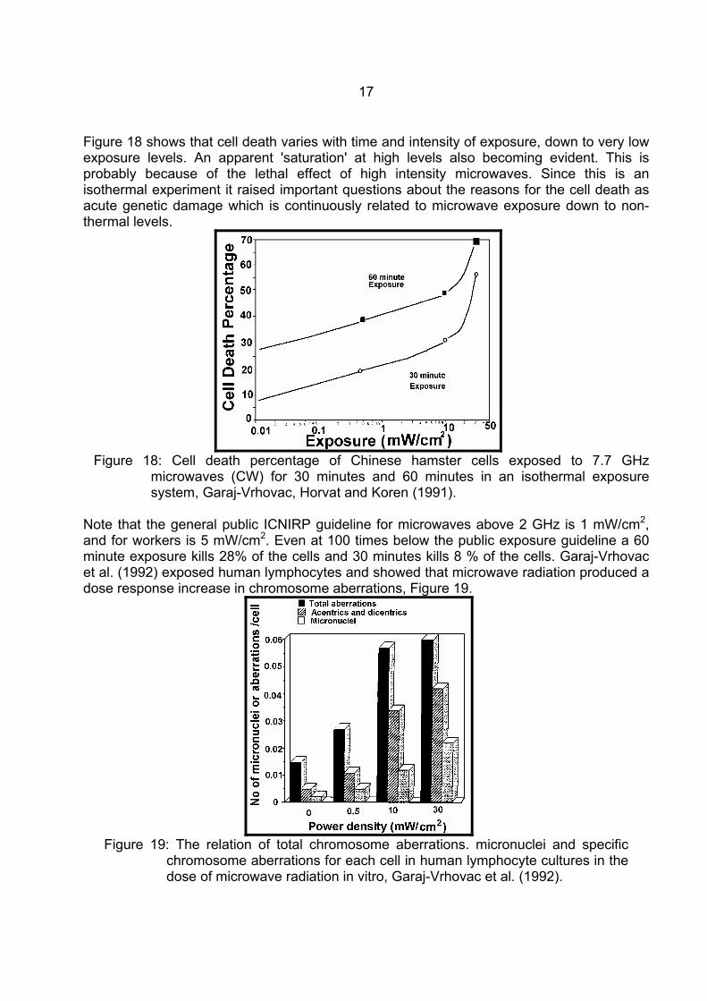
17
Figure 18 shows that cell death varies with time and intensity of exposure, down to very low exposure levels. An apparent 'saturation' at high levels also becoming evident. This is probably because of the lethal effect of high intensity microwaves. Since this is an isothermal experiment it raised important questions about the reasons for the cell death as acute genetic damage which is continuously related to microwave exposure down to non-thermal levels.
Figure 18: Cell death percentage of Chinese hamster cells exposed to 7.7 GHz
microwaves (CW) for 30 minutes and 60 minutes in an isothermal exposure system, Garaj-Vrhovac, Horvat and Koren (1991).
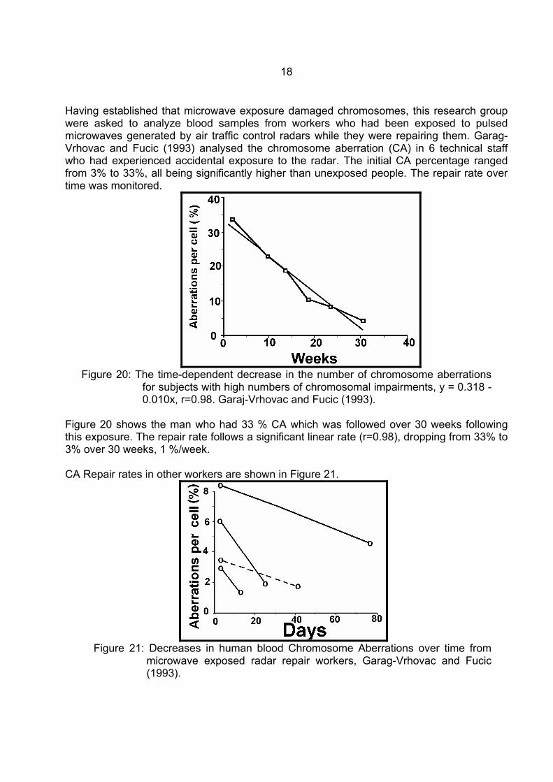
Note that the general public ICNIRP guideline for microwaves above 2 GHz is 1 mW/cm2, and for workers is 5 mW/cm2. Even at 100 times below the public exposure guideline a 60 minute exposure kills 28% of the cells and 30 minutes kills 8 % of the cells. Garaj-Vrhovac et al. (1992) exposed human lymphocytes and showed that microwave radiation produced a dose response increase in chromosome aberrations, Figure 19.
Figure 19: The relation of total chromosome aberrations. micronuclei and specific
chromosome aberrations for each cell in human lymphocyte cultures in the dose of microwave radiation in vitro, Garaj-Vrhovac et al. (1992).
18
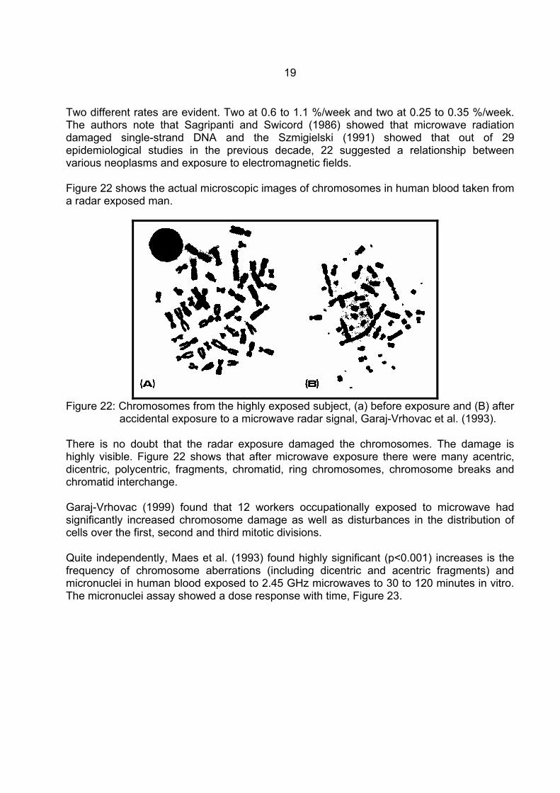
Having established that microwave exposure damaged chromosomes, this research group were asked to analyze blood samples from workers who had been exposed to pulsed microwaves generated by air traffic control radars while they were repairing them. Garag-Vrhovac and Fucic (1993) analysed the chromosome aberration (CA) in 6 technical staff who had experienced accidental exposure to the radar. The initial CA percentage ranged from 3% to 33%, all being significantly higher than unexposed people. The repair rate over time was monitored.
Figure 20: The time-dependent decrease in the number of chromosome aberrations
for subjects with high numbers of chromosomal impairments, y = 0.318 - 0.010x, r=0.98. Garaj-Vrhovac and Fucic (1993).
Figure 20 shows the man who had 33 % CA which was followed over 30 weeks following this exposure. The repair rate follows a significant linear rate (r=0.98), dropping from 33% to 3% over 30 weeks, 1 %/week. CA Repair rates in other workers are shown in Figure 21.
Figure 21: Decreases in human blood Chromosome Aberrations over time from
microwave exposed radar repair workers, Garag-Vrhovac and Fucic (1993).
19
Two different rates are evident. Two at 0.6 to 1.1 %/week and two at 0.25 to 0.35 %/week. The authors note that Sagripanti and Swicord (1986) showed that microwave radiation damaged single-strand DNA and the Szmigielski (1991) showed that out of 29 epidemiological studies in the previous decade, 22 suggested a relationship between various neoplasms and exposure to electromagnetic fields. Figure 22 shows the actual microscopic images of chromosomes in human blood taken from a radar exposed man.
Figure 22: Chromosomes from the highly exposed subject, (a) before exposure and (B) after
accidental exposure to a microwave radar signal, Garaj-Vrhovac et al. (1993). There is no doubt that the radar exposure damaged the chromosomes. The damage is highly visible. Figure 22 shows that after microwave exposure there were many acentric, dicentric, polycentric, fragments, chromatid, ring chromosomes, chromosome breaks and chromatid interchange. Garaj-Vrhovac (1999) found that 12 workers occupationally exposed to microwave had significantly increased chromosome damage as well as disturbances in the distribution of cells over the first, second and third mitotic divisions. Quite independently, Maes et al. (1993) found highly significant (p<0.001) increases is the frequency of chromosome aberrations (including dicentric and acentric fragments) and micronuclei in human blood exposed to 2.45 GHz microwaves to 30 to 120 minutes in vitro. The micronuclei assay showed a dose response with time, Figure 23.
20
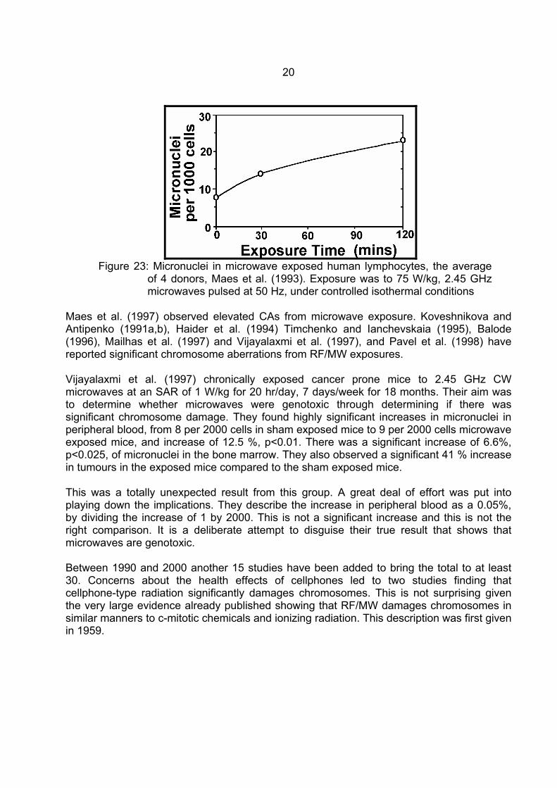
Figure 23: Micronuclei in microwave exposed human lymphocytes, the average
of 4 donors, Maes et al. (1993). Exposure was to 75 W/kg, 2.45 GHz microwaves pulsed at 50 Hz, under controlled isothermal conditions
Maes et al. (1997) observed elevated CAs from microwave exposure. Koveshnikova and Antipenko (1991a,b), Haider et al. (1994) Timchenko and Ianchevskaia (1995), Balode (1996), Mailhas et al. (1997) and Vijayalaxmi et al. (1997), and Pavel et al. (1998) have reported significant chromosome aberrations from RF/MW exposures. Vijayalaxmi et al. (1997) chronically exposed cancer prone mice to 2.45 GHz CW microwaves at an SAR of 1 W/kg for 20 hr/day, 7 days/week for 18 months. Their aim was to determine whether microwaves were genotoxic through determining if there was significant chromosome damage. They found highly significant increases in micronuclei in peripheral blood, from 8 per 2000 cells in sham exposed mice to 9 per 2000 cells microwave exposed mice, and increase of 12.5 %, p<0.01. There was a significant increase of 6.6%, p<0.025, of micronuclei in the bone marrow. They also observed a significant 41 % increase in tumours in the exposed mice compared to the sham exposed mice. This was a totally unexpected result from this group. A great deal of effort was put into playing down the implications. They describe the increase in peripheral blood as a 0.05%, by dividing the increase of 1 by 2000. This is not a significant increase and this is not the right comparison. It is a deliberate attempt to disguise their true result that shows that microwaves are genotoxic. Between 1990 and 2000 another 15 studies have been added to bring the total to at least 30. Concerns about the health effects of cellphones led to two studies finding that cellphone-type radiation significantly damages chromosomes. This is not surprising given the very large evidence already published showing that RF/MW damages chromosomes in similar manners to c-mitotic chemicals and ionizing radiation. This description was first given in 1959.
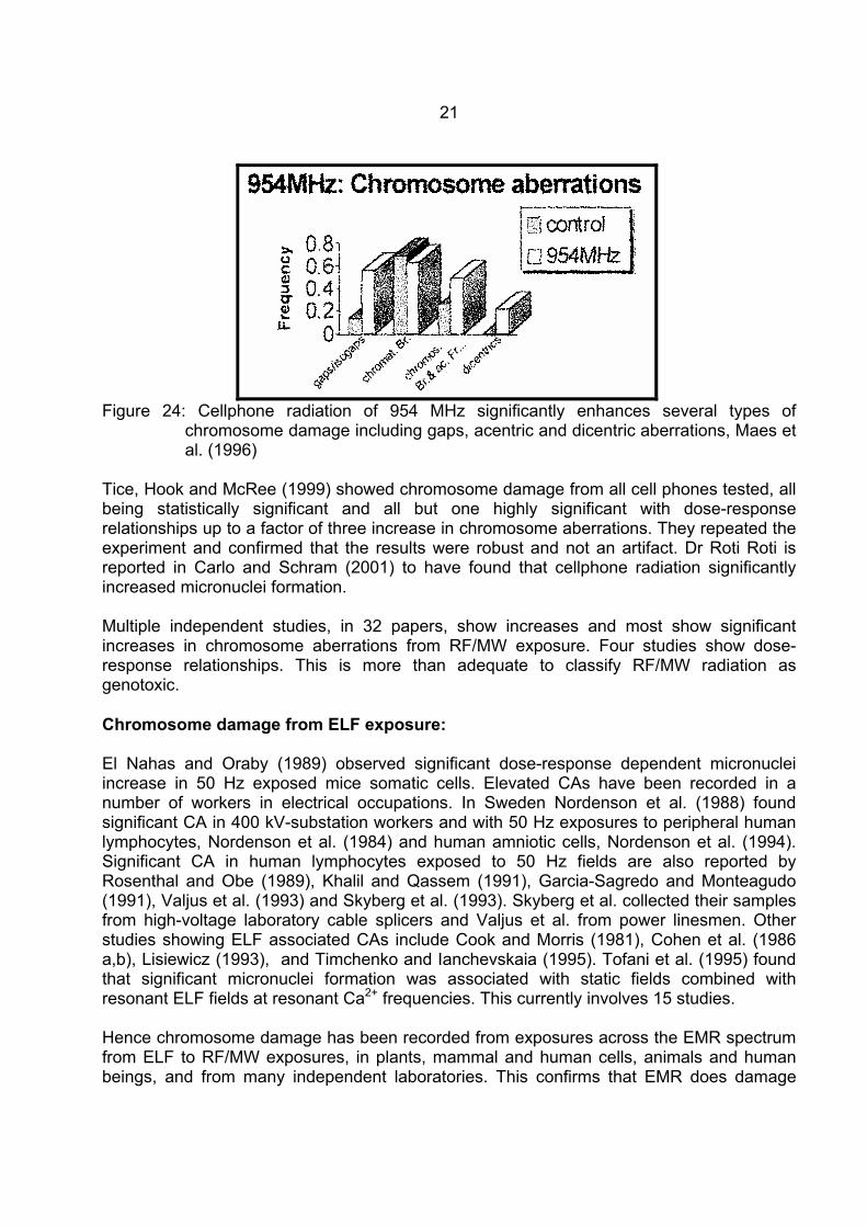
21
Figure 24: Cellphone radiation of 954 MHz significantly enhances several types of
chromosome damage including gaps, acentric and dicentric aberrations, Maes et al. (1996)
Tice, Hook and McRee (1999) showed chromosome damage from all cell phones tested, all being statistically significant and all but one highly significant with dose-response relationships up to a factor of three increase in chromosome aberrations. They repeated the experiment and confirmed that the results were robust and not an artifact. Dr Roti Roti is reported in Carlo and Schram (2001) to have found that cellphone radiation significantly increased micronuclei formation. Multiple independent studies, in 32 papers, show increases and most show significant increases in chromosome aberrations from RF/MW exposure. Four studies show dose-response relationships. This is more than adequate to classify RF/MW radiation as genotoxic. Chromosome damage from ELF exposure: El Nahas and Oraby (1989) observed significant dose-response dependent micronuclei increase in 50 Hz exposed mice somatic cells. Elevated CAs have been recorded in a number of workers in electrical occupations. In Sweden Nordenson et al. (1988) found significant CA in 400 kV-substation workers and with 50 Hz exposures to peripheral human lymphocytes, Nordenson et al. (1984) and human amniotic cells, Nordenson et al. (1994). Significant CA in human lymphocytes exposed to 50 Hz fields are also reported by Rosenthal and Obe (1989), Khalil and Qassem (1991), Garcia-Sagredo and Monteagudo (1991), Valjus et al. (1993) and Skyberg et al. (1993). Skyberg et al. collected their samples from high-voltage laboratory cable splicers and Valjus et al. from power linesmen. Other studies showing ELF associated CAs include Cook and Morris (1981), Cohen et al. (1986 a,b), Lisiewicz (1993), and Timchenko and Ianchevskaia (1995). Tofani et al. (1995) found that significant micronuclei formation was associated with static fields combined with resonant ELF fields at resonant Ca2+ frequencies. This currently involves 15 studies. Hence chromosome damage has been recorded from exposures across the EMR spectrum from ELF to RF/MW exposures, in plants, mammal and human cells, animals and human beings, and from many independent laboratories. This confirms that EMR does damage
22
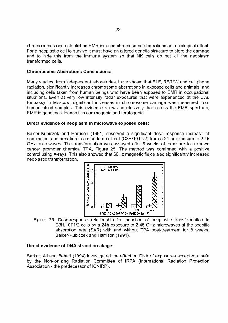
chromosomes and establishes EMR induced chromosome aberrations as a biological effect. For a neoplastic cell to survive it must have an altered genetic structure to store the damage and to hide this from the immune system so that NK cells do not kill the neoplasm transformed cells. Chromosome Aberrations Conclusions: Many studies, from independent laboratories, have shown that ELF, RF/MW and cell phone radiation, significantly increases chromosome aberrations in exposed cells and animals, and including cells taken from human beings who have been exposed to EMR in occupational situations. Even at very low intensity radar exposures that were experienced at the U.S. Embassy in Moscow, significant increases in chromosome damage was measured from human blood samples. This evidence shows conclusively that across the EMR spectrum, EMR is genotoxic. Hence it is carcinogenic and teratogenic. Direct evidence of neoplasm in microwave exposed cells: Balcer-Kubiczek and Harrison (1991) observed a significant dose response increase of neoplastic transformation in a standard cell set (C3H/10T1/2) from a 24 hr exposure to 2.45 GHz microwaves. The transformation was assayed after 8 weeks of exposure to a known cancer promoter chemical TPA, Figure 25. The method was confirmed with a positive control using X-rays. This also showed that 60Hz magnetic fields also significantly increased neoplastic transformation.
Figure 25: Dose-response relationship for induction of neoplastic transformation in
C3H/10T1/2 cells by a 24h exposure to 2.45 GHz microwaves at the specific absorption rate (SAR) with and without TPA post-treatment for 8 weeks, Balcer-Kubiczek and Harrison (1991).
Direct evidence of DNA strand breakage: Sarkar, Ali and Behari (1994) investigated the effect on DNA of exposures accepted a safe by the Non-ionizing Radiation Committee of IRPA (International Radiation Protection Association - the predecessor of ICNIRP).
23
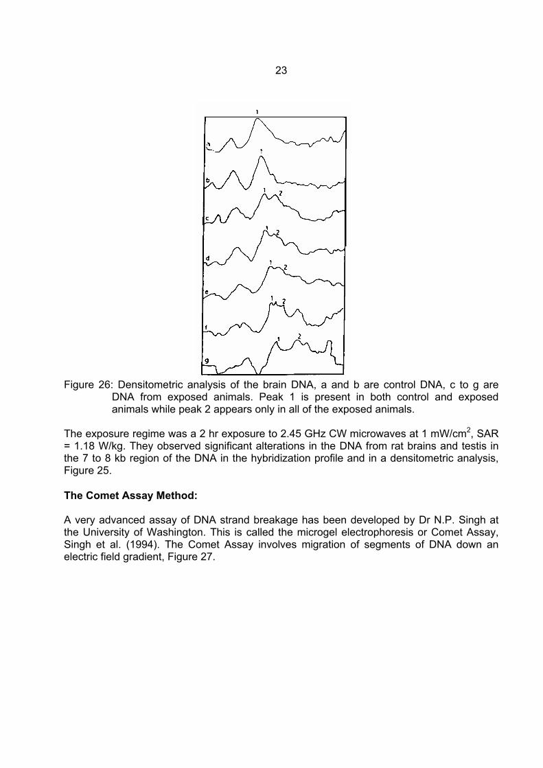
Figure 26: Densitometric analysis of the brain DNA, a and b are control DNA, c to g are
DNA from exposed animals. Peak 1 is present in both control and exposed animals while peak 2 appears only in all of the exposed animals.
The exposure regime was a 2 hr exposure to 2.45 GHz CW microwaves at 1 mW/cm2, SAR = 1.18 W/kg. They observed significant alterations in the DNA from rat brains and testis in the 7 to 8 kb region of the DNA in the hybridization profile and in a densitometric analysis, Figure 25. The Comet Assay Method: A very advanced assay of DNA strand breakage has been developed by Dr N.P. Singh at the University of Washington. This is called the microgel electrophoresis or Comet Assay, Singh et al. (1994). The Comet Assay involves migration of segments of DNA down an electric field gradient, Figure 27.
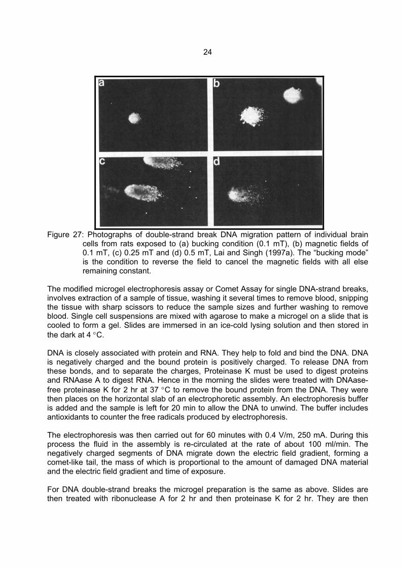
24
Figure 27: Photographs of double-strand break DNA migration pattern of individual brain
cells from rats exposed to (a) bucking condition (0.1 mT), (b) magnetic fields of 0.1 mT, (c) 0.25 mT and (d) 0.5 mT, Lai and Singh (1997a). The “bucking mode” is the condition to reverse the field to cancel the magnetic fields with all else remaining constant.
The modified microgel electrophoresis assay or Comet Assay for single DNA-strand breaks, involves extraction of a sample of tissue, washing it several times to remove blood, snipping the tissue with sharp scissors to reduce the sample sizes and further washing to remove blood. Single cell suspensions are mixed with agarose to make a microgel on a slide that is cooled to form a gel. Slides are immersed in an ice-cold lysing solution and then stored in the dark at 4 °C. DNA is closely associated with protein and RNA. They help to fold and bind the DNA. DNA is negatively charged and the bound protein is positively charged. To release DNA from these bonds, and to separate the charges, Proteinase K must be used to digest proteins and RNAase A to digest RNA. Hence in the morning the slides were treated with DNAase-free proteinase K for 2 hr at 37 °C to remove the bound protein from the DNA. They were then places on the horizontal slab of an electrophoretic assembly. An electrophoresis buffer is added and the sample is left for 20 min to allow the DNA to unwind. The buffer includes antioxidants to counter the free radicals produced by electrophoresis. The electrophoresis was then carried out for 60 minutes with 0.4 V/m, 250 mA. During this process the fluid in the assembly is re-circulated at the rate of about 100 ml/min. The negatively charged segments of DNA migrate down the electric field gradient, forming a comet-like tail, the mass of which is proportional to the amount of damaged DNA material and the electric field gradient and time of exposure. For DNA double-strand breaks the microgel preparation is the same as above. Slides are then treated with ribonuclease A for 2 hr and then proteinase K for 2 hr. They are then
25
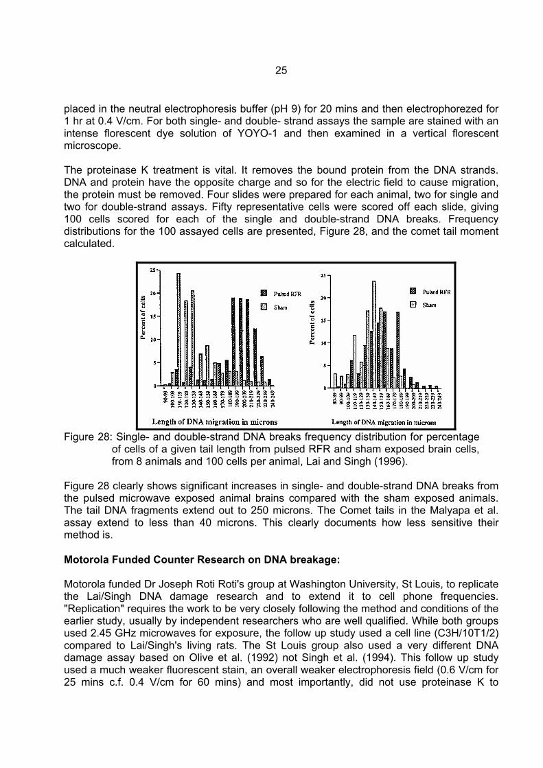
placed in the neutral electrophoresis buffer (pH 9) for 20 mins and then electrophorezed for 1 hr at 0.4 V/cm. For both single- and double- strand assays the sample are stained with an intense florescent dye solution of YOYO-1 and then examined in a vertical florescent microscope. The proteinase K treatment is vital. It removes the bound protein from the DNA strands. DNA and protein have the opposite charge and so for the electric field to cause migration, the protein must be removed. Four slides were prepared for each animal, two for single and two for double-strand assays. Fifty representative cells were scored off each slide, giving 100 cells scored for each of the single and double-strand DNA breaks. Frequency distributions for the 100 assayed cells are presented, Figure 28, and the comet tail moment calculated.
Figure 28: Single- and double-strand DNA breaks frequency distribution for percentage
of cells of a given tail length from pulsed RFR and sham exposed brain cells, from 8 animals and 100 cells per animal, Lai and Singh (1996).
Figure 28 clearly shows significant increases in single- and double-strand DNA breaks from the pulsed microwave exposed animal brains compared with the sham exposed animals. The tail DNA fragments extend out to 250 microns. The Comet tails in the Malyapa et al. assay extend to less than 40 microns. This clearly documents how less sensitive their method is. Motorola Funded Counter Research on DNA breakage: Motorola funded Dr Joseph Roti Roti's group at Washington University, St Louis, to replicate the Lai/Singh DNA damage research and to extend it to cell phone frequencies. "Replication" requires the work to be very closely following the method and conditions of the earlier study, usually by independent researchers who are well qualified. While both groups used 2.45 GHz microwaves for exposure, the follow up study used a cell line (C3H/10T1/2) compared to Lai/Singh's living rats. The St Louis group also used a very different DNA damage assay based on Olive et al. (1992) not Singh et al. (1994). This follow up study used a much weaker fluorescent stain, an overall weaker electrophoresis field (0.6 V/cm for 25 mins c.f. 0.4 V/cm for 60 mins) and most importantly, did not use proteinase K to
26
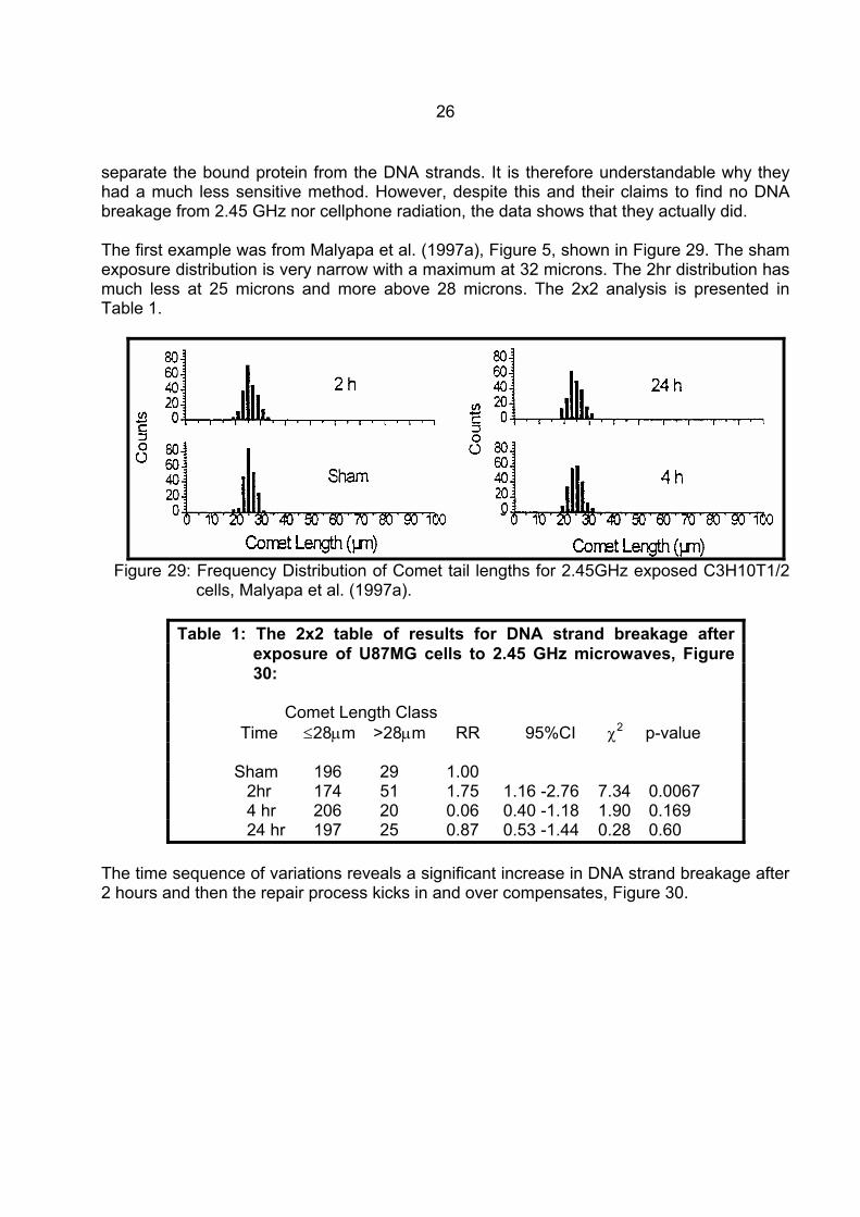
separate the bound protein from the DNA strands. It is therefore understandable why they had a much less sensitive method. However, despite this and their claims to find no DNA breakage from 2.45 GHz nor cellphone radiation, the data shows that they actually did. The first example was from Malyapa et al. (1997a), Figure 5, shown in Figure 29. The sham exposure distribution is very narrow with a maximum at 32 microns. The 2hr distribution has much less at 25 microns and more above 28 microns. The 2x2 analysis is presented in Table 1.
Figure 29: Frequency Distribution of Comet tail lengths for 2.45GHz exposed C3H10T1/2
cells, Malyapa et al. (1997a).
Table 1: The 2x2 table of results for DNA strand breakage after exposure of U87MG cells to 2.45 GHz microwaves, Figure 30:
Comet Length Class Time ≤28µm >28µm RR 95%CI χ2 p-value Sham 196 29 1.00 2hr 174 51 1.75 1.16 -2.76 7.34 0.0067 4 hr 206 20 0.06 0.40 -1.18 1.90 0.169 24 hr 197 25 0.87 0.53 -1.44 0.28 0.60
The time sequence of variations reveals a significant increase in DNA strand breakage after 2 hours and then the repair process kicks in and over compensates, Figure 30.
27
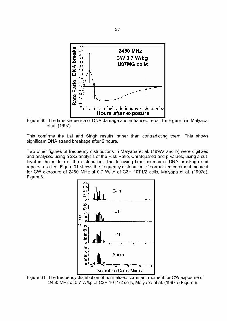
Figure 30: The time sequence of DNA damage and enhanced repair for Figure 5 in Malyapa
et al. (1997). This confirms the Lai and Singh results rather than contradicting them. This shows significant DNA strand breakage after 2 hours. Two other figures of frequency distributions in Malyapa et al. (1997a and b) were digitized and analysed using a 2x2 analysis of the Risk Ratio, Chi Squared and p-values, using a cut-level in the middle of the distribution. The following time courses of DNA breakage and repairs resulted. Figure 31 shows the frequency distribution of normalized comment moment for CW exposure of 2450 MHz at 0.7 W/kg of C3H 10T1/2 cells, Malyapa et al. (1997a), Figure 6.
Figure 31: The frequency distribution of normalized comment moment for CW exposure of
2450 MHz at 0.7 W/kg of C3H 10T1/2 cells, Malyapa et al. (1997a) Figure 6.
28
Table 2: The 2x2 table of results for DNA strand breakage after exposure
of C3H 10T1/2 cells to 2.45 GHz microwaves, Figure 32: Comet Moment Class Time ≤6 >6 RR 95%CI χ2 p-value Sham 194 75 1.00 2hr 176 101 1.31 1.02 -1.67 4.59 0.0321 4 hr 126 119 1.74 1.38 -2.20 23.31 0.0000014 24 hr 159 132 1.63 1.29 -2.05 18.30 0.0000189
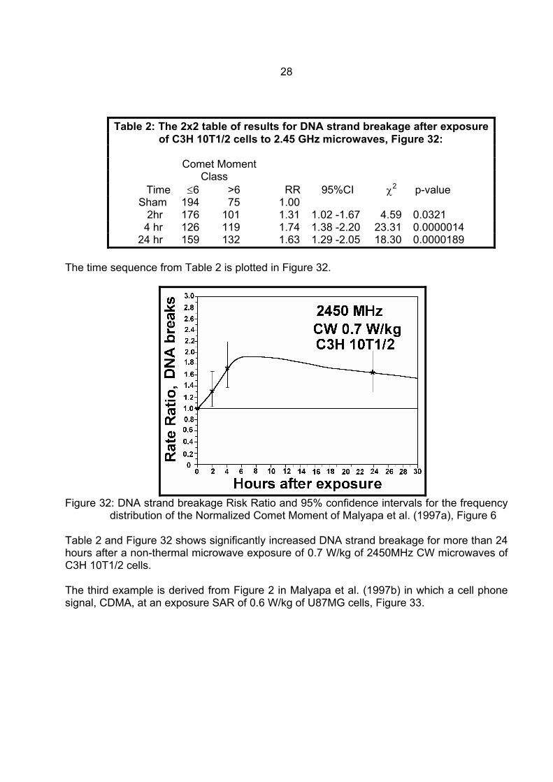
The time sequence from Table 2 is plotted in Figure 32.
Figure 32: DNA strand breakage Risk Ratio and 95% confidence intervals for the frequency
distribution of the Normalized Comet Moment of Malyapa et al. (1997a), Figure 6 Table 2 and Figure 32 shows significantly increased DNA strand breakage for more than 24 hours after a non-thermal microwave exposure of 0.7 W/kg of 2450MHz CW microwaves of C3H 10T1/2 cells. The third example is derived from Figure 2 in Malyapa et al. (1997b) in which a cell phone signal, CDMA, at an exposure SAR of 0.6 W/kg of U87MG cells, Figure 33.
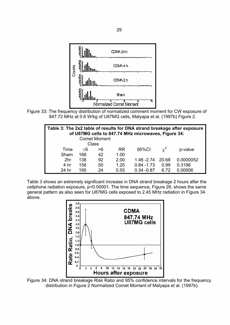
29
Figure 33: The frequency distribution of normalized comment moment for CW exposure of
847.72 MHz at 0.6 W/kg of U87MG cells, Malyapa et al. (1997b) Figure 2.
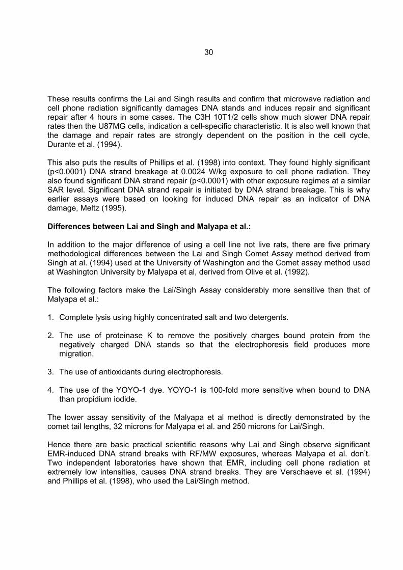
Table 3: The 2x2 table of results for DNA strand breakage after exposure of U87MG cells to 847.74 MHz microwaves, Figure 34:
Comet Moment Class Time ≤6 >6 RR 95%CI χ2 p-value Sham 168 42 1.00 2hr 138 92 2.00 1.46 -2.74 20.68 0.0000052 4 hr 158 50 1.20 0.84 -1.73 0.99 0.3196 24 hr 195 24 0.55 0.34 -0.87 6.72 0.00956
Table 3 shows an extremely significant increase in DNA strand breakage 2 hours after the cellphone radiation exposure, p<0.00001. The time sequence, Figure 26, shows the same general pattern as also seen for U87MG cells exposed to 2.45 MHz radiation in Figure 34 above.
Figure 34: DNA strand breakage Risk Ratio and 95% confidence intervals for the frequency
distribution in Figure 2 Normalized Comet Moment of Malyapa et al. (1997b).
30
These results confirms the Lai and Singh results and confirm that microwave radiation and cell phone radiation significantly damages DNA stands and induces repair and significant repair after 4 hours in some cases. The C3H 10T1/2 cells show much slower DNA repair rates then the U87MG cells, indication a cell-specific characteristic. It is also well known that the damage and repair rates are strongly dependent on the position in the cell cycle, Durante et al. (1994). This also puts the results of Phillips et al. (1998) into context. They found highly significant (p<0.0001) DNA strand breakage at 0.0024 W/kg exposure to cell phone radiation. They also found significant DNA strand repair (p<0.0001) with other exposure regimes at a similar SAR level. Significant DNA strand repair is initiated by DNA strand breakage. This is why earlier assays were based on looking for induced DNA repair as an indicator of DNA damage, Meltz (1995). Differences between Lai and Singh and Malyapa et al.: In addition to the major difference of using a cell line not live rats, there are five primary methodological differences between the Lai and Singh Comet Assay method derived from Singh at al. (1994) used at the University of Washington and the Comet assay method used at Washington University by Malyapa et al, derived from Olive et al. (1992). The following factors make the Lai/Singh Assay considerably more sensitive than that of Malyapa et al.: 1. Complete lysis using highly concentrated salt and two detergents. 2. The use of proteinase K to remove the positively charges bound protein from the
negatively charged DNA stands so that the electrophoresis field produces more migration.
3. The use of antioxidants during electrophoresis. 4. The use of the YOYO-1 dye. YOYO-1 is 100-fold more sensitive when bound to DNA
than propidium iodide. The lower assay sensitivity of the Malyapa et al method is directly demonstrated by the comet tail lengths, 32 microns for Malyapa et al. and 250 microns for Lai/Singh. Hence there are basic practical scientific reasons why Lai and Singh observe significant EMR-induced DNA strand breaks with RF/MW exposures, whereas Malyapa et al. don’t. Two independent laboratories have shown that EMR, including cell phone radiation at extremely low intensities, causes DNA strand breaks. They are Verschaeve et al. (1994) and Phillips et al. (1998), who used the Lai/Singh method.
31
The Comet Assay and EMR effects: Drs Lai and Singh have now shown that ELF and RF/MW radiation both cause single and double strand DNA breakage and are associated with free radical and reduced melatonin in living exposed rats. Lai and Singh (1995) observed a dose response increase in Single-strand DNA breakage in the rat’s brain and hippocampus that increased significantly after 4 hours, Figure 35. The increases in DNA single-strand breakage after 4 hrs is highly significant, p<0.001 and they show a dose-response relationship.
Figure 35: DNA single-strand breakage in cells from the rat brain and hippocampus,
immediately after a 2 hr exposure to a whole body SAR of 0.6 and 1.2 W/kg to 2.45 GHz microwave radiation, pulsed at 500 pps. N is the number of rats studied. Lai and Singh (1995).
The assay method was extended to measure DNA double-strand breakage. Lai and Singh (1996) reported that both continuous wave (CW) and pulsed microwaves caused significant (p<0.01) increased single-strand DNA breakage, and double-strand breakage, CW, p<0.05) and pulsed, p<0.01), Figure 36. This shows that both continuous and pulsed microwaves cause single and double DNA strand breakage, but pulsed microwaves cause more than continuous waves. Hence pulsed cell phone signals and radar signals are highly likely to cause DNA damage. This has been confirmed for radar and chromosome aberrations above and for cell phones by Phillips et al. (1998). In the mean time Lai and Singh (1997) investigated the mechanism which is involved with this genotoxic effect of RF/MW radiation. They treated the microwave exposed rats with melatonin and a spin-trap compound (PBN) to determine the role of free radicals. They showed that both melatonin and PBN eliminated the microwave induced DNA damage. Figure 37 shows the effect of melatonin for single- and double-strand DNA breaks and Figure 38 the same for PBN.
32
Figure 36: Single-strand (left) and double-strand (right) breaks in brain cells of rat after
exposure to pulsed or continuous-wave RFR. Each bar represents data from 8 rats, Lai and Singh (1996).
Figure 37: Effect of treatment with melatonin for RFR-induced increase in DNA single-
strand (left) and double-strand (right) breaks in rats brain cells. Data was analysed using the one-way ANOVA, which showed a significant treatment effect (p<0.001) for both cases. "vehicle" involves injecting with the physiological saline without the active substance. Lai and Singh (1997)
Lai and Singh (1997) conclude that if free radicals are involved in the RFR-induced DNA strand breaks in brain cells, the results of their study could have an important implication of the health effects of RFR exposure. Involvement of free radicals in human diseases, such as cancer and atherosclerosis, have been suggested. Free radicals also play an important role in aging processes, Reiter, (1995). They also point out that both melatonin and PBN can have other actions on cells in the brain that can decrease DNA damage. Therefore further support is necessary to interpret these results.
33
Figure 38: Effect of treatment with PBN for RFR-induced increase in DNA single-strand
(left) and double-strand (right) breaks in rats brain cells. Data was analysed using the one-way ANOVA, which showed a significant treatment effect (p<0.001) for both cases. "vehicle" involves injecting with the physiological saline without the active substance. Lai and Singh (1997).
Phelan et al. (1992) exposed B-16 melanoma cell line to pulsed 2.45 GHz, 100 pps, 1hr exposure SAR = 0.2 W/kg. This resulted in changes of membrane ordering. Their data indicated that a significant, specific alteration of the cell-membrane ordering followed microwave exposure and that the alteration was due at least part, to the generation of oxygen radicals. Hence there is independent support for the generation of free radicals by microwaves, as well as the Lai/Singh evidence that PBN and Melatonin reduce the RFR induced DNA damage. Two other laboratories have recorded RF/MW produced significant DNA stands breaks. Verschave et al. (1994), who used a GSM cell phone signal to expose human and rat peripheral blood lymphocytes, found significantly increased strand breaks at high, but non-thermal exposure levels. Phillips et al. (1998) exposed Molt-4 T-lymphoblastoid cells the a range of cell phone radiation in the SAR range 0.0024 W/kg to 0.026 W/kg for both iDEN and TDMA signals. Using the basic equations, these SARs at the 813-836 MHz range [SAR = σE2/2ρ, σ=1 S/m, ρ=800 kg/m3, and S = E2/3.77 µW/cm2, E: the electric field gradient in V/m and S the exposure in µW/cm2] result in 1.0 to 11.0µW/cm2. A 2 hr exposure to these low levels of cell phone radiation significantly increased (p<0.0001) or decreased (p<0.0001) the DNA damage. Decreased DNA damage is evidence of increased repair that is evidence of damage, Meltz (1995). Significance at these levels is often taken as causal. Hence RF/MW radiation has been confirmed to enhance DNA damage under RF/MW exposure from radar-like and cell phone exposures, including an exposure level which is 0.22% of the ICNIRP guideline.
34
ELF Exposure and DNA strand breakage: Four independent laboratories have also published data on ELF induced DNA strand breaks confirming that ELF EMR damages DNA strands; Lai and Singh (1997a), Svedenstal et al. (1998), Phillips et al. (1998a), and Ahuja et al. (1997). Lai and Singh (1997a) also demonstrate the involvement of free radicals and the protective effect of melatonin. With the evidence above that EMR reduces melatonin this confirms that reduced melatonin causes higher concentrations of free radicals which produce more DNA strand breaks from EMR exposure from ELF to RF/MW frequencies. Increased DNA strand breaks will result in increased chromosome aberrations. Multiple evidence from independent laboratories established that EMR from ELF to RF/MW causes DNA single- and double-strand breaks at very low, non-thermal exposure levels. This extends and confirms the genotoxic evidence from chromosome aberration studies. EMR Altered Gene Activity There is also evidence that EMR not only can damage chromosomes and DNA strands, but it is observed to alter cellular calcium ions and the activity levels of proto oncogenes (cancer genes). Blackman (1990) concluded that there was overwhelming evidence that EMR can alter normal calcium ion homeostasis and lead to changes in the response of biological systems to their environment. On of these changes is altered gene transcription and expression. The lowest published exposure level associated with significant EMR-induced alteration of cellular calcium ions occur is reported by Schwartz et al. (1990). It was 0.00015 W/kg in a 30 min exposure to a 240 MHz signal modulated at 16 Hz. The medium was frog hearts. This is equivalent to an exposure level of about 0.06 µW/cm2. Calcium ion fluxes occur in “windows” of exposure parameter combinations. Two studies associate EMR exposure alteration of gene transcription with exposure windows. Litovitz et al. (1990) identified amplitude (intensity) windows, and Wei et al. (1990) frequency windows in the range 15 to 150 Hz. They observed a peak effect in c-myc gene transcription at 45 Hz. Liburdy et al. (1993) show that c-myc induction occurs in a direct sequence from calcium ion influx. Increased c-myc gene transcripts by 50/60 Hz fields has also been observed, Goodman et al. (1989, 1992) and Lin et al. (1994). Phillips et al. (1992, 1993) observed time-dependent changes in the transcription of c-fos, c-jun, c-myc and protein kinase C, from 60 Hz exposure and a linear reduction in ras p21 expression by a 72 Hz signal. 50/60 Hz signals altered c-jun and c-fos gene expression as observed by and Lagroye and Poncy (1998) and c-fos expression by Rao and Henderson (1996) and Campbell-Beachler et al. (1998). The ppSom gene is very important in human neurological disorders, and is regulated by calcium ions Capone, Choi and Vertifuille (1998). Cell phone radiation (836.55 MHz) significantly altered c-jun transcript levels, Ivaschuk et al. (1997). Cell phone radiation significantly enhances the proto oncogene c-fos activity in C3H 10T 1/2 cells, from a 40 % (p=0.04) increase from a digital cell phone and a 2-fold increase (p=0.001) from an analogue cell phone, Goswami et al. (1999).
35
Hence proto oncogene activity is altered and enhanced in multiple independent experiments from ELF and RF/MW exposure, including cell phone radiation. Immune system impairment by EMR Impairment of the immune system is related to calcium ion efflux, Walleczek (1992) and to reduced melatonin, Reiter and Robinson (1995). Cossarizza et al. (1993) showed that ELF fields increased both the spontaneous and PHA and TPA- induced production of interleukin-1 and IL-6 in human peripheral blood. Rats exposed to microwaves showed a significant reduction in splenic activity of natural killer (NK) cells, Nakamura et al. (1997). Dmoch and Moszczynski (1998) found that microwave exposed workers had decreased NK cells and a lower value of the T-helper/T-suppressor ratio was found. Moszczynski et al. (1999) observed increased IgG and IgA and decreased lymphocytes and T8 cells in TV signal exposed workers. Quan et al. (1992) showed that microwave heating of human breast milk highly significantly suppressed the specific immune system factors for E.Coli bacteria compared with conventional heating. Chronic, 25 year, exposure to an extremely low intensity (<0.1µW/cm2) 156-162 MHz, 24.4 Hz pulse frequency, radar signal in Latvia produced significant alterations in the immune system factors of exposed villagers, Bruvere et al. (1998). Genotoxicity Conclusions: There is more than sufficient evidence of chromosome aberrations, DNA strand breakage altered oncogene activity and neoplastic transformation if cells to conclude that EMR across the spectrum from ELF to RF/MW is genotoxic. This is independently confirmed by the established biological mechanisms of calcium ion efflux and melatonin reduction. His is also totally independent of over a hundred occupational groups showing elevated cancer from EMR exposure, scores showing significantly to extremely significantly elevated cancer incidence and mortality, and dozens of dose response relationships.
Epidemiological dose-response relationships from RF/MW exposures: Dose-response relationships are shown here because they are very strong evidence of cause and effect and they give guidance as to the exposure levels involved. It should be noted however, that many other studies show significant increases in all of the cancer, cardiac, neurological and reproductive effects reported here. All occur at long-term mean exposure levels more than 100 times below the ICNIRP guideline, and residential studies involve mean exposures more than 1000 times lower than the public exposure guideline. The guidance given by Sir Austin Bradford Hill, Hill (1965) shows that even a consistent non-significant relationship can be assessed as a causal effect. When a dose response relationship is obtained then it is very strong evidence of a causal effect.
36
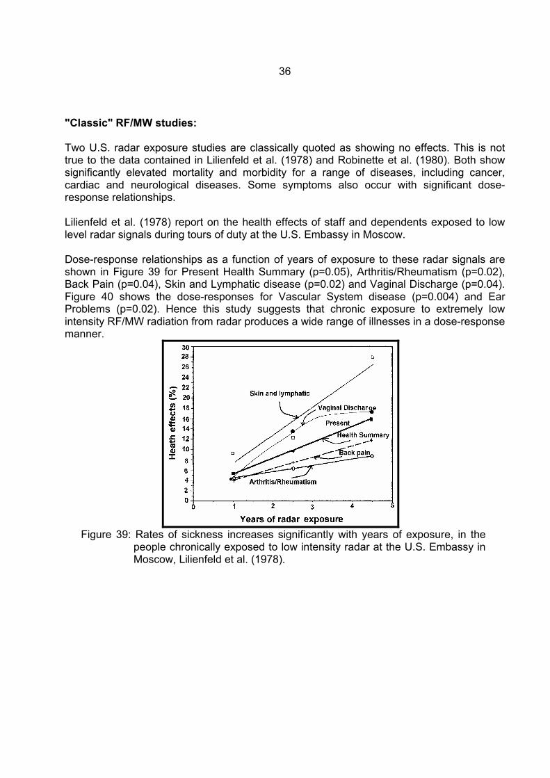
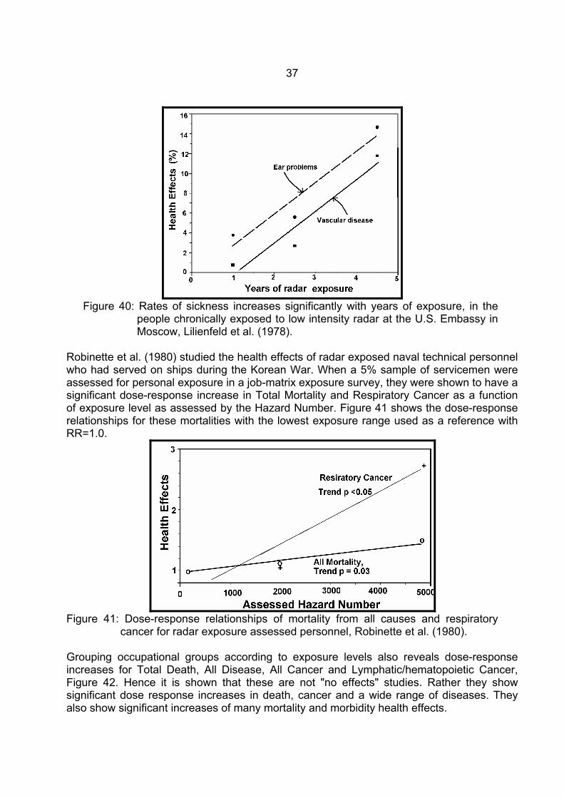
"Classic" RF/MW studies: Two U.S. radar exposure studies are classically quoted as showing no effects. This is not true to the data contained in Lilienfeld et al. (1978) and Robinette et al. (1980). Both show significantly elevated mortality and morbidity for a range of diseases, including cancer, cardiac and neurological diseases. Some symptoms also occur with significant dose-response relationships. Lilienfeld et al. (1978) report on the health effects of staff and dependents exposed to low level radar signals during tours of duty at the U.S. Embassy in Moscow. Dose-response relationships as a function of years of exposure to these radar signals are shown in Figure 39 for Present Health Summary (p=0.05), Arthritis/Rheumatism (p=0.02), Back Pain (p=0.04), Skin and Lymphatic disease (p=0.02) and Vaginal Discharge (p=0.04). Figure 40 shows the dose-responses for Vascular System disease (p=0.004) and Ear Problems (p=0.02). Hence this study suggests that chronic exposure to extremely low intensity RF/MW radiation from radar produces a wide range of illnesses in a dose-response manner.
Figure 39: Rates of sickness increases significantly with years of exposure, in the
people chronically exposed to low intensity radar at the U.S. Embassy in Moscow, Lilienfeld et al. (1978).
37
Figure 40: Rates of sickness increases significantly with years of exposure, in the
people chronically exposed to low intensity radar at the U.S. Embassy in Moscow, Lilienfeld et al. (1978).
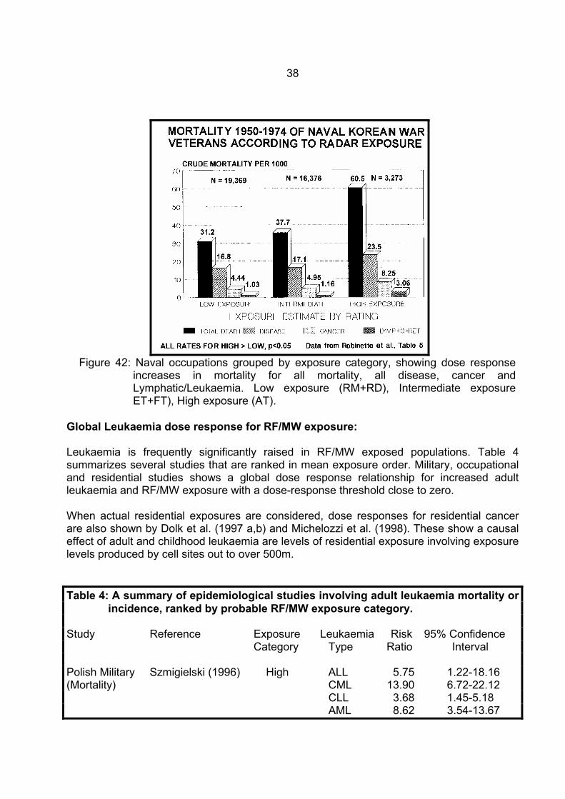
Robinette et al. (1980) studied the health effects of radar exposed naval technical personnel who had served on ships during the Korean War. When a 5% sample of servicemen were assessed for personal exposure in a job-matrix exposure survey, they were shown to have a significant dose-response increase in Total Mortality and Respiratory Cancer as a function of exposure level as assessed by the Hazard Number. Figure 41 shows the dose-response relationships for these mortalities with the lowest exposure range used as a reference with RR=1.0.
Figure 41: Dose-response relationships of mortality from all causes and respiratory
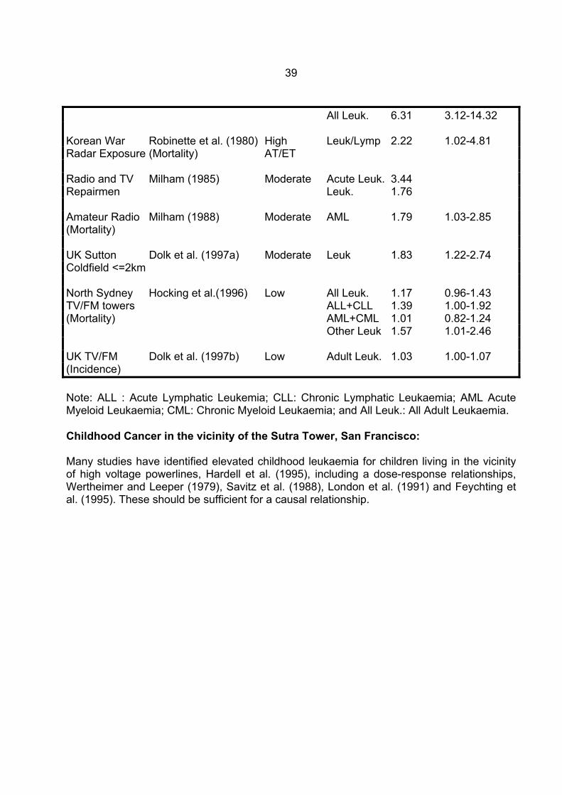
cancer for radar exposure assessed personnel, Robinette et al. (1980). Grouping occupational groups according to exposure levels also reveals dose-response increases for Total Death, All Disease, All Cancer and Lymphatic/hematopoietic Cancer, Figure 42. Hence it is shown that these are not "no effects" studies. Rather they show significant dose response increases in death, cancer and a wide range of diseases. They also show significant increases of many mortality and morbidity health effects.
38
Figure 42: Naval occupations grouped by exposure category, showing dose response
increases in mortality for all mortality, all disease, cancer and Lymphatic/Leukaemia. Low exposure (RM+RD), Intermediate exposure ET+FT), High exposure (AT).
Global Leukaemia dose response for RF/MW exposure: Leukaemia is frequently significantly raised in RF/MW exposed populations. Table 4 summarizes several studies that are ranked in mean exposure order. Military, occupational and residential studies shows a global dose response relationship for increased adult leukaemia and RF/MW exposure with a dose-response threshold close to zero. When actual residential exposures are considered, dose responses for residential cancer are also shown by Dolk et al. (1997 a,b) and Michelozzi et al. (1998). These show a causal effect of adult and childhood leukaemia are levels of residential exposure involving exposure levels produced by cell sites out to over 500m. Table 4: A summary of epidemiological studies involving adult leukaemia mortality or
incidence, ranked by probable RF/MW exposure category. Study Reference Exposure Leukaemia Risk 95% Confidence Category Type Ratio Interval Polish Military Szmigielski (1996) High ALL 5.75 1.22-18.16 (Mortality) CML 13.90 6.72-22.12 CLL 3.68 1.45-5.18 AML 8.62 3.54-13.67
39
All Leuk. 6.31 3.12-14.32 Korean War Robinette et al. (1980) High Leuk/Lymp 2.22 1.02-4.81 Radar Exposure (Mortality) AT/ET Radio and TV Milham (1985) Moderate Acute Leuk. 3.44 Repairmen Leuk. 1.76 Amateur Radio Milham (1988) Moderate AML 1.79 1.03-2.85 (Mortality) UK Sutton Dolk et al. (1997a) Moderate Leuk 1.83 1.22-2.74 Coldfield <=2km North Sydney Hocking et al.(1996) Low All Leuk. 1.17 0.96-1.43 TV/FM towers ALL+CLL 1.39 1.00-1.92 (Mortality) AML+CML 1.01 0.82-1.24 Other Leuk 1.57 1.01-2.46 UK TV/FM Dolk et al. (1997b) Low Adult Leuk. 1.03 1.00-1.07 (Incidence) Note: ALL : Acute Lymphatic Leukemia; CLL: Chronic Lymphatic Leukaemia; AML Acute Myeloid Leukaemia; CML: Chronic Myeloid Leukaemia; and All Leuk.: All Adult Leukaemia. Childhood Cancer in the vicinity of the Sutra Tower, San Francisco: Many studies have identified elevated childhood leukaemia for children living in the vicinity of high voltage powerlines, Hardell et al. (1995), including a dose-response relationships, Wertheimer and Leeper (1979), Savitz et al. (1988), London et al. (1991) and Feychting et al. (1995). These should be sufficient for a causal relationship.
40
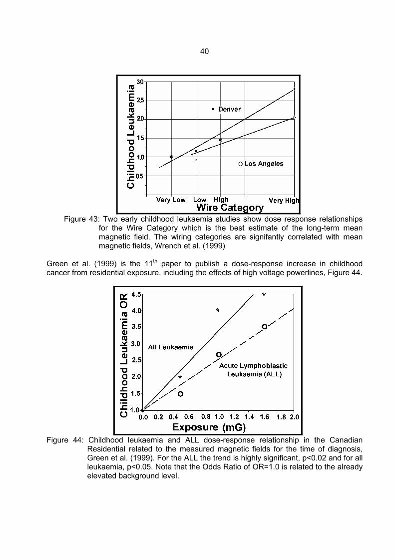
Figure 43: Two early childhood leukaemia studies show dose response relationships
for the Wire Category which is the best estimate of the long-term mean magnetic field. The wiring categories are signifantly correlated with mean magnetic fields, Wrench et al. (1999)
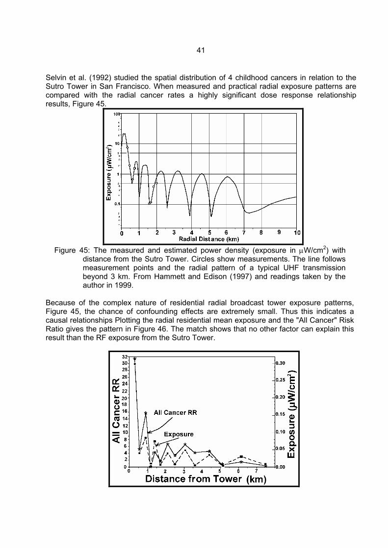
Green et al. (1999) is the 11th paper to publish a dose-response increase in childhood cancer from residential exposure, including the effects of high voltage powerlines, Figure 44.
Figure 44: Childhood leukaemia and ALL dose-response relationship in the Canadian
Residential related to the measured magnetic fields for the time of diagnosis, Green et al. (1999). For the ALL the trend is highly significant, p<0.02 and for all leukaemia, p<0.05. Note that the Odds Ratio of OR=1.0 is related to the already elevated background level.
41
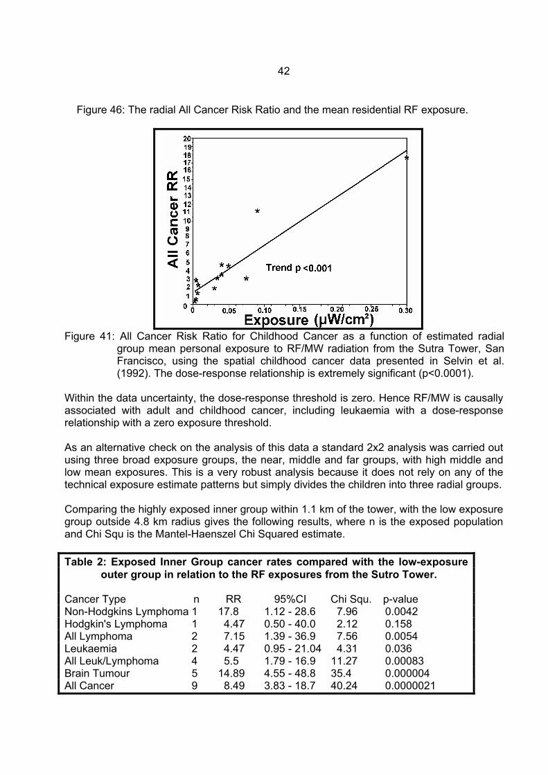
Selvin et al. (1992) studied the spatial distribution of 4 childhood cancers in relation to the Sutro Tower in San Francisco. When measured and practical radial exposure patterns are compared with the radial cancer rates a highly significant dose response relationship results, Figure 45.
Figure 45: The measured and estimated power density (exposure in µW/cm2) with
distance from the Sutro Tower. Circles show measurements. The line follows measurement points and the radial pattern of a typical UHF transmission beyond 3 km. From Hammett and Edison (1997) and readings taken by the author in 1999.
Because of the complex nature of residential radial broadcast tower exposure patterns, Figure 45, the chance of confounding effects are extremely small. Thus this indicates a causal relationships Plotting the radial residential mean exposure and the "All Cancer" Risk Ratio gives the pattern in Figure 46. The match shows that no other factor can explain this result than the RF exposure from the Sutro Tower.
42
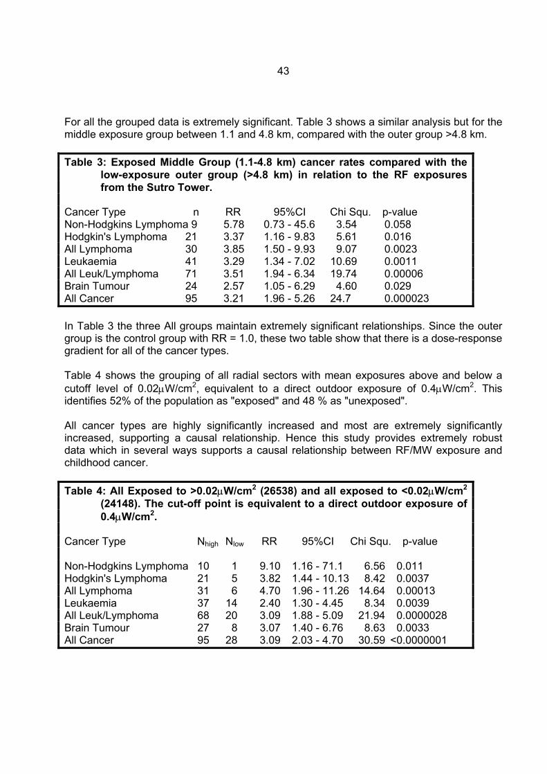
Figure 46: The radial All Cancer Risk Ratio and the mean residential RF exposure.
Figure 41: All Cancer Risk Ratio for Childhood Cancer as a function of estimated radial
group mean personal exposure to RF/MW radiation from the Sutra Tower, San Francisco, using the spatial childhood cancer data presented in Selvin et al. (1992). The dose-response relationship is extremely significant (p<0.0001).
Within the data uncertainty, the dose-response threshold is zero. Hence RF/MW is causally associated with adult and childhood cancer, including leukaemia with a dose-response relationship with a zero exposure threshold. As an alternative check on the analysis of this data a standard 2x2 analysis was carried out using three broad exposure groups, the near, middle and far groups, with high middle and low mean exposures. This is a very robust analysis because it does not rely on any of the technical exposure estimate patterns but simply divides the children into three radial groups. Comparing the highly exposed inner group within 1.1 km of the tower, with the low exposure group outside 4.8 km radius gives the following results, where n is the exposed population and Chi Squ is the Mantel-Haenszel Chi Squared estimate. Table 2: Exposed Inner Group cancer rates compared with the low-exposure
outer group in relation to the RF exposures from the Sutro Tower. Cancer Type n RR 95%CI Chi Squ. p-value Non-Hodgkins Lymphoma 1 17.8 1.12 - 28.6 7.96 0.0042 Hodgkin's Lymphoma 1 4.47 0.50 - 40.0 2.12 0.158 All Lymphoma 2 7.15 1.39 - 36.9 7.56 0.0054 Leukaemia 2 4.47 0.95 - 21.04 4.31 0.036 All Leuk/Lymphoma 4 5.5 1.79 - 16.9 11.27 0.00083 Brain Tumour 5 14.89 4.55 - 48.8 35.4 0.000004 All Cancer 9 8.49 3.83 - 18.7 40.24 0.0000021
43
For all the grouped data is extremely significant. Table 3 shows a similar analysis but for the middle exposure group between 1.1 and 4.8 km, compared with the outer group >4.8 km. Table 3: Exposed Middle Group (1.1-4.8 km) cancer rates compared with the
low-exposure outer group (>4.8 km) in relation to the RF exposures from the Sutro Tower.
Cancer Type n RR 95%CI Chi Squ. p-value Non-Hodgkins Lymphoma 9 5.78 0.73 - 45.6 3.54 0.058 Hodgkin's Lymphoma 21 3.37 1.16 - 9.83 5.61 0.016 All Lymphoma 30 3.85 1.50 - 9.93 9.07 0.0023 Leukaemia 41 3.29 1.34 - 7.02 10.69 0.0011 All Leuk/Lymphoma 71 3.51 1.94 - 6.34 19.74 0.00006 Brain Tumour 24 2.57 1.05 - 6.29 4.60 0.029 All Cancer 95 3.21 1.96 - 5.26 24.7 0.000023 In Table 3 the three All groups maintain extremely significant relationships. Since the outer group is the control group with RR = 1.0, these two table show that there is a dose-response gradient for all of the cancer types. Table 4 shows the grouping of all radial sectors with mean exposures above and below a cutoff level of 0.02µW/cm2, equivalent to a direct outdoor exposure of 0.4µW/cm2. This identifies 52% of the population as "exposed" and 48 % as "unexposed". All cancer types are highly significantly increased and most are extremely significantly increased, supporting a causal relationship. Hence this study provides extremely robust data which in several ways supports a causal relationship between RF/MW exposure and childhood cancer. Table 4: All Exposed to >0.02µW/cm2 (26538) and all exposed to <0.02µW/cm2
(24148). The cut-off point is equivalent to a direct outdoor exposure of 0.4µW/cm2.
Cancer Type Nhigh Nlow RR 95%CI Chi Squ. p-value Non-Hodgkins Lymphoma 10 1 9.10 1.16 - 71.1 6.56 0.011 Hodgkin's Lymphoma 21 5 3.82 1.44 - 10.13 8.42 0.0037 All Lymphoma 31 6 4.70 1.96 - 11.26 14.64 0.00013 Leukaemia 37 14 2.40 1.30 - 4.45 8.34 0.0039 All Leuk/Lymphoma 68 20 3.09 1.88 - 5.09 21.94 0.0000028 Brain Tumour 27 8 3.07 1.40 - 6.76 8.63 0.0033 All Cancer 95 28 3.09 2.03 - 4.70 30.59 <0.0000001
44
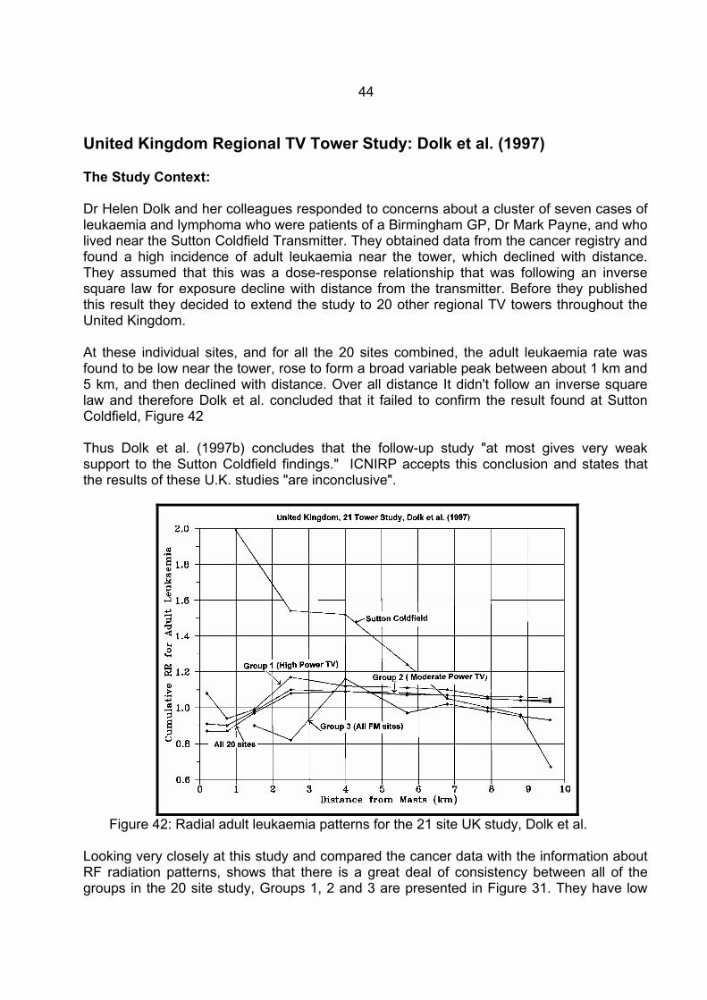
United Kingdom Regional TV Tower Study: Dolk et al. (1997) The Study Context: Dr Helen Dolk and her colleagues responded to concerns about a cluster of seven cases of leukaemia and lymphoma who were patients of a Birmingham GP, Dr Mark Payne, and who lived near the Sutton Coldfield Transmitter. They obtained data from the cancer registry and found a high incidence of adult leukaemia near the tower, which declined with distance. They assumed that this was a dose-response relationship that was following an inverse square law for exposure decline with distance from the transmitter. Before they published this result they decided to extend the study to 20 other regional TV towers throughout the United Kingdom. At these individual sites, and for all the 20 sites combined, the adult leukaemia rate was found to be low near the tower, rose to form a broad variable peak between about 1 km and 5 km, and then declined with distance. Over all distance It didn't follow an inverse square law and therefore Dolk et al. concluded that it failed to confirm the result found at Sutton Coldfield, Figure 42 Thus Dolk et al. (1997b) concludes that the follow-up study "at most gives very weak support to the Sutton Coldfield findings." ICNIRP accepts this conclusion and states that the results of these U.K. studies "are inconclusive".
Figure 42: Radial adult leukaemia patterns for the 21 site UK study, Dolk et al.
Looking very closely at this study and compared the cancer data with the information about RF radiation patterns, shows that there is a great deal of consistency between all of the groups in the 20 site study, Groups 1, 2 and 3 are presented in Figure 31. They have low
45
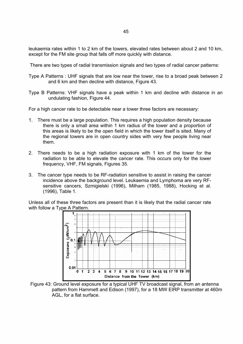
leukaemia rates within 1 to 2 km of the towers, elevated rates between about 2 and 10 km, except for the FM site group that falls off more quickly with distance. There are two types of radial transmission signals and two types of radial cancer patterns: Type A Patterns : UHF signals that are low near the tower, rise to a broad peak between 2
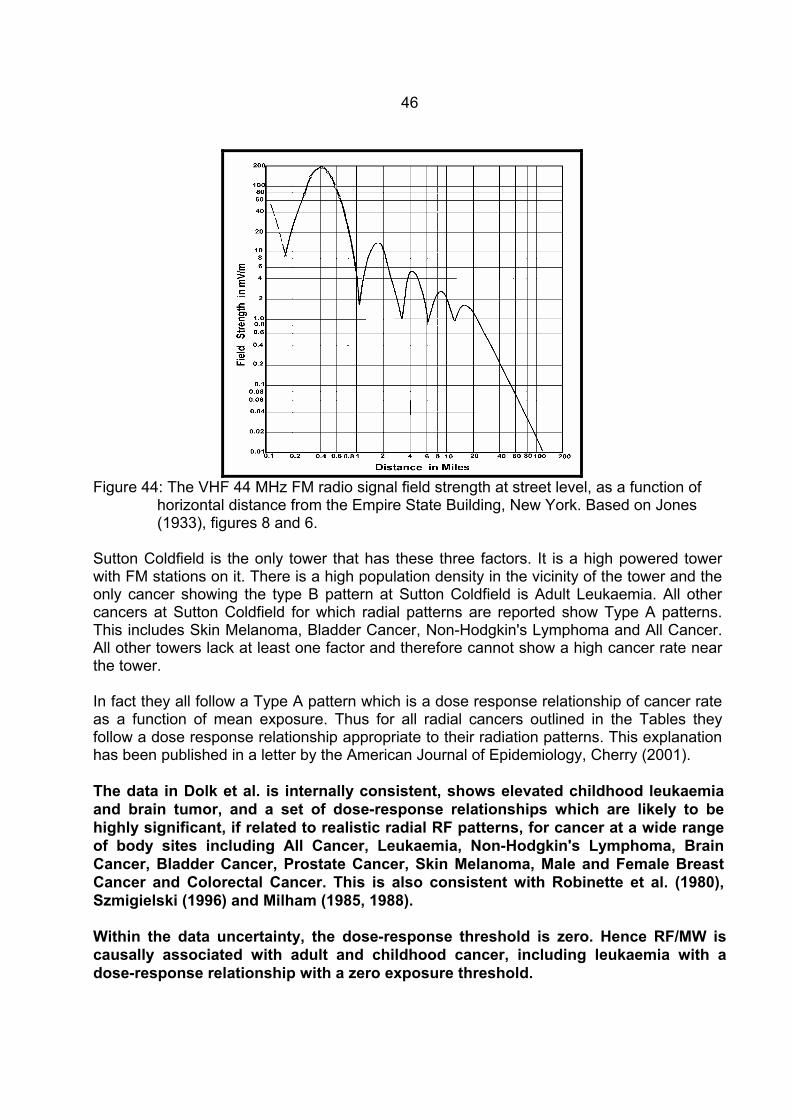
and 6 km and then decline with distance, Figure 43. Type B Patterns: VHF signals have a peak within 1 km and decline with distance in an
undulating fashion, Figure 44. For a high cancer rate to be detectable near a tower three factors are necessary: 1. There must be a large population. This requires a high population density because
there is only a small area within 1 km radius of the tower and a proportion of this areas is likely to be the open field in which the tower itself is sited. Many of the regional towers are in open country sides with very few people living near them.
2. There needs to be a high radiation exposure with 1 km of the tower for the
radiation to be able to elevate the cancer rate. This occurs only for the lower frequency, VHF, FM signals, Figures 35.
3. The cancer type needs to be RF-radiation sensitive to assist in raising the cancer
incidence above the background level. Leukaemia and Lymphoma are very RF-sensitive cancers, Szmigielski (1996), Milham (1985, 1988), Hocking et al. (1996), Table 1.
Unless all of these three factors are present than it is likely that the radial cancer rate with follow a Type A Pattern.
Figure 43: Ground level exposure for a typical UHF TV broadcast signal, from an antenna
pattern from Hammett and Edison (1997), for a 18 MW EIRP transmitter at 460m AGL, for a flat surface.
46
Figure 44: The VHF 44 MHz FM radio signal field strength at street level, as a function of
horizontal distance from the Empire State Building, New York. Based on Jones (1933), figures 8 and 6.
Sutton Coldfield is the only tower that has these three factors. It is a high powered tower with FM stations on it. There is a high population density in the vicinity of the tower and the only cancer showing the type B pattern at Sutton Coldfield is Adult Leukaemia. All other cancers at Sutton Coldfield for which radial patterns are reported show Type A patterns. This includes Skin Melanoma, Bladder Cancer, Non-Hodgkin's Lymphoma and All Cancer. All other towers lack at least one factor and therefore cannot show a high cancer rate near the tower. In fact they all follow a Type A pattern which is a dose response relationship of cancer rate as a function of mean exposure. Thus for all radial cancers outlined in the Tables they follow a dose response relationship appropriate to their radiation patterns. This explanation has been published in a letter by the American Journal of Epidemiology, Cherry (2001). The data in Dolk et al. is internally consistent, shows elevated childhood leukaemia and brain tumor, and a set of dose-response relationships which are likely to be highly significant, if related to realistic radial RF patterns, for cancer at a wide range of body sites including All Cancer, Leukaemia, Non-Hodgkin's Lymphoma, Brain Cancer, Bladder Cancer, Prostate Cancer, Skin Melanoma, Male and Female Breast Cancer and Colorectal Cancer. This is also consistent with Robinette et al. (1980), Szmigielski (1996) and Milham (1985, 1988). Within the data uncertainty, the dose-response threshold is zero. Hence RF/MW is causally associated with adult and childhood cancer, including leukaemia with a dose-response relationship with a zero exposure threshold.
47
Neurological effects: Brains are very electromagnetically sensitive because our sight, thoughts, memories, learning and emotions use complex electromagnetic signals. Research in Germany in the post war period proved that human brains detect and use extremely small natural low frequency (ELF) EMR signals, Wever (1974), Konig (1974). Since RF/MW signals induce higher currents in human tissues and low frequency signals it is inevitable that we will observe neurological effects from chronic RF/MW exposures. Recent studies have revealed some neurological dose response relationships for sleep disturbance, Multiple Sclerosis and Suicide at extremely low exposures to RF and ELF exposures. Beale et al. (1997) found significant dose response for psychological symptoms, including anxiety and depression, living in proximity to high voltage powerlines. This strongly confirms the sensitivity of human brains to EMR exposure. These studies have early roots in U.S. Embassy in Moscow study. Lilienfeld et al. (1978), showed significant neurological effects from chronic low level radar exposure, including Depression (p=0.004), Irritability (p=0.009), Memory Loss (p=0.008) and Difficulty in Concentrating (p=0.001). These are all symptoms related to melatonin reduction. The Korean War Study, Robinette et al. (1980) also found increased neurological health effects from personnel who were exposed to radar on ships during the Korean War. These symptoms between a high exposure group (FT+AT) and a low exposure group (ET), Mental Conditions, RR = 1.68, 95%CI: 1.13-2.50, p<0.01 and Neurological illness, RR = 1.42, 95%CI: 0.74-2.72. Mild et al. (1998) show significant dose-response relationships for cell phone usage and headaches, dizziness, memory loss, discomfort, fatigue, and loss of concentration. Dose responses were shown for both calls/day and minutes/day. Figures 36 and 37 show the minutes/day graphs for Norway and Sweden, respectively. Norway is dominantly analogue and Sweden digital. The analogue phones used in Norway typically have higher SAR levels than the digital phones used in Sweden. The sensation of warmth on an behind the ear is much stronger in Norway. The symptoms reported in Norway are somewhat more prevalent than in Sweden. For example, the Fatigue prevalence in Norway for more than 60 mins per day is 28% and in Sweden it is 20%. The difference is quite marked for all symptoms except Concentration and Memory Loss. These are the same symptoms that have frequently been reported as "Microwave Sickness Syndrome" or "Radiofrequency Sickness Syndrome", Baranski and Czerski (1976) and Johnson-Liakouris (1998).

48
Figure 45: Prevalence of symptoms for Norwegian mobile phone users, mainly analogue,
with various categories of length of calling time per day, Mild et al. (1998).
Figure 46: Prevalence of symptoms for Swedish mobile phone users, mainly digital, with
various categories of length of calling time per day, Mild et al. (1998). Sleep Disturbance near a Shortwave Radio Tower, Schwarzenburg, Switzerland: The Schwarzenburg Study, Alpeter et al. (1995) and Abelin (1999) showed a causal relationship of sleep disturbance with exposure to a short wave radio signal. The effect is assessed as causal because of the significant dose response relationship, the variation of sleep disturbance in two experiments, one involving changing the beams and one turning the transmitter off, and the identification of significant melatonin reduction. Professor Abelin told seminars in Christchurch that they had measured a significant increase in melatonin
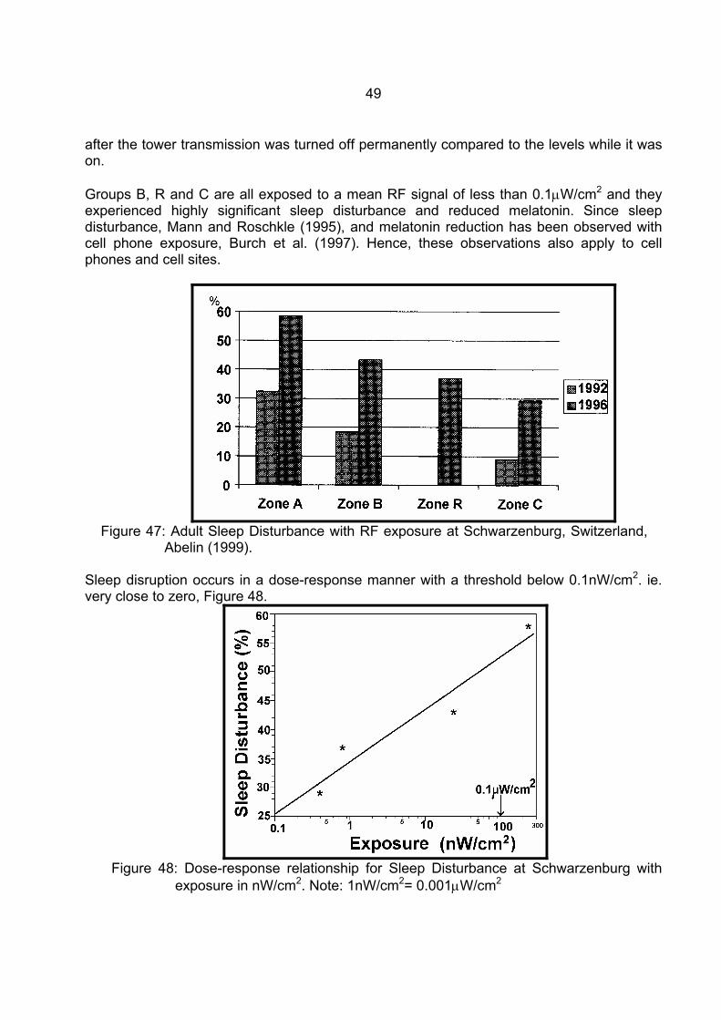
49
after the tower transmission was turned off permanently compared to the levels while it was on. Groups B, R and C are all exposed to a mean RF signal of less than 0.1µW/cm2 and they experienced highly significant sleep disturbance and reduced melatonin. Since sleep disturbance, Mann and Roschkle (1995), and melatonin reduction has been observed with cell phone exposure, Burch et al. (1997). Hence, these observations also apply to cell phones and cell sites.
Figure 47: Adult Sleep Disturbance with RF exposure at Schwarzenburg, Switzerland,
Abelin (1999). Sleep disruption occurs in a dose-response manner with a threshold below 0.1nW/cm2. ie. very close to zero, Figure 48.
Figure 48: Dose-response relationship for Sleep Disturbance at Schwarzenburg with
exposure in nW/cm2. Note: 1nW/cm2= 0.001µW/cm2
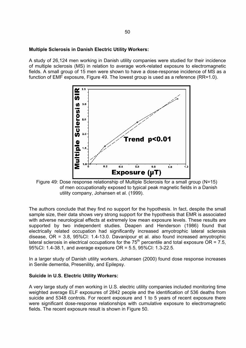
50
Multiple Sclerosis in Danish Electric Utility Workers: A study of 26,124 men working in Danish utility companies were studied for their incidence of multiple sclerosis (MS) in relation to average work-related exposure to electromagnetic fields. A small group of 15 men were shown to have a dose-response incidence of MS as a function of EMF exposure, Figure 49. The lowest group is used as a reference (RR=1.0).
Figure 49: Dose response relationship of Multiple Sclerosis for a small group (N=15)
of men occupationally exposed to typical peak magnetic fields in a Danish utility company, Johansen et al. (1999).
The authors conclude that they find no support for the hypothesis. In fact, despite the small sample size, their data shows very strong support for the hypothesis that EMR is associated with adverse neurological effects at extremely low mean exposure levels. These results are supported by two independent studies. Deapen and Henderson (1986) found that electrically related occupation had significantly increased amyotrophic lateral sclerosis disease, OR = 3.8, 95%CI: 1.4-13.0. Davanipour et al. also found increased amyotrophic lateral sclerosis in electrical occupations for the 75th percentile and total exposure OR = 7.5, 95%CI: 1.4-38.1, and average exposure OR = 5.5, 95%CI: 1.3-22.5. In a larger study of Danish utility workers, Johansen (2000) found dose response increases in Senile dementia, Presenility, and Epilepsy. Suicide in U.S. Electric Utility Workers: A very large study of men working in U.S. electric utility companies included monitoring time weighted average ELF exposures of 2842 people and the identification of 536 deaths from suicide and 5348 controls. For recent exposure and 1 to 5 years of recent exposure there were significant dose-response relationships with cumulative exposure to electromagnetic fields. The recent exposure result is shown in Figure 50.
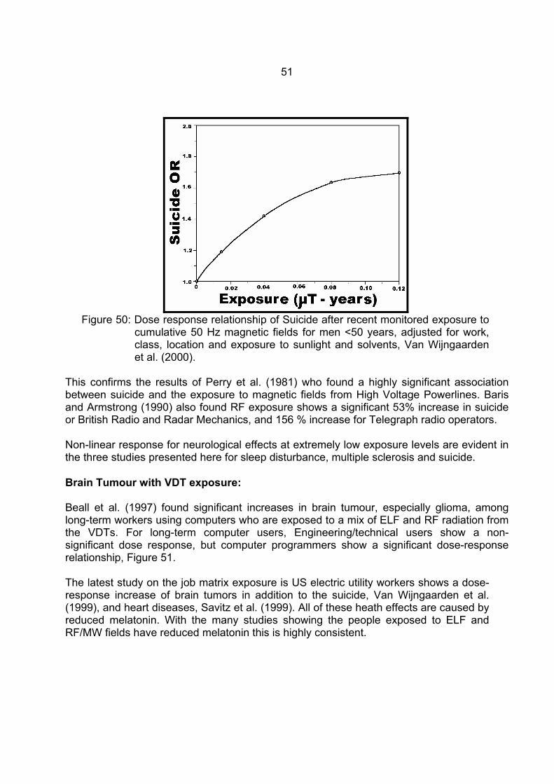
51
Figure 50: Dose response relationship of Suicide after recent monitored exposure to
cumulative 50 Hz magnetic fields for men <50 years, adjusted for work, class, location and exposure to sunlight and solvents, Van Wijngaarden et al. (2000).
This confirms the results of Perry et al. (1981) who found a highly significant association between suicide and the exposure to magnetic fields from High Voltage Powerlines. Baris and Armstrong (1990) also found RF exposure shows a significant 53% increase in suicide or British Radio and Radar Mechanics, and 156 % increase for Telegraph radio operators. Non-linear response for neurological effects at extremely low exposure levels are evident in the three studies presented here for sleep disturbance, multiple sclerosis and suicide. Brain Tumour with VDT exposure: Beall et al. (1997) found significant increases in brain tumour, especially glioma, among long-term workers using computers who are exposed to a mix of ELF and RF radiation from the VDTs. For long-term computer users, Engineering/technical users show a non-significant dose response, but computer programmers show a significant dose-response relationship, Figure 51. The latest study on the job matrix exposure is US electric utility workers shows a dose-response increase of brain tumors in addition to the suicide, Van Wijngaarden et al. (1999), and heart diseases, Savitz et al. (1999). All of these heath effects are caused by reduced melatonin. With the many studies showing the people exposed to ELF and RF/MW fields have reduced melatonin this is highly consistent.
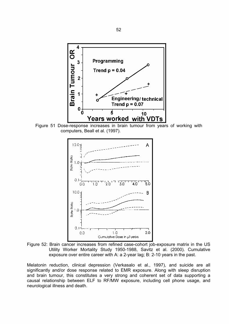
52
Figure 51 Dose-response increases in brain tumour from years of working with
computers, Beall et al. (1997).
Figure 52: Brain cancer increases from refined case-cohort job-exposure matrix in the US
Utility Worker Mortality Study 1950-1988, Savitz et al. (2000). Cumulative exposure over entire career with A: a 2-year lag; B: 2-10 years in the past.
Melatonin reduction, clinical depression (Verkasalo et al., 1997), and suicide are all significantly and/or dose response related to EMR exposure. Along with sleep disruption and brain tumour, this constitutes a very strong and coherent set of data supporting a causal relationship between ELF to RF/MW exposure, including cell phone usage, and neurological illness and death.
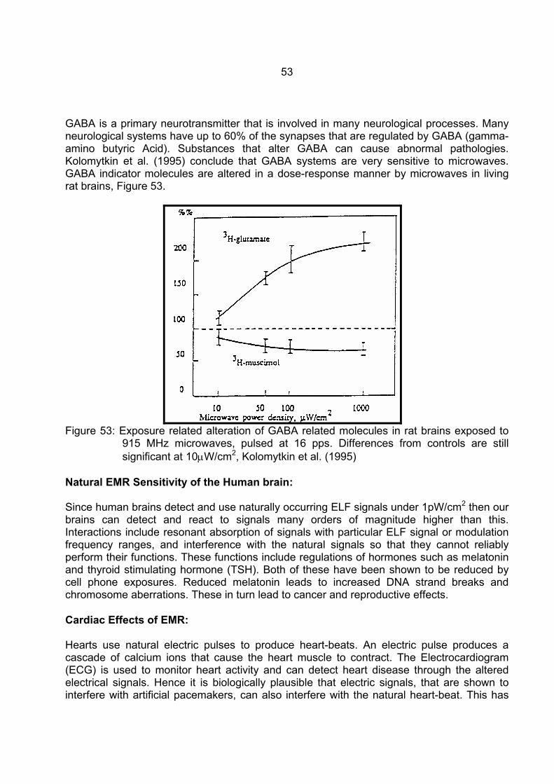
53
GABA is a primary neurotransmitter that is involved in many neurological processes. Many neurological systems have up to 60% of the synapses that are regulated by GABA (gamma-amino butyric Acid). Substances that alter GABA can cause abnormal pathologies. Kolomytkin et al. (1995) conclude that GABA systems are very sensitive to microwaves. GABA indicator molecules are altered in a dose-response manner by microwaves in living rat brains, Figure 53.
Figure 53: Exposure related alteration of GABA related molecules in rat brains exposed to
915 MHz microwaves, pulsed at 16 pps. Differences from controls are still significant at 10µW/cm2, Kolomytkin et al. (1995)
Natural EMR Sensitivity of the Human brain: Since human brains detect and use naturally occurring ELF signals under 1pW/cm2 then our brains can detect and react to signals many orders of magnitude higher than this. Interactions include resonant absorption of signals with particular ELF signal or modulation frequency ranges, and interference with the natural signals so that they cannot reliably perform their functions. These functions include regulations of hormones such as melatonin and thyroid stimulating hormone (TSH). Both of these have been shown to be reduced by cell phone exposures. Reduced melatonin leads to increased DNA strand breaks and chromosome aberrations. These in turn lead to cancer and reproductive effects. Cardiac Effects of EMR: Hearts use natural electric pulses to produce heart-beats. An electric pulse produces a cascade of calcium ions that cause the heart muscle to contract. The Electrocardiogram (ECG) is used to monitor heart activity and can detect heart disease through the altered electrical signals. Hence it is biologically plausible that electric signals, that are shown to interfere with artificial pacemakers, can also interfere with the natural heart-beat. This has

54
been shown in several studies in relation to reduction of the heart rate variability (HRV). This is a known risk factor for heart disease. Satre, Cook and Graham (1998) observed significantly reduced heart rate variability (HRV) in volunteers sleeping in 60Hz fields. Extrinsic EMR signals interfere with hearts and cause heart disease and death. Bortkiewicz et al. (1995, 1996, 1997) and Szmigielski et al. (1998) found that RF exposure altered heart rate variability and blood pressure. Forman et al.(1982) present case studies of microwave exposed personnel with induced hypertension. Braune et al. (1998) showed that cell phone significantly increased blood pressure. Savitz et al. (1999) found a highly significant dose response relationship for mortality from Arrhythmia related heart disease and heart attack (Acute Myocardial Infarction) for exposed electrical occupations and for individual occupations of electrician, lineman and power plant operator. Hamburger, Logue and Silverman (1983) observed significant dose responses for heart disease for male physiotherapists as a function of treatments per month with microwaves, OR = 2.51 (1.09-5.78), Trend p<0.05); shortwave, OR = 3.40 (1.56-7.39), trend p=0.005; and Combined Microwave and Shortwave, OR = 2.88 (1.21-6.70), trend p=0.025. This is a powerful set of epidemiological evidence showing that EMR across the spectrum increases the incidence and mortality from arrhythmia related heart disease and from heart attack. The following graph shows the dose-response curve for Acute Myocardial Infarction (Heart Attack) in electric utility workers, Figure 54.
Figure 54: Acute Myocardial Infarction as a function of cumulative exposure to 60 Hz fields
in U.S. electricity utility workers, Savitz et al. (1999). Savitz et al. (1999) show crude dose responses for Cardiac Arrhythmia related heart disease. Miscarriage in microwave exposed Physiotherapists, United States: Physiotherapists have been exposed to microwaves and shortwave radiation in the course of diathermy of patients. From a large survey group 6,684 women reported using microwave
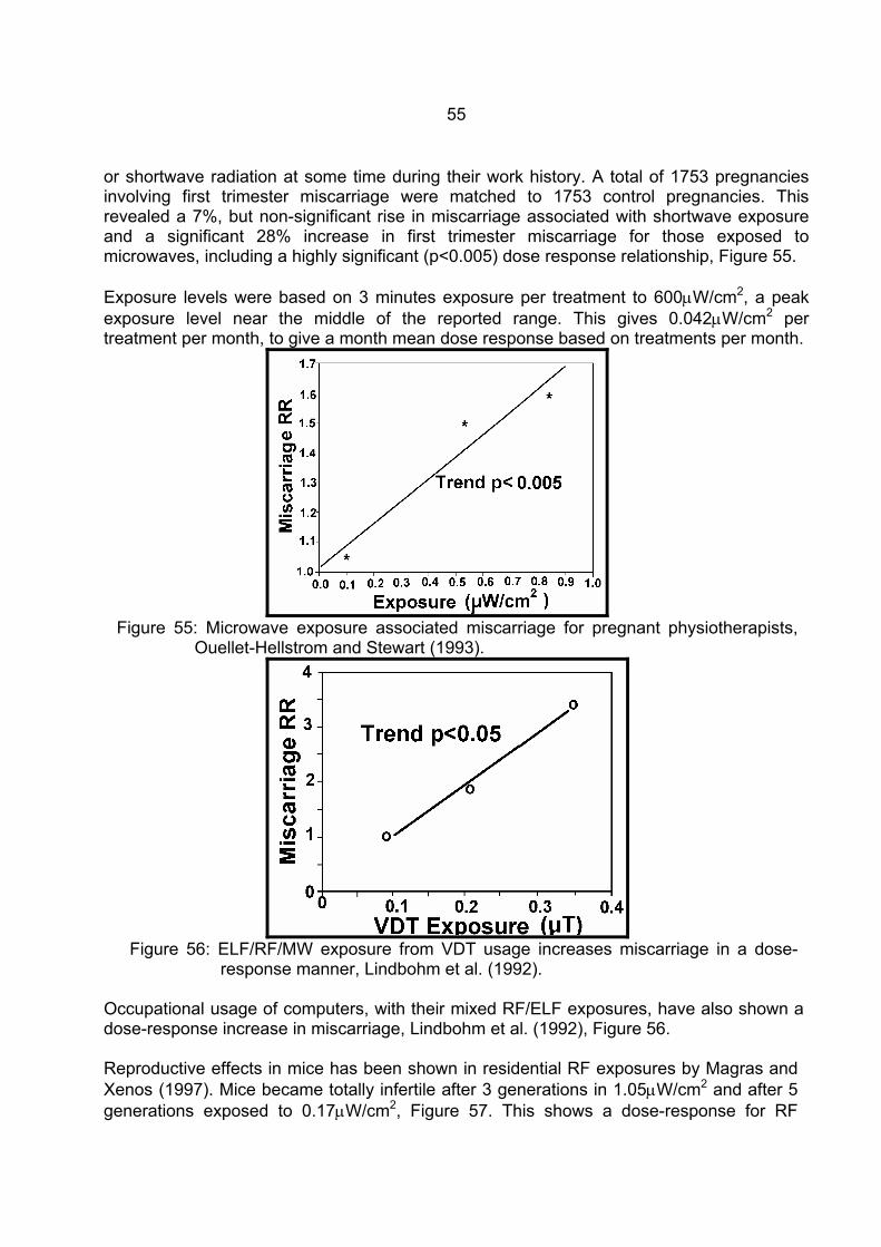
55
or shortwave radiation at some time during their work history. A total of 1753 pregnancies involving first trimester miscarriage were matched to 1753 control pregnancies. This revealed a 7%, but non-significant rise in miscarriage associated with shortwave exposure and a significant 28% increase in first trimester miscarriage for those exposed to microwaves, including a highly significant (p<0.005) dose response relationship, Figure 55. Exposure levels were based on 3 minutes exposure per treatment to 600µW/cm2, a peak exposure level near the middle of the reported range. This gives 0.042µW/cm2 per treatment per month, to give a month mean dose response based on treatments per month.
Figure 55: Microwave exposure associated miscarriage for pregnant physiotherapists,
Ouellet-Hellstrom and Stewart (1993).
Figure 56: ELF/RF/MW exposure from VDT usage increases miscarriage in a dose-
response manner, Lindbohm et al. (1992).
Occupational usage of computers, with their mixed RF/ELF exposures, have also shown a dose-response increase in miscarriage, Lindbohm et al. (1992), Figure 56. Reproductive effects in mice has been shown in residential RF exposures by Magras and Xenos (1997). Mice became totally infertile after 3 generations in 1.05µW/cm2 and after 5 generations exposed to 0.17µW/cm2, Figure 57. This shows a dose-response for RF
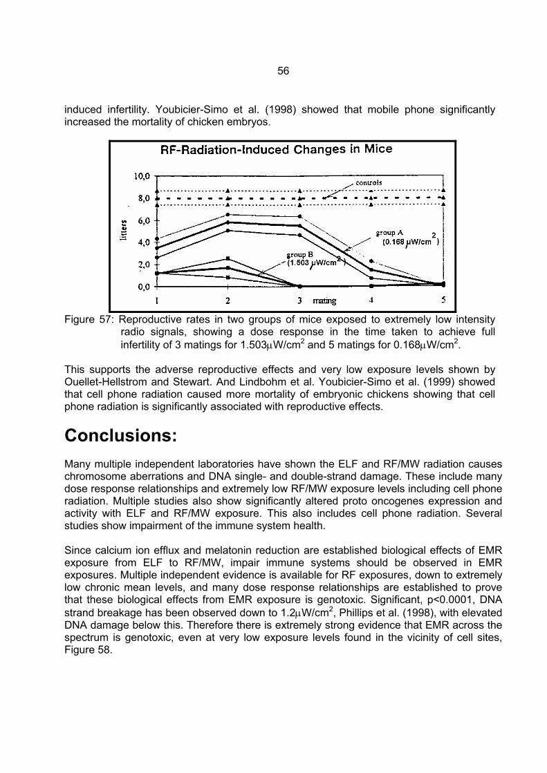
56
induced infertility. Youbicier-Simo et al. (1998) showed that mobile phone significantly increased the mortality of chicken embryos.
Figure 57: Reproductive rates in two groups of mice exposed to extremely low intensity
radio signals, showing a dose response in the time taken to achieve full infertility of 3 matings for 1.503µW/cm2 and 5 matings for 0.168µW/cm2.
This supports the adverse reproductive effects and very low exposure levels shown by Ouellet-Hellstrom and Stewart. And Lindbohm et al. Youbicier-Simo et al. (1999) showed that cell phone radiation caused more mortality of embryonic chickens showing that cell phone radiation is significantly associated with reproductive effects.
Conclusions: Many multiple independent laboratories have shown the ELF and RF/MW radiation causes chromosome aberrations and DNA single- and double-strand damage. These include many dose response relationships and extremely low RF/MW exposure levels including cell phone radiation. Multiple studies also show significantly altered proto oncogenes expression and activity with ELF and RF/MW exposure. This also includes cell phone radiation. Several studies show impairment of the immune system health. Since calcium ion efflux and melatonin reduction are established biological effects of EMR exposure from ELF to RF/MW, impair immune systems should be observed in EMR exposures. Multiple independent evidence is available for RF exposures, down to extremely low chronic mean levels, and many dose response relationships are established to prove that these biological effects from EMR exposure is genotoxic. Significant, p<0.0001, DNA strand breakage has been observed down to 1.2µW/cm2, Phillips et al. (1998), with elevated DNA damage below this. Therefore there is extremely strong evidence that EMR across the spectrum is genotoxic, even at very low exposure levels found in the vicinity of cell sites, Figure 58.
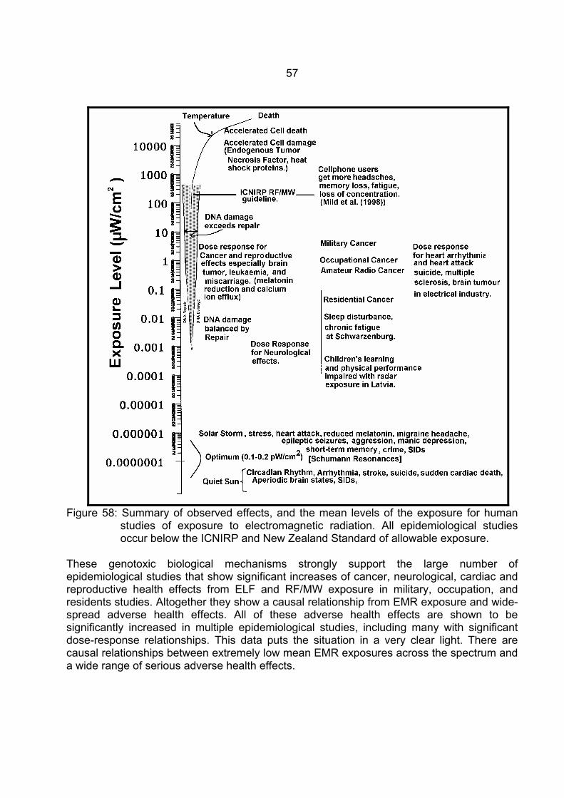
57
Figure 58: Summary of observed effects, and the mean levels of the exposure for human
studies of exposure to electromagnetic radiation. All epidemiological studies occur below the ICNIRP and New Zealand Standard of allowable exposure.
These genotoxic biological mechanisms strongly support the large number of epidemiological studies that show significant increases of cancer, neurological, cardiac and reproductive health effects from ELF and RF/MW exposure in military, occupation, and residents studies. Altogether they show a causal relationship from EMR exposure and wide-spread adverse health effects. All of these adverse health effects are shown to be significantly increased in multiple epidemiological studies, including many with significant dose-response relationships. This data puts the situation in a very clear light. There are causal relationships between extremely low mean EMR exposures across the spectrum and a wide range of serious adverse health effects.
58
Cell Phone Conclusions: Cell phones will high probably increase many neurological diseases and brain tumours over the next 10 to 20 years. Cell sites will highly probably increases sleep disturbance, reducing melatonin, damaging DNA, increasing rates of miscarriage, many cancers, suicide, neurological and cardiac diseases and death. Thousands of cell sites being installed in communities, are significantly raising the exposure of millions of people to RF/MW at levels that are know to cause serious adverse health effects. The problems are going to increase unless rapid, drastic and determined moves are made to reverse the trend and only install new sites in locations that produce extremely low mean residential exposures,
somewhat less than 10 nW/cm2 (0.01µW/cm2).
59
References: Alberts, B., Bray, D., Lewis, J., Raff, M., Roberts, K. and Watson, J.D., 1994: "Molecular Biology of the cell".
3rd edition, New York, Garland Publishing, 1994. Abelin, T, 1999: Sleep disruption with exposure to a Shortwave Radio transmission, Seminar at CRC. Ahissar, E., Haidarliu, S. and Zacksenhouse, M., 1997: "Decoding temporally encoded sensory input by
cortical oscillations and thalamic phase comparators". Proc Nat Acad Sci USA 94:11633-11638. Ahuja, Y.R., Bhargava, A., Sircar, S., Rizwani, W., Lima, S., Devadas, A.H. and Bhargava, S.C., 1997:
"Comet assay to evaluate DNA damage caused by magnetic fields". Proceed. "International Conference on Electromagnetic Interference and compatability", Dec 3-5, 1997, Hyderbad, India.
Altpeter, E.S., Krebs, Th., Pfluger, D.H., von Kanel, J., Blattmann, R., et al., 1995: "Study of health effects of
Shortwave Transmitter Station of Schwarzenburg, Berne, Switzerland". University of Berne, Institute for Social and Preventative Medicine, August 1995.
Antipenko, E.N., Koveshnikova, I.V. and Timchenko, O.I., 1984: "Effect of microwaves of nonthermal intensity
on the number of aberrant hepatocytes in rats". Radiobiologiia 24(3): 403-405. Arnetz, B.B. and Berg, M., 1996: "Melatonin and Andrenocorticotropic Hormone levels in video display unit
workers during work and leisure. J Occup Med 38(11): 1108-1110. Balcer-Kubiczek, E.K. and Harrison, G.H., 1991: "Neoplastic transformation of C3H/10T1/2 cells following
exposure to 120Hz modulated 2.45 GHz microwaves and phorbol ester tumor promoter". Radiation Research, 125: 65-72.
Balode, Z., 1996: “Assessment of radio-frequency electromagnetic radiation by the micronucleus test in
Bovine peripheral erythrocytes”. The Science of the Total Environment, 180: 81-86. Banerjee, R., Goldfeder, A. and Mitra, J., 1983a: "Sister chromatid exchanges and chromosome aberrations
induced by radiosensitizing agents in bone marrow cells of treated tumor-bearing mice". J Natl Cancer Inst 70(3): 517-521.
Banerjee, R., Goldfeder, A. and Mitra, J., 1983b: "Combined effects of misonidazole, microwave hyperthermia,
and X-rays on the in vivo sister chromatid exchanges and chromosomal abnormalities of the dbrB tumor and on the survival of its host". Cancer Res 43(5): 2210-2215.
Baranski, S. and Czerski, P., 1976: "Biological effects of microwaves". Publ. Dowden, Hutchison and Ross,
Inc. Stroudsburg, Pennsylvania. Baris, D. and Armstrong, B., 1990: "Suicide among electric utility workers in England and Wales". Br J Indust
Med 47:788-789. Bawin, S.M. and Adey, W.R., 1976: “Sensitivity of calcium binding in cerebral tissue to weak electric fields
oscillating at low frequency”. Proc. Natl. Acad. Sci. USA, 73: 1999-2003. Beale, I.L., Pearce, N.E., Conroy, D.M., Henning, M.A., and Murrell, K., A., 1997: "Psychological effects of
chronic exposure to 50 Hz magnetic fields in humans living near extra-high-voltage transmission lines". Bioelectromagnetics, 18(8): 584-94.
Beall, C., Delzell, E., Cole, P., and Brill, I., 1996: "Brain tumors among electronics industry workers".
Epidemiology, 7(2): 125-130. Beechey, C.V., Brooker, D., Kowalczuk, C.I., Saunders, R.D. and Searle, A.G., 1986: "Cytogenetic effects of
microwave irradiation on male germ cells of the mouse". Int J Radiat Biol Relat Stud Phys Chem Med 50(5): 909-918.
60
Berman, E., Carter, H.B. and House, D., 1980: "Tests of mutagenesis and reproduction in male rats exposed
to 2,450-MHz (CW) microwaves. Bioelectromagnetics 1(1): 65-76. Blackman, C.F., 1990: "ELF effects on calcium homeostasis". In "Extremely low frequency electromagnetic
fields: The question of cancer", BW Wilson, RG Stevens, LE Anderson Eds, Publ. Battelle Press Columbus: 1990; 187-208.
Bortkiewicz, A., Zmyslony, M., Palczynski, C., Gadzicka, E. and Szmigielski, S., 1995: "Dysregulation of
autonomic control of cardiac function in workers at AM broadcasting stations (0.738-1.503 MHz)". Electro- and Magnetobiology 14(3): 177-191.
Bortkiewicz, A., Gadzicka, E. and Zmyslony, M., 1996: "Heart rate in workers exposed to medium-frequency
electromagnetic fields". J Auto Nerv Sys 59: 91-97. Bortkiewicz, A., Zmyslony, M., Gadzicka, E., Palczynski, C. and Szmigielski, S., 1997: "Ambulatory ECG
monitoring in workers exposed to electromagnetic fields". J Med Eng and Tech 21(2):41-46. Braune, S., Wrocklage, C., Raczek, J., Gailus, T. and Lucking C.H., 1998: "Resting blood pressure increase
during exposure to a radio-frequency electromagnetic field". The Lancet, 351, June 20, 1988, 1857-1858.
Brueve, R., Feldmane, G., Heisele, O., Volrate, A. and Balodis, V., 1998: "Several immune system functions
of the residents from territories exposed to pulse radio-frequency radiation". Presented to the Annual Conference of the ISEE and ISEA, Boston Massachusetts July 1998.
Burch, J.B., Reif, J.S., Pittrat, C.A., Keefe, T.J. and Yost, M.G., 1997: "Cellular telephone use and excretion of
a urinary melatonin metabolite". In: Annual review of Research in Biological Effects of electric and magnetic fields from the generation, delivery and use of electricity, San Diego, CA, Nov. 9-13, P-52.
Burch, J.B., Reif, J.S., Yost, M.G., Keefe, T.J. and Pittrat, C.A., 1998: "Nocturnal excretion of urinary
melatonin metabolite among utility workers". Scand J Work Environ Health 24(3): 183-189. Burch, J.B., Reif, J.S., Yost, M.G., Keefe, T.J. and Pittrat, C.A., 1999a: "Reduced excretion of a melatonin
metabolite among workers exposed to 60 Hz magnetic fields" Am J Epidemiology 150(1): 27-36. Burch, J.B., Reif, J.S. and Yost, M.G., 1999b: "Geomagnetic disturbances are associated with reduced
nocturnal excretion of melatonin metabolite in humans". Neurosci Lett 266(3):209-212. Burch, J.B., Reif, J.S., Noonan, C.W. and Yost, M.G., 2000: "Melatonin metabolite levels in workers exposed
to 60-Hz magnetic fields: work in substations and with 3-phase conductors". J of Occupational and Environmental Medicine, 42(2): 136-142.
Burke, J., 1985: "The Day the Universe Changed". Publ. Little and Co., Boston. Campbell-Beachler, M., Ishida-Jones, T., Haggren, W. and Phillips, J.L., 1998: "Effect of 60 Hz magnetic field
exposure on c-fos expression in stimulated PC12 cells". Mol Cell Biochem 189(1-2): 107-111. Capone, G., Choi, C. and Vertifuille, J., 1998: "Regulation of the prepromsomatostatin gene by cyclic-AMP in
cerebrocortical neurons". Bran Res Mol Brain Res 60(2): 247-258. Catterall, W.A., 1992: "Cellular and molecular biology of voltage-gated sodium channels". Physiological
Reviews 72(4): S15-S48. Cossarizza, A., Angioni, S., Petraglia, F., Genazzani, A.R., Monti, D., Capri, M., Bersani, F., Cadossi, R. and
Franceschi, C., 1993: "Exposure to low frequency pulsed electromagnetic fields increases interleukin-
61
1 and interleukin-6 production by human peripheral blood mononuclear cells". Exp Cell Res 204(2):385-387.
Dmoch, A. and Moszczynski, P., 1998: "Levels of immunoglobulin and subpopulations of T lymphocytes and
NK cells in men occupationally exposed to microwave radiation in frequencies of 6-12GHz". Med Pr 49(1):45-49.
Dolk, H., Shaddick, G., Walls, P., Grundy, C., Thakrar, B., Kleinschmidt, I. and Elliott, P., 1997a: “Cancer
incidence near radio and television transmitters in Great Britain, I - Sutton-Colfield transmitter”. American J. of Epidemiology, 145(1):1-9.
Dolk, H., Elliott, P., Shaddick, G., Walls, P., Grundy, C., and Thakrar, B.,1997b: “Cancer incidence near radio
and television transmitters in Great Britain, II All high power transmitters”. American J. of Epidemiology, 145(1):10-17.
El Nahas, S.M. and Oraby, H.A., 1989: "Micronuclei formation in somatic cells of mice exposed to 50 Hz
electric fields. Environ Mol Mutagen 13(2):107-111. Feychting, M., Schulgen, G., Olsen, J.H., and Ahlbom, A., 1995: “Magnetic fields and childhood cancer-
pooled analysis of two Scandinavian studies”. European J. of Cancer, 31A (12): 2035-2039. Feychting, M, Schulgen, G., Olsen ,J.H. and Ahlbom, A., 1995: "Magnetic fields and childhood cancer - a
pooled analysis of two Scandinavian studies". Eur. J. Cancer 31A(12): 2035-2039. Forman, S.A., Holmes, C.K., McManamon, T.V., and Wedding, W.R., 1982: "Physiological Symptoms and
Intermittent Hypertension following acute microwave exposure". J. of Occup. Med. 24(11): 932-934. Frey, A.H., 1993: "Electromagnetic field interactions with biological systems". FASEB J., 7: 272-281. Frey, A.H., 1995: “An integration of the data on mechanisms with particular reference to cancer”, Chapter 2 in
“On the Nature of electromagnetic Field Interactions with Biological Systems”, Ed A.H. Frey, Publ. R.G. Landes Co. Medical Intelligence Unit, Austin, Texas.
Frey, A.H., 1998: "Headaches from cellular telephones: are they real and what are the impacts". Environ
Health Perspect 106(3):101-103. Garcia-Sagredo, J.M. and Monteagudo, J.L., 1991: "Effect of low-level pulsed electromagnetic fields on
human chromosomes in vitro: analysis of chromosome aberrations". Hereditas 115(1): 9-11. Garaj-Vrhovac, V., Horvat, D., Brumen-Mahovic and Racic, J., 1987: "Somatic mutations in persons
occupationally exposed to microwave radiation". Mutation Research 181: 321 Garaj-Vrhovac, V., Fucic, A, and Horvat, D., 1990: "Comparison of chromosome aberration and micronucleus
induction in human lymphocytes after occupational exposure to vinyl chloride monomer and microwave radiation"., Periodicum Biologorum, Vol 92, No.4, pp 411-416.
Garaj-Vrhovac, V., Horvat, D. and Koren, Z., 1990: "The effect of microwave radiation on the cell genome".
Mutat Res 243: 87-93 (1990). Garaj-Vrhovac, V., Horvat, D. and Koren, Z., 1991: "The relationship between colony-forming ability,
chromosome aberrations and incidence of micronuclei in V79 Chinese Hamster cells exposed to microwave radiation". Mutat Res 263: 143-149.
Garaj-Vrhovac, V., Fucic, A, and Horvat, D., 1992: The correlation between the frequency of micronuclei and
specific aberrations in human lymphocytes exposed to microwave radiation in vitro”. Mutation Research, 281: 181-186.
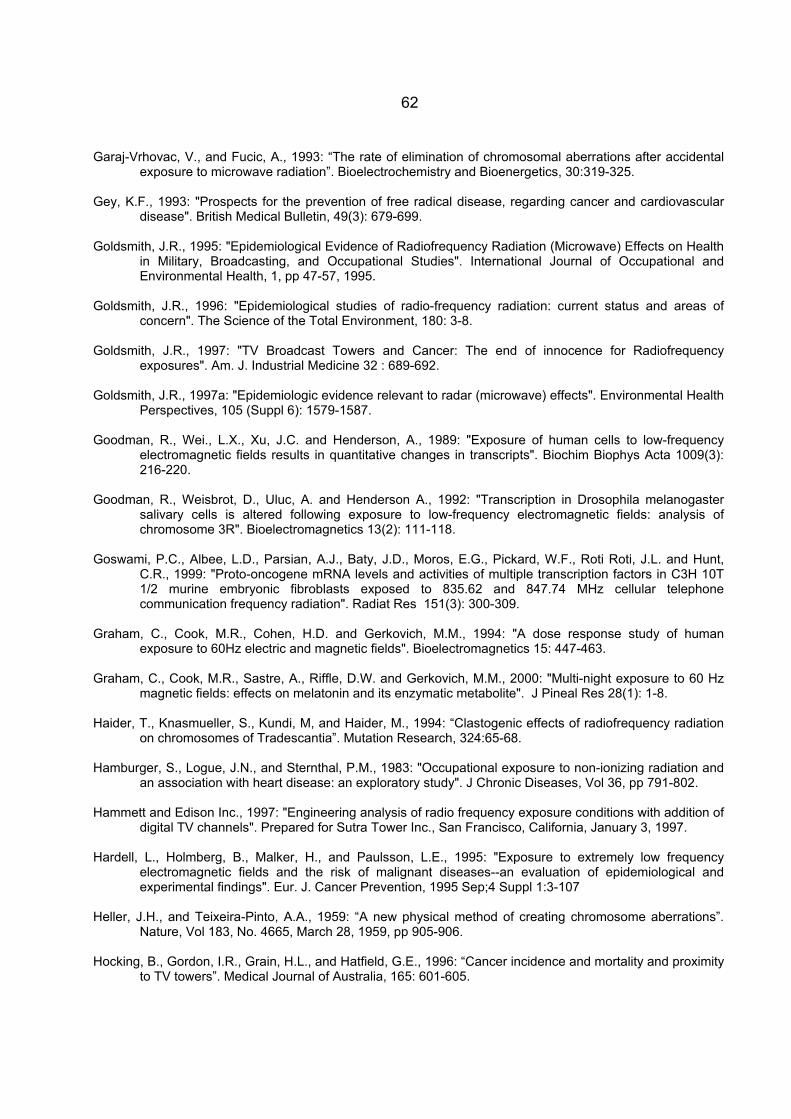
62
Garaj-Vrhovac, V., and Fucic, A., 1993: “The rate of elimination of chromosomal aberrations after accidental exposure to microwave radiation”. Bioelectrochemistry and Bioenergetics, 30:319-325.
Gey, K.F., 1993: "Prospects for the prevention of free radical disease, regarding cancer and cardiovascular
disease". British Medical Bulletin, 49(3): 679-699. Goldsmith, J.R., 1995: "Epidemiological Evidence of Radiofrequency Radiation (Microwave) Effects on Health
in Military, Broadcasting, and Occupational Studies". International Journal of Occupational and Environmental Health, 1, pp 47-57, 1995.
Goldsmith, J.R., 1996: "Epidemiological studies of radio-frequency radiation: current status and areas of
concern". The Science of the Total Environment, 180: 3-8. Goldsmith, J.R., 1997: "TV Broadcast Towers and Cancer: The end of innocence for Radiofrequency
exposures". Am. J. Industrial Medicine 32 : 689-692. Goldsmith, J.R., 1997a: "Epidemiologic evidence relevant to radar (microwave) effects". Environmental Health
Perspectives, 105 (Suppl 6): 1579-1587. Goodman, R., Wei., L.X., Xu, J.C. and Henderson, A., 1989: "Exposure of human cells to low-frequency
electromagnetic fields results in quantitative changes in transcripts". Biochim Biophys Acta 1009(3): 216-220.
Goodman, R., Weisbrot, D., Uluc, A. and Henderson A., 1992: "Transcription in Drosophila melanogaster
salivary cells is altered following exposure to low-frequency electromagnetic fields: analysis of chromosome 3R". Bioelectromagnetics 13(2): 111-118.
Goswami, P.C., Albee, L.D., Parsian, A.J., Baty, J.D., Moros, E.G., Pickard, W.F., Roti Roti, J.L. and Hunt,
C.R., 1999: "Proto-oncogene mRNA levels and activities of multiple transcription factors in C3H 10T 1/2 murine embryonic fibroblasts exposed to 835.62 and 847.74 MHz cellular telephone communication frequency radiation". Radiat Res 151(3): 300-309.
Graham, C., Cook, M.R., Cohen, H.D. and Gerkovich, M.M., 1994: "A dose response study of human
exposure to 60Hz electric and magnetic fields". Bioelectromagnetics 15: 447-463. Graham, C., Cook, M.R., Sastre, A., Riffle, D.W. and Gerkovich, M.M., 2000: "Multi-night exposure to 60 Hz
magnetic fields: effects on melatonin and its enzymatic metabolite". J Pineal Res 28(1): 1-8. Haider, T., Knasmueller, S., Kundi, M, and Haider, M., 1994: “Clastogenic effects of radiofrequency radiation
on chromosomes of Tradescantia”. Mutation Research, 324:65-68. Hamburger, S., Logue, J.N., and Sternthal, P.M., 1983: "Occupational exposure to non-ionizing radiation and
an association with heart disease: an exploratory study". J Chronic Diseases, Vol 36, pp 791-802. Hammett and Edison Inc., 1997: "Engineering analysis of radio frequency exposure conditions with addition of
digital TV channels". Prepared for Sutra Tower Inc., San Francisco, California, January 3, 1997. Hardell, L., Holmberg, B., Malker, H., and Paulsson, L.E., 1995: "Exposure to extremely low frequency
electromagnetic fields and the risk of malignant diseases--an evaluation of epidemiological and experimental findings". Eur. J. Cancer Prevention, 1995 Sep;4 Suppl 1:3-107
Heller, J.H., and Teixeira-Pinto, A.A., 1959: “A new physical method of creating chromosome aberrations”.
Nature, Vol 183, No. 4665, March 28, 1959, pp 905-906. Hocking, B., Gordon, I.R., Grain, H.L., and Hatfield, G.E., 1996: “Cancer incidence and mortality and proximity
to TV towers”. Medical Journal of Australia, 165: 601-605.
63
Ivaschuk, O.I., Jones, R.A., Ishida-Jones, T., Haggren, Q., Adey, W.R. and Phillips, J.L., 1997: "Exposure of nerve growth factor-treated PC12 rat pheochromscytoma cells to a modulated radiofrequency field at 836.55 MHz: effects on c-jun and c-fos expression". Bioelectromagnetics 18(3): 223-229.
Johanson C, Kock-Henriksen N, Rasmussen S, Olsen JH. 1999: "Multiple Sclerosis among utility workers".
Neurology 52: 1279-1282. Johnson-Liakouris, A.J.. 1998: "Radiofrequency (RF) Sickness in the Lilienfeld Study: an effect of modulated
microwaves". Arch Environ Heath 53(3):236-238. Juutilainen, J., Stevens, R.G., Anderson, L.E., Hansen, N.H., Kilpelainen, M., Laitinen, J.T., Sobel, E. and
Wilson, B.W., 2000: "Nocturnal 6-hydroxymelatonin sulphate excretion in female workers exposed to magnetic fields". J Pineal Res 28(2): 97-104.
Karasek, M., Woldanska-Okonska, M., Czernicki, J., Zylinska, K. and Swietoslawski, J., 1998: "Chronic
exposure to 2.9 mT, 40 Hz magnetic field reduces melatonin concentrations in humans". J Pineal Research 25(4): 240-244.
Khaili, A.M. and Qassem, W., 1991: "Cytogenetic effects of pulsing electromagnetic field on human
lymphocytes in vitro: chromosome aberrations, sister-chromatid exchanges and cell kinetics". Mutat Res 247: 141-146.
König HL. 1974, Behavioural changes in human subjects associated with ELF electric fields. In Persinger MA,
editor. ELF and VLF electromagnetic field effects. New York, Plenum Press. Kolomytkin, O., Kuznetsov, V., Yurinska, M, Zharikova, A., and Zharikov, S., 1995: “Response of brain
receptor systems to microwave energy exposure”. pp 195-206 in “On the nature of electromagnetic field interactions with biological systems”, Ed Frey, A.H., Publ. R.G. Landes Co.
Koveshnikov, I.V. and Antipenko, E.N., 1991a: "Quantitative patterns in the cytogenetic action of microwaves".
Radiobiologiia 31(1): 149-151. Koveshnikova, I.V. and Antipenko, E.N., 1991b: "The participation of thyroid hormones in modifying the
mutagenic effect of microwaves". Radiobiologiia 31(1):147-149. Kowalczuk, C.I., Saunders, R.D. and Stapleton, H.R., 1983: "Sperm count and sperm abnormality in male
mice after exposure to 2.45 GHz microwave radiation". Mutat Res 122(2):155-161. Lagroye, I, and Poncy J.L., 1998: "Influences of 50 Hz magnetic fields and ionizing radiation on c-jun and c-
fos oncoproteins". Bioelectromagnetics 19(2): 112-116. Lai, H. and Singh, N.P., 1995: “Acute low-intensity microwave exposure increases DNA single-strand breaks
in rat brain cells”. Bioelectromagnetics 16: 207-210. Lai, H. and Singh, N.P., 1996: “Single- and double-strand DNA breaks in rat brain cells after acute exposure
to radiofrequency electromagnetic radiation”. Int. J. Radiation Biology, 69 (4): 513-521. Lai, H. and Singh, N.P., 1996a: "Reply to "Comment on 'Acute low-intensity microwave exposure increases
DNA single-strand breaks in rat brain cells' ". Bioelectromagnetics 17: 166. Lai, H., and Singh, N.P., 1997a: “Melatonin and N-tert-butyl-a-phenylnitrone Block 60 Hz magnetic field-
induced DNA single- and double-strands Breaks in Rat Brain Cells.” J of Pineal Res. 22:152-162. Lai, H., and Singh, N.P., 1997b: “Melatonin and Spin-Trap compound Block Radiofrequency Electromagnetic
Radiation-induced DNA Strands Breaks in Rat Brain Cells.” Bioelectromagnetics 18:446-454.
64
Li, C.M., Chiang, H., Fu, Y.D., Shao, B.J., Shi, J.R. and Yao, G.D., 1999: "Effects of 50Hz magnetic fields on gap junction intercellular communication". Bioelectromagnetics 20(5):290-294.
Liburdy, R.P., Callahan, D.E., Harland, J., Dunham, E., Sloma, T.R. and Yaswen, P., 1993: "Experimental
evidence for 60 Hz magnetic fields operating through the signal transduction cascade - effects on calcium influx and c-MYC mRNA induction". FEBS Lett 334(3): 301-308.
Lilienfeld, A.M., Tonascia, J., and Tonascia S., Libauer, C.A., and Cauthen, G.M., 1978: “Foreign Service
health status study - evaluation of health status of foreign service and other employees from selected eastern European posts”. Final Report (Contr.No.6025-619073), U.S. Dept of State, July 31, 1978.
Lin, H., Goodman, R. and Shirley-Henderson, A., 1994: "Specific region of the c-myc promoter is responsible
for electric and magnetic fields". J Cell Biochem 54 30: 281-288. Lindbohm, M-L,, Hietanen, M., Kyyronen, P., Sallmen, M., von Nandelstadh, P., Taskinen, H., Pekkarinen, M.,
Ylikoski, M. and Hemminki, K., 1992: "Magnetic fields of video display terminals and spontaneous abortion". Am J Epidemiol 136:1041-1051.
Litovitz, T.A., Montrose, C.J., Goodman, R. and Elson, E.C., 1990: "Amplitude windows and transiently
augmented transcription from exposure to electromagnetic fields". Bioelectromagn. 11(4):297-312. London, S.J., Thomas, D.C., Bowman, J.D., Sobel, E., Chen, T.S. and Peters J.M., 1991: "Exposure to
residential electric and magnetic fields and risk of childhood leukemia". Am. J. Epid. 134 (9): 923-937. Maes, A., Verschaeve, L., Arroyo, A., De Wagter, C. and Vercruyssen, L., 1993: "In vitro effects of 2454 MHz
waves on human peripheral blood lymphocytes”. Bioelectromagnetics 14: 495-501. Maes, A., Collier, M., Slaets, D., and Verschaeve, L., 1996: “954 MHz Microwaves enhance the mutagenic
properties of Mitomycin C”. Environmental and Molecular Mutagenesis, 28: 26-30. Maes, A., Collier, M., Van Gorp, U., Vandoninck, S. and Verschaeve, L., 1997: "Cytogenetic effects of 935.2-
MHz (GSM) microwaves alone and in combination with mitomycin C". Mutat Res 393(1-2): 151-156. Mailhes, J.B., Young, D., Marino, A.A. and London, S.N., 1997: "Electromagnetic fields enhance chemically-
induced hyperploidy in mammalian oocytes". Mutagenesis 12(5): 347-351. Malyapa, R.S., Ahern, E.W., Bi, C., Straube, W/L/., LaRegina, M., Pickard, W.F. and Roti Roti, J.L., 1998:
"DNA damage in rat brain cells after in vivo exposure to 2450 MHz electromagnetic radiation and various methods of euthanasia". Radiation Research 149(6): 637-645.
Magras, I.N. and Xenos, T.D., 1997: "RF radiation-induced changes in the prenatal development of mice".
Bioelectromagnetics 18: 455-461. Manikowska-Czerska, E., Czerski, P. and Leach, W.M., 1985: "Effects of 2.45 GHz microwaves on meiotic
chromosomes of male CBA/CAY mice". J Hered 76(1): 71-73. Manikowska, E., Luciani, J.M., Servantie, B., Czerski, P., Obrenovitch, J. and Stahl, A., 1979: "Effects of 9.4
GHz microwave exposure on meiosis in mice". Experientia 35(3): 388-390. Mann, K.,and Roschkle, J, 1995: “Effects of pulsed high-frequency electromagnetic fields on human sleep”.
Neuropsychobiology, 33: 41-47. Meltz, M.L., 1995: "Biological effects versus health effects: an investigation of the genotoxicity of microwave
radiation". In: Radiofrequency Radiation Standards, NATO ASI Series (B.J. Klauebberg Ed). New York, Plenum Press, 1995: 235-241.
65
Michelozzi, P., Ancona, C., Fusco, D., Forastiere, F. and Perucci, C.A., 1998: "Risk of leukamia and residence near a radio transmitter in Italy". ISEE/ISEA 1998 Conference, Boston Mass. Paper 354 P., Abstract in Epidemiology 9(4):S111.
Mild, K.H., Oftedal, G., Sandstrom, M., Wilen, J., Tynes, T., Haugsdal, B. and Hauger E., 1998: "Comparison
of symptoms by users of analogue and digital mobile phones - A Swedish-Norwegian epidemiological study". National Institute for working life, 1998:23, Umea, Sweden, 84pp.
Milham S. 1985, Mortality in workers exposed to electromagnetic fields. Environ Health Persp. 62:297-300. Milham S. 1988, Increased mortality in amateur radio operators due to lymphatic and hematopoietic
malignancies. Am. J. Epidemiol 127(1): 50-54. Moszczynski, P., Lisiewicz, J., Dmoch, A., Zabinski, Z., Bergier, L., Rucinska, M. and Sasiadek, U., 1999:
"The effect of various occupational exposures to microwave radiation on the concentrations of immunoglobulins and T lymphocyte subsets". Wiad Lek 52(1-2):30-34.
Motluk, A., 1997: "Radio head: The brain has its own FM receiver". New Scientist, 25 October 1997, p17. Nakamura, H., Seto,T., Nagase, H., Yoshida, M., Dan, S. and Ogina, K., 1997: "Effects of exposure to
microwaves on cellular immunity and placental steroids in pregnant rats. Occup Environ Med 54(9):676-680.
Nawrot, P.S., McRee, D.I. and Staples, R.E., 1981: "Effects of 2.45 GHz CW microwave radiation on
embryofetal development in mice". Teratology 24(3): 303-314. Nordenson, I., Mild, K.H., Nordstrom, S., Sweins, A. and Birke, E., 1984: "Clastogenic effects in human
lymphocytes of power frequency electric fields". Radiat Environ Biophys 23(3): 191-201. Nordenson, I., Mild, K.H., Ostman, U. and Ljungberg, H., 1988: "Chromosome effects in lymphocytes of 400
kV-substation workers". Radiat Environ Biophys 27(1): 39-47. Nordenson, I., Mild, K.H., Andersson, G., and Sandstrom, M., 1994: “Chromosomal aberrations in human
amniotic cells after intermittent exposure to 50 Hz magnetic fields”. Bioelectromagn. 15(4):293-301. Ouellet-Hellstrom, R. and Stewart, W.F., 1993: “Miscarriages among Female Physical Therapists who report
using radio- and microwave- frequency electromagnetic radiation.” American J. of Epidemiology, 138 (10): 775-86.
Perry, F.S., Reichmanis, M., Marino, A. and Becker, R.O., 1981: "Environmental power-frequency magnetic
fields and suicide". Health Phys 41(2): 267-277. Pfluger, D.M. and Minder, C.E., 1996: "Effects of 16.7 Hz magnetic fields on urinary 6-hydroxymelatonin
sulfate excretion of Swiss railway workers". J Pineal Research 21(2): 91-100. Phelan, A.M., Lange, D.G., Kues, H.A, and Lutty, G.A., 1992: “Modification of membrane fluidity in Melanin-
containing cells by low-level microwave radiation”. Bioelectromagnetics, 13 : 131-146. Philips, J.L., Haggren, W., Thomas, W.J., Ishida-Jones, T. and Adey, W.R., 1992: "Magnetic field-induced
changes in specific gene transcription". Biochem Biophys Acta 1132(2): 140-144. Philips, J.L., Haggren, W., Thomas, W.J., Ishida-Jones, T. and Adey, W.R., 1993: "Effect of 72 Hz pulsed
magnetic field exposure on ras p21 expression in CCRF-CEM cells". Cancer Biochem Biophys 13(3): 187-193.
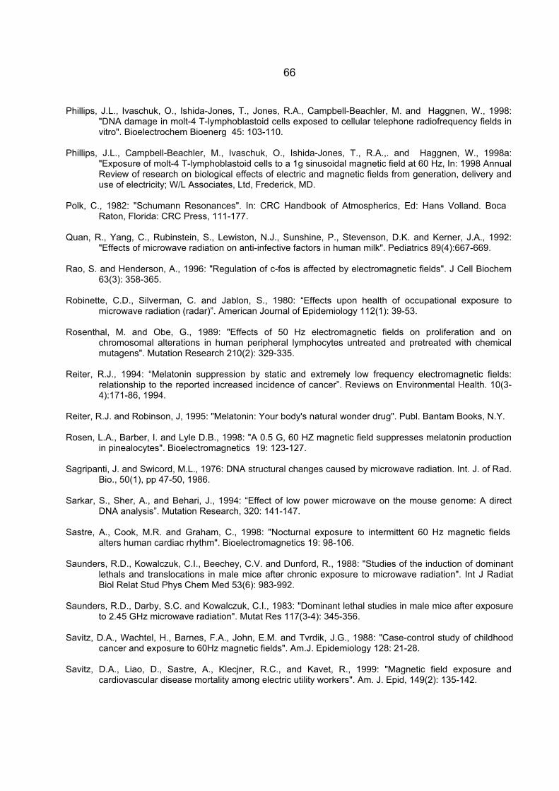
66
Phillips, J.L., Ivaschuk, O., Ishida-Jones, T., Jones, R.A., Campbell-Beachler, M. and Haggnen, W., 1998: "DNA damage in molt-4 T-lymphoblastoid cells exposed to cellular telephone radiofrequency fields in vitro". Bioelectrochem Bioenerg 45: 103-110.
Phillips, J.L., Campbell-Beachler, M., Ivaschuk, O., Ishida-Jones, T., R.A.,. and Haggnen, W., 1998a:
"Exposure of molt-4 T-lymphoblastoid cells to a 1g sinusoidal magnetic field at 60 Hz, In: 1998 Annual Review of research on biological effects of electric and magnetic fields from generation, delivery and use of electricity; W/L Associates, Ltd, Frederick, MD.
Polk, C., 1982: "Schumann Resonances". In: CRC Handbook of Atmospherics, Ed: Hans Volland. Boca
Raton, Florida: CRC Press, 111-177. Quan, R., Yang, C., Rubinstein, S., Lewiston, N.J., Sunshine, P., Stevenson, D.K. and Kerner, J.A., 1992:
"Effects of microwave radiation on anti-infective factors in human milk". Pediatrics 89(4):667-669. Rao, S. and Henderson, A., 1996: "Regulation of c-fos is affected by electromagnetic fields". J Cell Biochem
63(3): 358-365. Robinette, C.D., Silverman, C. and Jablon, S., 1980: “Effects upon health of occupational exposure to
microwave radiation (radar)”. American Journal of Epidemiology 112(1): 39-53. Rosenthal, M. and Obe, G., 1989: "Effects of 50 Hz electromagnetic fields on proliferation and on
chromosomal alterations in human peripheral lymphocytes untreated and pretreated with chemical mutagens". Mutation Research 210(2): 329-335.
Reiter, R.J., 1994: “Melatonin suppression by static and extremely low frequency electromagnetic fields:
relationship to the reported increased incidence of cancer”. Reviews on Environmental Health. 10(3-4):171-86, 1994.
Reiter, R.J. and Robinson, J, 1995: "Melatonin: Your body's natural wonder drug". Publ. Bantam Books, N.Y. Rosen, L.A., Barber, I. and Lyle D.B., 1998: "A 0.5 G, 60 HZ magnetic field suppresses melatonin production
in pinealocytes". Bioelectromagnetics 19: 123-127. Sagripanti, J. and Swicord, M.L., 1976: DNA structural changes caused by microwave radiation. Int. J. of Rad.
Bio., 50(1), pp 47-50, 1986. Sarkar, S., Sher, A., and Behari, J., 1994: “Effect of low power microwave on the mouse genome: A direct
DNA analysis”. Mutation Research, 320: 141-147. Sastre, A., Cook, M.R. and Graham, C., 1998: "Nocturnal exposure to intermittent 60 Hz magnetic fields
alters human cardiac rhythm". Bioelectromagnetics 19: 98-106. Saunders, R.D., Kowalczuk, C.I., Beechey, C.V. and Dunford, R., 1988: "Studies of the induction of dominant
lethals and translocations in male mice after chronic exposure to microwave radiation". Int J Radiat Biol Relat Stud Phys Chem Med 53(6): 983-992.
Saunders, R.D., Darby, S.C. and Kowalczuk, C.I., 1983: "Dominant lethal studies in male mice after exposure
to 2.45 GHz microwave radiation". Mutat Res 117(3-4): 345-356. Savitz, D.A., Wachtel, H., Barnes, F.A., John, E.M. and Tvrdik, J.G., 1988: "Case-control study of childhood
cancer and exposure to 60Hz magnetic fields". Am.J. Epidemiology 128: 21-28. Savitz, D.A., Liao, D., Sastre, A., Klecjner, R.C., and Kavet, R., 1999: "Magnetic field exposure and
cardiovascular disease mortality among electric utility workers". Am. J. Epid, 149(2): 135-142.
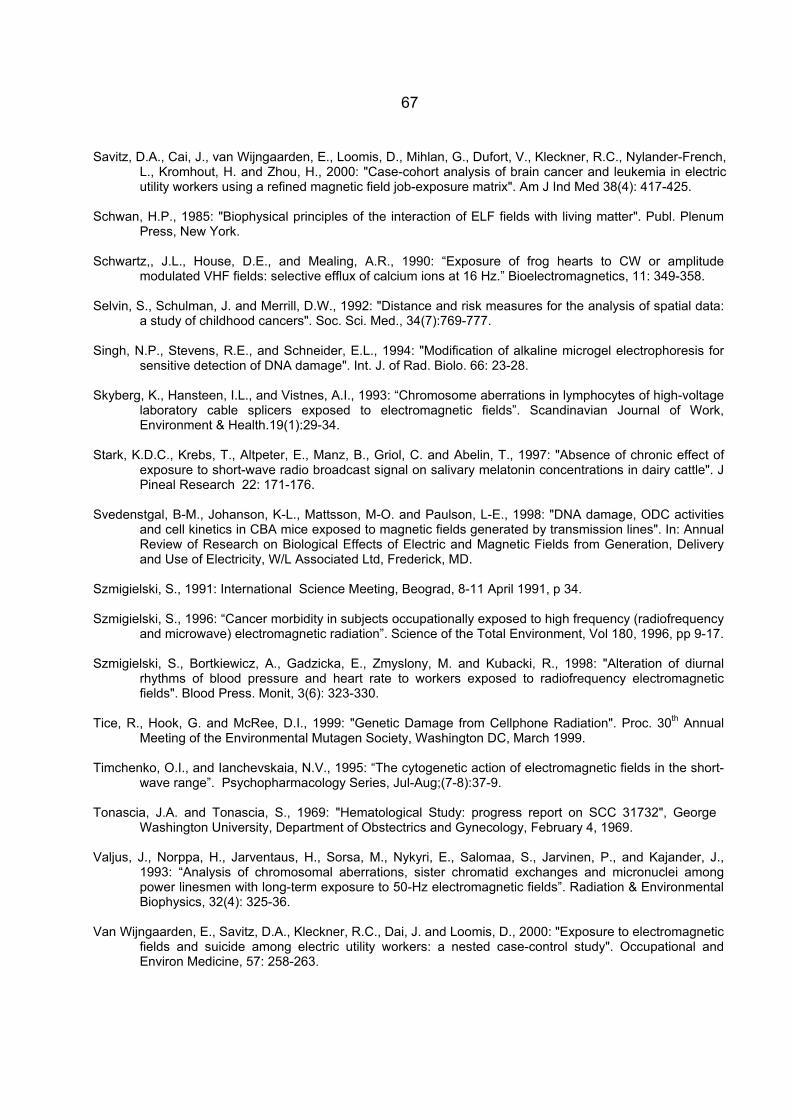
67
Savitz, D.A., Cai, J., van Wijngaarden, E., Loomis, D., Mihlan, G., Dufort, V., Kleckner, R.C., Nylander-French, L., Kromhout, H. and Zhou, H., 2000: "Case-cohort analysis of brain cancer and leukemia in electric utility workers using a refined magnetic field job-exposure matrix". Am J Ind Med 38(4): 417-425.
Schwan, H.P., 1985: "Biophysical principles of the interaction of ELF fields with living matter". Publ. Plenum
Press, New York. Schwartz,, J.L., House, D.E., and Mealing, A.R., 1990: “Exposure of frog hearts to CW or amplitude
modulated VHF fields: selective efflux of calcium ions at 16 Hz.” Bioelectromagnetics, 11: 349-358. Selvin, S., Schulman, J. and Merrill, D.W., 1992: "Distance and risk measures for the analysis of spatial data:
a study of childhood cancers". Soc. Sci. Med., 34(7):769-777. Singh, N.P., Stevens, R.E., and Schneider, E.L., 1994: "Modification of alkaline microgel electrophoresis for
sensitive detection of DNA damage". Int. J. of Rad. Biolo. 66: 23-28. Skyberg, K., Hansteen, I.L., and Vistnes, A.I., 1993: “Chromosome aberrations in lymphocytes of high-voltage
laboratory cable splicers exposed to electromagnetic fields”. Scandinavian Journal of Work, Environment & Health.19(1):29-34.
Stark, K.D.C., Krebs, T., Altpeter, E., Manz, B., Griol, C. and Abelin, T., 1997: "Absence of chronic effect of
exposure to short-wave radio broadcast signal on salivary melatonin concentrations in dairy cattle". J Pineal Research 22: 171-176.
Svedenstgal, B-M., Johanson, K-L., Mattsson, M-O. and Paulson, L-E., 1998: "DNA damage, ODC activities
and cell kinetics in CBA mice exposed to magnetic fields generated by transmission lines". In: Annual Review of Research on Biological Effects of Electric and Magnetic Fields from Generation, Delivery and Use of Electricity, W/L Associated Ltd, Frederick, MD.
Szmigielski, S., 1991: International Science Meeting, Beograd, 8-11 April 1991, p 34. Szmigielski, S., 1996: “Cancer morbidity in subjects occupationally exposed to high frequency (radiofrequency
and microwave) electromagnetic radiation”. Science of the Total Environment, Vol 180, 1996, pp 9-17. Szmigielski, S., Bortkiewicz, A., Gadzicka, E., Zmyslony, M. and Kubacki, R., 1998: "Alteration of diurnal
rhythms of blood pressure and heart rate to workers exposed to radiofrequency electromagnetic fields". Blood Press. Monit, 3(6): 323-330.
Tice, R., Hook, G. and McRee, D.I., 1999: "Genetic Damage from Cellphone Radiation". Proc. 30th Annual
Meeting of the Environmental Mutagen Society, Washington DC, March 1999. Timchenko, O.I., and Ianchevskaia, N.V., 1995: “The cytogenetic action of electromagnetic fields in the short-
wave range”. Psychopharmacology Series, Jul-Aug;(7-8):37-9. Tonascia, J.A. and Tonascia, S., 1969: "Hematological Study: progress report on SCC 31732", George
Washington University, Department of Obstectrics and Gynecology, February 4, 1969. Valjus, J., Norppa, H., Jarventaus, H., Sorsa, M., Nykyri, E., Salomaa, S., Jarvinen, P., and Kajander, J.,
1993: “Analysis of chromosomal aberrations, sister chromatid exchanges and micronuclei among power linesmen with long-term exposure to 50-Hz electromagnetic fields”. Radiation & Environmental Biophysics, 32(4): 325-36.
Van Wijngaarden, E., Savitz, D.A., Kleckner, R.C., Dai, J. and Loomis, D., 2000: "Exposure to electromagnetic
fields and suicide among electric utility workers: a nested case-control study". Occupational and Environ Medicine, 57: 258-263.
68
Verkasalo, P.K., Kaprio, J., Varjonen, J., Romanov, K., Heikkila, K., and Koskenvuo, M., 1997: "Magnetic fields of transmission lines and depression". Am. J. Epidemiology, 146(12): 1037-45.
Verschaeve, L., Slaets, D., Van Gorp, U., Maes, A. and Vanderkom, J., 1994: "In vitro and in vivo genetic
effects of microwaves from mobile phone frequencies in human and rat peripheral blood lymphocytes". Proceedings of Cost 244 Meetings on Mobile Communication and ELF field: Instrumentation and measurements in Bioelectromagnetics Research. Ed. D, Simunic, pp 74-83.
Vignati, M. and Giuliani, L., 1997: “Radiofrequency exposure near high-voltage lines”. Environmental Health
Perspectives, 105 (Suppl 6): 1569-1573. Vijayalaxmi, B.Z., Frei, M.R., Dusch, S.J., Guel, V., Meltz, M.L. and Jauchem, J.R., 1997: "Frequency of
micronuclei in the peripheral blood and bone marrow of cancer-prone mice chronically exposed to 2450 MHz radiofrequency radiation". Radiation Research, 147: 495-500.
Vijayalaxmi, B.Z., Frei, M.R., Dusch, S.J., Guel, V., Meltz, M.L. and Jauchem, J.R., 1997a: "Frequency of
micronuclei in the peripheral blood and bone marrow of cancer-prone mice chronically exposed to 2450 MHz radiofrequency radiation - a correction". Radiation Research, 148:
Walleczek, J., 1992: “Electromagnetic field effects on cells of the immune system: the role of calcium
signaling”. FASEB J., 6: 3176-3185. Wang, S.G. 1989: "5-HT contents change in peripheral blood of workers exposed to microwave and high
frequency radiation". Chung Hua Yu Fang I Hsueh Tsa Chih 23(4): 207-210. Wei, L.X., Goodman, R. and Henderson, A., 1990: "Changes in levels of c-myc and histone H2B following
exposure of cells to low-frequency sinusoidal electromagnetic fields: evidence for a window effect". Bioelectromagnetics, 11(4): 269-272.
Wertheimer, N. and Leeper, E.,1979: "Electrical wiring configurations and childhood cancer". Am. J.
Epidemiology 109: 273-284. Wever R. 1974, ELF-effects on Human Circadian Rhythms. In: Persinger MA editor. ELF and VLF
Electromagnetic Field Effects. New York, Plenum Press. p 101-144. Williams, G.M., 1996: "Comment on 'Acute low-intensity microwave exposure increases DNA single-strand
breaks in rat brain cells' "by Henry Lai and Narendra P. Singh. Bioelectromagnetics 17: 165. Wilson, B.W., Wright, C.W., Morris, J.E., Buschbom, R.L., Brown, D.P., Miller, D.L., Sommers-Flannigan, R.
and Anderson, L.E., 1990: "Evidence of an effect of ELF electromagnetic fields on human pineal gland function". J Pineal Research 9(4): 259-269.
Wood, A.W., Armstrong, S.M., Sait, M.L., Devine, L. and Martin, M.J., 1998: "Changes in human plasma
melatonin profiles in response to 50 Hz magnetic field exposure". J Pineal Research 25(2): 116-127. Yao, K.T., 1978: "Microwave radiation-induced chromosomal aberrations in corneal epithelium of Chinese
hamsters". J Hered 69(6): 409-412. Yao. K.T., 1982: "Cytogenetic consequences of microwave irradiation on mammalian cells incubated in vitro".
J Hered 73(2): 133-138. Youbicier-Simo, B.J., Lebecq, J.C.,and Bastide, M., 1999: "Mortality of chicken embryos exposed to EMFs
from mobile phones". Presented at the 20th Annual meeting of the Bioelectromagnetics Society, St Pete Beach, FL, June 1999.