Embed Size (px)
Citation preview
Please cite this article as follows: Gholizadeh N, Beygom Taheri J, Namazi Z, Mashhadiabbas F, Bakhtiari S, Rahimzamani A, et al. Excision of different oral benign exophytic lesions with a diode laser: A clinical Case Series. J Lasers Med Sci. 2020;11(4):502-508. doi:10.34172/jlms.2020.80.
Case Series
doi 10.34172/jlms.2020.80
Excision of Different Oral Benign Exophytic Lesions With a Diode Laser: A Clinical Case Series Narges Gholizadeh1, Jamileh Beygom Taheri2, Zahra Namazi3, Fatemeh Mashhadiabbas4, Seddigheh Bakhtiari5, Arezoo Rahimzamani6, Mohammad Asnaashari7*
1Associate Professor, Department of Oral and Maxillofacial Medicine, Faculty of Dentistry, Tehran University of Medical Sciences, Tehran, Iran2Professor, Department of Oral Medicine, Faculty of Dentistry, Shahid Beheshti University of Medical Sciences, Tehran, Iran3Assistant Professor, Department of Dental Biomaterials, Faculty of Dentistry, Shahid Beheshti University of Medical Sciences, Tehran, Iran4Associate Professor, Oral & Maxillofacial Pathology, School of Dentistry, Shahid Beheshti University of Medical Science, Tehran, Iran5Associate Professor of Oral & Maxillofacial Medicine, Department of Oral Diseases, School of Dentistry, Shahid Beheshti University of Medical Sciences, Tehran, Iran6Post-graduate Student of Oral & Maxillofacial Medicine, Department of Oral Diseases, School of Dentistry, Shahid Beheshti University of Medical Sciences, Tehran, Iran7Professor of Endodontics, Laser Application in Medical Sciences Research Center, Shahid Beheshti University of Medical Sciences, Tehran, Iran
AbstractIntroduction: Lasers are becoming the standard of care for many dental procedures, and are being introduced as a high-tech instrument. They are also becoming more routine in dentistry through the advent of office-based lasers, which are also simple to use within the oral cavity. Many studies have shown the competencies of laser technology for the management of benign oral lesions as these techniques allow for painless and bloodless oral surgery.Cases Report: Nine patients attending the Department of oral and maxillofacial Disease, Shahid Beheshti University of Medical Sciences, Tehran, Iran, underwent laser outpatient procedures for the surgical removal of a wide range of benign oral lesions. Regarding the history, present illnesses and clinical figures of all reported cases, the clinician ensured that the lesions were benign and after taking informed consent from every patient, he did complete the excision of lesions with diode lasers. Following the injection of local anesthesia, an 810 nm diode laser was applied for the excisional biopsy of oral lesions. The specimens were sent for histopathological evaluations and the patients were assessed on intraoperative and postoperative complications. The patients were followed up for postoperative complications at one week and 2 weeks post-treatment.Conclusions: According to our findings, a diode laser can be a choice for the outpatient treatment of oral mucosal benign lesions as this technique provides painless and almost bloodless treatment.Keywords: Oral lesions; Benign; Soft tissue; Laser.
*Correspondence toMohammad Asnaashari,Address: DDS, MS Professor of endodontics, Laser Application in Medical Sciences Research Center, Shahid Beheshti University of Medical Sciences, Tehran, Iran.Tel: +98 9121145860;Email: [email protected]
Published online October 3, 2020
Journal of
Lasersin Medical Sciences
J Lasers Med Sci 2020 Autumn;11(4):502-508
http://journals.sbmu.ac.ir/jlms
Introduction Over the last decade, lasers have been accepted as an alternative to traditional methods in surgery. This is due to the fact that many procedures can be performed more efficiently and with less morbidity using lasers as compared with scalps.1-3 A wide range of benign oral lesions exists as either an isolated oral finding or the manifestation of dermatologic conditions.4 These lesions can be removed by different surgical procedures such as the conventional scalpel, electrosurgical scalpel, or lasers.5,6 Lasers have beneficial and safe clinical applications for oral medicine specialists in both diagnosis
and treatment of different forms of oral mucosal lesions. Some of these applications include the treatment of facial pigmentation or vascular lesions, the excisional biopsy of oral lesions, frenectomy, epulis fissuratum, fibroma, gingival enlargements, gingivectomies, and certain crown lengthening procedures.1,7,8 In addition, surgery by the laser has become a reliable treatment option for oral mucosal lesions.9 Lasers that are frequently used in the oral surgical processes are Er:YAG (erbium-doped yttrium aluminium garnet laser), carbon dioxide (CO2), KTM Nd:YAG (neodymium-doped yttrium aluminum garnet), and diode, and are considered as an efficient
Journal of Lasers in Medical Sciences Volume 11, Number 4, Autumn 2020 503
Excision of Oral Benign Exophytic Lesions With a Diode Laser
instrument for some procedures within this specialty.10,11 We could consider a laser as a minimally invasive
technique with many advantages such as remote application, better visibility in the surgical field, precise cutting, hemostasis, minor cicatrization, better infection control, excellent wound healing, reduced postoperative pain and swelling, and patient acceptance.8,11,12
In this study, we report the removal of a benign exophytic lesion from the oral cavity by a laser to emphasize the features of this technique and assess its consequences on living tissues.
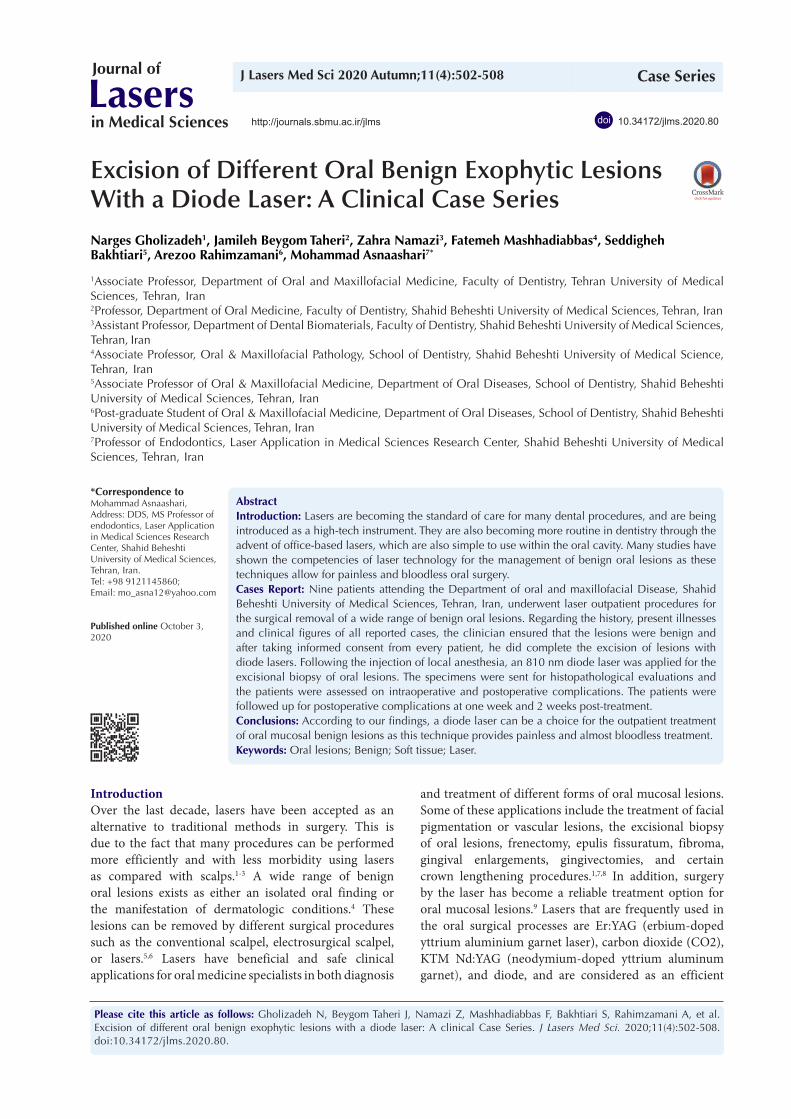
Case 1A 26-year-old pregnant woman in the third trimester was referred to the department of oral and maxillofacial diseases, Shahid Beheshti Dental School, with her chief complaint being soft tissue swelling of the anterior maxilla. Clinical examination showed that there was an exophytic smooth surface nodule with a sessile, red-colored, and popular surface near the tooth region, approximately 0.5 cm × 1 cm in dimension. The lesion bled by a little trauma and the patient had poor oral hygiene (Figure 1A). Her past medical history was clear. The lesion was excised completely from its base after local anesthesia injection by a diode laser with an 810 nm continuous wavelength and 3 W power and 300 µm-fiber tips for 180 seconds (Dr. Smile, Italia). The fiber tip got in contact with the lesion during surgery (Figure 1B). Histopathologic evaluations
showed pyogenic granuloma as the test result. In this case, the amount of bleeding was far less than expected (Figure 1C).
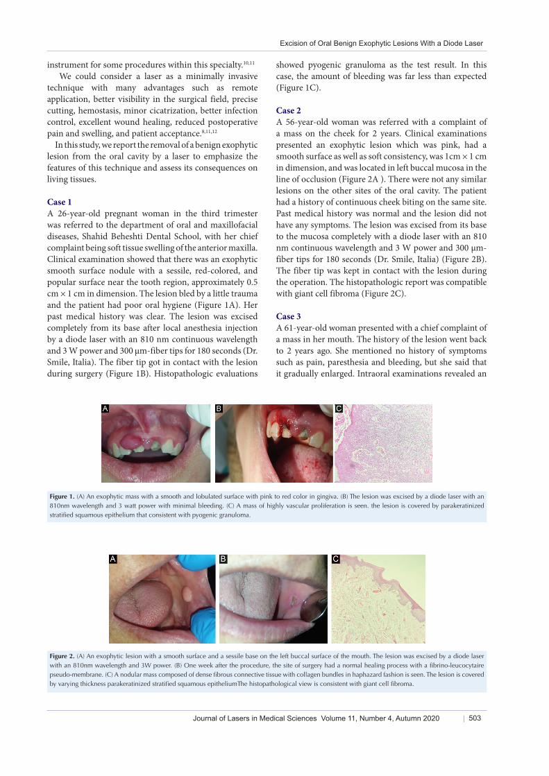
Case 2A 56-year-old woman was referred with a complaint of a mass on the cheek for 2 years. Clinical examinations presented an exophytic lesion which was pink, had a smooth surface as well as soft consistency, was 1cm × 1 cm in dimension, and was located in left buccal mucosa in the line of occlusion (Figure 2A ). There were not any similar lesions on the other sites of the oral cavity. The patient had a history of continuous cheek biting on the same site. Past medical history was normal and the lesion did not have any symptoms. The lesion was excised from its base to the mucosa completely with a diode laser with an 810 nm continuous wavelength and 3 W power and 300 µm-fiber tips for 180 seconds (Dr. Smile, Italia) (Figure 2B). The fiber tip was kept in contact with the lesion during the operation. The histopathologic report was compatible with giant cell fibroma (Figure 2C).
Case 3A 61-year-old woman presented with a chief complaint of a mass in her mouth. The history of the lesion went back to 2 years ago. She mentioned no history of symptoms such as pain, paresthesia and bleeding, but she said that it gradually enlarged. Intraoral examinations revealed an
Figure 1. (A) An exophytic mass with a smooth and lobulated surface with pink to red color in gingiva. (B) The lesion was excised by a diode laser with an 810nm wavelength and 3 watt power with minimal bleeding. (C) A mass of highly vascular proliferation is seen. the lesion is covered by parakeratinized stratified squamous epithelium that consistent with pyogenic granuloma.
Figure 2. (A) An exophytic lesion with a smooth surface and a sessile base on the left buccal surface of the mouth. The lesion was excised by a diode laser with an 810nm wavelength and 3W power. (B) One week after the procedure, the site of surgery had a normal healing process with a fibrino-leucocytaire pseudo-membrane. (C) A nodular mass composed of dense fibrous connective tissue with collagen bundles in haphazard fashion is seen. The lesion is covered by varying thickness parakeratinized stratified squamous epitheliumThe histopathological view is consistent with giant cell fibroma.
Gholizadeh et al
Journal of Lasers in Medical Sciences Volume 11, Number 4, Autumn 2020504
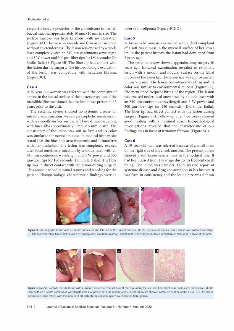
exophytic nodule posterior of the commissure in the left buccal mucosa, approximately 10 mm×10 mm in size. The surface mucosa was hyperkeratotic, with no ulceration (Figure 3A). The mass was sessile and firm in consistency, without any tenderness. The lesion was excised by a diode laser completely with an 810 nm continuous wavelength and 3 W power and 300 µm-fiber tips for 180 seconds (Dr. Smile, Italia) ( Figure 3B).The fiber tip had contact with the lesion during surgery. The histopathologic evaluation of the lesion was compatible with irritation fibroma (Figure 3C).
Case 4A 50-year-old woman was referred with the complaint of a mass in the buccal surface of the posterior section of the mandible. She mentioned that the lesion was present for 3 years prior to the visit.
The systemic review showed no systemic disease. In intraoral examinations, we saw an exophytic sessile lesion with a smooth surface on the left buccal mucosa, along with linea alba approximately 5 mm × 5 mm in size. The consistency of the lesion was soft to firm and its color was similar to the normal mucosa. In medical history, she stated that she bites this area frequently and it interferes with her occlusion. The lesion was completely excised after local anesthesia injection by a diode laser with an 810 nm continuous wavelength and 3 W power and 300 µm-fiber tips for 180 seconds (Dr. Smile, Italia). The fiber tip was in direct contact with the lesion during surgery. This procedure had minimal trauma and bleeding for the patient. Histopathologic characteristic findings were in
favor of fibrolipoma (Figure 4C&D).
Case 5A 34-year-old woman was visited with a chief complaint of a soft tissue mass in the mucosal surface of her lower lip. In the patient history, the lesion had developed from 2 years ago.
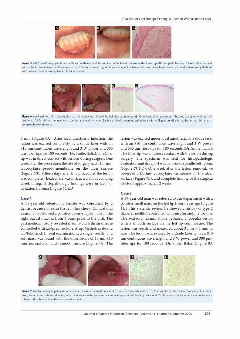
The systemic review showed appendectomy surgery 10 years ago. Intraoral examination revealed an exophytic lesion with a smooth and nodular surface on the labial mucosa of the lower lip. The lesion size was approximately 5 mm × 5 mm. The lesion consistency was firm and its color was similar to environmental mucosa (Figure 5A). She mentioned frequent biting of the region. The lesion was excised under local anesthesia by a diode laser with an 810 nm continuous wavelength and 3 W power and 300 µm-fiber tips for 180 seconds) (Dr. Smile, Italia). The fiber tip had direct contact with the lesion during surgery (Figure 5B). Follow-up after two weeks showed good healing with a minimal scar. Histopathological investigations revealed that the characteristic of our findings was in favor of irritation fibroma (Figure 5C).
Case 6A 19-year-old man was referred because of a small mass on the right side of his cheek mucosa. The present illness showed a soft tissue sessile mass in the occlusal line. It had been raised from 1 year ago due to his frequent cheek biting. The lesion was painless. There was no report of systemic disease and drug consumption in his history. It was firm in consistency and the lesion size was 5 mm×
Figure 3. (A) Exophytic lesion with a smooth surface on the left part of the buccal mucosa. (B) The excision of lesions with a diode laser without bleeding. (C) Fibrous connective tissue that covered by hyperplastic stratified squamous epithelium with collagen bundles in haphazard fashion is in favor of fibroma.
Figure 4. (A) An Exophytic sessile lesion with a smooth surface on the left buccal mucosa, along the occlusal line which was completely excised by a diode laser with an 810 nm continuous wavelength and 3 W power. (B) One month later, clinical follow-up showed complete healing of the tissue. (C&D) Fibrous connective tissue mixed with the lobules of fat cells ,this histopathologic view explained fibrolipoma.
Journal of Lasers in Medical Sciences Volume 11, Number 4, Autumn 2020 505
Excision of Oral Benign Exophytic Lesions With a Diode Laser
5 mm (Figure 6A). After local anesthesia injection, the lesion was excised completely by a diode laser with an 810 nm continuous wavelength and 3 W power and 300 µm-fiber tips for 180 seconds (Dr. Smile, Italia). The fiber tip was in direct contact with lesions during surgery. One week after the procedure, the site of surgery had a fibrino-leucocytaire pseudo-membrane on the ulcer surface (Figure 6B). Fifteen days after this procedure, the lesion was completely healed. He was instructed about avoiding cheek biting. Histopathologic findings were in favor of irritation fibroma (Figure 6C&D).
Case 7A 59-year-old edentulous female was consulted by a dentist because of extra tissue in her cheek. Clinical oral examination showed a painless dome-shaped mass in the right buccal mucosa from 3 years prior to the visit. The past medical history revealed rheumatoid arthritis disease controlled with tab/prednisolone, Amp. Methotrexate and tab/folic acid. In oral examinations, a single, sessile, and soft mass was found with the dimensions of 10 mm×10 mm, normal color and a smooth surface (Figure 7A). The
lesion was excised under local anesthesia by a diode laser with an 810 nm continuous wavelength and 3 W power and 300 µm-fiber tips for 180 seconds (Dr. Smile, Italia). The fiber tip was in direct contact with the lesion during surgery. The specimen was sent for histopathologic evaluation and its report was in favor of spindle cell lipoma (Figure 7C&D). One week after the lesion removal, we observed a fibrino-leucocytaire membrane on the ulcer surface (Figure 7B), and complete healing of the surgical site took approximately 2 weeks.
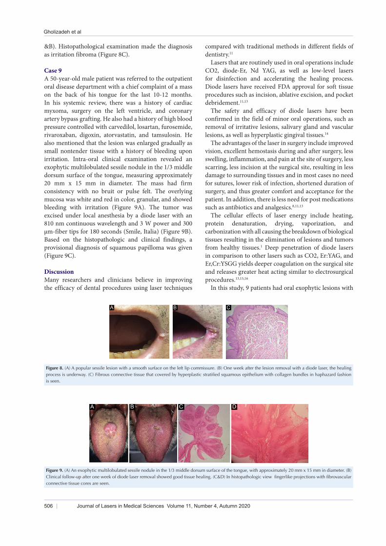
Case 8A 59-year-old man was referred to our department with a painless small mass on the left lip from 1 year ago (Figure 1). In his systemic review, he showed a history of type 2 diabetes mellitus controlled with insulin and metformin. The extraoral examinations revealed a popular lesion with a smooth surface on the left lip commissure. The lesion was sessile and measured about 3 mm × 4 mm in size. The lesion was excised by a diode laser with an 810 nm continuous wavelength and 3 W power and 300 µm-fiber tips for 180 seconds (Dr. Smile, Italia) (Figure 8A
Figure 5. (A) A small exophytic lesion with a smooth and nodular surface on the labial mucosa of the lower lip. (B) Complete healing of tissue after removal with a diode laser in one-month follow-up. (C) In histopathologc figure fibrous connective tissue that covered by hyperplastic stratified squamous epithelium with collagen bundles in haphazard fashion is seen.
Figure 6. (A) A painless, firm and sessile mass in the occlusal line of the right buccal mucosa. (B) One week after laser surgery, healing was good without any problem. (C&D) fibrous connective tissue that covered by hyperplastic stratified squamous epithelium with collagen bundles in haphazard fashion that is compatible with fibroma.
Figure 7. (A) An exophytic painless dome-shaped mass in the right buccal mucosa with a smooth surface. (B) One week after the lesion removal with a diode laser, we observed a fibrino-leucocytaire membrane on the ulcer surface indicating a normal healing process. (C & D) presence of lobules of mature fat cells intermixed with spindle cells in a myxoid stroma.
A B C D
Gholizadeh et al
Journal of Lasers in Medical Sciences Volume 11, Number 4, Autumn 2020506
&B). Histopathological examination made the diagnosis as irritation fibroma (Figure 8C).
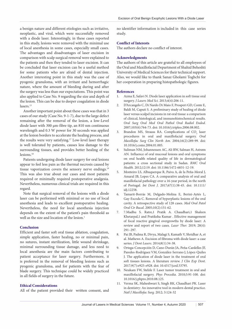
Case 9A 50-year-old male patient was referred to the outpatient oral disease department with a chief complaint of a mass on the back of his tongue for the last 10-12 months. In his systemic review, there was a history of cardiac myxoma, surgery on the left ventricle, and coronary artery bypass grafting. He also had a history of high blood pressure controlled with carvedilol, losartan, furosemide, rivaroxaban, digoxin, atorvastatin, and tamsulosin. He also mentioned that the lesion was enlarged gradually as small nontender tissue with a history of bleeding upon irritation. Intra-oral clinical examination revealed an exophytic multilobulated sessile nodule in the 1/3 middle dorsum surface of the tongue, measuring approximately 20 mm x 15 mm in diameter. The mass had firm consistency with no bruit or pulse felt. The overlying mucosa was white and red in color, granular, and showed bleeding with irritation (Figure 9A). The tumor was excised under local anesthesia by a diode laser with an 810 nm continuous wavelength and 3 W power and 300 µm-fiber tips for 180 seconds (Smile, Italia) (Figure 9B). Based on the histopathologic and clinical findings, a provisional diagnosis of squamous papilloma was given (Figure 9C).
DiscussionMany researchers and clinicians believe in improving the efficacy of dental procedures using laser techniques
compared with traditional methods in different fields of dentistry.11
Lasers that are routinely used in oral operations include CO2, diode-Er, Nd YAG, as well as low-level lasers for disinfection and accelerating the healing process. Diode lasers have received FDA approval for soft tissue procedures such as incision, ablative excision, and pocket debridement.11,13
The safety and efficacy of diode lasers have been confirmed in the field of minor oral operations, such as removal of irritative lesions, salivary gland and vascular lesions, as well as hyperplastic gingival tissues.14
The advantages of the laser in surgery include improved vision, excellent hemostasis during and after surgery, less swelling, inflammation, and pain at the site of surgery, less scarring, less incision at the surgical site, resulting in less damage to surrounding tissues and in most cases no need for sutures, lower risk of infection, shortened duration of surgery, and thus greater comfort and acceptance for the patient. In addition, there is less need for post medications such as antibiotics and analgesics.8,11,13
The cellular effects of laser energy include heating, protein denaturation, drying, vaporization, and carbonization with all causing the breakdown of biological tissues resulting in the elimination of lesions and tumors from healthy tissues.1 Deep penetration of diode lasers in comparison to other lasers such as CO2, Er:YAG, and Er,Cr:YSGG yields deeper coagulation on the surgical site and releases greater heat acting similar to electrosurgical procedures.13,15,16
In this study, 9 patients had oral exophytic lesions with
Figure 8. (A) A popular sessile lesion with a smooth surface on the left lip commissure. (B) One week after the lesion removal with a diode laser, the healing process is underway. (C) Fibrous connective tissue that covered by hyperplastic stratified squamous epithelium with collagen bundles in haphazard fashion is seen.
Figure 9. (A) An exophytic multilobulated sessile nodule in the 1/3 middle dorsum surface of the tongue, with approximately 20 mm x 15 mm in diameter. (B)Clinical follow-up after one week of diode laser removal showed good tissue healing. (C&D) In histopathologic view fingerlike projections with fibrovascular connective tissue cores are seen.
Journal of Lasers in Medical Sciences Volume 11, Number 4, Autumn 2020 507
Excision of Oral Benign Exophytic Lesions With a Diode Laser
a benign nature and different etiologies such as irritative, neoplastic, and viral, which were successfully removed with a diode laser. Interestingly, in these cases reported in this study, lesions were removed with the minimal use of local anesthesia in some cases, especially small cases. The advantages and disadvantages of laser excision in comparison with scalp surgical removal were explained to the patients and then they tended to laser excision. It can be concluded that laser excision can be a useful method for some patients who are afraid of dental injection. Another interesting point in this study was the case of pyogenic granuloma, with an irritant and hemorrhagic nature, where the amount of bleeding during and after the surgery was less than our expectations. This point was also applied to Case No. 9 regarding the size and depth of the lesion. This can be due to deeper coagulation in diode lasers.13
Another important point about these cases was that in 3 cases of our study (Case No. 9-1-7), due to the large defect remaining after the removal of the lesion, a low-Level diode laser with 300 µm-fibre tip, an 810 nm continuous wavelength and 0.3 W power for 30 seconds was applied at the lesion borders to accelerate the healing process, and the results were very satisfying.17 Low-level laser therapy is well tolerated by patients, causes less damage to the surrounding tissues, and provides better healing of the lesions.18
Patients undergoing diode laser surgery for oral lesions appear to feel less pain as the thermal necrosis caused by tissue vaporization covers the sensory nerve endings.19 This was also true about our cases and most patients required or minimally required postoperative analgesia. Nevertheless, numerous clinical trials are required in this regard.
Note that surgical removal of the lesions with a diode laser can be performed with minimal or no use of local anesthesia and leads to excellent postoperative healing. Nevertheless, the need for local anesthesia injection depends on the extent of the patient’s pain threshold as well as the size and location of the lesion.1
Conclusion Efficient and faster soft oral tissue ablation, coagulation, simple application, faster healing, no or minimal pain, no sutures, instant sterilization, little wound shrinkage, minimal surrounding tissue damage, and less need to local anesthesia are the main factors contributing to patient acceptance for laser surgery. Furthermore, it is preferred in the removal of bleeding lesions such as pyogenic granuloma, and for patients with the fear of blade surgery. This technique could be widely practiced in all fields of surgery in the future.
Ethical ConsiderationsAll of the patient provided their written consent, and
no identifier information is included in this case series study.
Conflict of InterestsThe authors declare no conflict of interest.
AcknowledgmentsThe authors of this article are grateful to all employees of the Oral and Maxillofacial Department of Shahid Beheshti University of Medical Sciences for their technical support. Also, we would like to thank Sanaz Gholami Toghchi for her cooperation in preparing histopathologic figures.
References1. Azma E, Safavi N. Diode laser application in soft tissue oral
surgery. J Lasers Med Sci. 2013;4(4):206-11. 2. D’Arcangelo C, Di Nardo Di Maio F, Prosperi GD, Conte E,
Baldi M, Caputi S. A preliminary study of healing of diode laser versus scalpel incisions in rat oral tissue: a comparison of clinical, histological, and immunohistochemical results. Oral Surg Oral Med Oral Pathol Oral Radiol Endod. 2007;103(6):764-73. doi: 10.1016/j.tripleo.2006.08.002.
3. Brandon MS, Strauss RA. Complications of CO2 laser procedures in oral and maxillofacial surgery. Oral Maxillofac Surg Clin North Am. 2004;16(2):289-99. doi: 10.1016/j.coms.2004.01.005.
4. Suliman NM, Johannessen AC, Ali RW, Salman H, Astrøm AN. Influence of oral mucosal lesions and oral symptoms on oral health related quality of life in dermatological patients: a cross sectional study in Sudan. BMC Oral Health. 2012;12:19. doi: 10.1186/1472-6831-12-19.
5. Monteiro LS, Albuquerque R, Paiva A, de la Peña-Moral J, Amaral JB, Lopes CA. A comparative analysis of oral and maxillofacial pathology over a 16-year period, in the north of Portugal. Int Dent J. 2017;67(1):38-45. doi: 10.1111/idj.12258.
6. Tamarit-Borrás M, Delgado-Molina E, Berini-Aytés L, Gay-Escoda C. Removal of hyperplastic lesions of the oral cavity. A retrospective study of 128 cases. Med Oral Patol Oral Cir Bucal. 2005;10(2):151-62.
7. 7.Madhu S. Ratre,1 Pratik A. Chaudhari,1 Shaleen Khetarpal,1 and Pratiksha Kumar . Effective management of focal reactive gingival overgrowths by diode laser: A review and report of two cases. Laser Ther. 2019; 28(4): 291–297.
8. Pai JB, Padma R, Divya, Malagi S, Kamath V, Shridhar A, et al. Mathews A. Excision of fibroma with diode laser: a case series. J Dent Lasers. 2014;8(1):34-38.
9. Ortega-Concepción D, Cano-Durán JA, Peña-Cardelles JF, Paredes-Rodríguez VM, González-Serrano J, López-Quiles J. The application of diode laser in the treatment of oral soft tissues lesions. A literature review. J Clin Exp Dent. 2017;9(7):e925-e928. doi: 10.4317/jced.53795.
10. Neukam FW, Stelzle F. Laser tumor treatment in oral and maxillofacial surgery. Phys Procedia. 2010;5:91-100. doi: 10.1016/j.phpro.2010.08.125.
11. Verma SK, Maheshwari S, Singh RK, Chaudhari PK. Laser in dentistry: An innovative tool in modern dental practice. Natl J Maxillofac Surg. 2012; 3:124-32
Gholizadeh et al
Journal of Lasers in Medical Sciences Volume 11, Number 4, Autumn 2020508
12. Pandurić DG, Bago I, Zore IF, Sušić M, Katanec D, Milenović A, et al. In: Kalantar Motamedi MH, editor. A textbook of advanced oral and maxillofacial surgery. Rijeka, Croatia: InTech; 2013. p. 341-382. doi: 10.5772/3316.
13. Seyyedi SA, Khashabi E, Falaki F. Laser application in periodontics. J Lasers Med Sci. 2012; 3(1):26-32.
14. Mazarei Sotoode S, Azimi S, Taheri SA, Asnaashari M, Kahlighi H, Rahmani S, et al. Diode laser in minor oral surgery: a case series of laser removal of different benign exophytic lesions. J Lasers Med Sci. 2015;6(3):133-138. doi:10.15171/jlms.2015.08.
15. Aoki A, Sasaki KM, Watanabe H, Ishikawa I. Lasers in non-surgical periodontal therapy. Periodontol 2000. 2004;36:59-97.
16. Ishikawa I, Aoki A, Takasaki AA, Mizutani K, Sasaki KM, Izumi Y. Application of lasers in periodontics: true
innovation or myth? Periodontol 2000. 2009;50:90-126. doi: 10.1111/j.1600-0757.2008.00283.x.
17. Schindl M, Kerschan K, Schindl A, Schon H, Heinzl H, Schindl L. Induction of complete wound healing in recalcitrant ulcers by low-intensity laser irradiation depends on ulcer cause and size. Photodermatol Photoimmunol Photomed. 1999;15(1):18-21. doi: 10.1111/j.1600-0781.1999.tb00047.x.
18. Najafi S, Sheykhbahaei N, khayamzadeh M, Gholizadeh N. The effect of low level laser on number of Candida albicans colonies in-vitro: a new finding. BMC Oral Health. 2019;19:4. doi: 10.1186/s12903-019-0814-5.
19. Wyman A, Duffy S, Sweetland HM, Sharp F, Rogers K. Preliminary evaluation of a new high power diode laser. Lasers Surg Med. 1992;12(5):506-9. doi: 10.1002/lsm.1900120509.