Embed Size (px)
Citation preview
1
Exosomes in Cancer Research
Sarah R. Vaiselbuh, MD
Children’s Cancer Center,
Staten Island University Hospital, Staten Island-New York
Corresponding Author:
Sarah R. Vaiselbuh, MD
Director Children’s Cancer Center
Staten Island University Hospital
Staten Island – New York 10305
USA
Tel: 718 226 6435
Assistant Investigator
The Feinstein Institute for Medical Research
350 Community Drive
Manhasset-NY 11030
USA

2
Abstract
Next generation sequencing has provided the ability to screen for novel microRNA (miR)
biomarkers in biofluids of patients with cancer. Extravesicular vesicles in the peripheral blood,
known as exosomes, provide a reliable source of miR for disease biomarker detection. The
molecular content of exosomes, readily available in body fluids such as blood, urine and saliva,
is highly specific and a powerful biomedical tool. Exosomes generated during tumorigenesis
and derived from cancer cells provide the perfect cancer fingerprint, detectable in peripheral
blood. In addition, since cancer exosomes are messengers for signaling and alteration of the
tumor microenvironment, it is no surprise that cancer features such as angiogenesis,
chemoresistance and metastasis are associated with them, and their ability to facilitate the
formation of a pre-metastatic niche as a primer for implantation of circulating tumor cells. The
aim of this paper is to provide a review of the state-of-the-art of exosomes in cancer research,
their role in cancer niche development with clinical correlation as biomarkers for cancer
diagnosis and prognosis, as well as their future use in exo-therapy in the era of precision
oncology medicine.

3
Introduction
Newer and less toxic treatments for cancer are desperately needed as the group of cancer
survivors teaches us about long-term effects of current standard chemotherapy. In addition,
identification of novel minimal invasive biomarkers for early cancer detection may improve
treatment outcomes. Cancer biomarkers are informative of the pathological status for
diagnosis and prognosis of a disease and have received increasing attention in cancer research
for screening, early diagnosis and detection of relapse of solid tumors.
One such biomarkers group is a class of small non-coding RNAs, termed microRNAs (miRs).
An intriguing new strategy for biomarker detection is analysis of miR content of exosomes,
isolated from body fluids of cancer patients. Exosomes are extracellular lipid microvesicles
(40-100 nm), secreted by nearly all cells in body fluids (1) such as peripheral blood (2), urine
(3), saliva, breast milk (4), cerebrospinal fluid and malignant effusions (5). They contain
functional biomolecules including oncogenic proteins, lipids, ssDNA and dsDNA (6) and are
perceived to be carriers of unique signaling molecules like miRs (7). By membrane
invagination of late endosomes, exosomal vesicles are formed that contain cytosolic content of
the cell of origin in healthy as well as pathological cells (8). Upon release in the body fluid
microenvironment, the exosomes represent the fingerprint of the releasing cell type that can be
horizontally transferred to target cells. The vesicle-associated RNA is called exosomal shuttle
RNA (esRNA) that includes miRs as well as mRNA and are functional in target cells where
they effectively silence genes (9, 10). Twelve years after the first major review (8), exosome
research is about to explode especially in the field of cancer pathophysiology (11, 12). As
vehicles of intercellular communication, exosomes divulge important roles in cancer
tumorigenesis and metastatic disease (13). Tumor-specific exosomes are mediators of

4
oncogenesis as they exhibit tissue-specific addresses and the ability to disturb the exquisite
precise regulatory mechanism of secretion and adhesion might contribute to cancer
pathogenesis (i.e. mail delivered to the wrong address). The spectrum of current scientific
interest in exosomes ranges from studying functions and pathways of exosomes to utilizing
them in diagnostics and development of new therapeutics (14, 15), some of which are already
in Phase I clinical trial for melanoma (16).
Exosomes biogenesis in cancer
Exosomes were first identified in 1985 as small vesicles floating in supernatant of in vitro
cultured adherent cell layers that were associated with plasma membrane enzymatic activity of
the parent cells (17, 18). Historically however in the late 70s, extracellular vesicles have been
isolated from physiological media such as seminal plasma and prostatic fluid, hence called
“prostasomes” after the organ of origin ie. prostate (19, 20). Prostasomes have been studied for
their dual role in normal reproduction as well as malignant prostate growth (21, 22). Exosome
biogenesis is a two step process with formation of endosomesintraluminal vesicles by
invaginationendocytic budding of the cell membrane with trapping of extracellular material
intraluminally. Consequently, a second inward budding with trapping of a portion of the cell’s
cytoplasm on multiple loci of the endosomal membrane, gives rise to intraluminal vesicles
(forming the of the endosomes with trapping of a portion of the cell’s cytoplasm,
multivesicular body,ies MVB) that, aftercontaining exosomes are formed. Upon fusion
between the membrane surrounding the MVB and with the cell surface plasma membrane, are
released as exosomes extracellularly. Eexosomes are shed in the extracellular environment
allowing for re-uptake by target cells to deliver their cargo (8) (Fig. 1). The 2013 Nobel Prize
Formatted: Highlight
Formatted: Highlight
Formatted: Highlight
Formatted: Highlight
Formatted: Highlight
5
in Medicine was awarded for the discovery of the molecular principles that govern how this
cargo is delivered to the right place at the right time
http://www.nobelprize.org/nobleprize/medicine/2013 (23-25). By repetitive ‘budding’ (the
vesicle pinches off from a ‘donor’ membrane) and ‘fusion’ (the membrane of the vesicle
merges with the target membrane) of exosomes, a lucrative vesicle transfer process is created
with an important role in intracellular and extracellular physiology (23). Exosome
concentrations are increased in body fluids of cancer patients compared to healthy controls,
implicating their role in tumor growth and metastatic spread (26, 27). Two mechanisms have
been proposed: 1/ cancer cells might increase their exosome biogenesis and shedding bydue to
upregulation of tumor activated pathway-1 (TSAP6 - a direct p53 transcriptional target gene)
(12, 28, 29); 2/ exosome secretion by tumors is induced via the exocytic pathway by Rab-
GTPases family members, more precisely the Rab27B protein (Fig.1) (30, 31).
Molecular markers of exosomes are the surface tetraspanins CD9, CD63, CD81, CD82 and
CD151 in addition to major histocompatibility complex (MHC) class I and MHC class II
proteins (32). Several detailed protocols are readily available that describe exosome isolation
methods using different techniques: ultracentrifugation, OptiPrep density gradient
centrifugation, ExoQuick and total exosome isolation precipitation (1, 33). Exosomal
equipment such as lipids, proteins, miR and messenger RNA (mRNA) are concealed by the
cancer source cell in exosomes and, once absorbed by target cells of different lineage, are
capable of inducing pathways involved in cancer initiation, support and progression (12). The
proteomic content of exosomes is very specific, combining plasma membrane (cell surface
receptors) and cytosolic proteins, such as heat shock proteins (HSP-70), which are known to
Formatted: Highlight
Formatted: Highlight
Formatted: Highlight
Formatted: Highlight
Formatted: Highlight

6
have immunological properties and the ability to induce dendritic cells. Therefore, exosome-
based vaccines may be a promising new strategy as immunotherapy in cancer (34, 35).
MicroRNAs are small, non-coding RNA molecules implicated in post-transcriptional gene
expression regulation. Since exosomal miR (exo-miR) can be transferred to target cells and
translated into functional proteins, they have appropriately been called “exosomal shuttle
RNA” (esRNA) (7). Moreover, exo-miRs are protected from degradation by RNAse enzymes
in the circulation (36), forming the perfect exo-missiles with long-distance range to alter gene
expression in the target cells (Fig.1). Because of the complexity of tissue/cell type specific
proteins present in exosomes a compendium called ExoCarta ((http://exocarta.ludwig.edu.au)
of the most commonly found exosome-related proteins (n=4,563), lipids (n=194), mRNAs
(n=1,639) and miRs (n=764) was established as a resource for exosomal research (37, 38). In
addition, two new recent initiatives are free available online as reference databases for
exosomes investigators: Vesiclepedia (a compendium for extracellular vesicles with continuous
community annotation) (39) and EVpedia (http://evpedia.info) (an integrated database of high-
throughput data for systemic analysis of extravesicular vesicles) (40, 41).
Hananan and Weinberg conceptualized the hallmarks of cancer transformation (growth,
apoptosis, migration and angiogenesis) as manifestations of a somatic mutational theory (42).
However, SSchonnenschein refuted this concept and promotes cancer as a tissue-based disease
whereby carcinogenesis is due to alteration of the reciprocal interaction between cells and their
microenvironment, which includes endothelial cells, immune cells, extracellular matrix,
fibroblasts and mesenchymal cells, mediated by signaling molecules such as RNA and miR
(43). End-stage disease in cancer patients seems to support the latter, where multiorgan failure
may originate from widespread dissemination of tumor cells derived from a single primary
Formatted: Highlight
Formatted: Highlight
Formatted: Highlight
Formatted: Highlight
Formatted: Highlight
Formatted: Highlight
Formatted: Highlight
Formatted: Highlight
7
tumor. Since cancer-derived exosomes carry both genomic and proteomic material, they may
interact as propagators of the cancer niche, with pathways supported by both theories (Fig. 2).
Exosomes: the language of the stem cell niche?
One of the proposed disparities between a normal stem cell and a cancer stem cell is the
difference in reliance on the stem cell niche, a specialized habitat to ensure their survival (44).
MiR regulates stem cell maintenance as well as oncogenesis within the niche, by promoting or
inhibiting proliferative signaling. Homing and recruitment molecular mechanisms from the
normal stem cell can be seized by cancer stem cells, as tools for invasion and metastasis (45,
46). Cancer-cell derived exo-miRs are cast within exosomes into the circulation and body
fluids. Bearers of the cancer signature to surrounding and long-distance cells, exosomes
communicate signals between stem cells and the microenvironment as translators of the
language of the cancer cell niche. This language is a two-way communication, whereby
according to the “seed and soil” theory (47), miR from the ‘seed’ can alter the ‘soil’ and vice
versa (46, 48).
The secretory Rab GTPases Rab3D, Rab27A and Rab27B are main regulators of exocytosis
(49). Rab GTPases constitutive exocytic trafficking has been involved in regulation of
exosomal matrix metalloproteases (MMP), which modulate the matrix in the target organ
necessary for cancer cell invasion (50). Since Rab27B exo-miR levels are significantly higher
in patients with invasive breast cancer (estrogen receptor-positive with nodal involvement),
they might serve as a biomarker of ER-positive breast cancer with poor prognosis (30).
In hematological malignancies, exosomes are shed by normal blood cells (platelets, endothelial
cells, leukocytes and monocytes) as well as by leukemia cells (51). B-cell chronic lymphatic
Formatted: Justified
8
leukemia (CLL) is a clonal B-cell disorder driven by constitutional expression of the receptor
tyrosine kinase AXL (RTK-AXL). Circulating exosomes in plasma of CLL patients are at
higher levels than in healthy controls and carry phosphorylated RTK-AXL, which are able to
activate the AKT-target rapamycin signaling pathway in CLL bone marrow stroma cells (27).
Chronic myeloid leukemia (CML) is a clonal myeloproliferative disease caused by Bcr-Abl
oncoprotein-driven tyrosine kinase activity (Philadelphia chromosome). Exosomal crosstalk
between CML cells and their bone marrow niche induces angiogenesis via exosomal release of
interleukin-8 in vitro (52). Moreover, transferred Bcr-Abl DNA from the K562-CML cell line
induced a CML-phenotype in mice, emphasizing the pathophysiological role of exosomal
tumor genes transfer in leukemogenesis (53).
In acute myeloid leukemia (AML), exosome trafficking alters the microenvironmental niche by
transferred mRNA, reprogramming the bone marrow during invasion of AML (54). Exosomes
also play a role in the immune surveillance by natural killer (NK) cells, building a protective
shield for human leukemia niche-invaders. NK cell surveillance is an important physiological
tool of cancer restraint that is curbed in patients with AML because blast-derived exosomes
suppress NK-cell activity using the TGF-1 pathway (55). Moreover, changes in exosomal
TGF-1 content may have clinical predictive value for the outcome of chemotherapy (56).
Exosomes in the pre-metastatic niche
Once tumor cells leave the protective niche of origin, additional conditions must be met for
successful sprouting in foreign soil i.e. growth of metastatic disease (57, 58). One of the
conditions is the presence of a long-distance signaling system conducive to the creation of an
adaptive environment for “traveling” tumor cells. This signaling system needs to be able to

9
alter expression of growth factors (VEGF-1), matrix metalloproteinases (MMP-9) and adhesion
molecules (integrin VLA-4) to allow the establishment of a pre-metastatic niche at a distance
from the primary tumor location (59). An emerging common theme in solid tumor oncogenesis
is the evidence that exosomes drive pre-metastatic niche formation (60, 61). Highly metastatic
melanoma exosomes can educate bone marrow progenitors towards a pro-vasculogenic
phenotype at a pre-metastatic site via the tyrosine kinase receptor MET. Peinado et al identified
an exosome-specific melanoma signature in patients with clinical correlation to advanced
disease stage and poor prognosis (62). Modulation of host extracellular matrix degradation by
tumor exosomes promotes motility and allows for recruitment and invasiveness of circulating
tumor cells to the pre-metastatic niche (63). Breast cancer exosomes are involved in leading
the ‘metastatic exodus to the promised niche’ (64). Exosomes produced by breast cancer cells
are taken up by stromal fibroblasts and reciprocally cancer-associated fibroblast-derived
exosomes stimulate breast cancer cell motility and metastatic behavior via autocrine Wnt-11
signaling (65-67). Tumor-initiating cells that express the mesenchymal cell marker CD105 in
human renal cell carcinoma release exosomes that stimulate the formation of the lung
metastatic niche in mice (68). Recent studies in cancer cell lines show that ovarian cancer cell
invasiveness correlates with excessive exosomal let-7 miR (69) and a metastatic gastric cancer
cell line releases let-7 miRs via exosomes in the extracellular environment to promote tumor
growth. In normal homeostasis, let-7 controls cell proliferation by negative regulation of Ras
GTPases but constitutively activated Ras mutations are common in solid tumors (70). The
exact mechanism by which exosomal let-7 regulates cancer growth remains to be elucidated.
Proteomic profiling of exosomes from human primary and metastatic colon cancer revealed
10
differential expression of key metastatic factors such as the tyrosine kinase receptor MET,
implementing their role in metastatic niche configuration (71).
Exosomes: novel biomarkers in cancer diagnostics
In order for biomarkers to be useful in the clinical setting of cancer patients, they must: 1/ be
easy accessible by minimal invasive procedures for repetitive sampling; 2/ represent the
signature of the cancer of origin; 3/ be distant travelers to deliver their protected cargo; 4/ gain
easy and specific entry into target cells; 5/ carry the machinery to interfere with intra- and
intercellular differentiation signaling; 6/ reflect disease-stage and exhibit changes in response
to initiated treatment and last but not least 7/ be amenable to high-output screening in larger
patient populations with a reference database available (Table 1).
Exosomes are key-players in intercellular communication, and since their host-cell derived
cargo can regulate cellular signaling, they are prime candidates for the role of traveling
messengers to deliver that cargo to local or distant target cells (13, 72). Noncoding transcripts
such as miRs are part of the exosome repertoireThe oncogenic cargo of cancer-related
exosomes contains bioactive molecules including DNA, mRNA and miRs (hence these cancer-
related exosomes are often referred to as oncosomes)(41)(73). Noncoding transcripts such as
miRs are part of the exosome repertoire by which onco-exosomes assist in the process of tumor
promotion by modulating the microenvironment into receptive metastatic niches. Proof of
concept that exosomes represent the signature of the cancer of origin was demonstrated in a
xenograft mouse model of human lung cancer cells, labeled with human CD63-green
fluorescent protein (GFP). hCD63-GFP exosomes were identified in blood and saliva of
Formatted: Highlight
Formatted: Highlight
Formatted: Highlight
Formatted: Highlight
Formatted: Highlight

11
tumor-bearing mice, suggestive of the link between distal tumor progression and biomarker
discovery in saliva (74). Entry into target cells seems not to be an at random event either.
Human brain tumors (gliomas) express an oncogenic form of the epidermal growth factor
receptor, known as EGFRvIII. EGFRvIII can be ‘shared’ by glioma cells via horizontal
transfer by oncosomes with transfer of oncogenic activity and promotion of the cancer
phenotype (75). However, identification of the exosomes-target anchorage and internalization
components remains greatly elusive, although heparin-glycan proteins and integrins on the
exosomal membrane surface have been suggested to play a role (76, 77). The packaging of
their molecular load is tissue-specific and strictly regulated by a group of proteins that form the
endosomal sorting complex, required for transport (ESCRT) (Fig.1) (78, 79). Because this
highly specific packaging process allows exosomes to carry the signature of the cancer cell of
origin into the blood stream and supporting stroma, it makes them perfect biomarkers for
cancer diagnostics with clinical correlation to disease stage (26, 80). Next-generation deep
sequencing (NGS) facilitates high-throughput profiling of miR in biological fluids making this
approach a viable screening tool to detect miR biomarkers. NGS has been used to profile miR
in exosomes, which appear to provide a consistent source of miRs suitable for cancer
biomarker detection (81). For example, NGS of exosomal transcripts in breast cancer cell lines
revealed their cell of origin, confirming their utility as potential biomarkers in breast cancer
(82). In addition, exosomal RNA is protected from degradation by circulating RNAses of the
blood stream assuring reproducibility and stability (36, 67). However, Argonaute2 (Ago2)
complexes carry a population of circulating miRs independent of exosomes in human plasma
and the circulating Ago2 complexes have been suggested as another mechanism responsible
for the stability of plasma miRs. This information is important for the development of
Formatted: Highlight
Formatted: Highlight
Formatted: Highlight
Formatted: Highlight
Formatted: Highlight
Formatted: Highlight

12
biomarker approaches based on analysis of circulating miRs (83). A consensus needs to be
reach on dependable isolation methods for exosome biomarker research (84). Reproducible
protocols that obtain the purest exosome fractions for downstream RNA profiling with lack of
contaminating Argo2-complexes would meet the standards of clinical care (33, 85, 86)(84).
Since exosomes can meet all proposed biomarker criteria, they have been recognized as
suitable biomarkers in cancer diagnostics for solid tumors as well as hematological
malignancies (Table 2).
Exosomes as cancer therapeutics – ExoDrug
Ideal cancer therapeutics should exhibit interference with tumor growth and invasiveness as
well as circumvene multidrug resistance (MDR) in order to obtain remission andas well as
effectiveness in cancer immune surveillance to prevent relapse. Exosomes have been
investigated as specific delivery tools of functional molecules to the microenvironment and
their uptake by target cells has been confirmed by labeling with fluorescent dyes (GFP, PKH,
DiOC18), immunofluorescence and flow cytometry (15). Because exosomes are capable of
instigating an immune response (likely through the actions of Hsp70 on the exosomal surface –
(87)), an exciting field of exosomal cancer immunotherapy is burgeoning (88-90). Most
studies evolve around the application of dendritic cell-derived exosomes (DEX) that have been
pulsed with a tumor antigen (12, 91). DEX harbor functional MHC complexes capable of
eliciting T-cell immune (CTL) responses and tumor rejection. This concept was translated in a
first Phase I clinical trial in late stage melanoma patients whereby autologous exosomes were
pulsed with MAGE3 peptides for vaccination (16). This study highlights the feasibility of
ExoDrug based on the properties of: 1/ large scale exosome production, 2/ safety of exosome
Formatted: Highlight
Formatted: Highlight
Formatted: Highlight
Formatted: Highlight
Formatted: Highlight
Formatted: Highlight
Formatted: Highlight

13
administration in a clinical setting and 3/ minimal toxicity. Antileukemia immunity was
demonstrated in vitro, when exosomes from NB4 cells (a human acute promyelocytic leukemia
(APL) cell line) presented leukemia antigens to dendritic cells with the help of ICAM1 and
Hsp70, hence inducing effective cytotoxic activity to kill leukemia cells (87). Ascites has been
proposed as another rich source of autologous tumor-exosomes (TEX) in ovarian cancer and
colorectal cancer patients (90, 92, 93). In colorectal cancer patients, exosomes plus
granulocyte monocytic colony stimulating factor (GM-CSF) injections induced beneficial
tumor-specific CTL responses, thereby demonstrating that clinical exo-therapy is well
tolerated. One of the main causes of disease relapse in cancer patients is phenotypic changes in
the tumor that result in multidrug resistance (MRD). Exosome-mediated communication of
drug resistance among tumor cells and between tumor cells and microenvironment has been
suggested as a mechanism of MRD (94, 95) and exosomal blockages of drug resistance-
transfer could mark an improved disease-free survival.
Another intriguing approach for ExoDrug application is to outfox the uptake of exosomes by
the target cells at the cancer niche (50), hence impeding TEX-initiating angiogenesis (96) in
premetastatic niche formation. Exosomal induced-PPARalpha-NF-kappaB-Akt pathways may
play a pivotal stimulatory role for neovascularization, which is crucial in tumor growth and
maintenance (97). In addition, exosomes can activate angiogenic programming of bone
marrow derived endothelial progenitor cells (EPC). Therefore, a potential method for
ExoDrug therapy could be exosomal blockage of EPC instead of activation. Since TEX
support tumor maintenance via several different mechanisms, mere removal of TEX from the
patient circulation by ultrafiltration through affinity binding filters with exosomal ligands and
TEX-specific antibodies has been proposed as adjuvant cancer therapy (98).
Formatted: Highlight
Formatted: Highlight
Formatted: Highlight
Formatted: Highlight
Formatted: Highlight

14
Nanomedicine explores the use of nano-particles for therapeutic and diagnostic application -
termed theranostics- and exosomes are active participants as nano-theranostic delivery agents
for gene therapy (99). The use of exosomes as drug delivery systems requires pharmacokinetic
studies of circulation time, biodistribution, stability, cellular interaction and cargo loading,
lessons that can be learned from the liposome field (100). RNA interference and gene transfer
cancer therapy looked promising as methods for effective interruption of the cancer apparatus.
However, one of the main problems to be overcome is successful conveyance of nucleic acids
across the cell’s plasma membrane. Bioengineered exosomes prepared through expression
vector transfection have declared themselves as perfect exo-missiles for targeted gene delivery
into cells derived from different lineages (7, 101, 102). Plasma exosomes have been used as
gene delivery vectors to transport exogenous small interference RNA (siRNA) that caused
selective gene silencing of mitogen-activated protein kinase 1 in human monocytes and
lymphocytes (14). The rapid and safe distribution of an exosome-encapsulated anti-
inflammatory drug, called curcumin, to the brain via intranasal administration, might open new
venues for easy and non-invasive drug delivery in neuro-oncology, bypassing the blood-brain
barrier. Fluorescent labeled intranasal delivered exosomes were visible in microglia,
suggesting cell uptake in the brain (103, 104) .
While the clinical application of exosomes for therapeutic drug delivery in oncology is still
immature, issues regarding the understanding of exosomal technology, large-scale production
and in vivo toxicity need to be addressed in order to develop lucrative and cost-effective
ExoDrug delivery systems.
Future Directions

15
Exosomes in cancer research is a promising new field in translational medicine. Their use as
clinical biomarkers for cancer staging is already in Phase I trial for melanoma. The
International Society for Extracellular Vesicles (ISEV) as a global society for researchers
around the world with interest in this field addresses best practice in EV isolation and RNA
packaging, low-input high throughput sequencing and digital PCR to stress the importance of a
unified approach for clinical application. Analysis and modification of cancer derived-
exosome content as well as creation of artificial-tailored exosomes for drug delivery may lead
to a novel era of cancer therapeutics.
Acknowledgement
The author wishes to thank Philip Roth, MD-PhD for his critical review of the intellectual
content of the manuscript.
Conflict of Interest
The author declares she has no competing conflict of interest.
Figures and Tables
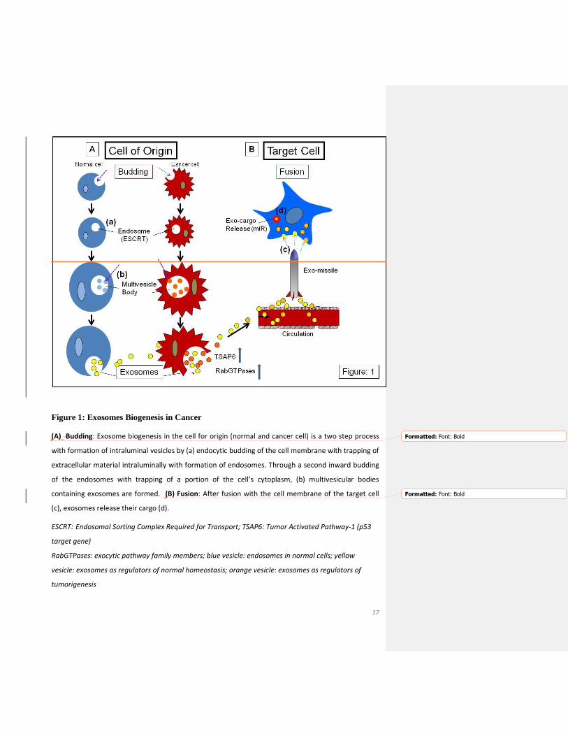
Figure 1: Exosomes Biogenesis in Cancer
Figure 2: Exosomes and the Cancer Niche
Table 1: Exosomes as Cancer Biomarkers
Table 2: Representative Studies on Exosomes as Biomarkers in Cancer Diagnostics

16
17
Figure 1: Exosomes Biogenesis in Cancer
(A) Budding: Exosome biogenesis in the cell for origin (normal and cancer cell) is a two step process
with formation of intraluminal vesicles by (a) endocytic budding of the cell membrane with trapping of
extracellular material intraluminally with formation of endosomes. Through a second inward budding
of the endosomes with trapping of a portion of the cell’s cytoplasm, (b) multivesicular bodies
containing exosomes are formed. (B) Fusion: After fusion with the cell membrane of the target cell
(c), exosomes release their cargo (d).
ESCRT: Endosomal Sorting Complex Required for Transport; TSAP6: Tumor Activated Pathway-1 (p53
target gene)
RabGTPases: exocytic pathway family members; blue vesicle: endosomes in normal cells; yellow
vesicle: exosomes as regulators of normal homeostasis; orange vesicle: exosomes as regulators of
tumorigenesis
Formatted: Font: Bold
Formatted: Font: Bold

18
Figure 2: Exosomes and the Cancer Niche
(A) Exosomes are secreted in the cancer niche by normal supportive cells of the microenvironment
(endothelial cells, mesenchymal stem cells, fibroblasts, bone marrow stem cells) as well as by the
cancer cells. (B) During the homeostatic process of self-renewal, exosomes may play a role in stem
cell maintenance as well as terminal differentiation. (C) Exosomes as propagators of oncogenesis (a) at
the primary tumor site, (b) at the pre-metastatic niche, and (c) in metastatic disease
Cancer niche: blue cancer cell indicates dormancy; Primary tumor environment: red cancer cell
indicates activation

19
Table 1: Exosomes as Cancer Biomarkers
Cancer Biomarker Criteria
Exosomes as Cancer Biomarkers
Easy accessible by minimal invasive
procedures for repetitive sampling
Body fluids (1, 9, 84)
Signature of the cancer of origin Exo-miRs (74, 105)
Distant travelers for safe cargo delivery Rab GTPases-regulated vesicle trafficking (49)
p53-regulated exosome release (106)
Exo-miRs in blood are RNase protected (36, 67)
Target cell-specific entry Phosphatidylserine exosome uptake signal in
NK cells (42)
Surface expression of adhesion tetraspanin-
integrin complexes on exosomes (64, 76, 77)
Interference with intra- and intercellular
differentiation signaling
Exosomal shuttle RNA (esRNA) (7)
Immunomodulation (89, 90)
Cancer dormancy (10)
Disease-stage specific and treatment
responsive Exo-predictive biomarker with chemo
responsiveness (80)
Exo-transfer of multidrug resistance (94, 95)
High-output screening tool NGS of exo-miR (81, 82)
Reference database available Exocarta (38)
Vesiclepedia (39)
EVpedia (40, 41)

20
Table 2: Representative Studies on Exosomes as Biomarkers in Cancer Diagnostics
Primary Tumor/ Exosome source
Biofluid Exosomal Biomarker Protein miRNA
References
Metastatic Melanoma
Serum MRD-9 and GFP78 TYRP2, VLA-4, HSP70, HSP90*
miR125b
Guan et al (107) Peinado et al (62) Alegre et al (108)
Lung Cancer Serum EGFR
miR-378a, miR-379, miR-139-5p, and miR-200b-5p
Cazzoli et al (109) Yamashita et al (110)
Pancreas Cancer Serum Saliva
CD44v6, Tspan8,EpCam MET, CD104 Apbb1ip, Aspn, BCO31781, Daf2, Foxp1, Gng2
miR-1246,miR-4644 miR-3976,miT-4306
Madhavan et al (111) Lau et al (112)
Ovarian Cancer Serum EpCam miR-21, miR-141, miR-200a, miR-200c, miR-200b, miR-203, miR-205, miR-214
Taylor et al (26)
Renal Cell Carcinoma
Urine MMP9, PODXL, CAIX, AQP1, CP
Raimondo et al (113)
Prostate Cancer Urine PCA-3, TMPRSS2:ERG miR34a*
Nilsson et al (114) Corcoran et al (80)
Breast Cancer Plasma miR-21 Corcoran et al (115)
Glioblastoma Serum Let-7a, miR15b,miR-16,miR-19b,miR-21,miR-26a,miR-27a,miR92,miR93, miR-320,miR-20
Skog et al (116)
Gastrointestinal Cancer
Serum Lindner et al (117)
Colon Cancer Serum Let-7a, miR-1229, miR-1246, miR-150, miR-21, miR-223, and miR-23a
Ogata-Kawata et al (118)
Acute Myeloid Leukemia
Plasma CD34 Hong et al (56)
*clinical trial

21
References
1. Thery C, Amigorena S, Raposo G, Clayton A. Isolation and characterization of exosomes from cell culture supernatants and biological fluids. Curr Protoc Cell Biol. 2006;Chapter(3):Unit 3.22. doi: 10.1002/0471143030.cb0322s30. 2. Caby MP, Lankar D, Vincendeau-Scherrer C, Raposo G, Bonnerot C. Exosomal-like vesicles are present in human blood plasma. Int Immunol. 2005;17(7):879-87. Epub 2005 May 20. 3. Huebner AR, Somparn P, Benjachat T, Leelahavanichkul A, Avihingsanon Y, Fenton RA, et al. Exosomes in urine biomarker discovery. Adv Exp Med Biol. 2015;845:43-58.(doi):10.1007/978-94-017-9523-4_5. 4. Lasser C, Alikhani VS, Ekstrom K, Eldh M, Paredes PT, Bossios A, et al. Human saliva, plasma and breast milk exosomes contain RNA: uptake by macrophages. J Transl Med. 2011;9:9.(doi):10.1186/479-5876-9-9. 5. Andre F, Schartz NE, Movassagh M, Flament C, Pautier P, Morice P, et al. Malignant effusions and immunogenic tumour-derived exosomes. Lancet. 2002;360(9329):295-305. 6. Thakur BK, Zhang H, Becker A, Matei I, Huang Y, Costa-Silva B, et al. Double-stranded DNA in exosomes: a novel biomarker in cancer detection. Cell Res. 2014;24(6):766-9. doi: 10.1038/cr.2014.44. Epub Apr 8. 7. Valadi H, Ekstrom K, Bossios A, Sjostrand M, Lee JJ, Lotvall JO. Exosome-mediated transfer of mRNAs and microRNAs is a novel mechanism of genetic exchange between cells. Nat Cell Biol. 2007;9(6):654-9. Epub 2007 May 7. 8. Thery C, Zitvogel L, Amigorena S. Exosomes: composition, biogenesis and function. Nat Rev Immunol. 2002;2(8):569-79. 9. Properzi F, Logozzi M, Fais S. Exosomes: the future of biomarkers in medicine. Biomark Med. 2013;7(5):769-78. doi: 10.2217/bmm.13.63. 10. Ono M, Kosaka N, Tominaga N, Yoshioka Y, Takeshita F, Takahashi RU, et al. Exosomes from bone marrow mesenchymal stem cells contain a microRNA that promotes dormancy in metastatic breast cancer cells. Sci Signal. 2014;7(332):ra63. doi: 10.1126/scisignal.2005231. 11. Inal JM, Fairbrother U, Heugh S. Microvesiculation and disease. Biochem Soc Trans. 2013;41(1):237-40. doi: 10.1042/BST20120258. 12. Henderson MC, Azorsa DO. The genomic and proteomic content of cancer cell-derived exosomes. Front Oncol. 2012;2:38.(doi):10.3389/fonc.2012.00038. eCollection 2012. 13. Hannafon BN, Ding WQ. Intercellular Communication by Exosome-Derived microRNAs in Cancer. Int J Mol Sci. 2013;14(7):14240-69. doi: 10.3390/ijms140714240. 14. Wahlgren J, De LKT, Brisslert M, Vaziri Sani F, Telemo E, Sunnerhagen P, et al. Plasma exosomes can deliver exogenous short interfering RNA to monocytes and lymphocytes. Nucleic Acids Res. 2012;40(17):e130. Epub 2012 May 22. 15. Nazarenko I, Rupp AK, Altevogt P. Exosomes as a potential tool for a specific delivery of functional molecules. Methods Mol Biol. 2013;1049:495-511.(doi):10.1007/978-1-62703-547-7_37. 16. Escudier B, Dorval T, Chaput N, Andre F, Caby MP, Novault S, et al. Vaccination of metastatic melanoma patients with autologous dendritic cell (DC) derived-exosomes: results of thefirst phase I clinical trial. J Transl Med. 2005;3(1):10. 17. Johnstone RM, Adam M, Hammond JR, Orr L, Turbide C. Vesicle formation during reticulocyte maturation. Association of plasma membrane activities with released vesicles (exosomes). J Biol Chem. 1987;262(19):9412-20. 18. Pan BT, Teng K, Wu C, Adam M, Johnstone RM. Electron microscopic evidence for externalization of the transferrin receptor in vesicular form in sheep reticulocytes. J Cell Biol. 1985;101(3):942-8. 19. Aalberts M, Stout TA, Stoorvogel W. Prostasomes: extracellular vesicles from the prostate. Reproduction. 2013;147(1):R1-14. doi: 0.1530/REP-13-0358. Print 2014 Jan. 20. Ronquist G, Hedstrom M. Restoration of detergent-inactivated adenosine triphosphatase activity of human prostatic fluid with concanavalin A. Biochim Biophys Acta. 1977;483(2):483-6. 21. Ronquist G, Nilsson BO. The Janus-faced nature of prostasomes: their pluripotency favours the normal reproductive process and malignant prostate growth. Prostate Cancer Prostatic Dis. 2004;7(1):21-31. 22. Sahlen G, Ahlander A, Frost A, Ronquist G, Norlen BJ, Nilsson BO. Prostasomes are secreted from poorly differentiated cells of prostate cancer metastases. Prostate. 2004;61(3):291-7.
22
23. Rothman JE. The principle of membrane fusion in the cell (nobel lecture). Angew Chem Int Ed Engl. 2014;53(47):12676-94. doi: 10.1002/anie.201402380. Epub 2014 Aug 1. 24. Schekman R, Orci L. Coat proteins and vesicle budding. Science. 1996;271(5255):1526-33. 25. Sudhof TC. The molecular machinery of neurotransmitter release (nobel lecture). Angew Chem Int Ed Engl. 2014;53(47):12696-717. doi: 10.1002/anie.201406359. Epub 2014 Oct 22. 26. Taylor DD, Gercel-Taylor C. MicroRNA signatures of tumor-derived exosomes as diagnostic biomarkers of ovarian cancer. Gynecol Oncol. 2008;110(1):13-21. doi: 10.1016/j.ygyno.2008.04.033. 27. Ghosh AK, Secreto CR, Knox TR, Ding W, Mukhopadhyay D, Kay NE. Circulating microvesicles in B-cell chronic lymphocytic leukemia can stimulate marrow stromal cells: implications for disease progression. Blood. 2010;115(9):1755-64. doi: 10.182/blood-2009-09-242719. Epub 2009 Dec 17. 28. Wan C, Fu J, Wang Y, Miao S, Song W, Wang L. Exosome-related multi-pass transmembrane protein TSAP6 is a target of rhomboid protease RHBDD1-induced proteolysis. PLoS One. 2012;7(5):e37452. doi: 10.1371/journal.pone.0037452. Epub 2012 May 18. 29. Lespagnol A, Duflaut D, Beekman C, Blanc L, Fiucci G, Marine JC, et al. Exosome secretion, including the DNA damage-induced p53-dependent secretory pathway, is severely compromised in TSAP6/Steap3-null mice. Cell Death Differ. 2008;15(11):1723-33. doi: 10.038/cdd.2008.104. Epub Jul 11. 30. Hendrix A, Westbroek W, Bracke M, De Wever O. An ex(o)citing machinery for invasive tumor growth. Cancer Res. 2010;70(23):9533-7. doi: 10.1158/0008-5472.CAN-10-3248. Epub 2010 Nov 23. 31. Ostenfeld MS, Jeppesen DK, Laurberg JR, Boysen AT, Bramsen JB, Primdal-Bengtson B, et al. Cellular disposal of miR23b by RAB27-dependent exosome release is linked to acquisition of metastatic properties. Cancer Res. 2014;74(20):5758-71. doi: 10.1158/0008-5472.CAN-13-3512. Epub 2014 Sep 26. 32. Zhang HG, Grizzle WE. Exosomes: a novel pathway of local and distant intercellular communication that facilitates the growth and metastasis of neoplastic lesions. Am J Pathol. 2014;184(1):28-41. doi: 10.1016/j.ajpath.2013.09.027. Epub Nov 21. 33. Van Deun J, Mestdagh P, Sormunen R, Cocquyt V, Vermaelen K, Vandesompele J, et al. The impact of disparate isolation methods for extracellular vesicles on downstream RNA profiling. J Extracell Vesicles. 2014;3.(doi):10.3402/jev.v3.24858. eCollection 2014. 34. Thery C, Duban L, Segura E, Veron P, Lantz O, Amigorena S, et al. Indirect activation of naive CD4+ T cells by dendritic cell-derived exosomes Exosomes: composition, biogenesis and function. Nat Immunol. 2002;3(12):1156-62. Epub 2002 Nov 11. 35. Yao Y, Wang C, Wei W, Shen C, Deng X, Chen L, et al. Dendritic cells pulsed with leukemia cell-derived exosomes more efficiently induce antileukemic immunities. PLoS One. 2014;9(3):e91463. doi: 10.1371/journal.pone.0091463. eCollection 2014. 36. Mitchell PS, Parkin RK, Kroh EM, Fritz BR, Wyman SK, Pogosova-Agadjanyan EL, et al. Circulating microRNAs as stable blood-based markers for cancer detection. Proc Natl Acad Sci U S A. 2008;105(30):10513-8. doi: 10.1073/pnas.0804549105. Epub 2008 Jul 28. 37. Mathivanan S, Simpson RJ. ExoCarta: A compendium of exosomal proteins and RNA. Proteomics. 2009;9(21):4997-5000. doi: 10.1002/pmic.200900351. 38. Mathivanan S, Fahner CJ, Reid GE, Simpson RJ, Kalra H. ExoCarta 2012: database of exosomal proteins, RNA and lipids ExoCarta as a resource for exosomal research. Nucleic Acids Res. 2012;40(Database issue):D1241-4. doi: 10.093/nar/gkr828. Epub 2011 Oct 11. 39. Kalra H, Simpson RJ, Ji H, Aikawa E, Altevogt P, Askenase P, et al. Vesiclepedia: a compendium for extracellular vesicles with continuous community annotation. PLoS Biol. 2012;10(12):e1001450. doi: 10.1371/journal.pbio.. Epub 2012 Dec 18. 40. Kim DK, Kang B, Kim OY, Choi DS, Lee J, Kim SR, et al. EVpedia: an integrated database of high-throughput data for systemic analyses of extracellular vesicles. J Extracell Vesicles. 2013;2.(doi):10.3402/jev.v2i0.20384. eCollection 2013. 41. Kim DK, Lee J, Kim SR, Choi DS, Yoon YJ, Kim JH, et al. EVpedia: a community web portal for extracellular vesicles research. Bioinformatics. 2014;10. 42. Hanahan D, Weinberg RA. The hallmarks of cancer. Cell. 2000;100(1):57-70. 43. Sonnenschein C, Soto AM. The aging of the 2000 and 2011 Hallmarks of Cancer reviews: a critique. J Biosci. 2013;38(3):651-63.
23
44. Li L, Neaves WB. Normal stem cells and cancer stem cells: the niche matters. Cancer Res. 2006;66(9):4553-7. 45. Spradling A, Drummond-Barbosa D, Kai T. Stem cells find their niche. Nature. 2001;414(6859):98-104. 46. Ma H, Liang C, Wang G, Jia S, Zhao Q, Xiang Z, et al. MicroRNA-mediated cancer metastasis regulation via heterotypic signals in the microenvironment. Curr Pharm Biotechnol. 2014;15(5):455-8. 47. Paget S. The distribution of secondary growths in cancer of the breast. 1889. Cancer Metastasis Rev. 1989;8(2):98-101. 48. Rana S, Malinowska K, Zoller M. Exosomal tumor microRNA modulates premetastatic organ cells. Neoplasia. 2013;15(3):281-95. 49. Ostrowski M, Carmo NB, Krumeich S, Fanget I, Raposo G, Savina A, et al. Rab27a and Rab27b control different steps of the exosome secretion pathway. Nat Cell Biol. 2010;12(1):19-30; sup pp 1-13. doi: 0.1038/ncb2000. Epub 9 Dec 6. 50. Thuma F, Zoller M. Outsmart tumor exosomes to steal the cancer initiating cell its niche. Semin Cancer Biol. 2014;28:39-50.(doi):10.1016/j.semcancer.2014.02.011. Epub Mar 12. 51. Aharon A, Rebibo-Sabbah A, Tzoran I, Levin C. Extracellular vesicles in hematological disorders. Rambam Maimonides Med J. 2014;5(4):e0032. doi: 10.5041/RMMJ.10166. eCollection 2014 Oct. 52. Corrado C, Raimondo S, Saieva L, Flugy AM, De Leo G, Alessandro R. Exosome-mediated crosstalk between chronic myelogenous leukemia cells and human bone marrow stromal cells triggers an interleukin 8-dependent survival of leukemia cells. Cancer Lett. 2014;348(1-2):71-6. doi: 10.1016/j.canlet.2014.03.009. Epub Mar 18. 53. Cai J, Wu G, Tan X, Han Y, Chen C, Li C, et al. Transferred BCR/ABL DNA from K562 extracellular vesicles causes chronic myeloid leukemia in immunodeficient mice. PLoS One. 2014;9(8):e105200. doi: 10.1371/journal.pone.0105200. eCollection 2014. 54. Huan J, Hornick NI, Shurtleff MJ, Skinner AM, Goloviznina NA, Roberts CT, Jr., et al. RNA trafficking by acute myelogenous leukemia exosomes. Cancer Res. 2013;73(2):918-29. doi: 10.1158/0008-5472.CAN-12-2184. Epub 012 Nov 13. 55. Szczepanski MJ, Szajnik M, Welsh A, Whiteside TL, Boyiadzis M. Blast-derived microvesicles in sera from patients with acute myeloid leukemia suppress natural killer cell function via membrane-associated transforming growth factor-beta1. Haematologica. 2011;96(9):1302-9. doi: 10.3324/haematol.2010.039743. Epub 2011 May 23. 56. Hong CS, Muller L, Whiteside TL, Boyiadzis M. Plasma exosomes as markers of therapeutic response in patients with acute myeloid leukemia. Front Immunol. 2014;5:160.(doi):10.3389/fimmu.2014.00160. eCollection 2014. 57. Kaplan RN, Rafii S, Lyden D. Preparing the "soil": the premetastatic niche. Cancer Res. 2006;66(23):11089-93. 58. Sceneay J, Smyth MJ, Moller A. The pre-metastatic niche: finding common ground. Cancer Metastasis Rev. 2013;32(3-4):449-64. doi: 10.1007/s10555-013-9420-1. 59. Kaplan RN, Riba RD, Zacharoulis S, Bramley AH, Vincent L, Costa C, et al. VEGFR1-positive haematopoietic bone marrow progenitors initiate the pre-metastatic niche. Nature. 2005;438(7069):820-7. 60. Peinado H, Lavotshkin S, Lyden D. The secreted factors responsible for pre-metastatic niche formation: old sayings and new thoughts. Semin Cancer Biol. 2011;21(2):139-46. doi: 10.1016/j.semcancer.2011.01.002. Epub Jan 18. 61. Alderton GK. Metastasis. Exosomes drive premetastatic niche formation. Nat Rev Cancer. 2012;12(7):447. doi: 10.1038/nrc3304. 62. Peinado H, Aleckovic M, Lavotshkin S, Matei I, Costa-Silva B, Moreno-Bueno G, et al. Melanoma exosomes educate bone marrow progenitor cells toward a pro-metastatic phenotype through MET. Nat Med. 2012;18(6):883-91. doi: 10.1038/nm.2753. 63. Mu W, Rana S, Zoller M. Host matrix modulation by tumor exosomes promotes motility and invasiveness. Neoplasia. 2013;15(8):875-87. 64. Hoffman RM. Stromal-cell and cancer-cell exosomes leading the metastatic exodus for the promised niche. Breast Cancer Res. 2013;15(3):310. doi: 10.1186/bcr3426.

24
65. Luga V, Zhang L, Viloria-Petit AM, Ogunjimi AA, Inanlou MR, Chiu E, et al. Exosomes mediate stromal mobilization of autocrine Wnt-PCP signaling in breast cancer cell migration. Cell. 2012;151(7):1542-56. doi: 10.016/j.cell.2012.11.024. 66. Suetsugu A, Honma K, Saji S, Moriwaki H, Ochiya T, Hoffman RM. Imaging exosome transfer from breast cancer cells to stroma at metastatic sites in orthotopic nude-mouse models. Adv Drug Deliv Rev. 2013;65(3):383-90. doi: 10.1016/j.addr.2012.08.007. Epub Aug 17. 67. Cheng L, Sharples RA, Scicluna BJ, Hill AF. Exosomes provide a protective and enriched source of miRNA for biomarker profiling compared to intracellular and cell-free blood. J Extracell Vesicles. 2014;3.(doi):10.3402/jev.v3.23743. eCollection 2014. 68. Grange C, Tapparo M, Collino F, Vitillo L, Damasco C, Deregibus MC, et al. Microvesicles released from human renal cancer stem cells stimulate angiogenesis and formation of lung premetastatic niche. Cancer Res. 2011;71(15):5346-56. doi: 10.1158/0008-5472.CAN-11-0241. Epub 2011 Jun 13. 69. Kobayashi M, Salomon C, Tapia J, Illanes SE, Mitchell MD, Rice GE. Ovarian cancer cell invasiveness is associated with discordant exosomal sequestration of Let-7 miRNA and miR-200. J Transl Med. 2014;12:4.(doi):10.1186/479-5876-12-4. 70. Johnson SM, Grosshans H, Shingara J, Byrom M, Jarvis R, Cheng A, et al. RAS is regulated by the let-7 microRNA family. Cell. 2005;120(5):635-47. 71. Ji H, Greening DW, Barnes TW, Lim JW, Tauro BJ, Rai A, et al. Proteome profiling of exosomes derived from human primary and metastatic colorectal cancer cells reveal differential expression of key metastatic factors and signal transduction components. Proteomics. 2013;13(10-11):1672-86. doi: 10.002/pmic.201200562. 72. Rana S, Yue S, Stadel D, Zoller M. Toward tailored exosomes: the exosomal tetraspanin web contributes to target cell selection. Int J Biochem Cell Biol. 2012;44(9):1574-84. doi: 10.016/j.biocel.2012.06.018. Epub Jun 19. 73. Principe S, Hui AB, Bruce J, Sinha A, Liu FF, Kislinger T. Tumor-derived exosomes and microvesicles in head and neck cancer: implications for tumor biology and biomarker discovery. Proteomics. 2013;13(10-11):1608-23. doi: 10.002/pmic.201200533. 74. Yang J, Wei F, Schafer C, Wong DT. Detection of tumor cell-specific mRNA and protein in exosome-like microvesicles from blood and saliva. PLoS One. 2014;9(11):e110641. doi: 10.1371/journal.pone.0110641. eCollection 2014. 75. Al-Nedawi K, Meehan B, Micallef J, Lhotak V, May L, Guha A, et al. Intercellular transfer of the oncogenic receptor EGFRvIII by microvesicles derived from tumour cells. Nat Cell Biol. 2008;10(5):619-24. doi: 10.1038/ncb725. Epub 2008 Apr 20. 76. Christianson HC, Svensson KJ, van Kuppevelt TH, Li JP, Belting M. Cancer cell exosomes depend on cell -surface heparan sulfate proteoglycans for their internalization and functional activity. Proc Natl Acad Sci U S A. 2013;110(43):17380-5. doi: 10.1073/pnas.1304266110. Epub 2013 Oct 7. 77. Clayton A, Turkes A, Dewitt S, Steadman R, Mason MD, Hallett MB. Adhesion and signaling by B cell-derived exosomes: the role of integrins. Faseb J. 2004;18(9):977-9. Epub 2004 Apr 1. 78. Colombo M, Moita C, van Niel G, Kowal J, Vigneron J, Benaroch P, et al. Analysis of ESCRT functions in exosome biogenesis, composition and secretion highlights the heterogeneity of extracellular vesicles. J Cell Sci. 2013;126(Pt 24):5553-65. doi: 10.1242/jcs.128868. Epub 2013 Oct 8. 79. Braicu C, Tomuleasa C, Monroig P, Cucuianu A, Berindan-Neagoe I, Calin GA. Exosomes as divine messengers: are they the Hermes of modern molecular oncology? Cell Death Differ. 2015;22(1):34-45. doi: 10.1038/cdd.2014.130. Epub Sep 19. 80. Corcoran C, Rani S, O'Driscoll L. miR-34a is an intracellular and exosomal predictive biomarker for response to docetaxel with clinical relevance to prostate cancer progression. Prostate. 2014;74(13):1320-34. doi: 10.002/pros.22848. Epub 2014 Jul 22. 81. Cheng L, Sun X, Scicluna BJ, Coleman BM, Hill AF. Characterization and deep sequencing analysis of exosomal and non-exosomal miRNA in human urine. Kidney Int. 2014;86(2):433-44. doi: 10.1038/ki.2013.502. Epub Dec 18. 82. Jenjaroenpun P, Kremenska Y, Nair VM, Kremenskoy M, Joseph B, Kurochkin IV. Characterization of RNA in exosomes secreted by human breast cancer cell lines using next-generation sequencing. PeerJ. 2013;1:e201.(doi):10.7717/peerj.201. eCollection 2013.

25
83. Arroyo JD, Chevillet JR, Kroh EM, Ruf IK, Pritchard CC, Gibson DF, et al. Argonaute2 complexes carry a population of circulating microRNAs independent of vesicles in human plasma. Proc Natl Acad Sci U S A. 2011;108(12):5003-8. doi: 10.1073/pnas.1019055108. Epub 2011 Mar 7. 84. van der Meel R, Krawczyk-Durka M, van Solinge WW, Schiffelers RM. Toward routine detection of extracellular vesicles in clinical samples. Int J Lab Hematol. 2014;36(3):244-53. doi: 10.1111/ijlh.12247. 85. Mestdagh P, Hartmann N, Baeriswyl L, Andreasen D, Bernard N, Chen C, et al. Evaluation of quantitative miRNA expression platforms in the microRNA quality control (miRQC) study. Nat Methods. 2014;11(8):809-15. doi: 10.1038/nmeth.3014. Epub 2014 Jun 29. 86. Kalra H, Adda CG, Liem M, Ang CS, Mechler A, Simpson RJ, et al. Comparative proteomics evaluation of plasma exosome isolation techniques and assessment of the stability of exosomes in normal human blood plasma. Proteomics. 2013;13(22):3354-64. doi: 10.1002/pmic.201300282. Epub 2013 Oct 18. 87. Shen C, Hao SG, Zhao CX, Zhu J, Wang C. Antileukaemia immunity: effect of exosomes against NB4 acute promyelocytic leukaemia cells. J Int Med Res. 2011;39(3):740-7. 88. Hao S, Moyana T, Xiang J. Review: cancer immunotherapy by exosome-based vaccines. Cancer Biother Radiopharm. 2007;22(5):692-703. 89. Mittelbrunn M, Gutierrez-Vazquez C, Villarroya-Beltri C, Gonzalez S, Sanchez-Cabo F, Gonzalez MA, et al. Unidirectional transfer of microRNA-loaded exosomes from T cells to antigen-presenting cells. Nat Commun. 2011;2:282.(doi):10.1038/ncomms285. 90. Peng P, Yan Y, Keng S. Exosomes in the ascites of ovarian cancer patients: origin and effects on anti-tumor immunity. Oncol Rep. 2011;25(3):749-62. doi: 10.3892/or.2010.1119. Epub 2010 Dec 22. 91. Pitt JM, Charrier M, Viaud S, Andre F, Besse B, Chaput N, et al. Dendritic cell-derived exosomes as immunotherapies in the fight against cancer. J Immunol. 2014;193(3):1006-11. doi: 10.4049/jimmunol.1400703. 92. Navabi H, Croston D, Hobot J, Clayton A, Zitvogel L, Jasani B, et al. Preparation of human ovarian cancer ascites-derived exosomes for a clinical trial. Blood Cells Mol Dis. 2005;35(2):149-52. 93. Dai S, Wei D, Wu Z, Zhou X, Wei X, Huang H, et al. Phase I clinical trial of autologous ascites-derived exosomes combined with GM-CSF for colorectal cancer. Mol Ther. 2008;16(4):782-90. doi: 10.1038/mt.2008.1. Epub Feb 5. 94. Corcoran C, Rani S, O'Brien K, O'Neill A, Prencipe M, Sheikh R, et al. Docetaxel-resistance in prostate cancer: evaluating associated phenotypic changes and potential for resistance transfer via exosomes. PLoS One. 2012;7(12):e50999. doi: 10.1371/journal.pone.0050999. Epub 2012 Dec 10. 95. Wang J, Hendrix A, Hernot S, Lemaire M, De Bruyne E, Van Valckenborgh E, et al. Bone marrow stromal cell-derived exosomes as communicators in drug resistance in multiple myeloma cells. Blood. 2014;124(4):555-66. doi: 10.1182/blood-2014-03-562439. Epub 2014 Jun 13. 96. Martinez MC, Andriantsitohaina R. Microparticles in angiogenesis: therapeutic potential. Circ Res. 2011;109(1):110-9. doi: 10.1161/CIRCRESAHA.110.233049. 97. Benameur T, Tual-Chalot S, Andriantsitohaina R, Martinez MC. PPARalpha is essential for microparticle-induced differentiation of mouse bone marrow-derived endothelial progenitor cells and angiogenesis. PLoS One. 2010;5(8):e12392. doi: 10.1371/journal.pone.0012392. 98. Marleau AM, Chen CS, Joyce JA, Tullis RH. Exosome removal as a therapeutic adjuvant in cancer. J Transl Med. 2012;10:134.(doi):10.1186/479-5876-10-134. 99. Tan A, Rajadas J, Seifalian AM. Exosomes as nano-theranostic delivery platforms for gene therapy. Adv Drug Deliv Rev. 2013;65(3):357-67. doi: 10.1016/j.addr.2012.06.014. Epub Jul 20. 100. van der Meel R, Fens MH, Vader P, van Solinge WW, Eniola-Adefeso O, Schiffelers RM. Extracellular vesicles as drug delivery systems: lessons from the liposome field. J Control Release. 2014;195:72-85.(doi):10.1016/j.jconrel.2014.07.049. Epub Aug 2. 101. El-Andaloussi S, Lee Y, Lakhal-Littleton S, Li J, Seow Y, Gardiner C, et al. Exosome-mediated delivery of siRNA in vitro and in vivo. Nat Protoc. 2012;7(12):2112-26. doi: 10.1038/nprot.2012.131. Epub Nov 15. 102. Lasser C. Exosomes in diagnostic and therapeutic applications: biomarker, vaccine and RNA interference delivery vehicle. Expert Opin Biol Ther. 2015;15(1):103-17. doi: 10.1517/14712598.2015.977250. Epub 2014 Nov 1. 103. Zhuang X, Xiang X, Grizzle W, Sun D, Zhang S, Axtell RC, et al. Treatment of brain inflammatory diseases by delivering exosome encapsulated anti-inflammatory drugs from the nasal region to the brain. Mol Ther. 2011;19(10):1769-79. doi: 10.038/mt.2011.164. Epub Sep 13.
26
104. Mistry A, Stolnik S, Illum L. Nanoparticles for direct nose-to-brain delivery of drugs. Int J Pharm. 2009;379(1):146-57. doi: 10.1016/j.ijpharm.2009.06.019. Epub Jun 23. 105. Rabinowits G, Gercel-Taylor C, Day JM, Taylor DD, Kloecker GH. Exosomal microRNA: a diagnostic marker for lung cancer. Clin Lung Cancer. 2009;10(1):42-6. doi: 10.3816/CLC.2009.n.006. 106. Yu X, Harris SL, Levine AJ. The regulation of exosome secretion: a novel function of the p53 protein. Cancer Res. 2006;66(9):4795-801. 107. Guan M, Chen X, Ma Y, Tang L, Guan L, Ren X, et al. MDA-9 and GRP78 as potential diagnostic biomarkers for early detection of melanoma metastasis. Tumour Biol. 2014;6:6. 108. Alegre E, Sanmamed MF, Rodriguez C, Carranza O, Martin-Algarra S, Gonzalez A. Study of circulating microRNA-125b levels in serum exosomes in advanced melanoma. Arch Pathol Lab Med. 2014;138(6):828-32. doi: 10.5858/arpa.2013-0134-OA. 109. Cazzoli R, Buttitta F, Di Nicola M, Malatesta S, Marchetti A, Rom WN, et al. microRNAs Derived from Circulating Exosomes as Noninvasive Biomarkers for Screening and Diagnosing Lung Cancer. J Thorac Oncol. 2013;8(9):1156-62. 110. Yamashita T, Kamada H, Kanasaki S, Maeda Y, Nagano K, Abe Y, et al. Epidermal growth factor receptor localized to exosome membranes as a possible biomarker for lung cancer diagnosis. Pharmazie. 2013;68(12):969-73. 111. Madhavan B, Yue S, Galli U, Rana S, Gross W, Muller M, et al. Combined evaluation of a panel of protein and miRNA serum-exosome biomarkers for pancreatic cancer diagnosis increases sensitivity and specificity. Int J Cancer. 2014;12(10):29324. 112. Lau C, Kim Y, Chia D, Spielmann N, Eibl G, Elashoff D, et al. Role of pancreatic cancer-derived exosomes in salivary biomarker development. J Biol Chem. 2013;288(37):26888-97. doi: 10.1074/jbc.M113.452458. Epub 2013 Jul 23. 113. Raimondo F, Morosi L, Corbetta S, Chinello C, Brambilla P, Della Mina P, et al. Differential protein profiling of renal cell carcinoma urinary exosomes. Mol Biosyst. 2013;9(6):1220-33. doi: 10.039/c3mb25582d. Epub 2013 Mar 19. 114. Nilsson J, Skog J, Nordstrand A, Baranov V, Mincheva-Nilsson L, Breakefield XO, et al. Prostate cancer-derived urine exosomes: a novel approach to biomarkers for prostate cancer. Br J Cancer. 2009;100(10):1603-7. doi: 10.038/sj.bjc.6605058. Epub 2009 Apr 28. 115. Corcoran C, Friel AM, Duffy MJ, Crown J, O'Driscoll L. Intracellular and extracellular microRNAs in breast cancer. Clin Chem. 2011;57(1):18-32. doi: 10.1373/clinchem.2010.150730. Epub 2010 Nov 8. 116. Skog J, Wurdinger T, van Rijn S, Meijer DH, Gainche L, Sena-Esteves M, et al. Glioblastoma microvesicles transport RNA and proteins that promote tumour growth and provide diagnostic biomarkers. Nat Cell Biol. 2008;10(12):1470-6. doi: 10.038/ncb800. Epub 2008 Nov 16. 117. Lindner K, Haier J, Wang Z, Watson DI, Hussey DJ, Hummel R. Circulating microRNAs: emerging biomarkers for diagnosis and prognosis in patients with gastrointestinal cancers. Clin Sci (Lond). 2015;128(1):1-15. doi: 0.1042/CS20140089. 118. Ogata-Kawata H, Izumiya M, Kurioka D, Honma Y, Yamada Y, Furuta K, et al. Circulating exosomal microRNAs as biomarkers of colon cancer. PLoS One. 2014;9(4):e92921. doi: 10.1371/journal.pone.0092921. eCollection 2014.
(End)


![The Role of Exosomes in Bone Remodeling: …downloads.hindawi.com/journals/dm/2019/9417914.pdfregulation [35]. 3.2. Exosomes from Osteoblasts. Ample data suggest that exosomes shed](https://img.pdfslide.net/doc/110x75/5f03c0c07e708231d40a9922/the-role-of-exosomes-in-bone-remodeling-regulation-35-32-exosomes-from-osteoblasts.jpg)